Embed Size (px)
Citation preview
ARTERY, VEIN, NERVE, LYMPHATICS
Artery
1) Haemangioma.
Answer. Definition: A benign skin lesion consisting of dense, usually elevated masses
of dilated blood vessels.
The hemangioma is a true vascular tumor that results from a overgrowth of normal
vascular tissue.
It exhibits relatively rapid early growth until approximately 6 to 8 months of age
(proliferative phase), followed by regression by 5 to 9 years of age (involutory
phase).
It grows by endothelial proliferation . During the rapid growth phase, an increased
number of mast cells is seen within the endothelial wall.
It is compressible.
Types of haemangioma:
Capillary Hemangioma:
o Salmon patch:
These are very common and occur in about 40% of all newborns.
They are usually small flat patches of pink or red skin with poorly defined borders.
They are commonly found at the nape of the neck (stork bite), on the forehead
between the eyebrows (angel's kiss) or on the eyelids.
They become more intense in colour and noticeable when the child is crying.
Most lesions will spontaneously disappear within the first year of life.
Stork bites tend to be more persistent and may remain unchanged into adult life in
50% of cases.
o Port wine stain:
Persists throughout the life.
A port wine stain is usually a large flat patch of purple or dark red skin with well-
defined borders.
At birth the surface of the port-wine stain is flat, but in time it becomes bumpy and
often more unsightly.
The face is most commonly affected although they can occur anywhere on the
body.
Where present, they generally appear on one side of the body with a sharp mid-line
cut-off.
o Strawberry angioma:
Strawberry red mark found on 1 out of 10 babies
Small as a freckle or large as a coaster
Consists of small closely packed blood vessels
95% disappear by the time the child is 10 years old
Cavernous (Deep) Hemangioma:
Deeply situated red-blue spongy mass of tissue filled with blood found on 2 out of
100 babies
Grows rapidly in the first six months
Composed of larger, more mature vascular elements
Some of these lesions disappear on their own
Compound Hemangioma:
Contains both superficial and deep parts
These are often the largest and the most spreading
Similar characteristics to both the strawberry hemangioma and the cavernous
hemangioma
Treatment should be considered if the hemangioma:
Ulcerates
Bleeds
Causes functional impairment
Causes infection
Grows rapidly and uncontrollably
Causes psychological problems
Treatment options:
Medical
o Steroid injection
o Interferon alfa-2a
Surgical
o Resection
o FPDL
o YAG laser
2) Arterio-venous fistula.
Answer. Introduction: An arteriovenous fistula is an abnormal connection or
passageway between an artery and a vein. It may be congenital, surgically created for
hemodialysis treatments, or acquired due to pathologic process, such as trauma or
erosion of an arterial aneurysm.
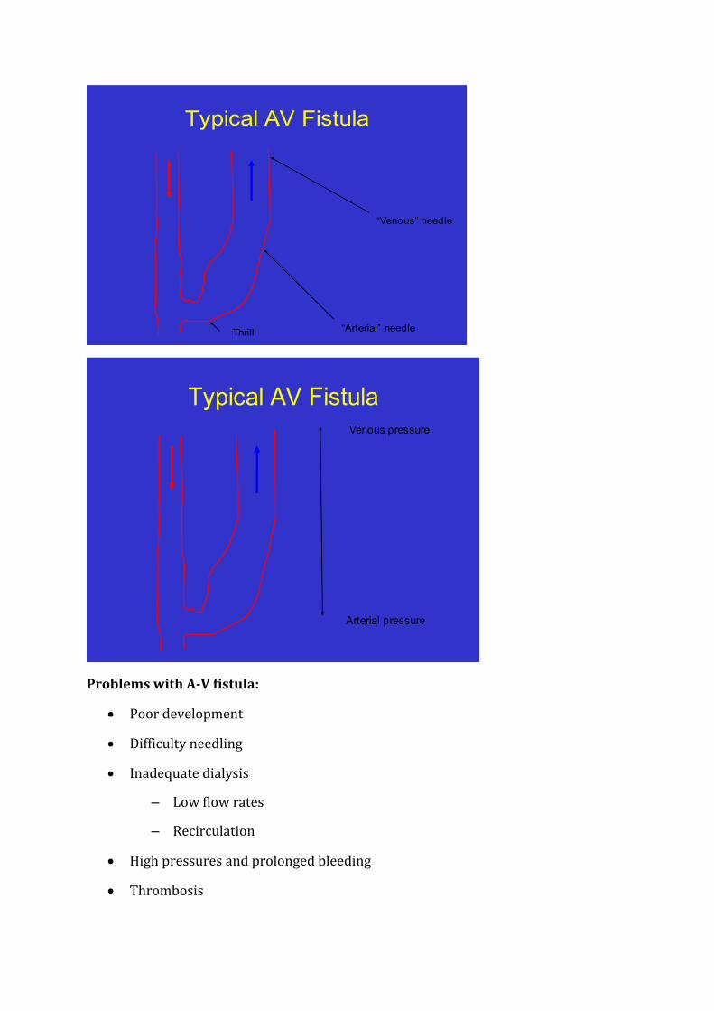
Typical AV Fistula
“Arterial” needle
“Venous” needle
Thrill
Typical AV Fistula
Arterial pressure
Venous pressure
Problems with A-V fistula:
Poor development
Difficulty needling
Inadequate dialysis
– Low flow rates
– Recirculation
High pressures and prolonged bleeding
Thrombosis
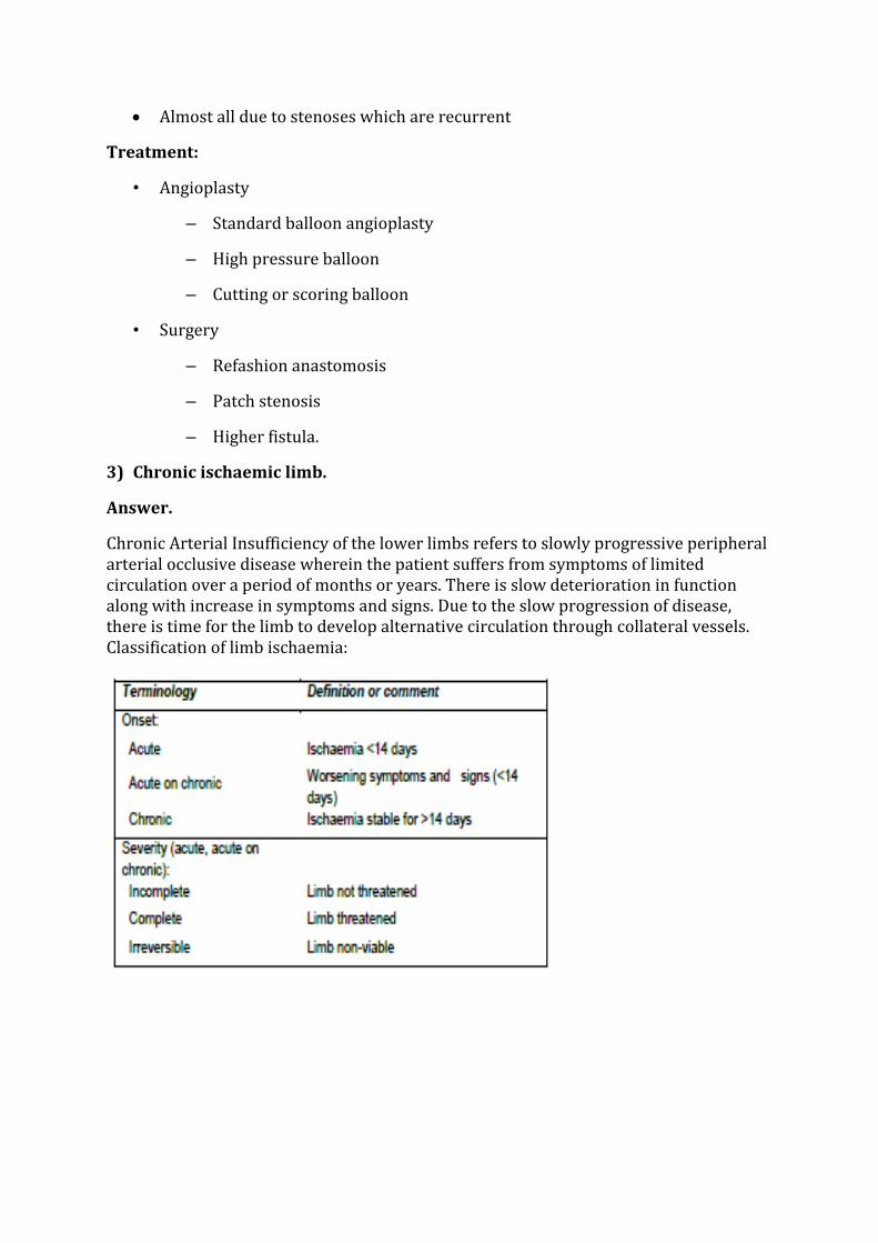
Almost all due to stenoses which are recurrent
Treatment:
• Angioplasty
– Standard balloon angioplasty
– High pressure balloon
– Cutting or scoring balloon
• Surgery
– Refashion anastomosis
– Patch stenosis
– Higher fistula.
3) Chronic ischaemic limb.
Answer.
Chronic Arterial Insufficiency of the lower limbs refers to slowly progressive peripheral
arterial occlusive disease wherein the patient suffers from symptoms of limited
circulation over a period of months or years. There is slow deterioration in function
along with increase in symptoms and signs. Due to the slow progression of disease,
there is time for the limb to develop alternative circulation through collateral vessels.
Classification of limb ischaemia:
Critical limb ischemia (CLI)
The term critical limb ischemia is used for all patients with chronic ischemic rest pain,
ulcers or gangrene attributable to objectively proven arterial occlusive disease. The
term CLI should only be used in the presence of symptoms for more than 2 weeks.
4) Define claudication. What are the grades of claudication? How will you manage
a case of Buerger s disease with dry gangrene of the foot?
Answer. This is a special character of pain described for arterial disorders. This is a
clinical condition where a cramping, aching or tightness like severe pain appears in the
leg affected during exercise, usually after a fixed level of exercise and is promptly
(within two to three minutes) relieved with rest. It Is due to the accumulation of
Substance P which fails to get washed away due to poor blood supply.
Boyd s classification of intermittent claudication:
Grade I Pain starts but if the patient continues to walk the metabolites increase the muscle
Blood flow and sweep away the P- substance produced by exercise and pain
disappears.
Grade 2 Pain continues but the patient can still walk with effort.
Grade 3 Pain compels the patient to take rest.
Grade 4 Pain compels the patient to take rest.
Management of a case of Buerger s disease with dry gangrene of the foot:
Investigations:
General:
Blood :
o Routine examination of blood including a hemoglobin percent (low Hb% can
decrease claudication distances and aggravate rest pain),
o Blood sugar examination as diabetics have worse prognosis, are essential.
o Erythrocyte sedimentation rate ESR is usually raised in Buerger s disease. o In patients with high suspicion of underlying connective tissue disorders, specific
test like RA factor, LE cell phenomenon etc. May be carried out.
o Lipid profile is mandatory in elderly patients with atherosclerosis.
Urine examination for sugar.
Plain X-ray of the abdomen will show the presence of arterial calcification and flecks
of calcium may outline an aneurysm.
ECG: an abnormality in ECG may influence the decision for surgery, in patients with
lower limb disease.
Tests of global Vascular Status:
Hand Held Doppler ultrasound: blood flow detection uses a continuous wave
ultrasound signal, beamed at an artery and the reflected beam is picked up by a
receiver. The changes of frequency in the reflected beam, as compared with the transmitted beam, are due to the―Doppler shift, resulting from passage of beam through moving blood. These frequency changes are converted to audio signals. This
investigation may be used effectively in cases where a differential diagnosis of
atherosclerosis is entertained showing the site of block and extent of distal run-off.
Ankle Brachial systolic blood pressure index (ABPI)
This measurement gives the quantitative assessment of the global limb arterial
perfusion.
Ankle Brachial Pressure Index
Segmental pressures: i.e. Differences in arterial blood pressure between segments
of limb can be detected to give indication of the sites of stenosis, specially as Buerger s is said to be a segmental disease. Toe Pressures Using Photoplethysmograph: These are used when the arterial
disease is suspected between the ankle and the toes.
Pole Test: This is used to determine the adequacy of lower limb bolld flow in
patients with incompressible vessels or who are unable to tolerate an ankle
pressure cuff.
Transcutaneous Oximtery (tcpo2): It is based on the principle that the partial
pressure of the oxygen which diffuses through to the surface of the skin reflects the
oxygen tension of the underlying tissues. It is time consuming and is best used in the
selection of amputation sites since it correlates well to subsequent stump healing.
Walk Test: The basis of this test is that measurement of ABPI before and after a
patient has walked can expose less severe or compensated peripheral vascular
disease.
Tests for Disease Localisation:
Duplex imaging: gives accurate information on the size of artery, the flow rate,
turbulence and the presence of stenosis. The combination of Doppler and color
mapping allows easy recognition of stenotic sites. This has been achieved by the use
of pulsed or continuous wave Doppler and the two- dimensional images produced
by the B- scan made either singly or in combination.
Intravascular Ultrasound: Gives details of arterial walls, luminal contents and
dimensions. This is not a routine investigation for peripheral arterial disease and as
yet is not cost effective.
Arteriography: This is an invasive technique which though has become much safer
in the recent years due to fine 3-4 F catheters, and remains the gold standard to
provide a road map required for vascualr surgeons expecially before surgery is
planned.
CT Angiography: The introduction of the helical (spiral) CT scanning and
multidetector CT which uses 2 or 4 helicals to scan the patient, CT imaging has been
revolutionized for vascular imaging wherein a single breathhold time is sufficient to
generate the scans from the aortic arch to the groins with imaging quality as good as
conventional angiography.
MR Angiography: MRI and Phase Contrast MRI were used to visualize moving
blood as a white image but the definition and clarity of the vessels was found to be
inferior to angiograms. More recently, Gadolinium Enhanced MRI (Gd-MRI) has
significantly improved this quality of image and made it comparable to conventional
angiography.
Treatment of Buerger s disease: Up to two- thirds of patients can be treated by conservative methods.
Abstinence from tobacco: The only proven treatment guideline to prevent disease
progression and avoiding an amputation is complete cessation of smoking or other
forms of tobacco.
Any form of continued usage of tobacco keeps the disease active. Repeated education and counseling is required for these patients. Raynaud s phenomenon or claudication may continue even after complete discontinuation of tobacco.
Explanation and advice: Many patients are worried by the presence of pain while
walking. Once told about the nature of disease and advice regarding methods to
improve their claudication distance e.g. By walking slowly or by improving underlying
systemic disorder like, anemia, congestive failure, the claudication distance can be
increased.
Adjustment of lifestyle: Adjustments to everyday habits of transport can increase
mobility within the claudication distance, e.g use of a bicycle etc.
Exercise & Diet: Taking regular exercise within limits of pain and control of weight in
case of obesity.
Care of feet, avoiding socks with holes and amateur chiropody, which can spark off
gangrene in the toes and heels, particularly in diabetic patients.
Heel raise: claudication distance may be improved by raising the heels of shoes by 1
cm. The work of the calf muscles is reduced thereby.
Analgesics and position: rest pain can be relieved to some extent in some patients by
use of analgesics, elevation of the head end of the bed Buerger s position and Buerger s exercises (repeated 2 minute elevation and dependency of limb).
Drugs: Despite the clear presence of inflammation in this disorder, anti-inflammatory
agents such as steroids have not been shown to be beneficial. Similarly, strategies of
anticoagulation (thinning of the blood with aspirin or other agents to prevent clots)
have not proven effective. Vasodilator drugs are usually started in these patients but
their role is equivocal.
Some of the drugs used are:
Prostaglandins: Prostacylin or PGI2 (Iloprost) has forty times antiplatelet and
vasodilator activity as compared to PGE1. They are effective in both cutaneous and
muscular vessels. Intravenous infusion of prostacyclin (Iloprost) can relieve rest pain.
Low molecular weight dextrans: dextrans of molecular weight 50000 are used during
acute attack of thromboangitis. They cause hemodilution, decrease viscosity of blood
and improve micro circulation. Intra-arterial infusion is said to be more effective than
intravenous.
Intra-arterial Thrombolytic therapy: Selective low dose intrarterial streptokinase
have been used in a very small group of patients with alteration of level of amputation.
Praxiline: (niftidrofuryl oxalate) may alter tissue metabolism, increasing the
claudication distance by allowing a greater oxygen debt to be incurred.
Trental: (oxypentifylline) has some effect on whole blood viscosity by reducing
rouleaux formation.
Aspirin in dispersible form may be prescribed for its anti-adhesive effect on platelets.
No proven benefit.
Direct Arterial Surgery: Surgical bypass or revascularization is rarely feasible in
patients with Buerger s disease because of occlusion of small and medium sized vessels, presence of segmental and skip lesions and absence of a distal target vessel for by pass.
Sympathectomy: Sympathectomy is not beneficial in intermittent claudication, but can
relieve rest pain and ulceration because the effect is mainly on skin and subcutaneous
blood vessels. For the same reason it helps in the healing of superficial ischaemic
ulcerations. It might aggravate claudication by stealing the blood from ichaemic muscles
and diverting it to the skin and therefore is a contraindication for sympathectomy. In
vessel wall, sympathectomy is done with following objective:
o To cause vasodilatation by decreasing sympathetic vasomotor tone.
o To abolish pain impulses carried by sympathetic fibers.
1. Surgical sympathectomy:
Lumbar sympathectomy: Open sympathectomy is done preferably through the
extraperitoneal approach.The sympathetic chain lies on the side of the body of
vertebrae, sometimes inside the psoas muscle sheath. In unilateral surgeries,
sympathetic ganglia, L1, L2, L3 and sometimes L4 are removed. In bilateral cases, L1 of
one side is preserved (to avoid retrograde ejaculation).
2. Chemical sympathectomy: This is an alternative to surgical sympathectomy but is
contraindicated in patients on anticoagulant therapy. A long 15 cm needle is inserted
with local infiltration, first to seek the side of the vertebral body and secondly to pass
alongside it to reach the lumbar sympathetic chain. 5 ml of phenol solution in water is
injected beside the bodies of 2nd, 3rd and 4th vertebrae. The procedure is done
preferably under fluoroscopic or ultrasound guidance and care is taken to avoid
penetrating the aorta or inferior vena cava.
Omental transposition: The procedure is based on the arterial arcade formed by the
anastomosis of right and left gastroepiploic arteries. For unilateral procedures, the
omental pedicle is based on the right gastroepiploic artery as it is a dominant artery and
has a longer length.
For bilateral procedures, both epiploics may be used, though sometimes a single vessel
is used as there is risk of gastric devasularization if both arteries are used. A subfascial
tunnel is made from the inferior end of the laparotomy incision to the inguinal and
further down to the ankle medially.
The omentum is lengthened based on the dominant artery in the pedicle and brought
down to the distal most portion of the affected limb through the subcutaneous tunnel.
Complications of this procedure include: gastric devascularization and necrosis,
paralytic ileus, gastric hemorrhage, omental necrosis and wound infection.
Autologous bone marrow – derived progenitor cell implantation into ischemic limbs
for potentiation of angiogenesis has been performed as an experimental alternative
option. Results have been satisfactory, with minimal complication rates.
Amputation of digit, if infection or gangrene occurs.
5) Raynaud s phenomenon.
Answer. Definition: repetitive episodes of biphasic colour change (at least 2 of pallor,
cyanosis, erythema), in either cold or normal environment.
Pathogenesis:
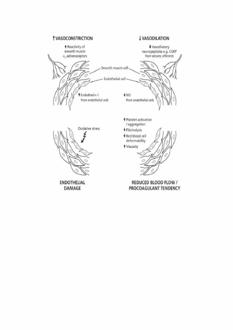
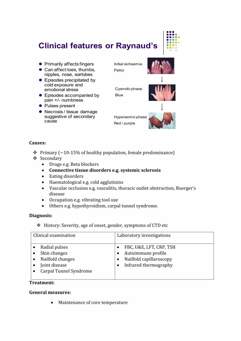
Clinical features or Raynaud’s
Primarily affects fingers
Can affect toes, thumbs, nipples, nose, earlobes
Episodes precipitated by cold exposure and emotional stress
Episodes accompanied by pain +/- numbness
Pulses present
Necrosis / tissue damage suggestive of secondary cause
Initial ischaemia
Pallor
Cyanotic phase
Blue
Hyperaemic phase
Red / purple
Causes:
Primary (~10-15% of healthy population, female predominance)
Secondary
Drugs e.g. Beta blockers
Connective tissue disorders e.g. systemic sclerosis
Eating disorders
Haematological e.g. cold agglutinins
Vascular occlusion e.g. vasculitis, thoracic outlet obstruction, Buerger s disease
Occupation e.g. vibrating tool use
Others e.g. hypothyroidism, carpal tunnel syndrome.
Diagnosis:
History: Severity, age of onset, gender, symptoms of CTD etc
Clinical examination
Laboratory investigations
Radial pulses
Skin changes
Nailfold changes
Joint disease
Carpal Tunnel Syndrome
FBC, U&E, LFT, CRP, TSH
Autoimmune profile
Nailfold capillaroscopy
Infrared thermography
Treatment:
General measures:
Maintenance of core temperature
Avoidance of cold exposure
Cessation of vasoconstrictive Rx e.g. B blockers
Gloves (heated)
Smoking cessation
Definitive treatment:
Promoting vasodilation Preventing vasoconstriction Novel treatments
Calcium channel blockers
o Dihydropyridine
Nifedipine better than
amlodipine
Nitrates
o Transdermal or oral
Prostaglandins
IV
Phosphodiesterase V inhibitors
ACEi and ARBs
o e.g. losartan
o May be better in primary RP
Alpha adrenoceptor blockade
o e.g. prazosin
SSRIs
o e.g. fluoxetine
Endothelin receptor
antagonists
o e.g. bosentan
Rho kinase inhibitors
o Responsible for cold-induced
expression of alpha-2
adrenoceptors
Statins
o In part due to Rho kinase
inhibition
Antiplatelet treatments?
o Current trial
6) Intermittent claudication.
Answer. Introduction: This is a clinical condition where a cramping, aching or
tightness like severe pain appears in the leg affected during exercise, usually after a
fixed level of exercise and is promptly (within two to three minutes) relieved with rest.
It Is due to the accumulation of Substance P which fails to get washed away due to poor
blood supply.
Boyd s classification of intermittent claudication:
Grade I Pain starts but if the patient continues to walk the metabolites increase the muscle
Blood flow and sweep away the P- substance produced by exercise and pain
disappears.
Grade 2 Pain continues but the patient can still walk with effort.
Grade 3 Pain compels the patient to take rest.
Grade 4 Pain compels the patient to take rest.
7) D.V.T.
Answer. Causes and features:
May develop in association with abnormalities of the vein wall, blood flow, or
constituents of blood (Virchow's triad).
May be due to vein compression or stasis (immobility, trauma, mass, bed rest,
surgery, paralysis, long distance travel including airline travel).
May be due to inherited hypercoaguability (factor V Leiden, protein C, protein S, or
antithrombin insufficiency).
May be due to acquired hypercoaguability (surgery, malignancy, polycythaemia,
smoking, hormone replacement therapy, OCP, dehydration).
Severity may vary from isolated asymptomatic tibial/calf thrombosis to severe
iliofemoral segment thrombosis with phlegmasia caerulea dolens (venous
gangrene).
Documented risk factors for DVT:
Increasing Age
Obesity
Prolonged Immobility
Varicose Veins
Stroke
Cardiac Dysfunction
Paralysis
Indwelling Central Venous
Catheters
Previous Venous
Thromboembolism
Inflammatory Bowel Disease
Cancer And Its Treatment
Nephrotic Syndrome
Major Surgery
Pregnancy Or Estrogen
Use.
Trauma
Clinical features:
Clinical manifestations may be absent.
Local features of venous engorgement and stasis:
Limb swelling;
Pain;
Erythema and warmth to the touch;
Mild fever and tachycardia resulting from release of inflammatory mediators;
Homan's sign calf pain on dorsiflexion of the foot is very unreliable and should
not be performed.
Complications:
Pulmonary embolism;
Venous gangrene (phlegmasia dolens)
Diagnosis and investigations:
Aim to confirm presence and extent of thrombosis (to decide on necessity and
type of treatment, risk of embolization).
Fibrin, Fibrinogen Assays:The basis of fibrin or fibrinogen can be assayed by
measuring the degradation of intravascular fibrin. The D-dimer test measures
cross-linked degradation products, which is a surrogate of plasmin's activity on
fibrin. It is shown that in combination with clinical evaluation and assessment,
the sensitivity exceeds 90% to 95%.
Ascending venography: rarely used now.
Duplex scan: investigation of choice. Visualizes anatomy and gives extent of
thrombosis. Relies on flow of blood and compressibility of vein. Is operator-
dependent and has lower sensitivity for calf DVT.
VQ scan: If suspicion or evidence of pulmonary embolism.
CT pulmonary angiography (CTPA): safest, most sensitive, and most specific
investigation for suspected pulmonary embolism.
Recommendations:
Low Risk Patients – No prophylaxis is needed other than early ambulation
Moderate Risk – Low dose unfractionated heparin (LDUH) (5000 U) BID or low
molecular weight heparin (LMWH) (< 3,400 U) QD or intermittent pneumatic
compression stocking (IPCS). There is some data suggesting multiple modalities
may be synergistic.
High Risk – LDUH (5000U) BID or TID or LMWH (< 3,400 U) QD or IPCS
Highest Risk – LDUH (5000U) BID or TID or LMWH (< 3,400 U) QD and IPCS
Treatment:
Prophylaxis.
Conservative measures: bed rest, elevation, and good hydration.
Uncomplicated DVT:
o Low molecular weight heparin (LMWH), initially in hospital; may be given on
an outpatient basis via a dedicated DVT clinic. Subsequent treatment is with
oral anticoagulation with warfarin for 3-6 months.
Complicated DVT:
o Initially with IV unfractionated heparin (UFH) whilst converting to oral
anticoagulation with warfarin.
Thrombolysis or surgical thrombectomy are reserved for severe thrombosis with
venous gangrene.
Vena caval filter percutaneously inserted via jugular vein into infrarenal IVC to
catch thromboemboli and prevent PE.
o Used for patients: with recurrent PEs despite treatment; at risk of major
central PE; requiring urgent surgery despite high risk that DVT is present.
o Risks include IVC obstruction, renal vein thrombosis, complications of
insertion.
8) Fat embolism.
Answer.
Aetiology:
95% of PE follows DVT in leg.
The source of embolus in the remaining 5% is from right ventricle, pelvic, renal or
hepatic veins.
Embolisms of foreign bodies (e.g. bullets) or septic material are clinical curiosities.
Clinical Features: Depend on the size of the emboli. A high index of suspicion is
required for
diagnosing this condition. A large majority of patients may be completely asymptomatic.
Amongst the symptomatic patients, the following symptoms may be noted.
Symptoms:The common symptoms, in decreasing order of frequency are,
Dyspnoea
Pleuritic chest pain
Cough
Hemoptysis
Signs: Are usually non-specific. These include
Tachypnoea (Respiratory rate > 20/min)
Localized crepitations (rales)
Loud P2 (second heart sound)
Tachycardia
Fever
Evidence of DVT
Features of massive pulmonary embolism;
These include syncope, disorientation or altered sensorium, central chest pain, central
cyanosis, raised JVP, and acute cor pulmonale.
Investigations: Routine investigations include ECG, chest X ray, and arterial blood gas
(ABG) analysis. Specific investigations include a ventilation/perfusion scan,
angiography or spiral CT scan. Duplex scanning of leg veins is added to confirm the
source of thromboembolism.
Chest X Ray The initial chest radiograph is rarely diagnostic, and often normal. Several
abnormalities may be noted. These include:
o Elevation of one dome of diaphragm
o Parenchymal infiltrates/infarction
o Oligemia of affected lungfield estermark s sign
o Pleural effusion
ECG is useful to exclude other causes of chest pain, notably myocardial infarction. It may
show the following:
o Sinus tachycardia
o T wave inversion (Leads V1-V4)and non-specific ST changes
o Right bundle branch block
o S1Q3T3 pattern with right axis deviation and RBBB (right bundle branch block)
is diagnostic, but found in less than 20% of cases.
ABG (arterial blood-gas analysis) may show low PaO2, with a normal or low PaCO2 and
acidosis. However a normal PaO2 does not exclude PE.
Ventilation/Perfusion lung scan (V/Q scan): This is the mainstay of diagnosis in patients
who are not acutely ill.
Ventilation–perfusion mismatch (normal perfusion but no ventilation) is classically
seen in a localized area of the lung.
Traditionally the perfusion scan is performed first, and if a perfusion defect is noted, the
ventilation scan is done. Failure of a segment of lung to show perfusion in the presence
of adequate ventilation is diagnostic of PE.
Pulmonary angiography is the most specific and accurate investigation in the diagnosis
of PE.
It is usually done in two settings: when the diagnosis is in doubt, or when massive PE is
suspected where a decision regarding surgical embolectomy or thrombolysis has to be
made urgently.
Spiral (Helical) CT scan. Contrast-enhances spiral CT is replacing V/Q scan for diagnosis
of PE in stable cases. It is said to be more reliable than the V/Q scan.
Treatment:
General supportive measures include oxygen therapy by mask or nasal prongs, pain
relief by intravenous morphine (3-5 mg), correction of acidosis, fluid therapy
(maintaining a CVP of about 12 mm Hg), and ionotropic support with dobutamine or
isoprenaline, if indicated.
Heparin remains the drug of choice for PE causing no or minimal haemodynamic
disturbances. A loading dose of 150-200 mg/kg is given, followed by continuous
infusion, maintaining the APTT to 1.5-2.5 times the normal.
Heparin is changed to oral anticoagulants after a few days, and these should be
continued for at least six months. An INR (international normalized ratio) of 2-3.5
should be maintained.
Thrombolytic therapy. Thrombolytic therapy is an attractive alternative, especially
in submassive and massive PE. It may also be used in patients of PE who do not
respond adequately to heparin therapy. It is also useful in patients with underlying
cardio-pulmonary disease, who have a prohibitive surgical risk (of dying from
surgical embolectomy).
Tissue plasminogen activator is probably better with fewer side effects and a better
clot lysis rate.
Pulmonary Embolectomy: This approach is still being practiced, especially in centers
where facilities for cardiopulmonary bypass are not available, even though the
mortality approaches 50%. An alternative (and better) surgical approach to
pulmonary artery is by median sternotomy with cardiopulmonary bypass. Some
form of pulmonary embolectomy (surgical, catheter aspiration) is indicated in
massive PE. In a patient with sudden collapse and no right-sided cardiac output,
emergency open surgical embolectomy can be life saving.
Catheter Embolectomy An embolectomy catheter with a suction-cup at its tip is
introduced via the jugular vein or femoral vein, and negotiated into the pulmonary
artery. The thrombus is sucked into the catheter and pulled back to the phlebotomy
incision, maintaining he suction on the cup. It is then delivered out of the
phlebotomy incision.
Inferior Vena Cava Filters These are an alternative to IVC (inferior vena cava)
ligation or plication in patients with repeated PE.
Indications for IVC Filter placement in Pulmonary Embolism:
Absolute Indications Relative indications
Anticoagulation (AC) contraindicated
Recurrent PE despite anticoagulation
Bleeding forcing discontinuation of
AC
After pulmonary embolectomy
Failure of IVC interruption
Large (>5 cm) free-floating iliac
thrombus
Propagating thrombus despite AC
Chronic PE with cor pulmonale
High-risk patient *
Septic PE
A high risk patient is one with significant COPD (chronic obstructive pulmonary
disease) with > 50% decrease of pulmonary bed who would not be able to tolerate even
minor PE from DVT.
Conclusion:
DVT is common in clinical practice. Awareness regarding the condition is lacking in the
country, even amongst physicians. Prophylaxis against DVT should be given where
indicated, because this decreases the incidence of pulmonary embolism. DVT should be
treated appropriately and aggressively. Low molecular weight heparins have made the
treatment of DVT simple, because this does not entail any laboratory monitoring.
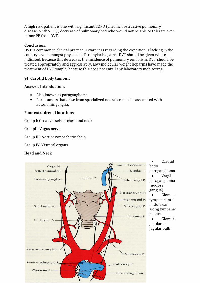
9) Carotid body tumour.
Answer. Introduction:
Also known as paraganglioma
Rare tumors that arise from specialized neural crest cells associated with
autonomic ganglia.
Four extradrenal locations
Group I: Great vessels of chest and neck
GroupII: Vagus nerve
Group III: Aorticosympathetic chain
Group IV: Visceral organs
Head and Neck
Carotid
body
paraganglioma
Vagal
paraganglioma
(nodose
ganglia)
Glomus
tympanicum -
middle ear
along tympanic
plexus
Glomus
jugulare -
jugular bulb
Aetiopathology:
1/30,000 head & neck tumors are paragangliomas
2-3% head/neck paragangliomas have functional hormone secretion
Usually benign- 6% CBTs reported to be malignant
Familial form (10-25%) – present younger and with multiple tumors
Presentation:
Average age = 45
Slow growing
Asymptomatic or mass-related effects
10% present with CN palsy
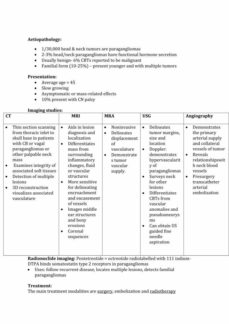
Imaging studies:
CT MRI
MRA USG Angiography
Thin section scanning
from thoracic inlet to
skull base in patients
with CB or vagal
paragangliomas or
other palpable neck
mass
Examines integrity of
associated soft tissues
Detection of multiple
lesions
3D reconstruction
visualizes associated
vasculature
Aids in lesion
diagnosis and
localization
Differentiates
mass from
surrounding
inflammatory
changes, fluid
or vascular
structures
More sensitive
for delineating
encroachment
and encasement
of vessels
Images middle
ear structures
and bony
erosions
Coronal
sequences
Noninvasive
Delineates
displacement
of
vasculature
Demonstrate
s tumor
vascular
supply.
Delineates
tumor margins,
size and
location
Doppler:
demonstrates
hypervascularit
y of
paragangliomas
Surveys neck
for other
lesions
Differentiates
CBTs from
vascular
anomalies and
pseudoaneurys
ms
Can obtain US
guided fine
needle
aspiration
Demonstrates
the primary
arterial supply
and collateral
vessels of tumor
Reveals
relationshipswit
h neck blood
vessels
Presurgery
transcatheter
arterial
embolization
Radionuclide imaging: Pentetreotide = octreotide radiolabelled with 111 indium-
DTPA binds somatostatin type 2 receptors in paragangliomas
Uses: follow recurrent disease, locates multiple lesions, detects familial
paragangliomas
Treatment:
The main treatment modalities are surgery, embolization and radiotherapy
10) Varicose vein.
Answer.
Definition
Varicose veins are defined as superficial veins, which permanently have lost its valvular
efficiency, and as a product of the resultant venous hypertension in the standing
position become dilated, tortuous and thickened.
Primary varicose veins
1 (95%) are caused by an increase in venous pressure in the superficial veins
of the leg due to damage to the venous valves between the deep and superficial venous
systems.
This increase may be at:
1. the sapheno-femoral junction between the long saphenous vein and the common
femoral vein in the groin
2. the sapheno-popliteal junction between the short saphenous vein and the popliteal
vein in the popliteal fossa
3. Other sites (when they are known as perforators).
Secondary varicose veins (5%) occur when the increased venous pressure in the
superficial venous system is due to a disturbance in venous blood flow elsewhere, for
example in:
1. Pelvic thrombosis
2. Extensive thrombosis of the veins in the leg (post phlebitis limb).
3. Arterioveonus malformations (congenital or acquired as a result of a fracture).
Secondary varicose veins are invariably associated with venous hypertension in deep
venoussystem with secondary involvement of superficial venous system.
Anatomy
One of the pitfalls in venous surgery lies in inadequate knowledge of the venous
physiology and anatomy. In contrast to the anatomy of the arteries, the venous anatomy
is characterized by numerous variations, which have a certain impact on the diagnosis
and surgery of varicose veins and chronic venous insufficiency
Some anatomical points of surgical importance:
The lower limb is drained by two sets of veins: superficial and deep. Perforator veins
connect these two systems.
The dorsal metatarsal veins collect blood from the digital veins of the foot and empty
into the dorsal venous arch which continues into the lesser and greater saphenous
veins on the lateral and medial sides of the foot respectively.
The greater and lesser saphenous veins are freely interconnected.
At the saphenopopliteal junction (SPJ), short saphenous vein commonly gives off an
upward
extension, called the Giacomini vein, which may run deep and parallel to the
profunda femoris vein, or superficially, curving round to join the lesser saphenous
vein via its posteromedial branch in the upper thigh.
The fibular branch of the LSV posterior arch vein (Leonardo"s vein) has connections
with the posterior fibular vein via Cockett´s perforating veins. Neglected
insufficiency in the posterior arch vein is quite often the cause of recurrence or even
venous ulcer in the lower leg.
There is marked variation in the anatomy of the superficial venous system involving
the origin, course, size, duplication and depth of truncal and tributary veins (TV), the
number and topography of perforating veins, the valvular distribution and the
arrangement of communicating veins.
From a surgical point of view, the most important variations occur at the venous
junctions, the saphenofemoral junction (SFJ) in the groin and the saphenopopliteal
junction (SPJ) in the popliteal fossa. The anatomical arrangement of the individual
tributaries at the SFJ, namely the superficial epigastric, iliac circumflex and external
pudendal veins as well as the lateral or medial accessory saphenous veins can be
very different from one leg to another.
The anatomy of the SSV is even more complicated, because not only the tributaries
may vary, but also the location of the saphenopopliteal junction. In only 50 to 70 %
of the cases is the saphenopopliteal junction located in the popliteal fossa, whereas
in about 10 % it is found below it. In the remaining 30% to 40 %, the SSV terminates
clearly above the popliteal fossa, with or without connection with the popliteal vein.
There are numerous perforating veins present on both sides of the leg and the thigh
which connect the superficial to the deep venous system, either directly to the main
axial veins (direct perforators) or indirectly to muscular tributaries or soleal venous
sinuses (indirect perforators). In the mid and distal calf the most important direct
medial perforators do notoriginate directly from the great saphenous vein (GSV).
The most significant calf perforators, termed the Cockett perforators, connect the
posterior arch vein to the paired posterior tibial veins. The next group of clinically
relevant perforating veins is the paratibial perforators, which connect the GSV and
its tributaries to the posterior tibial and popliteal veins. There are three additional
direct perforating veins that connect the GSV to the popliteal and femoral veins.
Boyd s perforator, just distal to the knee, connects the GS to the popliteal vein. Dodd s and Hunterian perforators are located in the thigh and connect the GSV to
the proximal popliteal or the femoral veins. In the distal calf, the small saphenous
vein is connected by direct perforators to the peroneal veins Bassi s perforators).
The indirect perforators connect tributaries of the small saphenous vein to either
the muscular venous sinuses of the gastrocnemius or soleus veins before entering
the deep axial system. Position of these perforators is highly variable from person to
person.
This is important to be stressed that incompetence in few perforators in the initial
part of disease will ultimately lead to incompetence in rest of the perforators. So
perforator surgery has to be radical in nature and isolated perforator ligation will
lead to nothing but recurrence.
There are perforators present in foot also which become important channels of
venous drainage in deep venous incompetence grade III and IV if superficial vein
surgery is taken.
Epidemiology
Venous disease, including varicose veins and chronic venous insufficiency (CVI), is one
of the most commonly reported chronic medical conditions and a substantial source of
morbidity in world.
Venous Hemodynamics and Pathophysiology
The Muscle Pump: Venous return against gravity is primarily dependent upon
muscle pumps located within the foot and calf. Muscular contraction (systole) within
facial compartments directs venous blood from sinusoidal intramuscular veins into
the deep stem veins and thence up the leg and thigh towards right atrium. Reverse
flow (reflux) in deep venous system during muscle relaxation (diastole) is prevented
by the closure of valves. Superficial veins collect blood from the superficial tissues,
and during diastole this blood enters the deep system via the perforating veins along
a pressure gradient. During systole, blood is prevented from re-entering the
superficial system through the closure of the perforating veins.
Ambulatory Venous Pressure: When standing motionless, with venous valves in the
neutral position, the pressure in the veins of the foot gradually increases until it equals
the hydrostatic pressure developed by the column of blood stretching between the foot
and the heart; in a person of average height, perhaps 90 mmHg. With active movement,
the muscle pumps and valves come into play and the venous column is divided into
several smaller columns, each at a lower pressure.
As a result, the pressure in the foot veins falls in health to less than 25 mmHg upon
walking – the ambulatory venous pressure (AVP). Patients with muscle pump and/or
venous valve failure, and/or venous outflow obstruction, demonstrate a raised AVP.
Such sustained venous hypertension is the main factor contributing to the development
of skin changes.
Venous Recirculation: In patients with varicose veins there is often a recirculation of
venous blood within the leg. During calf relaxation, abnormally large volumes of blood
enter the muscle pump from the superficial varices (increased preload). The muscle
pump expels blood from the leg only for it to re-enter the leg by refluxing down
superficial varices (akin to aortic regurgitation). This blood then re-enters the muscle
pump through perforating veins in the lower calf and so on.
By far the most powerful force propelling venous return flow is the musculovenous
pumping mechanism, which can handle large volumes rapidly and generate a force well
in excess of that required for venous return against gravity.8 When the limb is in the
dependent position, a normal set of valves in the deep and superficial veins will prevent
reflux of blood against the normal direction of venous flow.
Failure of competence in the venous valves at saphenofemoral and saphenopopliteal
junction will lead to retrograde flow down the limb when the patient stands up or after
exercise movement has resulted in slack veins in the lower part of the leg. In the
superficial veins, this is the basis of the most common venous disorder – simple
varicose veins.
Venous insufficiency is a condition of inadequate venous return and hypertension when
the patient is in an upright position. An increase in venous pressure results in a
corresponding increase in capillary pressure and characteristic changes in the skin and
subcutaneous tissue. Capillary transudation with protein molecules leads to deposition
of fibrin, which forms a barrier to nutritional exchange between the capillaries and the
surrounding tissue. Leukocytes are trapped in the capillaries causing further damage to
the endothelium and the vessel walls and slowing down microvascular circulation.
Extravasated hemosiderin gives the characteristic brown skin pigmentation. The
outflow of fluid and corpuscles from the capillaries into the interstitial tissue
initiates some of the mechanisms leading to symptoms of CVI.9 Swelling, venous eczema
anddermatitis, lipodermatosclerosis, pigmentation and finally venous ulcer take many
months, or even years, to develop. Sensory neuropathy is another feature of severe
chronic venous insufficiency, and its distribution is coincident with trophic changes.
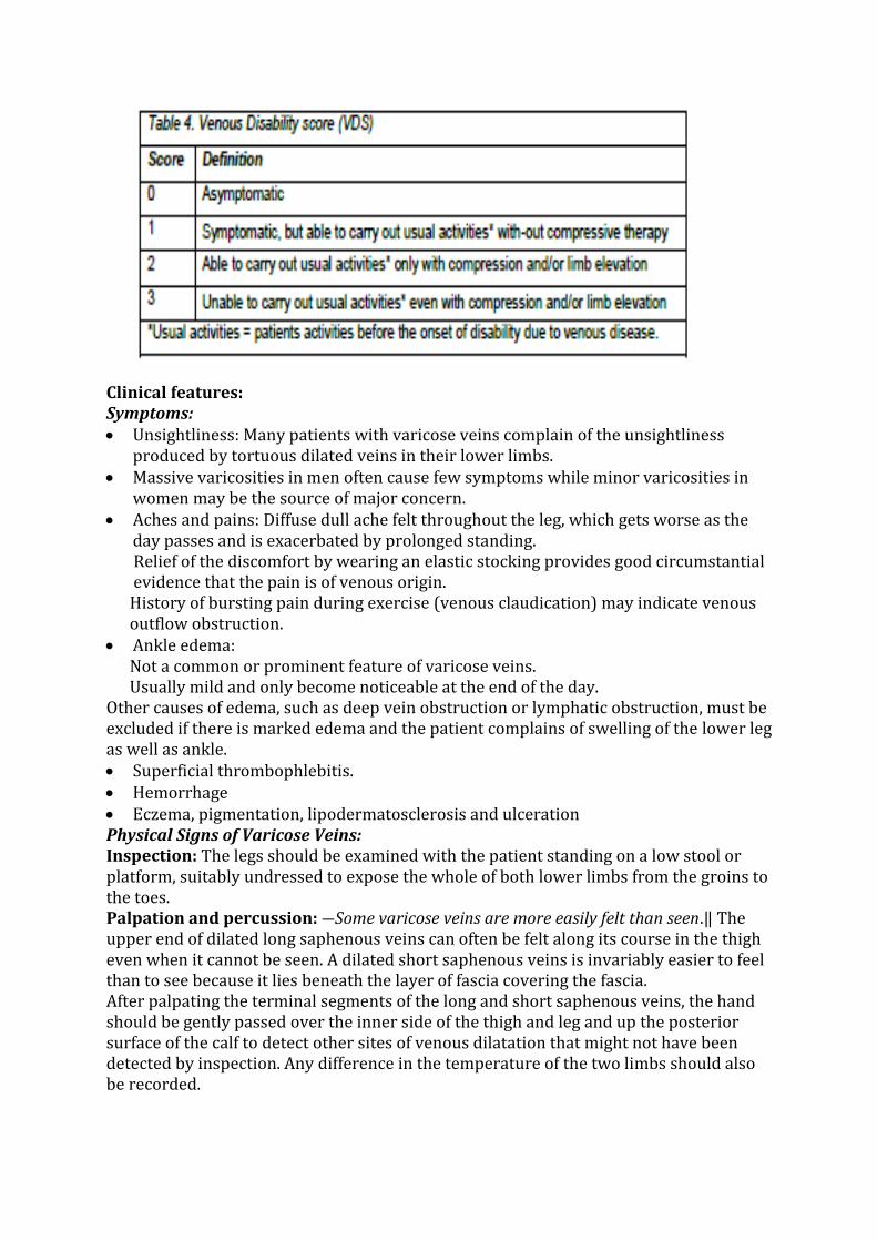
Classification of Chronic Venous Disease
An international Ad Hoc Committee of the American Venous Forum produced a
consensus document for the classification and grading of chronic venous disease, the
CEAP classification, which was formally endorsed by the American Venous Forum and
by the Joint Council of the Society for Vascular Surgery and the North American-
International Society for Cardiovascular Surgery (Table 1), limbs with chronic venous
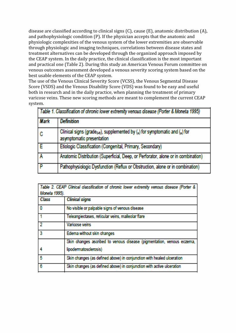
disease are classified according to clinical signs (C), cause (E), anatomic distribution (A),
and pathophysiologic condition (P). If the physician accepts that the anatomic and
physiologic complexities of the venous system of the lower extremities are observable
through physiologic and imaging techniques, correlations between disease states and
treatment alternatives can be developed through the organized approach imposed by
the CEAP system. In the daily practice, the clinical classification is the most important
and practical one (Table 2). During this study an American Venous Forum committee on
venous outcomes assessment developed a venous severity scoring system based on the
best usable elements of the CEAP system.
The use of the Venous Clinical Severity Score (VCSS), the Venous Segmental Disease
Score (VSDS) and the Venous Disability Score (VDS) was found to be easy and useful
both in research and in the daily practice, when planning the treatment of primary
varicose veins. These new scoring methods are meant to complement the current CEAP
system.
Clinical features:
Symptoms:
Unsightliness: Many patients with varicose veins complain of the unsightliness
produced by tortuous dilated veins in their lower limbs.
Massive varicosities in men often cause few symptoms while minor varicosities in
women may be the source of major concern.
Aches and pains: Diffuse dull ache felt throughout the leg, which gets worse as the
day passes and is exacerbated by prolonged standing.
Relief of the discomfort by wearing an elastic stocking provides good circumstantial
evidence that the pain is of venous origin.
History of bursting pain during exercise (venous claudication) may indicate venous
outflow obstruction.
Ankle edema:
Not a common or prominent feature of varicose veins.
Usually mild and only become noticeable at the end of the day.
Other causes of edema, such as deep vein obstruction or lymphatic obstruction, must be
excluded if there is marked edema and the patient complains of swelling of the lower leg
as well as ankle.
Superficial thrombophlebitis.
Hemorrhage
Eczema, pigmentation, lipodermatosclerosis and ulceration
Physical Signs of Varicose Veins:
Inspection: The legs should be examined with the patient standing on a low stool or
platform, suitably undressed to expose the whole of both lower limbs from the groins to
the toes.
Palpation and percussion: ―Some varicose veins are more easily felt than seen.‖ The upper end of dilated long saphenous veins can often be felt along its course in the thigh
even when it cannot be seen. A dilated short saphenous veins is invariably easier to feel
than to see because it lies beneath the layer of fascia covering the fascia.
After palpating the terminal segments of the long and short saphenous veins, the hand
should be gently passed over the inner side of the thigh and leg and up the posterior
surface of the calf to detect other sites of venous dilatation that might not have been
detected by inspection. Any difference in the temperature of the two limbs should also
be recorded.
The cough impulse test Morrissey‘s test : A visible or palpable venous expansion that
occurs on coughing indicates the absence of competent valves between the right atrium
and the veinunder examination. When this sign is detected in the groin over a large
saphena varix, it indicates long saphenous incompetence and may be accompanied by a
palpable thrill, indicating turbulent retrograde flow.
The tourniquet test (the Brodie-trendelenburg test): This simple bedside was
designed to assess the direction of blood flow and the source of refilling of the
superficial veins. The patient must be laid flat on the couch and the limb is elevated to at
least 45 degree to empty all the subcutaneous veins. When the veins have been emptied,
a narrow rubber tourniquet is applied around the thigh as close to the groin as possible.
It must be applied tightly to prevent all superficial vein reflux. Saphenofemoral
incompetence is indicated if the varices below the tourniquet remain collapsed for
between 15 and 30 seconds after the patient stands up, and rapidly refill when the
tourniquet is removed.
Multiple tourniquet test: Three tourniquets are tied after emptying the leg veins. First
tourniquet is tied just below the sapheno-femoral junction, second tourniquet is tied
just above knee and third tourniquet is tied just below knee. This helps in dividing
greater saphenous vein in multiple segments. Perforator incompetence in a particular
segment will lead to increase in size in veins in that particular segment. This can be
made more prominent by releasing tourniquet from below upwards.
Perthe‘s test / Perthe‘s maneuver: The Perthes maneuver is a traditional technique
intended to distinguish antegrade flow from retrograde flow in superficial varices.
Antegrade flow in a variceal system indicates that the system is a bypass pathway
around deep venous obstruction. This is critically important because if deep veins are
not patent, superficial varices are an important pathway for venous return and must not
be sclerosed or surgically removed.
To perform the Perthes maneuver, the affected lower extremity is wrapped with elastic
bandage. With the elastic bandage on, the patient is instructed to move around exercise.
Increase in the size of varices indicates incompetence of deep venous system. Severe
crampy pain is complained of if there is deep venous obstruction
Modified Perthe‘s test: A modification of Perthes' test in which a tourniquet is applied
round the upper part of the thigh after observing the veins. The patient is asked to walk
quickly with the touniquet in place. If the size of varicose veins decrease, the deep veins
are patent and competent. If they increase in size, the deep veins are incompetent.
Severe crampy pain is complained of if there is deep venous obstruction
Investigations:
Doppler examination: Confirmation of saphenofemoral or saphenopopliteal reflux
using a simple hand-held 8 MHz Doppler (HHD), also known as continuous wave (CW)
Doppler is increasingly replacing the use of tourniquet tests in the clinic. The
examination begins with the patient standing.
The probe is placed over the SFJ, which is found by insonating the femoral artery and
moving medially. Squeezing the calf will result in a prograde signal. In the presence of
SFJ incompetence, release of calf compression will result in a retrograde signal (greater
than 0.50 seconds) that is abolished by long saphenous vein (LSV) compression. The
directional Doppler ultrasound flow detector may also be used to detect sapheno
popliteal reflux down the short saphenous vein, and it has been used to determine the
exact site of the sapheno-popliteal junction which may vary considerably in position15.
Short saphenous incompetence is confirmed if retrograde flow after calf squeezing is
abolished by digital or tourniquet compression which occludes the upper end of the
short saphenous vein.
Duplex ultrasonography: Duplex ultrasound is capable of imaging the superficial and
deep veins of the leg and the communication between these two systems and is now
accepted as the best method of investigating cases of saphenous reflux and perforating
vein incompetence. It has also been suggested that before deciding upon treatment, all
patients with varicose veins should have a full Duplex examination. This represents a
considerable advance over the simple hand-held Doppler flow detector because it is
often very difficult to know exactly from which vessel the reflected ultrasound is
coming. This is not only very valuable in the groin and popliteal fossa, where the deep
and superficial systems meet, but especially helpful in the calf where duplex ultrasound
can be used to assess incompetence of the perforating veins and localize their position.
Duplex ultrasound has a good specificity but needs further assessment against a reliable
gold standard . Ascending phlebography is now no longer used to assess the size and incompetence of
calf communicating veins because, although its specificity is good, its sensitivity is poor.
Ascending Phlebography: Until recently, ascending phlebography has been the method
of choice to demonstrate patency and define the anatomy of veins. A second role has
been to detect incompetent perforating veins. It is still used as the ―gold standard‖ to establish the accuracy of new investigations that determine the presence or absence of
disease or its anatomic extent.
However, the development of several noninvasive tests, particularly duplex scanning,
now makes it unnecessary in most cases. Its current application is limited to cases in
which duplex scanning is unavailable, inadequate, or equivocal. Although phlebography
has been deemed the gold standard in the detection of the presence, site, and anatomic
extent of chronic venous obstruction, it cannot provide a quantitative functional
assessment of its severity or the adequacy of collateral veins.
Descending Phlebography: The aim of descending phlebography is to demonstrate
reflux in either the superficial or deep veins and to determine the points of leakage from
the pelvis to the lower limbs and from deep to superficial veins. It is also used to
provide information on the anatomic localization and morphology of the venous valves,
assess the extent of reflux, and delineate the venous anatomy in complex cases.
Descending phlebography is performed by puncturing femoral vein and injecting
contrast medium to assess retrograde venous flow. The lower limit of contrast
medium reflux is observed fluoroscopically and images taken of this. It can also assess
saphenofemoral incompetence.
Any reflux into this vessel is abnormal.
Five grades of reflux (0 to 4) have been described as follows:
Grade 0, indicates no reflux below the confluence of the superficial femoral and
profunda femoris veins;
Grade 1 – reflux down to the first valve below the site of injection
Grade 2 - reflux down to the upper third of the thigh
Grade 3 – reflux down to, but not below the knee joint
Grade 4 – reflux below the knee joint
Varicography: Varicography involves the direct injection of contrast medium through a
butterfly cannula into the superficial vein under investigation. It has a particularly
valuable clinical role in the elucidation of the anatomic connections of recurrent or
residual varicose veins as a ―road map to guide the surgeon. On the operating table, it
facilitates the use of minimal incisions and precise surgery. It is also used to define
abnormal drainage patterns in patients with venous malformations.
Varicograms show the superficial connections, the perforating veins, the tortuosity, the
dilatation and the extent if the varicose veins but they do not give any information about
valve function or venous reflux which must be assessed separately by duplex scanning
or descending phlebography.
Magnetic resonance venography: Magnetic resonance venography is safe, does not
involve ionizing radiation as does phlebography and is not operator dependent like
duplex ultrasound. At present Magnetic resonance venography is in its infancy but its
role will undoubtedly increase in future. The efficacy of Magnetic resonance venography
in deep vein thrombosis have been assessed against duplex ultrasound, contrast
phlebography or both using a variety of MR techniques and it has been shown that in
many cases MR can be as effective as or , on occasion, superior to these other imaging
studies. But, it is expensive and not available at all places.
Treatment
Compression therapy: Graduated compression hosiery is often the first line of
treatment in the management of varicose veins. Graduated compression leads to
multiple effects on the venous system in the leg, including decrease in edema, increase
in venous velocity, decrease in venous volume and decrease in venous return. This form
of therapy is relatively inexpensive, essentially risk free, and can be effective in
improving symptoms related to superficial venous reflux in and varicose veins.
Compression therapy is main modality of treatment in post phlebitic limb and grade
III and IV deep venous reflux. Compression therapy may relieve symptoms, conceal
veins and prevent deterioration of the skin changes associated with venous
hypertension.
Surgical treatment of varicose veins:
Indications for Treatment:
o When recognizable changes appear in the skin of the lower leg, i.e. presence of ankle
flare, lipodermatosclerosis or venous ulcer.
o If there are problems with hemorrhage or recurrent superficial thrombophlebitis.
o If the patient wishes to be treated for symptomatic or cosmetic reasons.
o Those who are medically unfit because of presence of varicose veins.
Objectives of Treatment:
Ablation of the hydrostatic forces of axial reflux i.e. disconnection of saphenofemoral
and saphenopopliteal junction and stripping of greater and lesser vein.
Removal the hydrodynamic forces of perforator vein reflux
Options available for surgical treatment of varicose veins are as follows:
o Ablation of saphenous vein reflux: greater or smaller
o Incompetent perforators interruptions
o Elimination of residual varicosities
Ablation of Saphenous Vein Reflux: Greater or Smaller
A. Saphenofemoral Junction Ligation: Saphenofemoral junction ligation alone, sometimes referred to as a ―Trendelenburg s procedure, is associated with a high rate of recurrence of varices. Recent research has shown that it is necessary to remove the
saphenous vein to ensure that as much venous reflux as possible is eliminated.
A few sound principles:
1. In a patient of normal build the SFJ lies directly beneath the groin crease; in the obese
it lies above. An incision made below the crease is likely to be too low.
2. Do not divide any vein until the SFJ has been unequivocally identified.
3. Beware of the superficial external pudendal artery that usually passes between GSV
and CFV but passes superficial to the GSV in 5% of cases.
4. Follow and divide all tributaries (Superficial circumflex iliac, superficial inferior
epigastric, superficial external pudendal) beyond secondary branch points. Failure to
do so leaves a network of superficial veins connecting the veins of the thigh with those
of the perineum, the lower abdominal wall and the iliac region. These cross groin
connections are a frequent cause of recurrence.
5. Ligate the GSV deep to all tributaries flush with the CFV.
6.Divide the deep external pudendal vein as it comes off the CFV
7. Retract the lower margin of the wound to identify and ligate the posteromedial thigh
branch that often joints the GSV high in the thigh. Failure to do so increases the risk
of haematoma formation after stripping above the bandage, as well as medial thigh
recurrence. A high anterolateral branch should be dealt with similarly.
B. Stripping: Several randomized trials have clearly shown that routinely stripping the
LSV reduces the risk of recurrence developing through the Hunterian perforating veins
and to remove a vein in the thigh which is difficult to treat later by sclerotherapy21
Stripping markedly reduces the risk of recurrence by:
1. Disconnecting the thigh perforators and saphenous tributaries
2. Preventing any neovascularisation arising from the saphenous stump
reconnecting with the GSV.
Perhaps the most common problem with conventional stripping of the GSV has been
that of saphenous nerve damage. Stripping the vein either to or from the ankle has long
beenrecognized as carrying a significant risk of this unpleasant complication.
Alternatives to stripping: New venous surgical techniques have been developed in an
effort to reduce the number and size of lower-extremity incisions and hematomas, to
eliminate postoperative discoloration, and to reduce the recuperation time.
Radio frequency (RF) ablation: The intervention employs radiofrequency (RF) energy
mediated heating of the vein wall to destroy the intima and denature collagen in the
media with resulting fibrous occlusion of the vein. The mechanics of the surgical
procedure are relatively straight forward with a few caveats. The treated vein should be
relatively straight, free of severe tortuosity or thrombus and without aneurysm.
Contraindications include a post phlebitic vein that cannot be accessed, a mega
saphenous vein (>12 mm), and significant dilation of the proximal saphenous vein with
an aneurismal SFJ.
Endovenous laser therapy: Endovenous laser therapy (EVLT) is similar to RF ablation,
but laser energy is used for ablation of the saphenous vein.
Foam Sclerotherapy: An increasing number of authors have recently reported
successful injection of incompetent GSV with 3% polidocanol in the form of foam.
C. Saphenopopliteal Ligation: Some surgeons advocate routine stripping of the short
saphenous vein should be disconnected and never stripped. The short saphenous vein
operation should be carried out first, if a long saphenous vein operation is to be
performed under the same anaesthetic.
Failure to mark the SPJ preoperatively will lead to a misplaced incision in a significant
number of cases that will necessitate further blind incisions or abandonment of the
procedure. Clinical examination and hand held Doppler are not reliable.
D. Ligation of the Lower Leg Perforating Veins: Surgery for these veins is usually
required in patients with lipodermatosclerosis or ulceration. The presence of
incompetent perforators in patients with advanced CVI (clinical classes 4 to 6) is an
indication for surgical treatment in a fit patient. Whereas open perforator ligation is
done only in those with healed ulceration, a clean, granulating open ulcer is not a
contraindication for subfascial endoscopic perforator vein surgery (SEPS).
Subfascial ligation of the medial communicating veins Linton‘s operation : In view of considerable wound complications associated with Linton s radical operation of subfascial ligation, which included long medial, anterolateral, and posterolateral calf
incisions, it was soon abandoned.
Extrafascial ligation of perforators Cockett‘s procedure : This operation is not
commonly employed today. The aim of surgery is to clear all the extrafascial enlarged
veins and to divide perforating veins.
Posterior approach (Robs procedure): This is done if the perforators on the lateral
side are also to be ligated. The incision is a posterior subfascial one and the perforators
on both the sides are ligated and divided. This procedure offers advantage in the fact
that the incision is away from the areas of ulceration and thus results in good healing.
Subfascial endoscopic perforator surgery (SEPS): The major drawback of open
procedure was a high incidence of wound complications.
Complications:
Major venous damage: Deep veins can be damaged during varicose veins surgery
through attempts to control bleeding and misidentification of anatomy. Complete
division of the common femoral vein is estimated to occur once in every 10,000
varicose veins operations.
Arterial damage.
Nerve damage. Popliteal dissection, stripping and distal avulsions may result in
damage to the divisions of the sciatic nerve (usually the common peroneal nerve),
saphenous and sural nerve.
Haematoma. This is the commonest cause of discomfort after varicose veins and can
be minimized by operating the patient in the head-down position, careful
hemostasis, and evacuation of all clots from the stripper tunnel and use of a
tourniquet.
Venous thromboembolism.
Necrosis of the wound edges: this is the most common and troublesome
complication of both the subfascial and extrafascial operations. It appears to occur
more frequently after the extrafascial operation
E. Elimination of Residual Varicosities
Sclerotherapy: The aim of injection sclerotherapy is to place a small volume of
sclerosant in the lumen of a vein empty of blood, and then appose the walls of that
vein with appropriate compression. The vein fibroses and gets closed without the
formation of clot. The sclerosant must remain localized within the segment of vein to
be treated. The vein must be kept empty of blood both during and after the injection.
Patients should be mobilized immediately afterwards and be encouraged to walk on a
daily basis. This measure allows symptoms and signs of allergic reactions to appear
and be treated. The comfort of elastic compression can be evaluated, and the deep
venous circulation is stimulated and any sclerosant that has entered from the
superficial injection is flushed. Immobility is a relative contraindication to
sclerotherapy.
Indications of sclerotherapy:
1. Telangiectasia
2. Reticular varicosities and reticular veins
3. Isolated varicosities
4. Below knee varicosities
5. Recurrent varicosities
Contraindications:
1. Presence of arterial occlusive disease
2. Patient immobility
3. Hypersensitivity to the drug
4. Acute thrombophlebitis
5. Huge varicosities with large communications to deep veins
Complications: The complications of injection sclerotherapy include:
1. Anaphylaxis.
2. Allergic reactions. Typically symptoms include urticaria, peri-orbital and oral
swelling, bronchospasm and migraine.
3. Ulceration. Ulceration follows extravascular injection. Commonly it is due to arterial
occlusion caused by sclerosant reaching a terminal arteriole. Another cause is reactive
vasospasm because of a large volume of injection.
Treatment is symptomatic. Unless the ulcer is obviously infected rare antibiotics have
no role.
4. Arterial injection. This is a serious complication that is accompanied by severe pain
distal to the injection site. The most vulnerable artery appears to be the posterior tibial
artery at the ankle. Treatment includes analgesia, cooling of the foot, and infusion of
heparin and dextran.
5. Pigmentation. Pigmentation is due to the deposition of haemosiderin, often following
superficial thrombophlebitis. Most commonly seen in those treated with sodium
tetradecyl sulphate and hypertonic saline and least common with polidocanol.
6. Superficial thrombophlebitis. This occurs when clot remains in the lumen of the
sclerosed vein and is largely due to inadequate compression. Localised haematoma is
particularly painful and may be eased by aspiration with a needle or scalpel under local
anaesthesia.
7. Deep venous thrombosis. The risk is reduced by careful patient selection and by
advising patients to walk immediately after injection treatment and thereafter on a
regular basis each day.
8. Nerve damage. Can occur due to approximate injection and/or pressure from
bandaging.
9. Telangiectatic matting: or neoangiogenesis is the new appearance of red
telangiectasias in a site of prior sclerotherapy. It is believed to be a complex process in
which new vessels grow in response to endothelial growth factors or platelet-derived
growth factors. Prevention is best achieved through use of dilute solutions and in small
volume.
11) Venous ulcer.
Answer. Introduction:
Chronic venous disease, including chronic venous insufficiency and chronic venous
ulceration, is a common and important medical problem that causes significant
morbidity. Venous ulcers are expensive to treat and adversely impact patient s quality of life.
Venous ulcers occur more commonly in the elderly, the peak prevalence occurring
between ages 60 and 80 years.
A venous leg ulcer can develop after a minor injury if there is a problem with
the circulation of blood in your leg veins. If this happens, the pressure inside
the veins increases.
Risk factors:
Obesity – this increases the risk of high pressure in the leg veins
Not being able to move for a long period of time – this can weaken the calf
muscles, which can affect circulation in the leg veins
Having previously had deep vein thrombosis (DVT)– blood clots that develop in
the leg, which can damage valves
Varicose veins – swollen and enlarged veins caused by malfunctioning valves
Previous injury to the leg, such as a broken or fractured bone, which may cause
DVT
Previous surgery to the leg, such as a hip replacement or knee replacement, which
can prevent you from moving about
Increasing age – as people generally find it harder to move about as they get older
Pathophysiology: (go through):
Venous hypertension: Deep vein thrombosis, perforator insufficiency,
superficial and deep vein insufficiencies, arteriovenous fistulas and calf
muscle pump insufficiencies lead to increased pressure in the distal veins of
the leg and finally venous hypertension.
Fibrin cuff theory: Fibrin gets excessively deposited around capillary beds
leading to elevated intravascular pressure. This causes enlargement of
endothelial pores resulting in further increased fibrinogen deposition in the
interstitium. The "fibrin cuff" which surrounds the capillaries in the dermis
decreases oxygen permeability 20-fold. This permeability barrier inhibits
diffusion of oxygen and other nutrients, leading to tissue hypoxia causing
impaired wound healing.
Inflammatory trap theory: Various growth factors and inflammatory cells,
which get trapped in the fibrin cuff promote severe uncontrolled inflammation
in surrounding tissue preventing proper regeneration of wounds. Leukocytes
get trapped in capillaries, releasing proteolytic enzymes and reactive oxygen
metabolites, which cause endothelial damage. These injured capillaries
become increasingly permeable to various macromolecules, accentuating
fibrin deposition. Occlusion by leukocytes also causes local ischemia thereby
increasing tissue hypoxia and reperfusion damage.
Dysregulation of various cytokines.
Dysregulation of various pro-inflammatory cytokines and growth factors like
tumor necrosis factor- TNF- , TGF- and matrix metalloproteinases lead to chronicity of the ulcers.
Miscellaneous: Thrombophilic conditions like factor V Leiden mutation,
prothrombin mutations, deficiency of antithrombin, presence of
antiphospholipid antibodies, protein C and S deficiencies and
hyperhomocysteinemia are also implicated.
Clinical features:
Venous leg ulcers are open, often painful, sores in the skin that take more than
four to six weeks to heal. They most often develop on the inside of the leg, just
above the ankle.
Symptoms:
Swollen ankles (oedema)
Discolouration and darkening of the skin around the ulcer
Hardened skin around the ulcer, which may make your leg feel hard and
resemble the shape of an upside-down champagne bottle
A heavy feeling in your legs
Aching or swelling in your legs
Red, flaky, scaly and itchy skin on your legs (varicose eczema)
Swollen and enlarged veins on your legs (varicose veins)
An unpleasant and foul-smelling discharge from the ulcer
Signs of an infection:
A venous leg ulcer can be susceptible to bacterial infection. Symptoms of an infected leg
ulcer can include:
Worsening pain
A green or unpleasant discharge coming from the ulcer
Redness and swelling of the skin around the ulcer
A high temperature (fever)
Nonsurgical Treatment
A. Infected ulcers
Necessitate treatment of the infection first.
Staphylococcus aureus, Streptococcus pyogenes, and Pseudomonas species are
responsible for most infections.
Usually treated with local wound care, wet-to-dry dressings, and oral antibiotics.
Topical antiseptics should be avoided.
Severe infections require intravenous antibiotics.
B. Leg elevation
Leg elevation can temporarily decrease edema and should be instituted when swelling
occurs. This should be done before a patient is fitted for stockings or boots.
C. Compression therapy
Compression therapy is the primary treatment for CVI.
Elastic compression stockings
o Fitted to provide a compression gradient from 30 to 40 mm Hg, with the greatest
compression at the ankle.
o Donned on arising from bed and removed at bedtime.
o Effective in healing ulcers but can take months to obtain good results.
o Stockings do not correct the abnormal venous hemodynamics and must be worn
after the ulcer has healed to prevent recurrence.
o Principal drawback is patient compliance.
Unna boots
o Paste gauze compression dressings that contain zinc oxide, calamine, and glycerin.
o Used to help prevent further skin breakdown.
o Provide nonelastic compression therapy.
o Changed once or twice a week.
o Healing time for ulcers is less than that of elastic compression alone.
Pneumatic compression devices
o Provide dynamic sequential compression.
o Used primarily in the prevention of deep vein thrombi in hospitalized patients.
o Also used successfully to treat venous insufficiency.
D. Topical medications
Largely ineffective as a stand-alone therapy for venous stasis ulcers.
Topical therapy is directed at absorbing wound drainage and avoiding
desiccation of the wound.
Antiseptics can be counterproductive. Hydrogen peroxide, povidone-iodine,
acetic acid, and sodium hypochlorite are toxic to cultured fibroblasts and should
be used for the shortest duration necessary to control ulcer infection.
Surgical Therapy: Skin grafting.
12) Classify ulcers. Discuss the pathology, clinical features, investigations and
management of venous ulcer.
Answer. Wagner Classification System:
Grade 0: Skin intact, but deformity present. Foot is "at risk".
Grade I: Localized superficial ulcer.
Grade II: Deep ulcer extending to tendon, joint and bone.
Grade III: Ulcer involving infection of bone.
Grade IV: Ulcer involving gangrene of toes or forefoot.
Grade V: Gangrene beyond forefoot.
UTSA Classification System:
A new and more comprehensive system has been developed by podiatrists at the
University of Texas at San Antonio (abbreviated here as "UTSA").
Wounds in this classification system are graded as follows:
First, the patient's medical condition is graded.
Grade A refers to a clean, non-infected wound in a patient with adequate blood
supply.
Grade B refers to patients with infected wounds but have adequate blood supply.
Grade C refers to patients with no infection in the wound, but has poor
circulation.
Grade D refers to patients with an infected wound and poor circulation.
Next, the wound is graded.
Grade 0 refers to pre- or post-ulcerative lesion that is completely healed.
Grade 1 refers to patients with a superficial wound not involving tendon, capsule
or bone.
Grade 2 refers to wounds penetrating to tendon or capsule.
Grade 3 refers to wounds extending to the bone or joint.
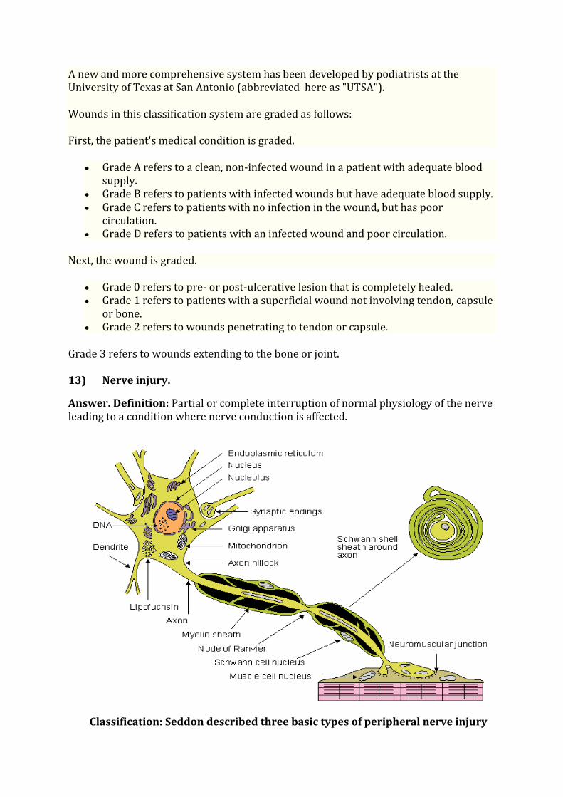
13) Nerve injury.
Answer. Definition: Partial or complete interruption of normal physiology of the nerve
leading to a condition where nerve conduction is affected.
Classification: Seddon described three basic types of peripheral nerve injury
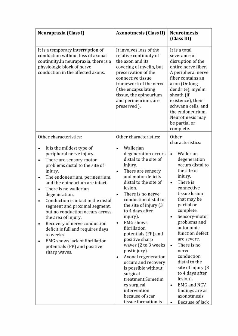
Neurapraxia (Class I)
Axonotmesis (Class II)
Neurotmesis
(Class III)
It is a temporary interruption of
conduction without loss of axonal
continuity.In neurapraxia, there is a
physiologic block of nerve
conduction in the affected axons.
It involves loss of the
relative continuity of
the axon and its
covering of myelin, but
preservation of the
connective tissue
framework of the nerve
( the encapsulating
tissue, the epineurium
and perineurium, are
preserved ).
It is a total
severance or
disruption of the
entire nerve fiber.
A peripheral nerve
fiber contains an
axon (Or long
dendrite), myelin
sheath (if
existence), their
schwann cells, and
the endoneurium.
Neurotmesis may
be partial or
complete.
Other characteristics:
It is the mildest type of
peripheral nerve injury.
There are sensory-motor
problems distal to the site of
injury.
The endoneurium, perineurium,
and the epineurium are intact.
There is no wallerian
degeneration.
Conduction is intact in the distal
segment and proximal segment,
but no conduction occurs across
the area of injury.
Recovery of nerve conduction
deficit is full,and requires days
to weeks.
EMG shows lack of fibrillation
potentials (FP) and positive
sharp waves.
Other characteristics:
Wallerian
degeneration occurs
distal to the site of
injury.
There are sensory
and motor deficits
distal to the site of
lesion.
There is no nerve
conduction distal to
the site of injury (3
to 4 days after
injury).
EMG shows
fibrillation
potentials (FP),and
positive sharp
waves (2 to 3 weeks
postinjury).
Axonal regeneration
occurs and recovery
is possible without
surgical
treatment.Sometim
es surgical
intervention
because of scar
tissue formation is
Other
characteristics:
Wallerian
degeneration
occurs distal to
the site of
injury.
There is
connective
tissue lesion
that may be
partial or
complete.
Sensory-motor
problems and
autonomic
function defect
are severe.
There is no
nerve
conduction
distal to the
site of injury (3
to 4 days after
lesion).
EMG and NCV
findings are as
axonotmesis.
Because of lack
required.
of nerve repair,
surgical
intervention is
necessary.
Sunderland s classification:
First-degree (Class I)
Seddon's neurapraxia and first-degree are the same.
Second-degree (Class II)
Seddon's axonotmesis and second-degree are the same.
Third-degree (Class II)
Sunderland's third-degree is a nerve fiber interruption. In third-degree injury, there is a
lesion of the endoneurium, but the epineurium and perineurium remain intact.
Recovery from a third-degree injury is possible, but surgical intervention may be
required.
Fourth-degree (Class II)
In fourth-degree injury, only the epineurium remains intact. In this case, surgical repair
is required.
Fifth-degree (Class III)
Fifth-degree lesion is a complete transection of the nerve. Recovery is not possible
without an appropriate surgical treatment.
Aetiology:
• Acute:
• Chronic:
o Fracture
o Wrong posture
o Surgery
o Electrical burn
o Tight nerve passage
o Tumors
Presentation:
• Pain • Loss of motion • Loss of reflexes • Trophic changes
• Loss of
sensation
• Loss of power • Wasting Contractures
Diagnostic aids:
• X-RAY
• EMG
• NCS – Nerve conduction study.
• MRI
Clinical examples:
• Erb s palsy
• Carpal tunnel syndrome (median nv)
• Radial nerve injury
• Ulnar nerve injury
• Sciatic nerve injury
• Lateral popliteal nerve injury
Nerve regeneration therapy:
Electrical stimulation can promote nerve regeneration. The frequency of stimulation is
an important factor in the success of both quality and quantity of axon regeneration as
well as growth of the surrounding myelin and blood vessels that support the axon.
Histological analysis and measurement of regeneration showed that low frequency
stimulation had a more successful outcome than high frequency stimulation on
regeneration of damaged sciatic nerves.
The use of autologous nerve grafting procedures that involve redirection of
regenerative donor nerve fibers into the graft conduit has been successful in restoring
target muscle function. Localized delivery of soluble neurotrophic factors may help
promote the rate of axon regeneration observed within these graft conduits.
14) Ulnar nerve injury:
Answer. Causes:There are many causes of ulnar nerve injuries, including pressure,
trauma and illness. In some cases, ulnar nerve injuries may arise without a known
cause.
The most common cause of ulnar nerve injury is extended pressure on the ulnar
nerve, known as ulnar nerve entrapment. As the ulnar nerve travels from the
shoulder to the hand, it passes through two tunnels of tissue, the cubital tunnel behind the elbow and Guyon s canal in the wrist. Both tunnels are common locations at which the ulnar nerve can be compressed and injured. The ulnar nerve may also
be compressed at the neck or beneath the collarbone.
Entrapment of the ulnar nerve may result from swelling of soft tissue, cysts, or
damage to the bones in the arms. Bone damage causing ulnar nerve injuries include
arthritis, elbow dislocations, elbow and wrist fractures, and bone spurs. Repetitive
motions of the arm and hand, extensive bending of the elbow, and long-term
pressure on the palm of the hand may also cause ulnar nerve injuries.
Ulnar nerve injuries may also be the result of direct trauma to the nerve. Finally, any
whole body illness that is known to cause nerve damage, such as diabetes or
hypothyroidism, can affect the ulnar nerve.
Risk factors:
Activities in which your elbow or wrist is bent or twisted for prolonged periods
Alcohol abuse
Brachial plexus injury (injury to the bundle of nerves that transmit signals from the
spine to the shoulder, arm and hand)
Diabetes chronic disease that affects your body s ability to use sugar for energy)
Elbow and wrist abnormalities
Hypothyroidism (underactive thyroid)
Nerve entrapment or compression, such as of the ulnar nerve in the arm
Rheumatoid arthritis (chronic autoimmune disease characterized by joint
inflammation)
Sleeping positions that put pressure on the ulnar nerve.
Symptoms:
Abnormal sensations in the little finger and part of the ring finger, usually on the palm
side.
Loss of coordination of the fingers
Numbness, decreased sensation
Pain
Tingling, burning sensation
Weakness and clumsiness of the hand
Clinical examinations and Tests:
An examination of the hand and wrist may show:
"Claw-like" deformity (in severe cases)
Difficulty moving the fingers
Wasting of the hand muscles (in severe cases)
Weakness of hand flexing
Tests may be needed, depending on the history, symptoms, and findings from the
physical exam. These tests may include:
Blood tests MRI of the
neck
Nerve conduction
tests
Recording of the
electrical activity in
muscles (EMG)
Imaging scans Nerve
ultrasound
X-rays
Treatment:
The goal of treatment is to allow you to use the hand and arm as much as possible. The
cause should be identified and treated. Sometimes, no treatment is needed and you will
get better on your own.
Medications may include:
Over-the-counter pain relievers or prescription pain medications to control pain
(neuralgia)
Other medications, including gabapentin, phenytoin, carbamazepine, or tricyclic
antidepressants such as amitriptyline or duloxetine, to reduce stabbing pains
Corticosteroids injected into the area to reduce swelling and pressure on the nerve.
A supportive splint at either the wrist or elbow can help prevent further injury and
relieve the symptoms. You may need to wear it all day and night, or only at night. If the
ulnar nerve is injured at the elbow, wearing a pad may help protect the nerve from
further injury. Be careful to avoid leaning on the elbow.
Surgery to relieve pressure on the nerve may help if the symptoms get worse, or if there
is proof that part of the nerve is wasting away.
Other treatments may include:
Physical therapy exercises to help maintain muscle strength
Occupational counseling or occupational therapy for changes you can make at work, or
retraining.
![Veins and Lymphatics - Tagungsmanagement · Veins and Lymphatics 2013; volume 2:e1 [Veins and Lymphatics 2013; 2:e1] [page 1] Stiffness of compression devices Giovanni Mosti Angiology](https://img.pdfslide.net/doc/110x75/5f0ee5c27e708231d44179f9/veins-and-lymphatics-veins-and-lymphatics-2013-volume-2e1-veins-and-lymphatics.jpg)