Embed Size (px)
Citation preview
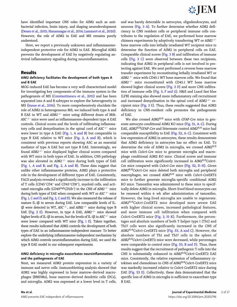
ARTICLE
AIM2 controls microglial inflammation to preventexperimental autoimmune encephalomyelitisChunmei Ma1*, Sheng Li1*, Yingchao Hu1, Yan Ma2, Yuqing Wu1, Chunyan Wu1, Xue Liu1, Bingwei Wang2, Gang Hu2,Jiawei Zhou3,4, and Shuo Yang1
The role of the PYHIN family member absent in melanoma 2 (AIM2), another important inflammasome sensor, in EAE remainsunclear. In this study, we found that AIM2 negatively regulates the pathogenesis of EAE independent of inflammasomeactivation. AIM2 deficiency enhanced microglia activation and infiltration of peripheral immune cells into the CNS, therebypromoting neuroinflammation and demyelination during EAE. Mechanistically, AIM2 negatively regulates the DNA-PK–AKT3 inmicroglia to control neuroinflammation synergistically induced by cGAS and DNA-PK. Administration of a DNA-PK inhibitorreduced the severity of the EAE. Collectively, these findings identify a new role for AIM2 in controlling the onset of EAE.Furthermore, delineation of the underlying inflammasome-independent mechanism highlights cGAS and DNA-PK signaling aspotential targets for the treatment of heterogeneous MS.
IntroductionMultiple sclerosis (MS) is a chronic inflammatory autoimmunedisease of the central nervous system (CNS) and affects ∼2.5million people globally. MS causes the dysfunction in motor,sensory, visual, and autonomic systems of patients, and itsneuropathological features include inflammatory cell infiltra-tion, chronic axonal damage, and demyelination of the CNS(Dendrou et al., 2015). As a multifactorial, heterogeneous dis-ease, MS is difficult to treat. Thus, the etiology and underlyingbasis of this heterogeneous disease remain largely unknown.
Experimental autoimmune encephalomyelitis (EAE) is a com-monly used experimental animal model of MS study and re-capitulates many of neuropathological features of MS (Ransohoff,2012). Myelin oligodendrocyte glycoprotein (MOG)–induced EAEin C57BL/6 mice is often used to explore multiple facets ofthe mechanism surrounding immune-mediated demyelination,especially in transgenic/KO mice (Constantinescu et al., 2011;Procaccini et al., 2015). Front-line clinical treatments for MSincluding IFN-β, glatiramer acetate, anti–VLA-4 integrin mAb,and fumaric acid esters have been developed, tested, and vali-dated in the MOG-induced EAE model (Aharoni et al., 2008;Galligan et al., 2010; Humphries et al., 2020; Mindur et al., 2014).Such models can be induced in different ways through innateimmune activation. Low doses of the adjuvant (mycobacteria[Mtb]) are able to induce a subtype of EAE termed type A, whichis NLRP3 inflammasome dependent. In contrast, high doses of
Mtb or acute virus infection can induce a subtype of EAE termedtype B, which occurs independently of NLRP3 inflammasome(Inoue et al., 2016). Thus, to elucidate the key molecules and un-derlying mechanisms involved in the distinct MOG-EAE subtypesmay provide new insights into developing diagnostic indicatorsand treatments for heterogeneous MS.
NLRP3 is assembled into multimeric complexes withapoptosis-associated speck-like protein containing CARD (ASC)and pro-inflammatory caspases (caspase-1 and -11) known as aninflammasome. Inflammasome assembly leads to caspase au-toactivation and the subsequent cleavage of pro–IL-1β andpro–IL-18 precursors into their mature forms. Inflammasomeactivation also results in an inflammatory form of cell deathknown as pyroptosis (Rathinam et al., 2012). Several indepen-dent studies have reported a critical role for the NLRP3 in-flammasome and its related proteins such as ASC andgasdermin D in the development of type A EAE (Inoue et al.,2012; Li et al., 2019b; Martin et al., 2016). Absent in melanoma2 (AIM2) is a PYHIN (pyrin and HIN domain containing) familymember and an important inflammasome sensor that detectscytosolic double-stranded DNA via its HIN200 domain(Hornung et al., 2009). The AIM2 inflammasome is also es-sential for host defense against bacterial and viral pathogens,such as Francisella tularensis, vaccinia virus, and mouse cyto-megalovirus (Rathinam et al., 2010). In addition, some studies
.............................................................................................................................................................................1Department of Immunology, Key Laboratory of Immunological Environment and Disease, State Key Laboratory of Reproductive Medicine, Center for Global Health, NanjingMedical University, Nanjing, China; 2Department of Pharmacology, Nanjing University of Chinese Medicine, Nanjing, China; 3Institute of Neuroscience, State KeyLaboratory of Neuroscience, Center for Excellence in Brain Science and Intelligence Technology, Shanghai Institutes for Biological Sciences, Chinese Academy of Sciences,Shanghai, China; 4School of Future Technology, University of Chinese Academy of Sciences, Beijing, China.
*C. Ma and S. Li contributed equally to this paper; Correspondence to Shuo Yang: [email protected]; Jiawei Zhou: [email protected]; Gang Hu: [email protected].
© 2021 Ma et al. This article is available under a Creative Commons License (Attribution 4.0 International, as described at https://creativecommons.org/licenses/by/4.0/).
Rockefeller University Press https://doi.org/10.1084/jem.20201796 1 of 17
J. Exp. Med. 2021 Vol. 218 No. 5 e20201796
have identified important CNS roles for AIM2 such as anti-bacterial infection, brain injury, and shaping neurodevelopment(Denes et al., 2015; Hanamsagar et al., 2014; Lammert et al., 2020).However, the role of AIM2 in EAE and MS remains poorlyunderstood.
Here, we report a previously unknown and inflammasome-independent protective role for AIM2 in EAE. Microglial AIM2prevents the development of EAE by negatively regulating an-tiviral inflammatory signaling during neuroinflammation.
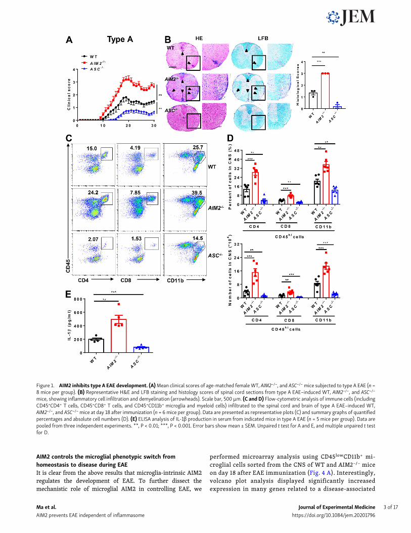
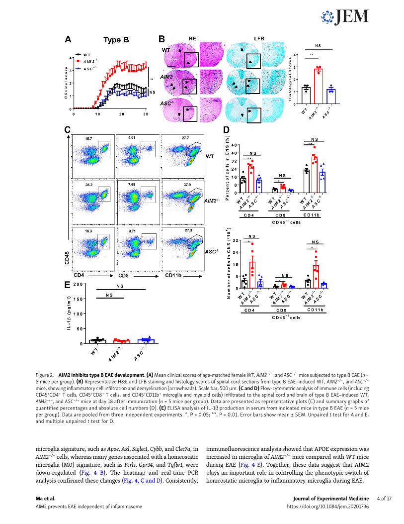
ResultsAIM2 deficiency facilitates the development of both types Aand B EAEMOG-induced EAE has become a very well characterized modelfor investigating key components of the immune system in thepathogenesis of MS (Constantinescu et al., 2011) and has beenseparated into A and B subtypes to explore the heterogeneity inMS (Inoue et al., 2016). To more comprehensively elucidate therole of AIM2 in heterogeneous MS, we induced both type A andB EAE in WT and AIM2−/− mice using different doses of Mtb.ASC−/− mice were used as inflammasome-dependent type A EAEcontrols. Clinical scores and the levels of infiltrating inflamma-tory cells and demyelination in the spinal cord of ASC−/− micewere lower in type A EAE (Fig. 1, A and B) but comparable intype B EAE relative to WT mice (Fig. 2, A and B), which isconsistent with previous reports showing ASC as an essentialmediator of type A EAE but not type B EAE. Interestingly, wefound AIM2−/− mice displayed higher clinical scores comparedwith WT mice in both types of EAE. In addition, CNS pathologywas also elevated in AIM2−/− mice during both types of EAE(Fig. 1, A and B; and Fig. 2, A and B). These data suggest thatunlike other inflammasome proteins, AIM2 plays a protectiverole in the development of different types of EAE. Consistently,FACS analysis revealed a significantly increase in the infiltrationof T cells (CD45+CD4+ and CD45+CD8+), myeloid cells, and acti-vated microglia cells (CD45highCD11b+) in the CNS of AIM2−/− miceduring both types of EAE when compared with WT or ASC−/− mice(Fig. 1, C andD; and Fig. 2, C andD).We alsomeasured the release ofmature IL-1β in serum during EAE. Low comparable levels of IL-1β were detected in WT, ASC−/−, and AIM2−/− mice during type BEAE (Fig. 2 E). However, in type A EAE, AIM2−/− mice showedhigher levels of IL-1β in serum, but the levels of IL-1β in ASC−/−micewere lower compared with WT mice (Fig. 1 E). Taken together,these results indicated that AIM2 controls the development of bothtypes of EAE in an inflammasome-independent manner. To betterexplore the underlying inflammasome-independent mechanism bywhich AIM2 controls neuroinflammation during EAE, we used thetype B EAE model in our subsequent experiments.
AIM2 deficiency in microglia exacerbates neuroinflammationand the pathogenesis of EAENext, we measured AIM2 protein expression in a variety ofimmune and nerve cells. Immunoblotting analysis showed thatAIM2 was highly expressed in bone marrow–derived macro-phages (BMDMs), bone marrow–derived dendritic cells (DCs),and microglia. AIM2 was expressed at a lower level in T cells,
and was barely detectable in astrocytes, oligodendrocytes, andneurons (Fig. 3 A). To further determine whether AIM2 defi-ciency in CNS resident cells or peripheral immune cells con-tributes to the regulation of EAE, we performed bone marrowchimera experiments by adoptively transferring WT or AIM2−/−
bone marrow cells into lethally irradiated WT recipient mice todetermine the function of AIM2 in peripheral cells on EAE.Comparable clinical scores (Fig. 3 B) and infiltration of immunecells (Fig. 3 C) were observed between these two recipients,indicating that AIM2 in peripheral cells is not involved in pro-tecting against EAE. We next performed a reverse bone marrowtransfer experiment by reconstituting lethally irradiated WT orAIM2−/− mice with CD45.1 WT bone marrow cells. We found thatAIM2−/− mice reconstituted with CD45.1 WT bone marrowshowed higher clinical scores (Fig. 3 D) and more CNS infiltra-tion of immune cells (Fig. 3, F and G). H&E and Luxol fast blue(LFB) staining also showed more inflammatory cell recruitmentand increased demyelination in the spinal cord of AIM2−/− re-cipient mice (Fig. 3 E). Thus, these results suggested that AIM2deficiency in CNS-resident cells promotes the pathogenesisof EAE.
We also crossed AIM2fl/fl mice with GFAP-Cre mice to gen-erate astrocyte conditional AIM2 KO mice (Fig. S1, A–C). DuringEAE, AIM2fl/flGFAP-Cre and littermate control AIM2fl/fl mice hadcomparable susceptibility to EAE (Fig. S2, A–C). Consistent withlow expression of AIM2 in astrocytes, these results demonstratethat AIM2 deficiency in astrocytes has no effect on EAE. Todetermine the role of AIM2 in microglia, we crossed AIM2fl/fl
mice with Cx3cr1-Cre mice to generate microglia and macro-phage conditional AIM2 KO mice. Clinical scores and immunecell infiltration were significantly increased in AIM2fl/flCx3cr1-Cre mice compared with Cx3cr1-Cre mice (Fig. S2, D–H). GivenAIM2fl/flCx3cr1-Cre mice deleted both microglia and peripheralmacrophages, we crossed AIM2fl/fl mice with Cx3cr1-CreERT2mice to further generate microglia-specific conditional AIM2KO mice. Tamoxifen was administered to these mice to specif-ically delete AIM2 inmicroglia. Short-lived bloodmonocytes canbe renewed within 6 wk after administration of tamoxifen.However, the long-lived microglia are unable to regenerate.AIM2fl/flCx3cr1-CreERT2 mice developed more severe EAEwith higher clinical scores, increased pathological features,and more immune cell infiltration when compared withCx3cr1-CreERT2 mice (Fig. 3, H–K). Furthermore, the percen-tages and absolute numbers of both T helper type 1 (Th1) andTh17 cells were also significantly increased in the CNS ofAIM2fl/flCx3cr1-CreERT2 mice (Fig. S3, A and C). However, theabsolute numbers of Th1 and Th17 cells in the spleen ofAIM2fl/flCx3cr1-CreERT2 mice were decreased, while percentageswere comparable to control mice (Fig. S3, B and D). Thus, theseresults suggest that the recruitment of pathogenic T cells into theCNS is substantially enhanced in AIM2fl/flCx3cr1-CreERT2 EAEmice. Consistently, the relative expression of inflammatory cy-tokines and chemokines in CNS of AIM2fl/flCx3cr1-CreERT2 micewas markedly increased relative to Cx3cr1-CreERT2 mice duringEAE (Fig. S3 E). Collectively, these data demonstrated that thespecific loss of AIM2 in microglia is sufficient to exacerbate typeB EAE.
Ma et al. Journal of Experimental Medicine 2 of 17
AIM2 prevents EAE independent of inflammasome https://doi.org/10.1084/jem.20201796
AIM2 controls the microglial phenotypic switch fromhomeostasis to disease during EAEIt is clear from the above results that microglia-intrinsic AIM2regulates the development of EAE. To further dissect themechanistic role of microglial AIM2 in controlling EAE, we
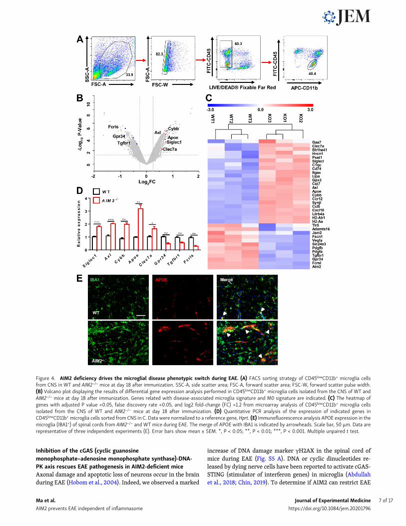
performed microarray analysis using CD45lowCD11b+ mi-croglial cells sorted from the CNS of WT and AIM2−/− miceon day 18 after EAE immunization (Fig. 4 A). Interestingly,volcano plot analysis displayed significantly increasedexpression in many genes related to a disease-associated
Figure 1. AIM2 inhibits type A EAE development. (A)Mean clinical scores of age-matched femaleWT, AIM2−/−, and ASC−/−mice subjected to type A EAE (n =8 mice per group). (B) Representative H&E and LFB staining and histology scores of spinal cord sections from type A EAE–induced WT, AIM2−/−, and ASC−/−
mice, showing inflammatory cell infiltration and demyelination (arrowheads). Scale bar, 500 µm. (C and D) Flow-cytometric analysis of immune cells (includingCD45+CD4+ T cells, CD45+CD8+ T cells, and CD45+CD11b+ microglia and myeloid cells) infiltrated to the spinal cord and brain of type A EAE–induced WT,AIM2−/−, and ASC−/−mice at day 18 after immunization (n = 6 mice per group). Data are presented as representative plots (C) and summary graphs of quantifiedpercentages and absolute cell numbers (D). (E) ELISA analysis of IL-1β production in serum from indicated mice in type A EAE (n = 5 mice per group). Data arepooled from three independent experiments. **, P < 0.01; ***, P < 0.001. Error bars showmean ± SEM. Unpaired t test for A and E, and multiple unpaired t testfor D.
Ma et al. Journal of Experimental Medicine 3 of 17
AIM2 prevents EAE independent of inflammasome https://doi.org/10.1084/jem.20201796
microglia signature, such as Apoe, Axl, Siglec1, Cybb, and Clec7a, inAIM2−/− cells, whereas many genes associated with a homeostaticmicroglia (M0) signature, such as Fcrls, Gpr34, and Tgfbr1, weredown-regulated (Fig. 4 B). The heatmap and real-time PCRanalysis confirmed these changes (Fig. 4, C and D). Consistently,
immunofluorescence analysis showed that APOE expression wasincreased in microglia of AIM2−/− mice compared with WT miceduring EAE (Fig. 4 E). Together, these data suggest that AIM2plays an important role in controlling the phenotypic switch ofhomeostatic microglia to inflammatory microglia during EAE.
Figure 2. AIM2 inhibits type B EAE development. (A)Mean clinical scores of age-matched femaleWT, AIM2−/−, and ASC−/−mice subjected to type B EAE (n =8 mice per group). (B) Representative H&E and LFB staining and histology scores of spinal cord sections from type B EAE–induced WT, AIM2−/−, and ASC−/−
mice, showing inflammatory cell infiltration and demyelination (arrowheads). Scale bar, 500 µm. (C and D) Flow-cytometric analysis of immune cells (includingCD45+CD4+ T cells, CD45+CD8+ T cells, and CD45+CD11b+ microglia and myeloid cells) infiltrated to the spinal cord and brain of type B EAE–induced WT,AIM2−/−, and ASC−/− mice at day 18 after immunization (n = 5 mice per group). Data are presented as representative plots (C) and summary graphs ofquantified percentages and absolute cell numbers (D). (E) ELISA analysis of IL-1β production in serum from indicated mice in type B EAE (n = 5 miceper group). Data are pooled from three independent experiments. *, P < 0.05; **, P < 0.01. Error bars show mean ± SEM. Unpaired t test for A and E,and multiple unpaired t test for D.
Ma et al. Journal of Experimental Medicine 4 of 17
AIM2 prevents EAE independent of inflammasome https://doi.org/10.1084/jem.20201796
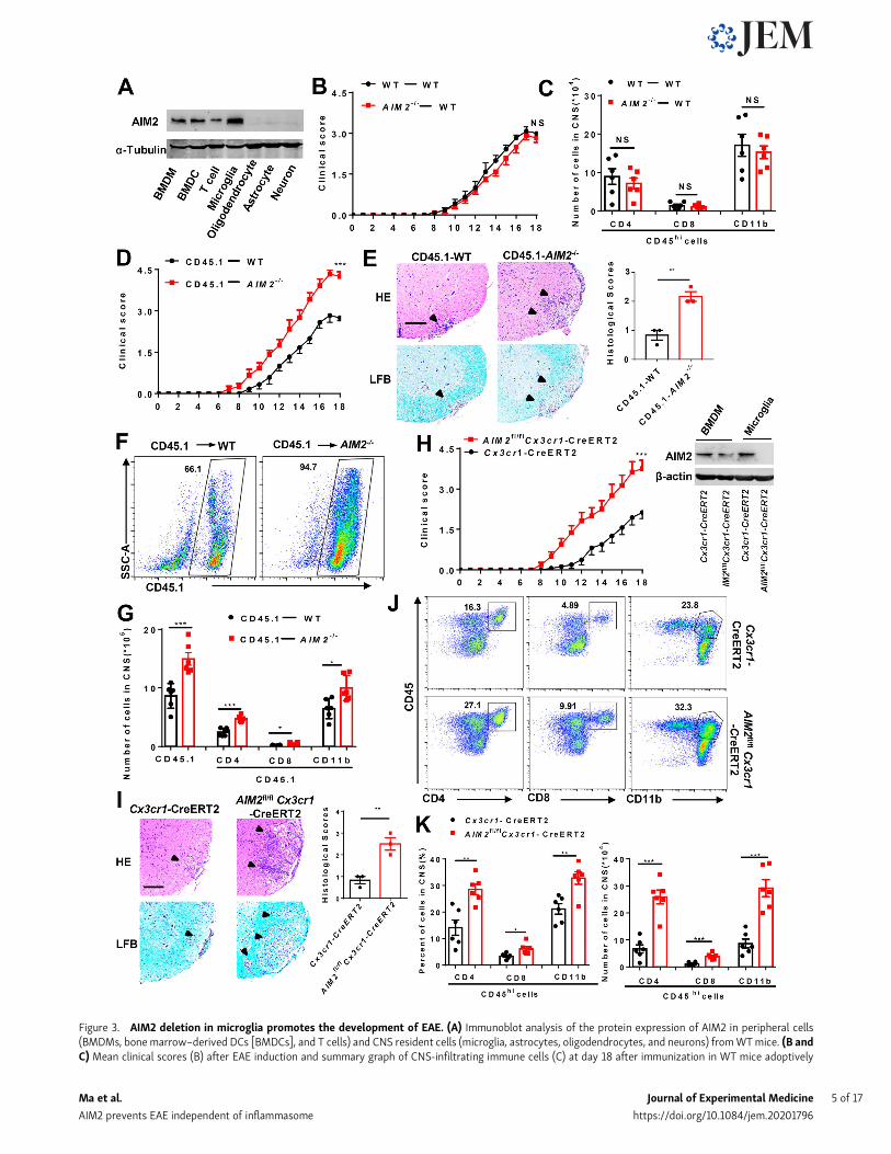
Figure 3. AIM2 deletion in microglia promotes the development of EAE. (A) Immunoblot analysis of the protein expression of AIM2 in peripheral cells(BMDMs, bonemarrow–derived DCs [BMDCs], and T cells) and CNS resident cells (microglia, astrocytes, oligodendrocytes, and neurons) fromWTmice. (B andC) Mean clinical scores (B) after EAE induction and summary graph of CNS-infiltrating immune cells (C) at day 18 after immunization in WT mice adoptively
Ma et al. Journal of Experimental Medicine 5 of 17
AIM2 prevents EAE independent of inflammasome https://doi.org/10.1084/jem.20201796
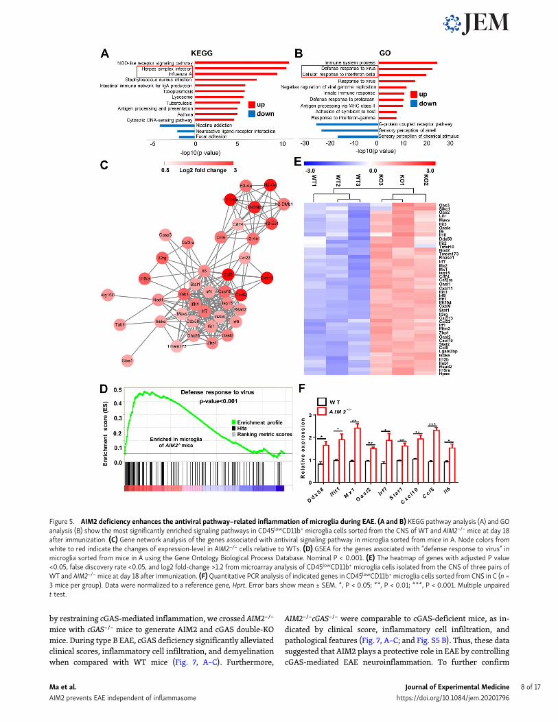
AIM2 deficiency enhances the microglial antiviralpathway–related inflammation during EAETo further identify the pathways involved in AIM2 controllingmicroglial inflammation, we did in-depth analysis of the mi-croarray data. Notably, Kyoto Encyclopedia of Genes andGenomes (KEGG) analysis showed the up-regulation of manyviral-related pathways in AIM2−/−CD45lowCD11b+ cells, in-cluding herpes simplex infection, influenza A, and cytosolicDNA–sensing pathway (Fig. 5 A). Gene ontology biologicalprocess (GO) analysis further confirmed the top biologicalprocesses up-regulated in AIM2−/− microglia were relatedwith defense response to virus and cellular response tointerferon-β (Fig. 5 B). Additionally, gene network and geneset enrichment analysis (GSEA) highlighted the regulatedrole of AIM2 in genes involved in the defense response tovirus pathway (Fig. 5, C and D). Consistently, the heatmapand real-time PCR analysis displayed significantly increasedexpression of many genes associated with antiviral pathwaysand DNA-sensing pathways, such as Ddx58, Ifit1, Mx1, Oasl2,Irf7, Stat1, Cxcl10, Ccl5, and Il6 in AIM2−/− cells (Fig. 5, E andF). Thus, these genomic analyses clearly demonstrate thatAIM2 restricts antiviral inflammatory signaling pathwaysin microglia.
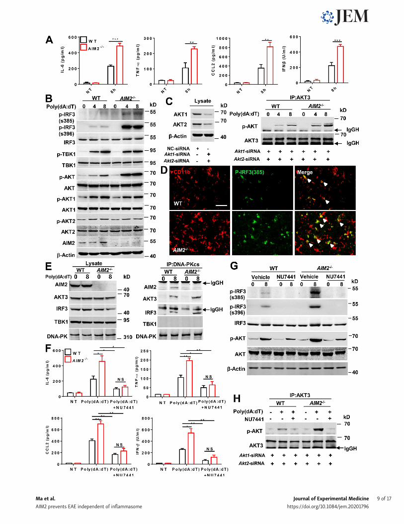
AIM2 targets the DNA-PK (DNA-dependent proteinkinase)–AKT3 axis to inhibit the microglial antiviral pathway-related inflammationPoly(dA:dT) (poly(deoxyadenylic-deoxythymidylic)) transfec-tion can strongly activate antiviral inflammatory pathways(Lees-Miller et al., 1990; Li et al., 2013), but barely do AIM2inflammasome activation without first signal to prime(Hornung et al., 2009). We next investigated theinflammasome-independent effect of AIM2 in microglia onantiviral pathway–associated inflammation in response topoly(dA:dT) transfection. Indeed, we detected higher levels ofantiviral-related cytokines and chemokines, such as IFN-β,CXCL10, CCL5, CCL2, IL-6, and TNF-α, in response to poly(dA:dT) in primarymicroglia from AIM2−/− mice than those fromWTmice (Fig. 6 A; and Fig. S4, A and B). AIM2 has been reported torestrict colon tumorigenesis by suppression of the DNA-PK–AKTaxis in an inflammasome-independent manner (Wilson et al.,2015). Additionally, a recent study reported that AKT3 canphosphorylate IRF3 at Ser385, which facilitates TBK1-inducedphosphorylation of IRF3 on Ser396 and enhances the antiviralinflammatory signaling (Xiao et al., 2020). We speculated that
AIM2 in microglia may negatively regulate the DNA-PK–AKT3axis to control antiviral pathway–related inflammation. To ver-ify this hypothesis, we treated primary microglia from WT andAIM2−/− mice with poly(dA:dT) and analyzed the phosphoryla-tion of IRF3, TBK1, and AKTs. We observed an increase in thephosphorylation of IRF3 at Ser385, Ser396, and AKT, but notthe phosphorylation of TBK1, AKT1, and AKT2 (Fig. 6 B) inAIM2−/− microglia compared with controls after treatment.Since there is no commercial antibody to specifically detectp-AKT3, we silenced AKT1 and AKT2 in microglia, andimmunoprecipitated AKT3 to detect AKT3 phosphorylation byusing an anti-phosphorylated AKT antibody after poly(dA:dT)treatment. We found that AIM2 deficiency markedly en-hanced poly(dA:dT)-induced AKT3 phosphorylation (Fig. 6 C),suggesting a negative role for AIM2 in regulating the activa-tion of AKT3. Consistently, we also observed the enhancedphosphorylation of IRF3 at Ser385 in microglia from AIM2−/−
during EAE (Fig. 6 D). To further dissect the mechanism ofAIM2 regulation of AKT3, we examined the interaction of theDNA-PK catalytic subunit (DNA-PKcs) with AIM2 and AKT3in microglia following poly(dA:dT) treatment. Poly(dA:dT)induced the interaction of DNA-PKcs with AIM2, AKT3, andIRF3 but not with TBK1 in WT microglia. Interestingly, theinteraction of DNA-PKcs with AKT3 and IRF3 was signifi-cantly increased in AIM2−/− microglia (Fig. 6 E), suggestingthat AIM2 suppresses recruitment of AKT3 and IRF3 to DNA-PK. Consistently, we observed great colocalization betweenAIM2 and DNA-PK in the spinal cord of EAE mice comparedwith untreated controls by using in situ hybridization (Fig.S4 H). To further verify that AIM2 targets the DNA-PK–AKT3axis, the DNA-PK inhibitor NU7441 (Leahy et al., 2004) wasused. WT and AIM2−/− microglia were treated with NU7441followed by poly(dA:dT) treatment. Treatment with NU7441significantly reduced the elevated production of IFN-β,CXCL10, CCL5, CCL2, IL-6, and TNF-α in AIM2−/− microglialcells (Fig. 6 F; and Fig. S4, C and D). In addition, NU7441 sig-nificantly reduced the phosphorylation of IRF3 at Ser385 andSer396, and AKT3 in AIM2−/−microglia to comparable levels inNU7441-treated WTs (Fig. 6, G and H). Moreover, we observedAKT3 knockdown significantly reduced the elevated produc-tion of IL-6, IFN-β, CXCL10, and CCL5 and in AIM2−/− micro-glial cells to comparable levels in AKT3-silenced WTs (Fig. S4,E–G). These data suggest AIM2 can inhibit the microglial an-tiviral inflammatory signaling by negatively regulating therecruitment of AKT3 and IRF3 to DNA-PK.
transferred with WT and AIM2−/− bone marrow cells (n = 6 mice per group). (D) Mean clinical scores after EAE induction in WT and AIM2−/− mice adoptivelytransferred with CD45.1 WT bone marrow cells (n = 6 mice per group). (E) Representative H&E and LFB staining and histology score of spinal cord sectionsharvested from the mice in D, showing inflammatory cell infiltration and demyelination (arrowheads). Scale bar, 200 µm. (F and G) Representative plot ofCD45.1+ cells (F) and summary graph of CNS-infiltrating immune cells from FACS analysis of the mice in D (n = 6 mice per group; G). SSC-A, side scatter area.(H) Mean clinical scores of AIM2fl/flCx3cr1-CreERT2 and Cx3cr1-CreERT2 mice after EAE induction (n = 8 mice per group). Immunoblot analysis of AIM2 ex-pression in microglia from the indicated mice. (I) Representative H&E and LFB staining and histology score of spinal cord sections harvested from the mice in H,showing inflammatory cell infiltration and demyelination (arrowheads). Scale bar, 200 µm. (J and K) FACS analysis of CNS-infiltrating immune cells (n = 6 miceper group) from the mice in H. Data in are presented as a representative plot (J), quantified percentage, and absolute cell numbers (K). Data are pooled fromthree independent experiments. *, P < 0.05; **, P < 0.01; ***, P < 0.001. Error bars show mean ± SEM. Unpaired t test for B, D, and H, and multiple unpairedt test for C, G, and K.
Ma et al. Journal of Experimental Medicine 6 of 17
AIM2 prevents EAE independent of inflammasome https://doi.org/10.1084/jem.20201796
Inhibition of the cGAS (cyclic guanosinemonophosphate–adenosine monophosphate synthase)-DNA-PK axis rescues EAE pathogenesis in AIM2-deficient miceAxonal damage and apoptotic loss of neurons occur in the brainduring EAE (Hobom et al., 2004). Indeed, we observed a marked
increase of DNA damage marker γH2AX in the spinal cord ofmice during EAE (Fig. S5 A). DNA or cyclic dinucleotides re-leased by dying nerve cells have been reported to activate cGAS-STING (stimulator of interferon genes) in microglia (Abdullahet al., 2018; Chin, 2019). To determine if AIM2 can restrict EAE
Figure 4. AIM2 deficiency drives the microglial disease phenotypic switch during EAE. (A) FACS sorting strategy of CD45lowCD11b+ microglia cellsfrom CNS in WT and AIM2−/− mice at day 18 after immunization. SSC-A, side scatter area; FSC-A, forward scatter area; FSC-W, forward scatter pulse width.(B) Volcano plot displaying the results of differential gene expression analysis performed in CD45lowCD11b+ microglia cells isolated from the CNS of WT andAIM2−/− mice at day 18 after immunization. Genes related with disease-associated microglia signature and M0 signature are indicated. (C) The heatmap ofgenes with adjusted P value <0.05, false discovery rate <0.05, and log2 fold-change (FC) >1.2 from microarray analysis of CD45lowCD11b+ microglia cellsisolated from the CNS of WT and AIM2−/− mice at day 18 after immunization. (D) Quantitative PCR analysis of the expression of indicated genes inCD45lowCD11b+ microglia cells sorted from CNS in C. Data were normalized to a reference gene, Hprt. (E) Immunofluorescence analysis APOE expression in themicroglia (IBA1+) of spinal cords from AIM2−/− and WT mice during EAE. The merge of APOE with IBA1 is indicated by arrowheads. Scale bar, 50 µm. Data arerepresentative of three independent experiments (E). Error bars show mean ± SEM. *, P < 0.05; **, P < 0.01; ***, P < 0.001. Multiple unpaired t test.
Ma et al. Journal of Experimental Medicine 7 of 17
AIM2 prevents EAE independent of inflammasome https://doi.org/10.1084/jem.20201796
by restraining cGAS-mediated inflammation, we crossed AIM2−/−
mice with cGAS−/− mice to generate AIM2 and cGAS double-KOmice. During type B EAE, cGAS deficiency significantly alleviatedclinical scores, inflammatory cell infiltration, and demyelinationwhen compared with WT mice (Fig. 7, A–C). Furthermore,
AIM2−/−cGAS−/− were comparable to cGAS-deficient mice, as in-dicated by clinical score, inflammatory cell infiltration, andpathological features (Fig. 7, A–C; and Fig. S5 B). Thus, these datasuggested that AIM2 plays a protective role in EAE by controllingcGAS-mediated EAE neuroinflammation. To further confirm
Figure 5. AIM2 deficiency enhances the antiviral pathway–related inflammation of microglia during EAE. (A and B) KEGG pathway analysis (A) and GOanalysis (B) show the most significantly enriched signaling pathways in CD45lowCD11b+ microglia cells sorted from the CNS of WT and AIM2−/− mice at day 18after immunization. (C) Gene network analysis of the genes associated with antiviral signaling pathway in microglia sorted from mice in A. Node colors fromwhite to red indicate the changes of expression-level in AIM2−/− cells relative to WTs. (D) GSEA for the genes associated with “defense response to virus” inmicroglia sorted from mice in A using the Gene Ontology Biological Process Database. Nominal P < 0.001. (E) The heatmap of genes with adjusted P value<0.05, false discovery rate <0.05, and log2 fold-change >1.2 from microarray analysis of CD45lowCD11b+ microglia cells isolated from the CNS of three pairs ofWT and AIM2−/−mice at day 18 after immunization. (F)Quantitative PCR analysis of indicated genes in CD45lowCD11b+ microglia cells sorted from CNS in C (n =3 mice per group). Data were normalized to a reference gene, Hprt. Error bars show mean ± SEM. *, P < 0.05; **, P < 0.01; ***, P < 0.001. Multiple unpairedt test.
Ma et al. Journal of Experimental Medicine 8 of 17
AIM2 prevents EAE independent of inflammasome https://doi.org/10.1084/jem.20201796
Ma et al. Journal of Experimental Medicine 9 of 17
AIM2 prevents EAE independent of inflammasome https://doi.org/10.1084/jem.20201796
that AIM2 controls EAE by inhibiting the DNA-PK–AKT3 axis,we administered WT and AIM2−/− mice by i.p. injection with theDNA-PK inhibitor NU7441. Treatment of WT mice with NU7441significantly attenuated the severity of EAE as indicated by lowerclinical score, less inflammatory cell infiltration in CNS, and lessdemyelination (Fig. 7, D–G). Notably, NU7441 treatment alsoeliminated the augmentation of EAE severity in AIM2−/− mice(Fig. 7, D–G). Overall, these data suggest that AIM2 protectsagainst neuroinflammation by suppressing cGAS and DNA-PK–mediated inflammation.
DiscussionPublic RNA sequencing (RNA-seq) and microarray datasets inGEO (accession nos. GSE123496, GSE26927, and GSE138614)showed that AIM2 expression is increased in the brain tissues ofMS patients when compared with healthy donors (Fig. S5, C–F),suggesting that AIM2 is induced duringMS andmay be involvedin regulating the progression of MS (Durrenberger et al., 2015;Elkjaer et al., 2019; Voskuhl et al., 2019). Therefore, we used theEAE model to comprehensively dissect the role of AIM2 in thedevelopment of MS in this study.
Emerging evidence suggests the importance of in-flammasome components of peripheral immune cells in the de-velopment of EAE by triggering T cell responses to initiateneuroinflammation (Inoue et al., 2012; Li et al., 2019b; Martinet al., 2016). However, in this study we found that AIM2 offers aprotective function against EAE neuroinflammation via an un-known inflammasome-independent role, which differs fromprevious reports on the function of other inflammasome com-ponents in promoting EAE-induced neuroinflammation. More-over, we found that AIM2 inmicroglia but not in peripheral cellsis indispensable for its protective role in EAE, whereas otherinflammasome proteins such as ASC and gasdermin D regulateEAE in T cells and peripheral myeloid cells (Li et al., 2019b;Martin et al., 2016). Although peripheral macrophages and DCsare involved in the onset of EAE, the amount of macrophages andDCs in the CNS tissue during EAE is much less than that of mi-croglia (Bailey et al., 2007; Mrdjen et al., 2018). Moreover, mi-croglia are the main CNS parenchyma-localized APCs requiredfor infiltration and amplification of T cells during EAE
(Archambault et al., 2005; Kawakami et al., 2004).Moreover, thepublic RNA-seq (GEO accession no. GSE52564) and single-cellRNA-seq (http://bis.zju.edu.cn/MCA/) data showed that AIM2is highly expressed in microglia compared with other CNS celltypes in mice, including neurons, astrocytes, and oligoden-drocytes (Han et al., 2018; Zhang et al., 2014). Consistent withthese published studies, our immunoblot analysis showed theexpression of AIM2 in microglia was higher than in macro-phages, DCs, astrocytes, and neurons. Thus, microglia-specificAIM2 plays a dominant role in modulating the pathogenesis ofEAE. Although inflammasome proteins such as ASC, caspase-1,and AIM2 are enriched in microglial cells (Singhal et al., 2014),the AIM2 inflammasome requires a higher threshold of DNA foractivation to occur (Choubey, 2019; Jones et al., 2010). Con-versely, lower levels of DNA can be sensed directly by cGAS andDNA-PK (Ablasser and Chen, 2019; Choubey, 2019; Lees-Milleret al., 1990). The amount of DNA released by damaged neuronsduring EAE nerve injury may not be sufficient to stimulateAIM2-dependent inflammasome activation, which may explainwhy AIM2 in microglia functions in EAE independently of itsinflammasome function.
Notably, we performed microarray analysis of microglia(CD45lowCD11b+ cells) isolated from the CNS of WT and AIM2−/−
mice of EAE and revealed that the most prominent up-regulatedresponses in AIM2−/− microglia from EAE-treated mice wereassociated with defense against viral infection. Correspondingly,a recent study showed an inflammasome-independent role ofAIM2 in obesity and insulin resistance that is mediated by theup-regulation of Ifi202b and antiviral IFN signaling (Gong et al.,2019). Previous studies have implicated viral infections as trig-gers of neurodegenerative diseases such as Parkinson’s disease,Alzheimer’s disease, and amyotrophic lateral sclerosis (Amoret al., 2010; Deleidi and Isacson, 2012). Consistent with this,the antiviral cytokine IFN-β levels increase with age (Baruchet al., 2014; Yu et al., 2015). Additionally, viruses have beenconsidered to be etiological agents of MS involved in demyeli-nation (Libbey et al., 2014). Although IFN-β is the first-linetreatment of relapsed and remitting MS at present, ∼50% ofMS patients are nonresponsive to IFN-β. Furthermore, in somecases, IFN-β can exacerbate MS and consistently worsens neu-romyelitis optica (Axtell et al., 2011; Rıo et al., 2006). Indeed, it
Figure 6. AIM2 inhibits the microglial antiviral pathway–related inflammation by suppressing the DNA-PK–AKT3 signaling axis. (A) ELISA analysis ofTNF-α, IL-6, CCL2, and IFN-β in the supernatants of WT and AIM2−/− microglia transfected with poly(dA:dT) for 8 h. (B) Immunoblot analysis of p-IRF3(S385),p-IRF3(S396), IRF3, p-TBK1, TBK1, p-AKT, AKT, p-AKT1, AKT1, p-AKT2, AKT2, AIM2, and β-actin (loading control) in WT and AIM2−/− microglia transfected withpoly(dA:dT) for 4 and 8 h. (C) WT and AIM2−/− microglia were transfected with siRNAs to silence Akt1 and Akt2 for 48 h, and then were transfected withpoly(dA:dT) for 4 and 8 h. Cell lysates were immunoprecipitated (IP) with anti-AKT3 antibody followed by immunoblotting with p-AKT and AKT3. (D) Im-munofluorescent labeling of p-IRF3(S385) (green) and CD11b (red) in the spinal cord of EAE-induced WT mice at day 18 after immunization. The merge ofp-IRF3(S385) with CD11b is indicated by arrowheads. Scale bar, 50 µm. (E) WT and AIM2−/− microglia were transfected with poly(dA:dT) for 8 h. Then celllysates were immunoprecipitated with anti-DNA-PK antibody followed by immunoblotting with DNA-PKcs, TBK1, IRF3, AKT3, and AIM2. (F) WT and AIM2−/−
microglia were treated with DNA-PK inhibitor NU7441 (1 nM) and then were transfected with poly(dA:dT) for 8 h. The production of indicated cytokines andchemokines was analyzed by ELISA. (G)WT and AIM2−/−microglia were treated with DNA-PK inhibitor NU7441 (1 nM) and then were transfected with poly(dA:dT) for 8 h. The expression of p-IRF3(S385), p-IRF3(S396), IRF3, p-AKT, AKT, and β-actin (loading control) was analyzed by immunoblot. (H) WT and AIM2−/−
microglia were transfected with siRNAs to silence Akt1 and Akt2 for 48 h, and then treated with DNA-PK inhibitor NU7441 (1 nM) followed by poly(dA:dT)transfection for 8 h. Cell lysates were immunoprecipitated with anti-AKT3 antibody followed by immunoblotting with p-AKT and AKT3. Data are pooled fromthree independent experiments for A and F. Data are representative of three (B and G) or two (C, E, and H) independent experiments. *, P < 0.05; **, P < 0.01;***, P < 0.001. Error bars show mean ± SEM. Unpaired t test. NT, no treatment.
Ma et al. Journal of Experimental Medicine 10 of 17
AIM2 prevents EAE independent of inflammasome https://doi.org/10.1084/jem.20201796
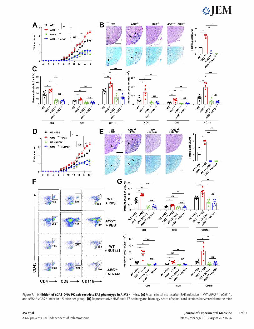
Figure 7. Inhibition of cGAS-DNA-PK axis restricts EAE phenotype in AIM2−/− mice. (A)Mean clinical scores after EAE induction in WT, AIM2−/−, cGAS−/−,and AIM2−/−cGAS−/− mice (n = 5 mice per group). (B) Representative H&E and LFB staining and histology score of spinal cord sections harvested from the mice
Ma et al. Journal of Experimental Medicine 11 of 17
AIM2 prevents EAE independent of inflammasome https://doi.org/10.1084/jem.20201796
has been reported that acute virus infection can induce the de-velopment of type B EAE in mice (Inoue et al., 2016). Significantincreases in the mRNA levels of antiviral inflammatory genessuch as Ddx58, Ifit1, Oasl2, Irf7, Il6, Il12, Ccl22, Cxcl10, and Ccl5wereidentified in microglia from AIM2−/− EAE–treated mice. The in-creased expression of chemokines and cytokines such as Cxcl10,Ccl22, Ccl5, and Il6 in AIM2−/− cells plays a crucial role in T cellinfiltration and cell proliferation during EAE (Balashov et al.,1999; Quandt and Dorovini-Zis, 2004; Takeda et al., 1998),which is critical step for the inflammation and pathogenesis ofEAE or MS. Additionally, changes in the inflammatory stateof microglia can affect its polarization and switching betweenM0 and microglial neurodegenerative phenotype (MGnD;Krasemann et al., 2017). We found the enhancement of MGnDgenes (e.g., Siglec1, Axl, Cybb, Clec7a, and Apoe) and the sup-pression of M0 genes (e.g., Siglec1, Gpr34, Tgfbr1, and Fcrsl) inAIM2−/− microglia during EAE. A previous study has suggestedthat MGnD microglia might play a detrimental role in EAEdevelopment (Krasemann et al., 2017). Moreover, the expres-sion of genes involved in antigen processing and presentation,such as H2-DMa, H2-DMb, H2-Aa, H2-Ab, and H2-Oa, were alsoup-regulated in AIM2−/− microglia of EAE. These MHC hap-lotypes have been established in EAE susceptibility (Iacobaset al., 2007; Welsh et al., 2016).
DNA-PK–associated proteins are expressed at the highestlevel in brain tissue (Moll et al., 1999), indicating an importantrole for the DNA-PK pathway in CNS. We revealed that AIM2associates with DNA-PK in microglia and limits DNA-PK–mediated AKT3 recruitment and phosphorylation, therebyinhibiting downstream IRF3 activation and antiviral inflamma-tory signaling in neuroinflammation. The administration of aDNA-PK inhibitor dramatically attenuated the pathogenesis ofEAE. Dying nerve cells have been reported to release DNA orcyclic dinucleotides to activate cGAS-STING–mediated inflam-mation in microglia (Abdullah et al., 2018). We found that theabsence of cGAS precluded the augmented EAE pathogenesisthat is observed in AIM2−/− mice. Overall, our studies suggestthat the development of therapeutic strategies to specificallytarget cGAS and DNA-PK signaling might be useful for thetreatment of heterogeneous MS.
In summary, our study describes an unexpected regulatoryrole of AIM2 in the development of EAE. We propose a model inwhich AIM2 can interact with DNA-PK to block the binding ofDNA-PK with AKT3, which suppresses AKT3 activation anddownstream IRF3 Ser385 phosphorylation. This interactionlimits cGAS and DNA-PK IRF3 activation and transcription of
inflammatory cytokines. Thus, AIM2 in microglial cells nega-tively regulates the DNA-PK–AKT3 axis to inhibit neuro-inflammation and protect against the onset of EAE (Fig. 8). Ourstudy uncovers an entirely new regulatory mechanism bywhichAIM2 modulates neuroinflammation and neurodegenerativediseases.
Materials and methodsMiceFemale mice with the C57BL/6 background were used in thisstudy. The AIM2−/− and ASC−/− mice were a gift from Dr. V. Dixit(Genentech, South San Francisco, CA). The cGAS−/− mice werefrom The Jackson Laboratory. The AIM2fl/fl mice were generatedusing conditional gene targeting methods by Biocytogen. Aim2conditional KO mice were generated by the CRISPR/Cas9-basedapproach. Briefly, two single guide RNAs (sgRNAs) were de-signed by a CRISPR design tool from the Feng Zhang lab (http://crispr.mit.edu; Platt et al., 2014) to target either a regionupstream or downstream of exon 5, and then were screened foron-target activity using the Universal CRISPR Activity Assay(Biocytogen). To minimize random integrations, we employ acircular donor vector. The gene-targeting vector containing 59homologous arm, target fragment (exon 5), 39 homologous armwas used as a template to repair the double-strand breaks gen-erated by Cas9/sgRNA. The two LoxP sites were precisely in-serted in both sides of the target fragment of the Aim2 gene. T7promoter sequence was added to the Cas9 or sgRNA template byPCR amplification in vitro. Cas9 mRNA, targeting vector, andsgRNAs were coinjected into the cytoplasm of one-cell-stagefertilized C57BL/6N eggs. The injected zygotes were transferredinto oviducts of Kunming pseudopregnant females to generateF0 (numerous founder 0) mice. F0 mice with the expectedgenotype confirmed by tail genomic DNA PCR and sequencingwere mated with C57BL/6N mice to establish germline-transmitted F1 heterozygous mice. F1 heterozygous mice weregenotyped by tail genomic PCR, Southern blot, and DNA se-quencing. AIM2 floxed mice were crossed with Cx3cr1-Cre (TheJackson Laboratory) to generate mononuclear phagocyte–conditional AIM2 KO mice (AIM2fl/fl Cx3cr1-Cre) or with GFAP-Cre (The Jackson Laboratory) to generate astrocyte-conditionalAIM2 KO mice (AIM2fl/fl GFAP-Cre). To generate the microglia-conditional AIM2 KOmice, AIM2 floxed mice were crossed withCx3cr1-CreERT2-EYFP mice (The Jackson Laboratory), and thenthe mice were i.p injected with 3 mg tamoxifen (T5648; Sigma-Aldrich) dissolved in 200 µl corn oil (C8267; Sigma-Aldrich) for
in A, showing inflammatory cell infiltration and demyelination (arrowheads). Scale bar, 200 µm. (C) Flow-cytometric analysis of immune cells (includingCD45+CD4+ T cells, CD45+CD8+ T cells, and CD45+CD11b+ microglia and myeloid cells) infiltrated to the spinal cord and brain of EAE-induced WT, AIM2−/−,cGAS−/−, and AIM2−/−cGAS−/− mice at day 18 after immunization (n = 5 mice per group). Data are presented as summary graphs of quantified percentages andabsolute cell numbers. (D)Mean clinical scores after EAE induction in WT and AIM2−/−mice administrated with PBS or NU7441 at a dose of 10 mg/kg daily (n =5mice per group). (E) Representative H&E and LFB staining and histology score of spinal cord sections harvested from themice in D, showing inflammatory cellinfiltration and demyelination (arrowheads). Scale bar, 200 µm. (F and G) Flow-cytometric analysis of immune cells (including CD45+CD4+ T cells, CD45+CD8+
T cells, and CD45+CD11b+ microglia and myeloid cells) infiltrated to the spinal cord and brain of EAE-induced WT and AIM2−/− mice administrated with PBS orNU7441 at a dose of 10 mg/kg daily at day 18 after immunization (n = 5 mice per group). Data are presented as representative plots (F) and summary graphs ofquantified percentages and absolute cell numbers (G). Data are pooled from three independent experiments. *, P < 0.05; **, P < 0.01; ***, P < 0.001. Error barsshow means ± SEM. Unpaired t test for A and D, and multiple unpaired t test for C and G.
Ma et al. Journal of Experimental Medicine 12 of 17
AIM2 prevents EAE independent of inflammasome https://doi.org/10.1084/jem.20201796
five consecutive days to induce the expression of Cre re-combinase. After 6 wk, the tamoxifen-induced mice weremicroglia-conditional AIM2 KO mice, and these mice were usedfor the EAE study. All mice were kept in a barrier facility, and allanimal experiments were conducted in accordance with theprocedure approved by the Ethical Review Committee for Lab-oratory Animal Welfare of Nanjing Medical University.
Antibodies and reagentsAntibodies to AIM2 (13095s), p-IRF3-S396 (4947s), IRF3 (4302s),p-TBK1 (5483s), TBK1 (3013s), p-AKT1 (9018s), AKT1 (2938s),p-AKT2 (8599s), AKT2 (3063s), p-AKT (4060P), and AKT3(14982s) were purchased from Cell Signaling Technology. Anti-bodies to p-IRF3-S385 (D151514) were purchased from SangonBiotech. Anti–ionized calcium binding adapter molecule 1 (Iba1;019–19741) was fromWako. Anti-Apoe (ab1906) and anti-γH2AX(ab11174) were from Abcam. Anti-CD11b (101204) was from Bi-olegend. Anti-AKT (21054) was from SAB. Antibodies to actin(A1978) and DNA-PK (SAB4502385) were purchased fromSigma-Aldrich. Anti–CD45-FITC (30-F11,11-0451-82), anti–CD45-Alexa Fluor 700 (30-F11,56–0451-82), anti–CD8a-PE (53–6.7,12-0081-83), anti–CD11b-APC (M1/70,17-0112-82), anti–IL17-PE(eBio18B7,12-7177-81), anti–IFNγ-PerCP-Cyanine5.5 (XMG1.2,85-45-7311-82), anti–IFNγ-APC (XMG1.2,17-7311-82), and FixableViability Dye (FVD) eFluor 506 were from eBioscience. Anti–CD4-APC-Cy7 (GK1.5,100414) was from Biolegend. Pertussistoxin (#180) was from List Biological Laboratories. Mycobacte-rium tuberculosis H37Ra (231141) was from BD. Incomplete
Freund’s adjuvant (F5506) was from Sigma-Aldrich. MOG35-55peptide (residues 35–55, Met-Glu-Val-Gly-Trp-Tyr-Arg-Ser-Pro-Phe-Ser-Arg-Val-Val-His-Leu-Tyr-Arg-Asn-Gly-Lys) was synthe-sized by Sangon Biotech (Shanghai).
Induction and assessment of EAETo induce type A EAE,MOG35-55 peptide (300 µg permouse) wasemulsified with complete Freund’s adjuvant (200 µg M. tuber-culosis H37Ra [Mtb] per mouse), and then subcutaneously in-jected in the flanks and neck of mice on day 0 for oneimmunization. Pertussis toxin (250 ng per mouse) was appliedintravenously on days 0 and 2 after immunization. The type BEAE was induced as described above except that M. tuberculosiswas administrated at 400 µg per mice. Mice were assessed dailyfor clinical signs of EAE in a blinded fashion. EAE score wasevaluated as follows: 0.5, partial tail paralysis; 1, tail paralysis;1.5, reversible corrective reflex impairment; 2, corrective refleximpairment; 2.5, one hindlimb paralysis; 3, both hindlimbs pa-ralysis; 3.5, both hindlimbs paralysis and one forelimb paralysis;4, hindlimb and forelimb paralysis; and 5, death.
Bone marrow chimerasThe recipient mice were subjected to lethal-dose irradiation (10Gy), and 1 d later, bone marrow cells (10 × 106) derived from thetibiae and femurs of donor mice were i.v. injected into lethallyirradiated mice. Under these conditions, the radio-resistantCNS-resident cells would be retained, whereas bone marrowand peripheral immune cells would be eliminated and replaced
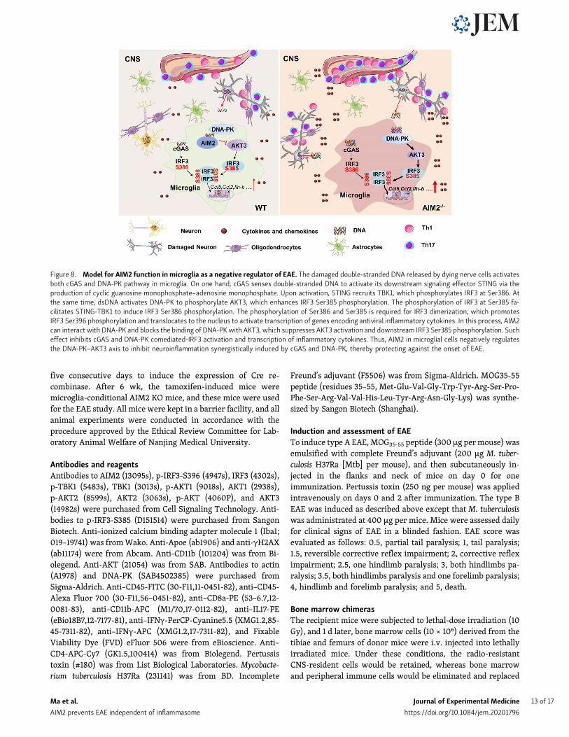
Figure 8. Model for AIM2 function in microglia as a negative regulator of EAE. The damaged double-stranded DNA released by dying nerve cells activatesboth cGAS and DNA-PK pathway in microglia. On one hand, cGAS senses double-stranded DNA to activate its downstream signaling effector STING via theproduction of cyclic guanosine monophosphate–adenosine monophosphate. Upon activation, STING recruits TBK1, which phosphorylates IRF3 at Ser386. Atthe same time, dsDNA activates DNA-PK to phosphorylate AKT3, which enhances IRF3 Ser385 phosphorylation. The phosphorylation of IRF3 at Ser385 fa-cilitates STING-TBK1 to induce IRF3 Ser386 phosphorylation. The phosphorylation of Ser386 and Ser385 is required for IRF3 dimerization, which promotesIRF3 Ser396 phosphorylation and translocates to the nucleus to activate transcription of genes encoding antiviral inflammatory cytokines. In this process, AIM2can interact with DNA-PK and blocks the binding of DNA-PKwith AKT3, which suppresses AKT3 activation and downstream IRF3 Ser385 phosphorylation. Sucheffect inhibits cGAS and DNA-PK comediated-IRF3 activation and transcription of inflammatory cytokines. Thus, AIM2 in microglial cells negatively regulatesthe DNA-PK–AKT3 axis to inhibit neuroinflammation synergistically induced by cGAS and DNA-PK, thereby protecting against the onset of EAE.
Ma et al. Journal of Experimental Medicine 13 of 17
AIM2 prevents EAE independent of inflammasome https://doi.org/10.1084/jem.20201796
by bonemarrow cells from donor mice. After 8 wk, the chimericmice were then subjected to EAE induction.
Histological analysisAll spinal cord tissue sections used here were 5 µm thick. Forparaffin-embedded tissue, spinal cords collected from PBS-perfused mice were fixed in 4% paraformaldehyde overnight.Sections were stained with H&E for evaluation of leukocyteinfiltration or with LFB for assessing demyelination. Histologywas scored in a double-blinded fashion as followed: 0, no in-flammatory cell infiltration and no demyelination; 1, slight in-flammatory cell infiltration or demyelination observed; 2,moderate inflammatory cell infiltration or demyelination inseveral spots; and 3, substantial inflammatory cell infiltrationand large area of demyelination.
RNA in situ hybridizationSpinal cord tissue samples were fixed in formalin for 48 h,embedded in paraffin, and cut into 6-µm sections. In situ hy-bridization was performed according to the manufacturer’s in-structions (G3017; Servicebio). Probes recognizing Aim2 RNA(NM_001013779) were multiplexed with probes recognizingDNAPK RNA (NM_011159).
Immunofluorescence stainingTissue sections were incubated at 4°C overnight with primaryantibody to IBA1, APOE, and CD11b. Slides were then incubatedwith indicated secondary antibodies. The nuclei were counter-stained with DAPI (Sigma-Aldrich). Slides were dried andmounted using ProLong Antifade mounting medium (BeyotimeBiotechnology). Slides were visualized by a Nikon 50i fluores-cent microscope.
Isolation of CNS immune cells and FACS analysis and sortingFor preparation of immune cells, brains and spinal cords fromMOG35-55–immunized mice were excised and digested at 37°Cwith DNase I (10 U/ml; Roche) and collagenase type IV (0.5 mg/ml; Sigma-Aldrich) in RPMI 1640 under agitation (200 rpm)conditions for 60 min. Single-cell suspensions were obtained bygrinding through a 70-µm cell strainer. Subsequently, homo-geneous cell suspensions were centrifuged over the Percolldensity gradient (GE Healthcare) and separated by collecting theinterface fractions between 37 and 70% Percoll. Mononuclearcells were isolated from the interface. After intensive washing,single-cell suspensions were stained with FVD eFluor 506, anti-CD45, anti-CD4, anti-CD8, and anti-CD11b for FACS analysis. Forintracellular cytokine staining, cells were stimulated withphorbol 12-myristate 13-acetate (Multi Sciences), ionomycin(Multi Sciences), and brefeldin A (Invitrogen) for 4 h of culture.Cells were fixed and permeabilized with the Intracellular Fixa-tion & Permeabilization Buffer Set (eBioscience) and then sub-jected to cytokine staining flow cytometry analyses. All flowcytometry was performed on an Attune NxT flow cytometer(Thermo Fisher Scientific), and data were analyzed by FlowJo10.0.7 software. For FACS sorting, single cell suspensions werestained with FVD eFluor 506, anti-CD45, and anti-CD11b andsorted on a BD FACS Aria.
Primary microglia cultureCerebral cortices from neonatal mice age 1–3 d were collectedand carefully stripped of their meninges and the blood vessels.Following dissection, the tissues were digested with 0.25%trypsin–EDTA and washed in HBSS containing FBS. Thenthe single-cell suspensions were obtained by passing through acell strainer (70 µm). The cell suspensions were seeded intopoly-D-lysine–precoated flasks and cultured in DMEM/F12supplemented with 10% FBS at 37°C and 5% CO2. Medium wasreplaced every 4–5 d. After 10–14 d, microglia were separatedfrom the underlying astrocytic layer by gentle shaking of theflask, plated overnight in poly-D-lysine–precoated plates, andtransfected with Lipofectamine 2000 (2 µl/ml)–complexedpoly(dA:dT) (1 µg/106 cells). For the DNA-PK inhibition experi-ment, the microglia were primed with 1 nM NU7441 (DNA-PKinhibitor) for 1h before poly(dA:dT) transfection. The condi-tioned media were collected and measured for cytokine pro-duction by ELISA, and the cells were collected for cytokine geneexpression by RT quantitative PCR (RT-qPCR) or protein acti-vation by Western blotting.
siRNA-mediated gene silences in microgliaPrimarymicroglia were plated in poly-D-lysine–precoated platesand were transfected with siRNA using Lipofectamine RNAi-MAX (Invitrogen) according to the manufacturer’s guidelines.The siRNA sequences were: siAKT1-1 (sense: 59-CCAUGAACGAGUUUGAGA-39; antisense: 59-GGUACUUGCUCAAACUCAU-39),siAKT1-2 (sense: 59-CUUCCUCCUCAAGAACGAU-39; antisense:59-GAAGGAGGAGUUCUUGCUA-39), siAKT2-1 (sense: 59-GGAGGUAGCUGUCAACAAG-39; antisense: 59-CUUGUUGACAGCUACCUCC-39), siAKT2-2 (sense: 59-GCAAAGAGGGCAUCAGUGA-39; antisense: 59-UCACUGAUGCCCUCUUUGC-39); and siAKT3(sense: 59-GCUCAUUCAUAGGCUAUAA-39; antisense: 59-UUAUAGCCUAUGAAUGAGC-39). The siRNA and negative controlsiRNA were from GenePharma.
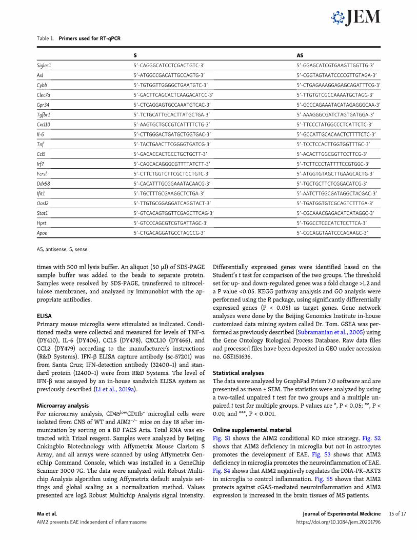
RT-qPCRTotal RNA was extracted by using TRIzol reagent (Life Tech-nologies) and subjected to cDNA synthesis. RT-qPCR was per-formed using SYBR Green Supermix (Vazyme). The expressionof a single gene was calculated by a standard curve method andstandardized to the expression of Hprt. The primers used arelisted in Table 1.
Immunoprecipitation and immunoblot analysisPrimary microglia were transfected with Lipofectamine 2000(2 µl/ml)–complexed poly(dA:dT) (1 µg/106 cells). Cells werecollected in NP-40 lysis buffer (20 mM Tris-HCl, pH 7.4, con-taining 150 mM NaCl, 0.5% [vol/vol] IGEPAL, 10% [wt/vol]glycerol, 50 mM NaF, 1 mM Na3VO4, 1 mM dithiothreitol, 1 mMphenylmethylsulphonyl fluoride, and complete protease inhibi-tor cocktail [Sigma-Aldrich]), followed by incubation for 40 minat 4°C. The lysates were centrifuged for 15 min at 14,000 rpm forremoval of cell debris and nuclei. Supernatants were incubatedwith the indicated antibody overnight at 4°C. Then an aliquot(40 µl) of protein A/G-agarose was added to each sample, fol-lowed by incubation for 4 h at 4°C. The beads were washed three
Ma et al. Journal of Experimental Medicine 14 of 17
AIM2 prevents EAE independent of inflammasome https://doi.org/10.1084/jem.20201796
times with 500 ml lysis buffer. An aliquot (50 µl) of SDS-PAGEsample buffer was added to the beads to separate protein.Samples were resolved by SDS-PAGE, transferred to nitrocel-lulose membranes, and analyzed by immunoblot with the ap-propriate antibodies.
ELISAPrimary mouse microglia were stimulated as indicated. Condi-tioned media were collected and measured for levels of TNF-α(DY410), IL-6 (DY406), CCL5 (DY478), CXCL10 (DY466), andCCL2 (DY479) according to the manufacturer’s instructions(R&D Systems). IFN-β ELISA capture antibody (sc-57201) wasfrom Santa Cruz; IFN-detection antibody (32400–1) and stan-dard protein (12400–1) were from R&D Systems. The level ofIFN-β was assayed by an in-house sandwich ELISA system aspreviously described (Li et al., 2019a).
Microarray analysisFor microarray analysis, CD45lowCD11b+ microglial cells wereisolated from CNS of WT and AIM2−/− mice on day 18 after im-munization by sorting on a BD FACS Aria. Total RNA was ex-tracted with Trizol reagent. Samples were analyzed by BeijingCnkingbio Biotechnology with Affymetrix Mouse Clariom SArray, and all arrays were scanned by using Affymetrix Gen-eChip Command Console, which was installed in a GeneChipScanner 3000 7G. The data were analyzed with Robust Multi-chip Analysis algorithm using Affymetrix default analysis set-tings and global scaling as a normalization method. Valuespresented are log2 Robust Multichip Analysis signal intensity.
Differentially expressed genes were identified based on theStudent’s t test for comparison of the two groups. The thresholdset for up- and down-regulated genes was a fold change >1.2 anda P value <0.05. KEGG pathway analysis and GO analysis wereperformed using the R package, using significantly differentiallyexpressed genes (P < 0.05) as target genes. Gene networkanalyses were done by the Beijing Genomics Institute in-housecustomized data mining system called Dr. Tom. GSEA was per-formed as previously described (Subramanian et al., 2005) usingthe Gene Ontology Biological Process Database. Raw data filesand processed files have been deposited in GEO under accessionno. GSE151636.
Statistical analysesThe data were analyzed by GraphPad Prism 7.0 software and arepresented as mean ± SEM. The statistics were analyzed by usinga two-tailed unpaired t test for two groups and a multiple un-paired t test for multiple groups. P values are *, P < 0.05; **, P <0.01; and ***, P < 0.001.
Online supplemental materialFig. S1 shows the AIM2 conditional KO mice strategy. Fig. S2shows that AIM2 deficiency in microglia but not in astrocytespromotes the development of EAE. Fig. S3 shows that AIM2deficiency inmicroglia promotes the neuroinflammation of EAE.Fig. S4 shows that AIM2 negatively regulates the DNA-PK–AKT3in microglia to control inflammation. Fig. S5 shows that AIM2protects against cGAS-mediated neuroinflammation and AIM2expression is increased in the brain tissues of MS patients.
Table 1. Primers used for RT-qPCR
S AS
Siglec1 59-CAGGGCATCCTCGACTGTC-39 59-GGAGCATCGTGAAGTTGGTTG-39
Axl 59-ATGGCCGACATTGCCAGTG-39 59-CGGTAGTAATCCCCGTTGTAGA-39
Cybb 59-TGTGGTTGGGGCTGAATGTC-39 59-CTGAGAAAGGAGAGCAGATTTCG-39
Clec7a 59-GACTTCAGCACTCAAGACATCC-39 59-TTGTGTCGCCAAAATGCTAGG-39
Gpr34 59-CTCAGGAGTGCCAAATGTCAC-39 59-GCCCAGAAATACATAGAGGGCAA-39
Tgfbr1 59-TCTGCATTGCACTTATGCTGA-39 59-AAAGGGCGATCTAGTGATGGA-39
Cxcl10 59-AAGTGCTGCCGTCATTTTCTG-39 59-TTCCCTATGGCCCTCATTCTC-39
Il-6 59-CTTGGGACTGATGCTGGTGAC-39 59-GCCATTGCACAACTCTTTTCTC-39
Tnf 59-TACTGAACTTCGGGGTGATCG-39 59-TCCTCCACTTGGTGGTTTGC-39
Ccl5 59-GACACCACTCCCTGCTGCTT-39 59-ACACTTGGCGGTTCCTTCG-39
Irf7 59-CAGCACAGGGCGTTTTATCTT-39 59-TCTTCCCTATTTTCCGTGGC-39
Fcrsl 59-CTTCTGGTCTTCGCTCCTGTC-39 59-ATGGTGTAGCTTGAAGCACTG-39
Ddx58 59-CACATTTGCGGAAATACAACG-39 59-TGCTGCTTCTCGGACATCG-39
Ifit1 59-TGCTTTGCGAAGGCTCTGA-39 59-AATCTTGGCGATAGGCTACGAC-39
Oasl2 59-TTGTGCGGAGGATCAGGTACT-39 59-TGATGGTGTCGCAGTCTTTGA-39
Stat1 59-GTCACAGTGGTTCGAGCTTCAG-39 59-CGCAAACGAGACATCATAGGC-39
Hprt 59-GTCCCAGCGTCGTGATTAGC-39 59-TGGCCTCCCATCTCCTTCA-39
Apoe 59-CTGACAGGATGCCTAGCCG-39 59-CGCAGGTAATCCCAGAAGC-39
AS, antisense; S, sense.
Ma et al. Journal of Experimental Medicine 15 of 17
AIM2 prevents EAE independent of inflammasome https://doi.org/10.1084/jem.20201796
AcknowledgmentsWe thank Dr. V. Dixit (Genentech) for AIM2−/− and ASC−/− mice.
This work was supported by the National Natural ScienceFoundation of China (82070567/91742116/81771773/81570499 toS. Yang, 91742116 to J. Zhou, 81991523 to G. Hu, 81802393 to B.Wang, and 81901227 to C. Ma), the Start Fund for SpeciallyAppointed Professor of Jiangsu Province (to S. Yang), the MajorProject of the Nanjing Medical University Science and Technol-ogy Development Fund (NMUD2018003 to S. Yang), the culti-vation project of “High Level Young Scientific and TechnologicalTalents” of Nanjing Medical University (NMUR2019003 to S.Yang), and the Natural Science Youth Foundation of JiangsuProvince (BK20180679 to C. Ma).
Author contributions: C. Ma, S. Li, Y. Hu, Y. Ma, Y. Wu, C.Wu, and X. Liu designed and performed the experiments, ana-lyzed the data, and prepared the figures; B. Wang, G. Hu, andJ. Zhou provided the key technique mentoring, research re-agents, and mice; and C. Ma, S. Li, and S. Yang wrote the paper.
Disclosures: The authors declare no competing interests exist.
Submitted: 19 August 2020Revised: 13 November 2020Accepted: 2 February 2021
ReferencesAbdullah, A., M. Zhang, T. Frugier, S. Bedoui, J.M. Taylor, and P.J. Crack.
2018. STING-mediated type-I interferons contribute to the neuro-inflammatory process and detrimental effects following traumaticbrain injury. J. Neuroinflammation. 15:323. https://doi.org/10.1186/s12974-018-1354-7
Ablasser, A., and Z.J. Chen. 2019. cGAS in action: Expanding roles in im-munity and inflammation. Science. 363:eaat8657. https://doi.org/10.1126/science.aat8657
Aharoni, R., A. Herschkovitz, R. Eilam, M. Blumberg-Hazan, M. Sela, W.Bruck, and R. Arnon. 2008. Demyelination arrest and remyelinationinduced by glatiramer acetate treatment of experimental autoimmuneencephalomyelitis. Proc. Natl. Acad. Sci. USA. 105:11358–11363. https://doi.org/10.1073/pnas.0804632105
Amor, S., F. Puentes, D. Baker, and P. van der Valk. 2010. Inflammation inneurodegenerative diseases. Immunology. 129:154–169. https://doi.org/10.1111/j.1365-2567.2009.03225.x
Archambault, A.S., J. Sim, M.A.T. Gimenez, and J.H. Russell. 2005. Definingantigen-dependent stages of T cell migration from the blood to thecentral nervous system parenchyma. Eur. J. Immunol. 35:1076–1085.https://doi.org/10.1002/eji.200425864
Axtell, R.C., C. Raman, and L. Steinman. 2011. Interferon-β exacerbates Th17-mediated inflammatory disease. Trends Immunol. 32:272–277. https://doi.org/10.1016/j.it.2011.03.008
Bailey, S.L., B. Schreiner, E.J. McMahon, and S.D. Miller. 2007. CNS myeloidDCs presenting endogenous myelin peptides ‘preferentially’ polarizeCD4+ T(H)-17 cells in relapsing EAE. Nat. Immunol. 8:172–180. https://doi.org/10.1038/ni1430
Balashov, K.E., J.B. Rottman, H.L. Weiner, and W.W. Hancock. 1999. CCR5(+)and CXCR3(+) T cells are increased in multiple sclerosis and their li-gands MIP-1α and IP-10 are expressed in demyelinating brain lesions.Proc. Natl. Acad. Sci. USA. 96:6873–6878. https://doi.org/10.1073/pnas.96.12.6873
Baruch, K., A. Deczkowska, E. David, J.M. Castellano, O. Miller, A. Kertser, T.Berkutzki, Z. Barnett-Itzhaki, D. Bezalel, T. Wyss-Coray, et al. 2014.Aging. Aging-induced type I interferon response at the choroid plexusnegatively affects brain function. Science. 346:89–93. https://doi.org/10.1126/science.1252945
Chin, A.C. 2019. Neuroinflammation and the cGAS-STING pathway.J. Neurophysiol. 121:1087–1091. https://doi.org/10.1152/jn.00848.2018
Choubey, D. 2019. Type I interferon (IFN)-inducible Absent in Melanoma2 proteins in neuroinflammation: implications for Alzheimer’s disease.J. Neuroinflammation. 16:236. https://doi.org/10.1186/s12974-019-1639-5
Constantinescu, C.S., N. Farooqi, K. O’Brien, and B. Gran. 2011. Experimentalautoimmune encephalomyelitis (EAE) as a model for multiple sclerosis(MS). Br. J. Pharmacol. 164:1079–1106. https://doi.org/10.1111/j.1476-5381.2011.01302.x
Deleidi, M., and O. Isacson. 2012. Viral and inflammatory triggers of neuro-degenerative diseases. Sci. Transl. Med. 4:121ps3. https://doi.org/10.1126/scitranslmed.3003492
Dendrou, C.A., L. Fugger, and M.A. Friese. 2015. Immunopathology of mul-tiple sclerosis. Nat. Rev. Immunol. 15:545–558. https://doi.org/10.1038/nri3871
Denes, A., G. Coutts, N. Lenart, S.M. Cruickshank, P. Pelegrin, J. Skinner, N.Rothwell, S.M. Allan, and D. Brough. 2015. AIM2 and NLRC4 in-flammasomes contribute with ASC to acute brain injury independentlyof NLRP3. Proc. Natl. Acad. Sci. USA. 112:4050–4055. https://doi.org/10.1073/pnas.1419090112
Durrenberger, P.F., F.S. Fernando, S.N. Kashefi, T.P. Bonnert, D. Seilhean, B.Nait-Oumesmar, A. Schmitt, P.J. Gebicke-Haerter, P. Falkai, E. Grün-blatt, et al. 2015. Common mechanisms in neurodegeneration andneuroinflammation: a BrainNet Europe gene expression microarraystudy. J. Neural Transm. (Vienna). 122:1055–1068. https://doi.org/10.1007/s00702-014-1293-0
Elkjaer, M.L., T. Frisch, R. Reynolds, T. Kacprowski, M. Burton, T.A. Kruse,M. Thomassen, J. Baumbach, and Z. Illes. 2019. Molecular signature ofdifferent lesion types in the brain white matter of patients with pro-gressive multiple sclerosis. Acta Neuropathol. Commun. 7:205. https://doi.org/10.1186/s40478-019-0855-7
Galligan, C.L., L.M. Pennell, T.T. Murooka, E. Baig, B. Majchrzak-Kita, R.Rahbar, and E.N. Fish. 2010. Interferon-β is a key regulator of proin-flammatory events in experimental autoimmune encephalomyelitis.Mult. Scler. 16:1458–1473. https://doi.org/10.1177/1352458510381259
Gong, Z., X. Zhang, K. Su, R. Jiang, Z. Sun, W. Chen, E. Forno, E.S. Goetzman,J. Wang, H.H. Dong, et al. 2019. Deficiency in AIM2 induces inflam-mation and adipogenesis in white adipose tissue leading to obesity andinsulin resistance. Diabetologia. 62:2325–2339. https://doi.org/10.1007/s00125-019-04983-x
Han, X., R. Wang, Y. Zhou, L. Fei, H. Sun, S. Lai, A. Saadatpour, Z. Zhou, H.Chen, F. Ye, et al. 2018. Mapping the mouse cell atlas by microwell-seq.Cell. 172:1091–1107.e17. https://doi.org/10.1016/j.cell.2018.02.001
Hanamsagar, R., A. Aldrich, and T. Kielian. 2014. Critical role for the AIM2inflammasome during acute CNS bacterial infection. J. Neurochem. 129:704–711. https://doi.org/10.1111/jnc.12669
Hobom,M., M.K. Storch, R.Weissert, K.Maier, A. Radhakrishnan, B. Kramer,M. Bahr, and R. Diem. 2004. Mechanisms and time course of neuronaldegeneration in experimental autoimmune encephalomyelitis. BrainPathol. 14:148–157. https://doi.org/10.1111/j.1750-3639.2004.tb00047.x
Hornung, V., A. Ablasser, M. Charrel-Dennis, F. Bauernfeind, G. Horvath,D.R. Caffrey, E. Latz, and K.A. Fitzgerald. 2009. AIM2 recognizes cy-tosolic dsDNA and forms a caspase-1-activating inflammasome withASC. Nature. 458:514–518. https://doi.org/10.1038/nature07725
Humphries, F., L. Shmuel-Galia, N. Ketelut-Carneiro, S. Li, B. Wang, V.V.Nemmara, R. Wilson, Z. Jiang, F. Khalighinejad, K. Muneeruddin, et al.2020. Succination inactivates gasdermin D and blocks pyroptosis. Sci-ence. 369:1633–1637. https://doi.org/10.1126/science.abb9818
Iacobas, D.A., S. Iacobas, P. Werner, E. Scemes, and D.C. Spray. 2007. Al-teration of transcriptomic networks in adoptive-transfer experimentalautoimmune encephalomyelitis. Front. Integr. Nuerosci. 1:10. https://doi.org/10.3389/neuro.07.010.2007
Inoue, M., K.L. Williams, M.D. Gunn, and M.L. Shinohara. 2012. NLRP3 in-flammasome induces chemotactic immune cell migration to the CNS inexperimental autoimmune encephalomyelitis. Proc. Natl. Acad. Sci. USA.109:10480–10485. https://doi.org/10.1073/pnas.1201836109
Inoue, M., P.H. Chen, S. Siecinski, Q.J. Li, C. Liu, L. Steinman, S.G. Gregory, E.Benner, and M.L. Shinohara. 2016. An interferon-β-resistant andNLRP3 inflammasome-independent subtype of EAE with neuronaldamage. Nat. Neurosci. 19:1599–1609. https://doi.org/10.1038/nn.4421
Jones, J.W., N. Kayagaki, P. Broz, T. Henry, K. Newton, K. O’Rourke, S. Chan,J. Dong, Y. Qu, M. Roose-Girma, et al. 2010. Absent in melanoma 2 isrequired for innate immune recognition of Francisella tularensis. Proc.Natl. Acad. Sci. USA. 107:9771–9776. https://doi.org/10.1073/pnas.1003738107
Kawakami, N., S. Lassmann, Z. Li, F. Odoardi, T. Ritter, T. Ziemssen, W.E.Klinkert, J.W. Ellwart, M. Bradl, K. Krivacic, et al. 2004. The activation
Ma et al. Journal of Experimental Medicine 16 of 17
AIM2 prevents EAE independent of inflammasome https://doi.org/10.1084/jem.20201796
status of neuroantigen-specific T cells in the target organ determinesthe clinical outcome of autoimmune encephalomyelitis. J. Exp. Med. 199:185–197. https://doi.org/10.1084/jem.20031064
Krasemann, S., C. Madore, R. Cialic, C. Baufeld, N. Calcagno, R. El Fatimy, L.Beckers, E. O’Loughlin, Y. Xu, Z. Fanek, et al. 2017. The TREM2-APOEpathway drives the transcriptional phenotype of dysfunctional mi-croglia in neurodegenerative diseases. Immunity. 47:566–581.e9. https://doi.org/10.1016/j.immuni.2017.08.008
Lammert, C.R., E.L. Frost, C.E. Bellinger, A.C. Bolte, C.A. McKee, M.E. Hurt,M.J. Paysour, H.E. Ennerfelt, and J.R. Lukens. 2020. AIM2 in-flammasome surveillance of DNA damage shapes neurodevelopment.Nature. 580:647–652. https://doi.org/10.1038/s41586-020-2174-3
Leahy, J.J., B.T. Golding, R.J. Griffin, I.R. Hardcastle, C. Richardson, L. Rig-oreau, and G.C. Smith. 2004. Identification of a highly potent and se-lective DNA-dependent protein kinase (DNA-PK) inhibitor (NU7441) byscreening of chromenone libraries. Bioorg. Med. Chem. Lett. 14:6083–6087. https://doi.org/10.1016/j.bmcl.2004.09.060
Lees-Miller, S.P., Y.R. Chen, and C.W. Anderson. 1990. Human cells contain aDNA-activated protein kinase that phosphorylates simian virus 40 Tantigen, mouse p53, and the human Ku autoantigen. Mol. Cell. Biol. 10:6472–6481. https://doi.org/10.1128/MCB.10.12.6472
Li, X.-D., J. Wu, D. Gao, H. Wang, L. Sun, and Z.J. Chen. 2013. Pivotal roles ofcGAS-cGAMP signaling in antiviral defense and immune adjuvant ef-fects. Science. 341:1390–1394. https://doi.org/10.1126/science.1244040
Li, J., C. Ma, F. Long, D. Yang, X. Liu, Y. Hu, C. Wu, B. Wang, M. Wang, Y.Chen, et al. 2019a. Parkin Impairs Antiviral Immunity by Suppressingthe Mitochondrial Reactive Oxygen Species-Nlrp3 Axis and AntiviralInflammation. iScience. 16:468–484. https://doi.org/10.1016/j.isci.2019.06.008
Li, S., Y. Wu, D. Yang, C. Wu, C. Ma, X. Liu, P.N. Moynagh, B. Wang, G. Hu,and S. Yang. 2019b. Gasdermin D in peripheral myeloid cells drivesneuroinflammation in experimental autoimmune encephalomyelitis.J. Exp. Med. 216:2562–2581. https://doi.org/10.1084/jem.20190377
Libbey, J.E., M.F. Cusick, and R.S. Fujinami. 2014. Role of pathogens inmultiple sclerosis. Int. Rev. Immunol. 33:266–283. https://doi.org/10.3109/08830185.2013.823422
Martin, B.N., C. Wang, C.J. Zhang, Z. Kang, M.F. Gulen, J.A. Zepp, J. Zhao, G.Bian, J.S. Do, B. Min, et al. 2016. T cell-intrinsic ASC critically promotesT(H)17-mediated experimental autoimmune encephalomyelitis. Nat.Immunol. 17:583–592. https://doi.org/10.1038/ni.3389
Mindur, J.E., N. Ito, S. Dhib-Jalbut, and K. Ito. 2014. Early treatmentwith anti-VLA-4 mAb can prevent the infiltration and/or development of path-ogenic CD11b+CD4+ T cells in the CNS during progressive EAE. PLoSOne. 9:e99068. https://doi.org/10.1371/journal.pone.0099068
Moll, U., R. Lau, M.A. Sypes, M.M. Gupta, and C.W. Anderson. 1999. DNA-PK,the DNA-activated protein kinase, is differentially expressed in normaland malignant human tissues. Oncogene. 18:3114–3126. https://doi.org/10.1038/sj.onc.1202640
Mrdjen, D., A. Pavlovic, F.J. Hartmann, B. Schreiner, S.G. Utz, B.P. Leung, I.Lelios, F.L. Heppner, J. Kipnis, D. Merkler, et al. 2018. High-dimensionalsingle-cell mapping of central nervous system immune cells revealsdistinct myeloid subsets in health, aging, and disease. Immunity. 48:380–395.e6. https://doi.org/10.1016/j.immuni.2018.01.011
Platt, R.J., S. Chen, Y. Zhou, M.J. Yim, L. Swiech, H.R. Kempton, J.E. Dahlman,O. Parnas, T.M. Eisenhaure, M. Jovanovic, et al. 2014. CRISPR-Cas9knockin mice for genome editing and cancer modeling. Cell. 159(2):440–455. https://doi.org/10.1016/j.cell.2014.09.014
Procaccini, C., V. De Rosa, V. Pucino, L. Formisano, and G. Matarese. 2015.Animal models of multiple sclerosis. Eur. J. Pharmacol. 759:182–191.https://doi.org/10.1016/j.ejphar.2015.03.042
Quandt, J., and K. Dorovini-Zis. 2004. The beta chemokines CCL4 and CCL5enhance adhesion of specific CD4+ T cell subsets to human brain en-dothelial cells. J. Neuropathol. Exp. Neurol. 63:350–362. https://doi.org/10.1093/jnen/63.4.350
Ransohoff, R.M. 2012. Animal models of multiple sclerosis: the good, the badand the bottom line. Nat. Neurosci. 15:1074–1077. https://doi.org/10.1038/nn.3168
Rathinam, V.A., Z. Jiang, S.N. Waggoner, S. Sharma, L.E. Cole, L. Waggoner,S.K. Vanaja, B.G. Monks, S. Ganesan, E. Latz, et al. 2010. The AIM2inflammasome is essential for host defense against cytosolic bacteriaand DNA viruses. Nat. Immunol. 11:395–402. https://doi.org/10.1038/ni.1864
Rathinam, V.A., S.K. Vanaja, and K.A. Fitzgerald. 2012. Regulation of in-flammasome signaling. Nat. Immunol. 13:333–342. https://doi.org/10.1038/ni.2237
Rıo, J., C. Nos, M. Tintore, N. Tellez, I. Galan, R. Pelayo, M. Comabella, and X.Montalban. 2006. Defining the response to interferon-β in relapsing-remitting multiple sclerosis patients. Ann. Neurol. 59:344–352. https://doi.org/10.1002/ana.20740
Singhal, G., E.J. Jaehne, F. Corrigan, C. Toben, and B.T. Baune. 2014. In-flammasomes in neuroinflammation and changes in brain function: afocused review. Front. Neurosci. 8:315. https://doi.org/10.3389/fnins.2014.00315
Subramanian, A., P. Tamayo, V.K. Mootha, S. Mukherjee, B.L. Ebert, M.A. Gil-lette, A. Paulovich, S.L. Pomeroy, T.R. Golub, E.S. Lander, and J.P. Mesirov.2005. Gene set enrichment analysis: a knowledge-based approach forinterpreting genome-wide expression profiles. Proc. Natl. Acad. Sci. USA.102:15545–15550. https://doi.org/10.1073/pnas.0506580102
Takeda, K., T. Kaisho, N. Yoshida, J. Takeda, T. Kishimoto, and S. Akira. 1998.Stat3 activation is responsible for IL-6-dependent T cell proliferationthrough preventing apoptosis: generation and characterization of Tcell-specific Stat3-deficient mice. J. Immunol. 161:4652–4660.
Voskuhl, R.R., N. Itoh, A. Tassoni, M.A. Matsukawa, E. Ren, V. Tse, E. Jang,T.T. Suen, and Y. Itoh. 2019. Gene expression in oligodendrocytesduring remyelination reveals cholesterol homeostasis as a therapeutictarget in multiple sclerosis. Proc. Natl. Acad. Sci. USA. 116:10130–10139.https://doi.org/10.1073/pnas.1821306116
Welsh, R.A., N. Song, A. Kim, Y. Poluektov, and S. Sadegh-Nasseri. 2016.Exploring a possible role for the MHC Class II accessory molecule H2-Oin the development of experimental autoimmune encephalomyelitis.J. Immunol. 196:46.10.
Wilson, J.E., A.S. Petrucelli, L. Chen, A.A. Koblansky, A.D. Truax, Y. Oyama,A.B. Rogers, W.J. Brickey, Y. Wang, M. Schneider, et al. 2015.Inflammasome-independent role of AIM2 in suppressing colon tu-morigenesis via DNA-PK and Akt.Nat. Med. 21:906–913. https://doi.org/10.1038/nm.3908
Xiao, J., W. Li, X. Zheng, L. Qi, H. Wang, C. Zhang, X. Wan, Y. Zheng, R.Zhong, X. Zhou, et al. 2020. Targeting 7-Dehydrocholesterol ReductaseIntegrates Cholesterol Metabolism and IRF3 Activation to EliminateInfection. Immunity. 52:109–122.e6. https://doi.org/10.1016/j.immuni.2019.11.015
Yu, Q., Y.V. Katlinskaya, C.J. Carbone, B. Zhao, K.V. Katlinski, H. Zheng, M.Guha, N. Li, Q. Chen, T. Yang, et al. 2015. DNA-damage-induced type Iinterferon promotes senescence and inhibits stem cell function. CellRep. 11:785–797. https://doi.org/10.1016/j.celrep.2015.03.069
Zhang, Y., K. Chen, S.A. Sloan, M.L. Bennett, A.R. Scholze, S. O’Keeffe, H.P.Phatnani, P. Guarnieri, C. Caneda, N. Ruderisch, et al. 2014. An RNA-sequencing transcriptome and splicing database of glia, neurons, andvascular cells of the cerebral cortex. J. Neurosci. 34:11929–11947. https://doi.org/10.1523/JNEUROSCI.1860-14.2014
Ma et al. Journal of Experimental Medicine 17 of 17
AIM2 prevents EAE independent of inflammasome https://doi.org/10.1084/jem.20201796
Supplemental material
Ma et al. Journal of Experimental Medicine S1
AIM2 prevents EAE independent of inflammasome https://doi.org/10.1084/jem.20201796
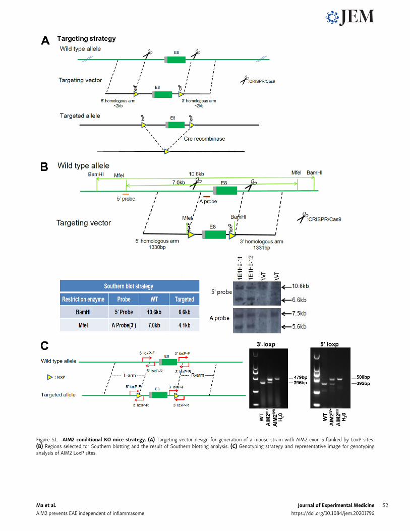
Figure S1. AIM2 conditional KO mice strategy. (A) Targeting vector design for generation of a mouse strain with AIM2 exon 5 flanked by LoxP sites.(B) Regions selected for Southern blotting and the result of Southern blotting analysis. (C) Genotyping strategy and representative image for genotypinganalysis of AIM2 LoxP sites.
Ma et al. Journal of Experimental Medicine S2
AIM2 prevents EAE independent of inflammasome https://doi.org/10.1084/jem.20201796
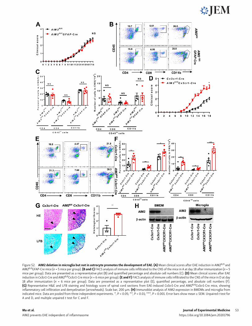
Figure S2. AIM2 deletion inmicroglia but not in astrocyte promotes the development of EAE. (A)Mean clinical scores after EAE induction in AIM2fl/fl andAIM2fl/flGFAP-Cre mice (n = 5 mice per group). (B and C) FACS analysis of immune cells infiltrated to the CNS of the mice in A at day 18 after immunization (n = 5mice per group). Data are presented as a representative plot (B) and quantified percentage and absolute cell numbers (C). (D) Mean clinical scores after EAEinduction in Cx3cr1-Cre and AIM2fl/flCx3cr1-Cremice (n = 6mice per group). (E and F) FACS analysis of immune cells infiltrated to the CNS of the mice in D at day18 after immunization (n = 6 mice per group). Data are presented as a representative plot (E), quantified percentage, and absolute cell numbers (F).(G) Representative H&E and LFB staining and histology score of spinal cord sections from EAE-induced Cx3cr1-Cre and AIM2fl/flCx3cr1-Cre mice, showinginflammatory cell infiltration and demyelination (arrowheads). Scale bar, 200 µm. (H) Immunoblot analysis of AIM2 expression in BMDMs and microglia fromindicated mice. Data are pooled from three independent experiments. *, P < 0.05; **, P < 0.01; ***, P < 0.001. Error bars showmean ± SEM. Unpaired t test forA and D, and multiple unpaired t test for C and F.
Ma et al. Journal of Experimental Medicine S3
AIM2 prevents EAE independent of inflammasome https://doi.org/10.1084/jem.20201796
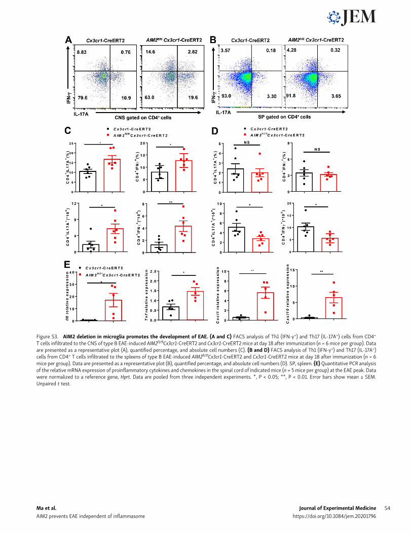
Figure S3. AIM2 deletion in microglia promotes the development of EAE. (A and C) FACS analysis of Th1 (IFN-γ+) and Th17 (IL-17A+) cells from CD4+
T cells infiltrated to the CNS of type B EAE-induced AIM2fl/flCx3cr1-CreERT2 and Cx3cr1-CreERT2 mice at day 18 after immunization (n = 6 mice per group). Dataare presented as a representative plot (A), quantified percentage, and absolute cell numbers (C). (B and D) FACS analysis of Th1 (IFN-γ+) and Th17 (IL-17A+)cells from CD4+ T cells infiltrated to the spleens of type B EAE-induced AIM2fl/flCx3cr1-CreERT2 and Cx3cr1-CreERT2 mice at day 18 after immunization (n = 6mice per group). Data are presented as a representative plot (B), quantified percentage, and absolute cell numbers (D). SP, spleen. (E)Quantitative PCR analysisof the relative mRNA expression of proinflammatory cytokines and chemokines in the spinal cord of indicated mice (n = 5 mice per group) at the EAE peak. Datawere normalized to a reference gene, Hprt. Data are pooled from three independent experiments. *, P < 0.05; **, P < 0.01. Error bars show mean ± SEM.Unpaired t test.
Ma et al. Journal of Experimental Medicine S4
AIM2 prevents EAE independent of inflammasome https://doi.org/10.1084/jem.20201796
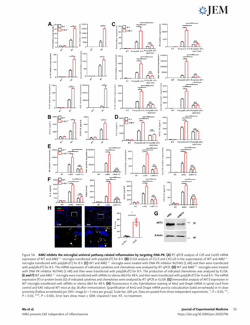
Figure S4. AIM2 inhibits the microglial antiviral pathway-related inflammation by targeting DNA-PK. (A) RT-qPCR analysis of Ccl5 and Cxcl10 mRNAexpression of WT and AIM2−/− microglia transfected with poly(dA:dT) for 8 h. (B) ELISA analysis of CCL5 and CXCL10 in the supernatants of WT and AIM2−/−
microglia transfected with poly(dA:dT) for 8 h. (C) WT and AIM2−/− microglia were treated with DNA-PK inhibitor NU7441 (1 nM) and then were transfectedwith poly(dA:dT) for 8 h. The mRNA expression of indicated cytokines and chemokines was analyzed by RT-qPCR. (D)WT and AIM2−/− microglia were treatedwith DNA-PK inhibitor NU7441 (1 nM) and then were transfected with poly(dA:dT) for 8 h. The production of indicated chemokines was analyzed by ELISA.(E and F)WT and AIM2−/−microglia were transfected with siRNAs to silence Akt3 for 48 h, and thenwere transfected with poly(dA:dT) for 4 and 8 h. ThemRNAexpression (F) or protein levels (G) of indicated cytokines and chemokines were analyzed by RT-qPCR or ELISA. (G) Immunoblot analysis of AKT3 expression inWT microglia transfected with siRNAs to silence Akt3 for 48 h. (H) Fluorescence in situ hybridization staining of Aim2 and Dnapk mRNA in spinal cord fromcontrol and EAE-induced WT mice at day 18 after immunization. Quantification of Aim2 and Dnapk mRNA puncta colocalization (solid arrowheads) or in closeproximity (hollow arrowheads) per 200× image (n = 3 mice per group). Scale bar, 100 µm. Data are pooled from three independent experiments. *, P < 0.05; **,P < 0.01; ***, P < 0.001. Error bars show mean ± SEM. Unpaired t test. NT, no treatment.
Ma et al. Journal of Experimental Medicine S5
AIM2 prevents EAE independent of inflammasome https://doi.org/10.1084/jem.20201796
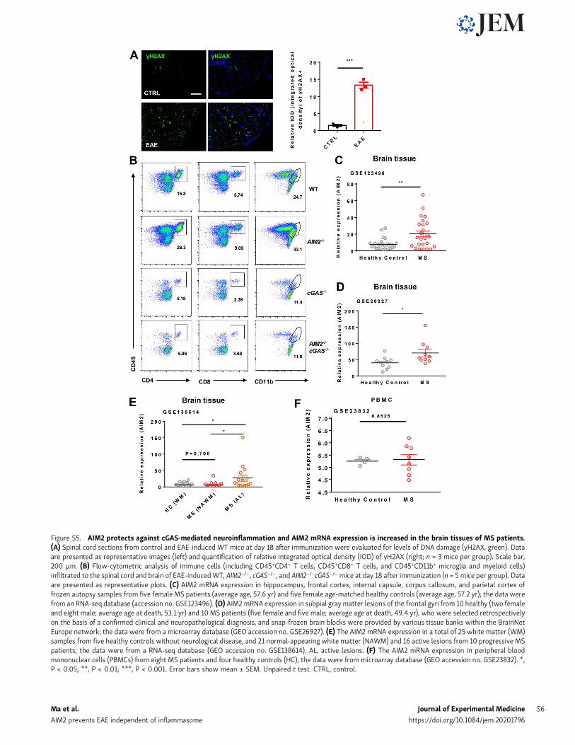
Figure S5. AIM2 protects against cGAS-mediated neuroinflammation and AIM2 mRNA expression is increased in the brain tissues of MS patients.(A) Spinal cord sections from control and EAE-induced WT mice at day 18 after immunization were evaluated for levels of DNA damage (γH2AX, green). Dataare presented as representative images (left) and quantification of relative integrated optical density (IOD) of γH2AX (right; n = 3 mice per group). Scale bar,200 µm. (B) Flow-cytometric analysis of immune cells (including CD45+CD4+ T cells, CD45+CD8+ T cells, and CD45+CD11b+ microglia and myeloid cells)infiltrated to the spinal cord and brain of EAE-inducedWT, AIM2−/−, cGAS−/−, and AIM2−/−cGAS−/−mice at day 18 after immunization (n = 5 mice per group). Dataare presented as representative plots. (C) AIM2 mRNA expression in hippocampus, frontal cortex, internal capsule, corpus callosum, and parietal cortex offrozen autopsy samples from five female MS patients (average age, 57.6 yr) and five female age-matched healthy controls (average age, 57.2 yr); the data werefrom an RNA-seq database (accession no. GSE123496). (D) AIM2mRNA expression in subpial gray matter lesions of the frontal gyri from 10 healthy (two femaleand eight male; average age at death, 53.1 yr) and 10 MS patients (five female and five male; average age at death, 49.4 yr), who were selected retrospectivelyon the basis of a confirmed clinical and neuropathological diagnosis, and snap-frozen brain blocks were provided by various tissue banks within the BrainNetEurope network; the data were from a microarray database (GEO accession no. GSE26927). (E) The AIM2 mRNA expression in a total of 25 white matter (WM)samples from five healthy controls without neurological disease, and 21 normal-appearing white matter (NAWM) and 16 active lesions from 10 progressive MSpatients; the data were from a RNA-seq database (GEO accession no. GSE138614). AL, active lesions. (F) The AIM2 mRNA expression in peripheral bloodmononuclear cells (PBMCs) from eight MS patients and four healthy controls (HC); the data were from microarray database (GEO accession no. GSE23832). *,P < 0.05; **, P < 0.01; ***, P < 0.001. Error bars show mean ± SEM. Unpaired t test. CTRL, control.
Ma et al. Journal of Experimental Medicine S6
AIM2 prevents EAE independent of inflammasome https://doi.org/10.1084/jem.20201796


![Review Article The Role of Microglia in Diabetic Retinopathydownloads.hindawi.com/journals/joph/2014/705783.pdf · change in microglial morphology [ ]. erefore, microglial responses](https://img.pdfslide.net/doc/110x75/5f0513557e708231d411234b/review-article-the-role-of-microglia-in-diabetic-change-in-microglial-morphology.jpg)