Embed Size (px)
Citation preview
Cellular & Molecular Immunology 107
Article
Volume 3 Number 2 April 2006
Target Organ Protection from a Novel Angiotensin II Receptor (AT1) Vaccine ATR12181 in Spontaneously Hypertensive Rats Feng Zhu1, Yuhua Liao1, 2, Liudong Li1, Min Cheng1, Fen Wei1, Yumiao Wei1 and Ming Wang1
Hypertension produces pathophysiological changes that are often responsible for the mortality associated with the disease. It is evident that overactive renin-angiotensin systems play a central role in the development of hyper- tension and target organ damage associated with hypertension. We have previously found that a novel angio- tensin II receptor (AT1) vaccine-ATR12181 attenuated the development of high blood pressure (BP) in spontaneously hypertensive rat (SHR) model of human essential hypertension. Our objective was to determine whether this attenuation of high BP is associated with prevention of target organ damage induced by hypertensive state. SHRs were immunized against a peptide (coded ATR12181) from the extracelluar portion of the AT1A receptor by repeated subcutaneous injections of peptide-tetanus-toxoid complex in combination with Freund’s adjuvant. A 64 weeks long-term observation was performed. Repeated vaccinations resulted in the induction of anti-ATR12181 antibodies. At the end of observation, vaccinated SHRs manifested lower BP, decreased cardiac hypertrophy and attenuation of kidney injuries. mRNA levels of c-fos and c-jun in heart and kidneys were decreased in vaccinated SHRs. Since a self antigen was used, safety of vaccine was concerned. However, the signs of autoimmune diseases were not observed in the sections of heart and kidney. These data demonstrated that repeated immunization against a domain of the extracellular portion of the AT1 receptor was able to cause a target organ protection against hypertension. Active immunization against the AT1 receptor may be considered as a promising new strategy in the treatment of hypertension. Cellular & Molecular Immunology. 2006;3(2):107-114. Key Words: AT1, hypertension, vaccine, immune system Introduction A role for the renin-angiotensin system (RAS) in the development of hypertension is well established in both human and animal models such as the spontaneously hypertensive rat (SHR) (1, 2). Interruption of the RAS pathway, either by preventing the formation of angiotensin II (i.e., angiotensinconverting enzyme inhibitor) or by blocking its actions at the level of the peptide receptor [i.e., angiotensin II type 1 (AT1) receptor antagonists], has been proved to be highly successful in the treatment and mana- gement of hypertension in a significant population of
hypertensive patients (3). In spite of this success, the traditional pharmacological therapy targeted to inhibit specific components of the RAS suffers from many signi- ficant disadvantages. Chronic administration of traditional therapies is necessary for long-term anti-hypertensive benefits. Required daily dosing and undesirable side effects such as coughing, angioedema, renal dysfunction and hyper- kalemia diminish patient compliance (4, 5). Thus, other specific ways of suppressing the RAS function in hyper- tension, for example, immunological approaches may have advantages over current therapy.
The renin-angiotensin vaccine is an old dream of the immunologist and cardiologist (6). In comparison with the chemical drugs, vaccine has potential benefits for controlling hypertension. The potential benefits include improvements in compliance management, smooth and progressive onset of action for patients, improved diurnal control of blood pressure and reduction in drug interactions associated with conventional drug polypharmacy. These advantages are due to the vaccine mode of action, where infrequent doses induce a biological response, overcoming the need for one or more daily tablets. Since the discovery of renin, renin was first used to immunize the animals as a vaccine against RAS (7). Michel JB et al. reported that active immunization against mouse renin could lower the blood pressure (BP) of Wistar
1Laboratory of Cardiovascular Immunology, Institute of Cardiology, Union Hospital, Tongji Medical College, Huazhong Science & Technology University, Wuhan 430022, China; 2Corresponding to: Dr. Yuhua Liao, Laboratory of Cardiovascular Immunology, Institute of Cardiology, Union Hospital, Tongji Medical College of Huazhong University of Science and Technology, 1277 Jiefang Road, Wuhan 430022, China. Fax: +86-27-8572-7140, E-mail: [email protected].
Received Jan 19, 2006. Accepted Mar 16, 2006. Copyright © 2006 by The Chinese Society of Immunology
108 Vaccine ATR12181 Cause a Target Organ Protection Against Hypertension
Volume 3 Number 2 April 2006
rats and spontaneously hypertensive rats (SHRs), and antibodies generated in the rats against renin could inhibit catalytic activity of renin (8). Similar results were found in the experiment of marmoset (9). Unfortunately, the effect on BP was accompanied by autoimmune disease of kidneys, which are major sites of production, storage and release of renin (8, 9). Since the safety concern, other components of RAS including angiotensin I (Ang I), angiotensin II (Ang II) and peptide from the AT1 receptor were used to immunize the animals (10-12). According to the results of previous studies, Ang I peptide analogue vaccines may be the most successful. Ang I peptide analogue vaccines selectively reduced the pressor effects of exogenous Ang I in conscious rat (10). Further experiments were done in the man with essential hypertension, and a clinical trial has been initiated since 2001(11, 13).
We have previously established active immunization with a peptide coded ATR12181 from the extracelluar portion of rat AT1A receptor attenuating the development of high BP in SHRs (14). In present study, SHRs were vaccinated against ATR12181-tetanus-toxoid complex repeatedly and a 64 weeks long-term observation was performed. Our objective was to determine whether this attenuation of high BP was associated with the prevention of cardiovascular and renal pathological changes induced by the hypertensive state. Materials and Methods Preparation of the AT1A receptor vaccine Two AT1 receptor subtypes have been described in rodents, AT1A and AT1B, with more than 95% amino acid sequence identity. AT1A receptors are abundantly expressed in vascular smooth muscle cells (VSMC) (15). We designed the peptide ATR12181 based on amino acids sequences of extracellular parts of the AT1A receptor. The sequence of the
peptide ATR12181 was shown in Table 1. The peptide was synthesized by PSSM-8 peptide synthesizer (Shimadzu, Japan). The carrier protein in solution of tetanus toxoid (TT) was purchased from Institute of Biological Products of Wuhan. To conjugate, the appropriate amount of the peptide was weighed out, dissolved in phosphate buffered saline (PBS) and mixed with TT. Following this, 0.3% glutaraldehyde solution was added to mixture of peptides and TT, and incubated for 2 hours at room temperature. Then glycerine was added into solution and incubated for 30 min at room temperature to the reaction. The mixtures were dialyzed against PBS at 4°C overnight. The conjugate was sterilized using 0.2 mm filters (Millipore, U.K.), and 100 μl of the conjugate containing 0.1 mg of peptide was used as a dose for each immunization of animal. Handling of animals All the animal protocols and procedures were approved by the Institutional Animal Care and Use Committee. Male SHRs aged 6 weeks (body weight 145 ± 10 g) were immunized by subcutaneous injection with peptide-TT conjugate (peptide, 0.1 mg for each dose). The conjugate was coupled with the same volume Freund’s adjuvant (Yafa Biological Technology Ltd., China). Complete Freund’s adjuvant was used in the initial injection and incomplete Freund’s adjuvant for subsequent procedures. SHRs were received repeated immunizations at 0, 4th, 8th, 12th, 16th, 24th, 32nd, 40th and 52nd week, and raised for 64 weeks. The homo-age male SHRs received 0.9% sodium chloride in a same schedule was used as control. ELISA Blood samples were taken every 4 weeks to monitor antibody titer by using an enzyme-linked immunosorbent assay (ELISA). To determine antibody responses to each peptide, 96-well micro-titer plates were coated with each peptide (1 μg/well) in 0.1 M Na2CO3 solution (pH 9.6). After adsorption overnight, the coating antigen was removed and the plates were washed for 3 × 5 min with PBS containing 3% bovine serum albumin (BSA) (PBS-A). To prevent non- specific binding, the plates were incubated with 200 μl PBS-A/well for 1 h at 37°C. Serum samples (100 μl/well) diluted in PBS, 0.1% BSA and 0.1% Tween-20 (TBS-T), were added in doubling dilutions from 1:80 to 1:10,240 and incubated at 37°C for 45 min. After three additional washings, horseradish peroxidase-conjugated anti-rat IgG antibodies 1:2,000 were added for 30 min at 37°C. The plates were then washed three more times, the substrate (0.01% H2O2 and 0.1% 3’-3’-5’-5’-tetramethyl benzidine) was added for 10 min, and the reaction was ended with 2 mol/L H2SO4. Optical density (OD) was measured at 450 nm in a microplate reader (Bio-tek type EX2800). The titre end-point was taken to be the dilution that no longer gave an absorbance value greater than twice that of the negative control. The negative control was the serum taken from the same rat prior to immunization. Echocardiography analysis
Table 1. Amino-acid sequences of the peptide coded ATR12181 Code ATR12181
Sequence AFHYESR* Molecular weight 908.97 Molecular formula H56C41N12O12 Location the second extracellular
loop of rat AT1A receptor (181-187)
Compared with AT1B protein AFHYESQ** Compared with rat AT2 receptor FRDVRTI Compared with human AT1 receptor AFHYESQ** Compared with human AT2 protein FRDVRTI
*ATR12181 is a seven-amino-acid sequence from rat AT1A receptor. **The letter underlined indicates the different amino-acid sequences of rat AT1B receptor or human AT1 receptor in comparison with rat AT1A receptor.
Cellular & Molecular Immunology 109
Volume 3 Number 2 April 2006
Before sacrifice, two-dimensional echocardiography was performed with a phased-array echocardiographic machine (GE type-Vivid, USA), using an 11.4-MHz (short focus) transducer. Short-axis view of the left ventricle was obtained. BP measurement Indirect systolic blood pressure (SBP) was monitored by a standard tail-cuff technique using Powerlab system (ADInstruments, Australia) every 4 weeks. Before the study, all the rats were trained to become accustomed to the tail-cuff procedure for 3 days. The three determinations were averaged to provide each measurement recorded. Before the sacrifice of rats, invasive BP measure was done. Rats were anaesthetized (sodium pentobarbital 45 mg/kg, i.p.) and a catheter was implanted in the left carotid artery. Intravascular BP was measured by using Powerlab system. Organ weight and morphometric analysis After measurement of BP, all rats were sacrificed. Heart and kidneys were excised and rinsed in 0.9% physiological saline, and total weights of heart and kidneys were recorded. The
weight of heart was normalized to body weight, and the ratio was compared among groups. Heart and kidneys were further dissected, and the fixed transverse sections of the tissues were embedded in paraffin, cut into 5-μm sections, and stained with Masson’s trichrome. Parts of tissues of renal cortex were immediately fixed in 0.25% glutaraldehyde for transmission electron microscopy (TEM) analysis. RT-PCR analysis Total RNA from left ventricle tissue and kidneys was prepared using Trizol reagent (Invitrogen, USA). The expressions of c-fos and c-jun were assessed by reverse transcriptase-polymerase chain reaction (RT-PCR). With the One-step RT-PCR Kit (TakaRa, Japan), the procedure followed the protocol of the manufacturer. Primers were synthesized by Shanghai Sangon with the following sequences: GAPDH 5’-AAT GCA TCC TGC ACC ACC AA-3’ and 5’-GTA GCC ATA TTC ATT GTC ATA-3’; c-fos 5’-TCC CAG AGG AGA TGT CTG TG-3’ and 5’-GGC TCC AGC TCT GTG ACC AT’-3, c-jun 5’-TAG TCC CTC CCG TGG TTG-3’ and 5’-TCT AGG AGT CGT CAG AAT CC-3’. Each PCR product was electrophoresed on a 2% agarose gel and stained with ethidium bromide (1 μg/ml). The density of each PCR band was measured and analyzed by ImageMaster VDS analysis software (Pharmacia Biotech). To quantify the amount of mRNA product, we used GAPDH mRNA as internal control. Statistical analysis Results are expressed as mean ± SEM. Statistical significance was estimated by using the Student’s t test. p < 0.05 was considered significant. Results Titer and BP Figure 1 showed the changes of antibody titre. After second booster, the titre reached a platform (1:2,560-1:10,240). The
A
B
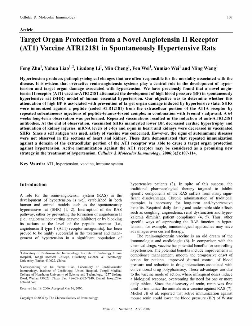
Figure 1. Plots of effect of vaccinations in spontaneously hypertensive rats. (A) Time dependent appearance of antibodiesagainst peptide ATR12181. P, primary injection; B, booster injection. The titer of antibodies against the peptide appeared in the circulation after primary injection and reached a platform after the second booster. (B) Changes in the systolic blood pressure (SBP) in ATR12181 group significantly differ from that in the control group after the 8th week of experiment (Student’s t test p < 0.05), and 17mmHg decrease of SBP was achieved at the end of observation.
Table 2. Blood pressure, heart rate and cardiac patho-physiological parameters of vaccinated and control animals at the 64th week Control group ATR12181 group SBP (mmHg) 197 ± 4 180 ± 6** DBP (mmHg) 142 ± 4 136 ± 5 HR (beats/min) 388 ± 23 392 ± 22 Body weight (g) 383 ± 18 538 ± 36** Heart weight (g) 1.59 ± 0.06 1.33 ± 0.11** HW/BW (mg/g) 4.2 ± 0.2 2.5 ± 0.1** IVSd (cm) 0.27 ± 0.01 0.22 ± 0.02** LVIDd (cm) 0.68 ± 0.03 0.58 ± 0.04** LVPWd (cm) 0.28 ± 0.02 0.23 ± 0.02** EF (%) 79.4 ± 3.0 81.5 ± 3.1
N = 6 per group. **p < 0.01 vs control group.
110 Vaccine ATR12181 Cause a Target Organ Protection Against Hypertension
Volume 3 Number 2 April 2006
platform could maintain by repeated immunization. The peak titre was 1:10,240. ELISA results demonstrated that vacci- nation was able to induce immune responses against peptide ATR12181.
Before the primary injection, SBP of two groups was at same level, and the average SBP of all SHR was 139.3 ± 9.4 mmHg. SBP of both groups increased and reached a plateau at the 8th week. In comparison with SBP of control group, there was a significant decrease in SBP level at the 8th week in the ATR12181 group (Figure 1). Before the sacrifice, a 17 mmHg reduction of systolic blood pressure was observed in vaccinated SHRs at the 64th week (Table 2). However, the level of BP did not reach target level (140/90 mmHg). Effects of vaccine-ATR12181 on cardiac remodeling Figure 2 showed representative short-axis echocardiographic views of heart from a control animal and a vaccinated rat.
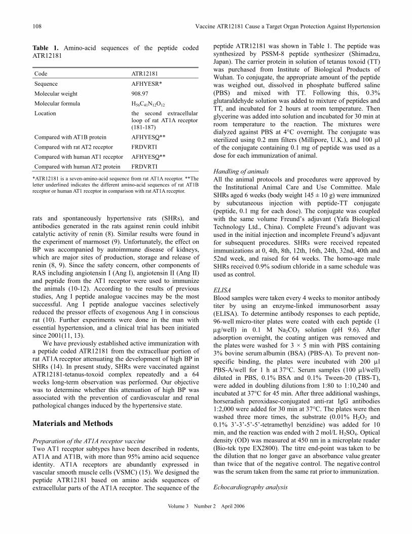
The left ventricle thickening of control group was obvious. As data shown in Table 2, compared to control group, LVH in vaccinated group was significantly attenuated, as evidenced by decreased end-diastolic interventricular septum (IVSd), left ventricular end-diastolic interdiameter (LVIDd, p < 0.01) and left ventricular end-diastolic posterior wall (LVPWd, p < 0.01) (Table 2). SHRs in both groups did not develop heart failure, as evidenced by normal ejection factor (EF) values in both groups at 64th week (p > 0.05) (Table 2). Echocardiography indicated that active immunization with the ATR12181 vaccine markedly attenuated LVH.
Necropsy of the rats in control also revealed LVH. In contrast, LVH in ATR12181 group was significantly attenuated (Figure 2). Mean body weights (BW) of the vaccine-treated SHRs were 40.4% higher than those of untreated SHRs (Table 2). In contrast, heart weights (HW) of the vaccine-treated SHR were 16.4% lower than those of
Figure 2. Effects of vaccine-ATR12181 on cardiac and renal pathological changes associated with hypertension. (A) Representative short-axis echocardiographic views of heart from a control animal and a vaccinated rat. Right ventricle (RV), left ventricle (LV), anterior interventricular septum (AIVS) and left ventricular posterior wall (LVPWd) are indicated. The wall of LV in control group is thicker than that in ATR12181 group. (B) Representative macroscopic graphs of hearts from control and vaccinated groups. Left ventricular hypertrophy (LVH) is evident in control group. LVH is attenuated in ATR12181 group. (C) Photomicrographs of sections of left ventricular from SHR of control group and ATR12181 group. Collagen deposition (blue) was determined by Masson’s trichrome staining. Multiple cardiac fibrosis is found in control group, but this phenomenon is not seen in hearts of the ATR12181 group. Magnification × 400. (D) Photomicrographs of sections of renal cortex from SHR of control group and ATR12181 group. Collagen deposition (blue) was determined by Masson’s trichrome staining. Glomerular damages and interstitial fibrosis in kidney are evident in control group, but these changes are not found in vaccinated group. Magnification × 200. (E and F) Electron photomicrographs of renal cortex from SHR of control group and ATR12181 group. (E) Thickening of basement membrane and interstitial fibrosis are evident in control group. (F) Thickening of basement membrane is only present in the part of glomeruluses in ATR12181 group.

Cellular & Molecular Immunology 111
Volume 3 Number 2 April 2006
untreated SHRs, and similar results in HW/BW were found (Table 2). Cardiac hypertrophy was significant prevented by the vaccine ATR12181. Multifocal areas of fibrosis in the myocardium are another characteristic of hypertension in this model. Fibrosis was clearly evident in control rat, but was rarely seen in ATR12181 group (Figure 2). All the morphometric results suggest that cardiac pathological changes in the genetic hypertension model attenuated. Effects of the vaccine ATR12181 on kidneys Kidney is an important target organ in hypertension. This organ has abundant arterioles and capillary, where the AT1A receptors are abundantly expressed. Whether or not there were vaccine-induced cellular or antibody autoimmune damages in kidneys need to be determined. In control SHRs, glomerular damages and interstitial fibrosis were evident at 64th week (Figure 2). In contrast, treatment with vaccine- ATR12181 significantly attenuated the advance of kidney damages in hypertension. Any signs of autoimmune damages were not found under microscope. For further observation, the ultrastructure changes were evaluated by TEM. In control group, interstitial fibrosis was observed. In addition, thickening of basement membrane was also found, but there were not electron-dense depositions present in basement membrane. Similar change was also present in vaccine- treated SHR, but thickening of basement membrane was present only part of renal glomeruluses. These findings indicated vaccine-ATR12181 afforded renal protection in hypertensive state. Change of c-fos and c-jun expression in target organs Finally, we tested the expressions of c-fos and c-jun, which were the main factors to promote proliferation induced by
Ang II through AT1 receptor. In comparison with the control group, the expressions of c-fos and c-jun mRNA in hearts and kidneys of ATR1218 group were decreased significantly (Figure 3). Discussion The results of this study demonstrate that repeated immunizations with a novel angiotensin II receptor (AT1) vaccine-ATR12181 attenuates the development of hemo- dynamic and pathological alterations associated with hyper- tension in SHRs. Primary injection and booster injections of vaccine in SHRs decreased SBP level, LVH, and attenuated renal injures. These novel data suggest that interruption of AT1 receptor in SHR, with an immunological approach, may be used to prevent hypertension and its associated cardiovascular and renal risk factors.
Vaccine-ATR12181 is complex of peptide-ATR12181 and TT in combination with Freund’s adjuvant. The sequence of peptide ATR12181 is located in 181-187 amino acids of the second loop of rat AT1A receptor. Wallukart, et al. first described the same sequence of human AT1 receptor. Their team first found that the patients with preeclampsia developed an agonistic autoantibody against human AT1 receptor (16). The autoantibody was able to specifically recognize the extracellular second loop of AT1 receptors and develop an agonistic effect similar to that of Ang II. The agonistic AT1 receptor autoantibody in the sera of preeclampsia patients could be neutralized by the peptide 181-187 from the second loop of human AT1 receptor (16, 17). It indicated that this amino-acid sequence was an important epitope for autoantibody, and might be a functional domain of AT1 receptor. In our previous work, active immunization with peptide ATR12181 in Wistar rats did not develop hypertension or cardiac and extracardiac organ damages (14). Those results indicated that antibody against ATR12181 did not display agonistic characters. AT1 receptor belongs to the family of seven transmembrane receptors, and the extracellular parts are the N-terminal part and three extracellular loops. Amino acids in the AT1 receptor are essential for Ang II binding, and Ang II binds primarily to the extracellular region of AT1 receptor (18). Each of the extracellular parts can be an antigen and produce an antibody (19). Our hypothesis is that some antibody produced by a synthetic peptide corresponding to extracellular region of receptor, which has no agonistic effect, and affect Ang II binding to the receptor. Zelezna B et al. reported that the antibody against the peptide 14-23 from N-terminal part of AT1 receptor was able to attenuate hypertension development in young SHRs (20). When administered intracerebroven- tricularly, the antibody against the peptide could antagonize Ang II-induced increase of BP and water drinking (21). However, active immunization with that peptide in adult SHR with established hypertension had no significant effect on BP (12). In the sequence of ATR12181 (181-187 amino acids in the second extracellular loop of AT1 receptor), His183 was an essential residue for agonist binding (18). We
A B
Figure 3. Detection of oncogenes c-fos and c-jun in mRNA level. (A) Representative RT-PCR products of RNA extracted from left ventricle and renal cortex of vaccinated SHRs and control SHRs. (B) Bar graphs show optical densities of c-fos and c-jun bands relative to GAPDH. Data of mRNA were normalized to GAPDH mRNA. In comparison with the control group, the mRNA level of c-fos and c-jun mRNA in heart and kidneys of ATR1218 groupsdecreased significantly. *p < 0.05, versus normal (n = 6).
112 Vaccine ATR12181 Cause a Target Organ Protection Against Hypertension
Volume 3 Number 2 April 2006
infer that specific antibody produced by ATR12181 peptide could recognize the same amino-acid sequence located in the second extracelluar loop of AT1A receptor and affect Ang II binding the receptor. To reveal the accurate effects of antibody against ATR12181 on activation of AT1A receptor, we intend to purify epitope specific IgG to evaluate its pharmacological character in further study.
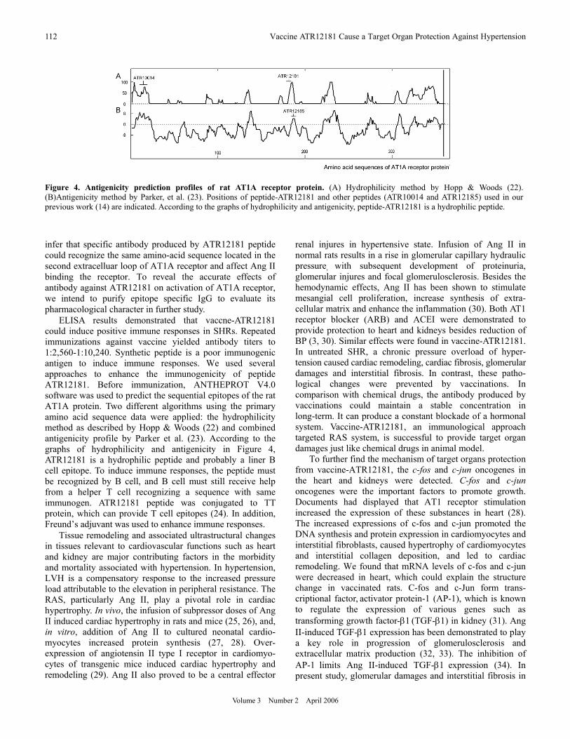
ELISA results demonstrated that vaccne-ATR12181 could induce positive immune responses in SHRs. Repeated immunizations against vaccine yielded antibody titers to 1:2,560-1:10,240. Synthetic peptide is a poor immunogenic antigen to induce immune responses. We used several approaches to enhance the immunogenicity of peptide ATR12181. Before immunization, ANTHEPROT V4.0 software was used to predict the sequential epitopes of the rat AT1A protein. Two different algorithms using the primary amino acid sequence data were applied: the hydrophilicity method as described by Hopp & Woods (22) and combined antigenicity profile by Parker et al. (23). According to the graphs of hydrophilicity and antigenicity in Figure 4, ATR12181 is a hydrophilic peptide and probably a liner B cell epitope. To induce immune responses, the peptide must be recognized by B cell, and B cell must still receive help from a helper T cell recognizing a sequence with same immunogen. ATR12181 peptide was conjugated to TT protein, which can provide T cell epitopes (24). In addition, Freund’s adjuvant was used to enhance immune responses.
Tissue remodeling and associated ultrastructural changes in tissues relevant to cardiovascular functions such as heart and kidney are major contributing factors in the morbidity and mortality associated with hypertension. In hypertension, LVH is a compensatory response to the increased pressure load attributable to the elevation in peripheral resistance. The RAS, particularly Ang II, play a pivotal role in cardiac hypertrophy. In vivo, the infusion of subpressor doses of Ang II induced cardiac hypertrophy in rats and mice (25, 26), and, in vitro, addition of Ang II to cultured neonatal cardio- myocytes increased protein synthesis (27, 28). Over- expression of angiotensin II type I receptor in cardiomyo- cytes of transgenic mice induced cardiac hypertrophy and remodeling (29). Ang II also proved to be a central effector
renal injures in hypertensive state. Infusion of Ang II in normal rats results in a rise in glomerular capillary hydraulic pressure, with subsequent development of proteinuria, glomerular injures and focal glomerulosclerosis. Besides the hemodynamic effects, Ang II has been shown to stimulate mesangial cell proliferation, increase synthesis of extra- cellular matrix and enhance the inflammation (30). Both AT1 receptor blocker (ARB) and ACEI were demonstrated to provide protection to heart and kidneys besides reduction of BP (3, 30). Similar effects were found in vaccine-ATR12181. In untreated SHR, a chronic pressure overload of hyper- tension caused cardiac remodeling, cardiac fibrosis, glomerular damages and interstitial fibrosis. In contrast, these patho- logical changes were prevented by vaccinations. In comparison with chemical drugs, the antibody produced by vaccinations could maintain a stable concentration in long-term. It can produce a constant blockade of a hormonal system. Vaccine-ATR12181, an immunological approach targeted RAS system, is successful to provide target organ damages just like chemical drugs in animal model. To further find the mechanism of target organs protection from vaccine-ATR12181, the c-fos and c-jun oncogenes in the heart and kidneys were detected. C-fos and c-jun oncogenes were the important factors to promote growth. Documents had displayed that AT1 receptor stimulation increased the expression of these substances in heart (28). The increased expressions of c-fos and c-jun promoted the DNA synthesis and protein expression in cardiomyocytes and interstitial fibroblasts, caused hypertrophy of cardiomyocytes and interstitial collagen deposition, and led to cardiac remodeling. We found that mRNA levels of c-fos and c-jun were decreased in heart, which could explain the structure change in vaccinated rats. C-fos and c-Jun form trans- criptional factor, activator protein-1 (AP-1), which is known to regulate the expression of various genes such as transforming growth factor-β1 (TGF-β1) in kidney (31). Ang II-induced TGF-β1 expression has been demonstrated to play a key role in progression of glomerulosclerosis and extracellular matrix production (32, 33). The inhibition of AP-1 limits Ang II-induced TGF-β1 expression (34). In present study, glomerular damages and interstitial fibrosis in
Figure 4. Antigenicity prediction profiles of rat AT1A receptor protein. (A) Hydrophilicity method by Hopp & Woods (22). (B)Antigenicity method by Parker, et al. (23). Positions of peptide-ATR12181 and other peptides (ATR10014 and ATR12185) used in our previous work (14) are indicated. According to the graphs of hydrophilicity and antigenicity, peptide-ATR12181 is a hydrophilic peptide.
Cellular & Molecular Immunology 113
Volume 3 Number 2 April 2006
kidney were prevented by vaccinations. Decreased mRNA levels of c-fos and c-jun might explain the pathylogical changes in kidney.
Since a self antigen was used, the safety of vaccine- ATR12181 is concern. In the history of RAS vaccine, the renin-vaccine was the most effective. Unfortunately, anti- renin antibody deposition was found in the juxtaglomerular apparatus, and progressive interstilial inflammatory injury of kidney (8, 9). In present study, we didn’t find any signs of autoimmune diseases in the sections of heart. Kidney has abundant arterioles and capillary, where the AT1A receptors are abundantly expressed. There were no signs of peri- vascular cellular inflammation. Although the thickening basement membrane of glomcruluses was found under TEM, control group also developed similar ultrastructure changes that were more serious. In addition, there were no signs of depositions in basement membrane or other places of glomcruluses in vaccinated group.
Vaccine-ATR12181 attenuated the development of hemodynamic alterations of hypertension, and 17 mmHg decrease of SBP was achieved. However, SHRs still were regarded as hypertension. Hypertension is a complex disease, and many factors contribute to elevation of BP. The vaccine only targets one of them, so it is difficult to decrease the BP to target level.
A role for RAS in the development or maintenance of hypertension is well established (1, 2). Our results confirm a fundamental role for AT1 receptors in both the development of high BP and the production of pathological organ damage. The interruption of the RAS pathway by using an angiotensin II receptor (AT1) vaccine ATR12181 offers the potential to prevent the development of hypertension and its associated pathophysiological alterations with infrequent admini- strations. Of course, there are still several questions need to be answered. How does the antibody against vaccine- ATR12181 lower BP? Is it safe to use a self antigen? Studies are now under way to determine these doubts. Acknowledgment This work was funded by National Natural Science Foundation of China (No. 30300133). References
1. Whelton PK. Epidemiology of hypertension. Lancet. 1944;344: 101-106.
2. Ruzicka M, Leenen FH. Update on local cardiac renin-angiotensin system. Curr Opin Cardiol. 1997;12:347-353.
3. Dahlof B, Deveruex RB, Kjeldsen SE, et al. Cardiovascular morbidity and mortality in the Losartan Intervention For Endpoint reduction in hypertension study (LIFE): a randomised trial against atenolol. Lancet. 2002;359:995-1003.
4. Sunman W, Sever PS. Non-angiotensin effects of angiotensin- converting enzyme inhibitors. Clin Sci. 1993;85:661-670.
5. Moser M. Angiotensin-converting enzyme inhibitors, angiotensin II receptor antagonists and calcium channel blocking agents: a review of potential benefits and possible adverse reactions. J
Am Coll Cardiol. 1997;29:1414-1422. 6. Michel JB. Renin-angiotensin vaccine: old story, new project
‘efficacy versus safety’. Clin Sci. 2000;10:145-147. 7. Deodhar SD, Haas E, Goldblatt H. Production of antirenin to
homologous renin and its effect on experimental renal hypertension. J Exp Med. 1964;139:425-435.
8. Michel JB, Sayah S, Guettier C, et al. Physiological and immunopathological consequences of active immunization of spontaneously hypertensive and normotensive rats against murine rennin. Circulation. 1990;81:1899-1910.
9. Michel JB, Guettier C, Philippe M, Galen FX, Corvol P, Menard J. Active immunization against renin in normotensive marmoset. Proc Natl Acad Sci U S A. 1987;204:281-288.
10. Gardiner SM, Auton TR, Downham MR, et al. Active immunization with angiotensin I peptide analogue vaccines selectively reduces the pressor effects of exogenous angiotensin I in conscious rats. Br J Pharmacol. 2000;129:1178-1182.
11. Brown MJ, Coltart J, Gunewardena K, Ritter JM, Auton TR, Glover JF. Randomized double-blind placebo-controlled study of an angiotensin immunotherapeutic vaccine (PMD3117) in hypertensive subjects. Clin Sci (Lond). 2004;107:167-173.
12. Zelezna B, Veselsky L, Velek J, Dobesova Z, Zicha J, Kunes J. Influence of active immunization against angiotensin AT1 or AT2 receptor on hypertension development in young and adult SHR. Physiol Res. 1999;48:259-265.
13. Downham MR, Auton TR, Rosul A, et al. Evaluation of two carrier protein–angiotensin I conjugate vaccines to assess their future potential to control high blood pressure (hypertension) in man. Br J Clin Pharmacol. 2003;56:505-512.
14. Li LD, Liao YH, Zhou ZH, et al. Effects of active immunization with agiotensin II receptor type 1 peptides on blood pressure and cardiovascular remodeling in spontaneous hypertensive rats. Zhongguo Mian Yi Xue Za Zhi. 2005;21:372-376.
15. Burson JM, Aguilera G, Gross KW, Sigmund CD. Differential expression of angiotensin receptor 1A and 1B in mouse. Am J Physiol. 1994;276:E260-E267.
16. Wallukat G, Homuth V, Fischer T, et al. Patients with preeclampsia develop agonistic antibodies against the angiotension AT1 receptor. J Clin Invest. 1999;103:945-952.
17. Thway TM, Shlykov SG, Day MC, et al. Antibodies from preeclamptic patients stimulate increased intracellular Ca2+ mobilization through angiotensin receptor activation. Circulation. 2004;110:1612-1619.
18. de Gasparo M, Catt KJ, Inagami T, Wright JW, Unger TH. International union of pharmacology. XXIII. The angiotensin II receptors. Pharmacol Rev. 2000;52:415-472.
19. Fu ML, Schulze W, Wallukat G, et al. Immunohistochemical localization of angiotensin II receptors (AT1) in the heart with anti-peptide antibodies showing a positive chronotropic effect. Receptors Channels. 1998;6:99-111.
20. Zelezna B, Velek J, Veselsky L, Zicha J, Dobesova Z, Kunes J. Blockade of AT1 receptors by specific antibody attenuated hypertension development in young spontaneously hypertensive rats. Physiol Res.1996;45:475-477.
21. Richards EM, Lu D, Zelezna B, et al. Inhibition of central angiotensin responses by angiotensin type-1 receptor antibody. Hypertension. 1993;21:1062-1065.
22. Hopp TP, Woods KR. Prediction of protein antigenic determinants from amino acid sequences. Proc Natl Acad Sci U S A. 1981;78:3824-3828.
23. Parker JMR, Guo D, Hodges RS. New hydrophilic scale derived from high-performance liquid chromatography peptide retention data: correlation of predicted surface residues with antigenicity and X-ray-derived accessible sites. Biochemistry. 1986;25:
114 Vaccine ATR12181 Cause a Target Organ Protection Against Hypertension
Volume 3 Number 2 April 2006
5426-5432. 24. Demotz S, Lanzavecchia A, Eisel U, Niemann H, Widmann C,
Corradin G. Delineation of several DR-restricted tetanus toxin T cell epitopes. J Immumol. 1989;142:394-402.
25. Harada K, Komuro I, Shiojima I, et al. Pressure overload induces cardiac hypertrophy in angiotensin II type 1A receptor knockout mice. Circulation.1998;97:1952-1959.
26. Susic D, Nunez E, Frohlich ED, Prakash O. Angiotensin II increases left ventricular mass without affecting myosin isoform mRNAs. Hypertension. 1996;28:265-268.
27. Booz GW, Baker KM. Role of type 1 and type 2 angiotensin receptors in angiotensin II-induced cardiomyocyte hypertrophy. Hypertension. 1996;28:635-640.
28. Sadoshima J, Xu Y, Slayter HS, Izumo S. Autocrine release of angiotensin II mediates stretch-induced hypertrophy of cardiac myocytes in vitro. Cell. 1993;75:977-984.
29. Paradis P, Dali-Youcef N, Paradis FW, Thibault G, Nemer M. Overexpression of angiotensin II type I receptor in cardio-
myocytes induces cardiac hypertrophy and remodeling. Proc Natl Acad Sci U S A. 2000;97:931-936.
30. Taal MW, Brenner BM. Renoprotective benefits of RAS inhibition: from ACEI to angiotensin II antagonists. Kidney Int. 2000;57:1803-1817.
31. Kim SJ, Angel P, Lafyatis R, et al. Autoinduction of transforming growth factor β1 is mediated by the AP-1 complex. Mol Cell Biol. 1990;10:1492-1497.
32. Border WA, Ruoslahti E. Transforming growth factor-β in disease: The dark side of tissue repair. J Clin Invest. 1992;90: 1-7.
33. Border WA, Noble NA. Transforming growth factor β in tissue fibrosis. N Engl J Med. 1994;331:1286-1292.
34. Birchenall-Roberts MC, Ruscetti FW, Kasper J, et al. Transcriptional regulation of the transforming growth factor β1 promoter by v-src gene products is mediated through the AP-1 complex. Mol Cell Biol. 1990;10:4978-4983.