Embed Size (px)
Citation preview
Artifacts, Faults and Failures: a Review © Leica Microsystems 2012
Artifacts, Faults and Failures: a Review
Presented by: Geoffrey Rolls and Fiona Tarbet
Leica Microsystems, Biosystems Division, Melbourne, Australia
AIMS NSM Darwin 2012
Artifacts, Faults and Failures: a Review © Leica Microsystems 2012
1878Page 2
Artifacts, Faults and Failures: a Review © Leica Microsystems 2012
Thomas Davies 1878
Page 3 Artifacts, Faults and Failures: a Review © Leica Microsystems 2012 Page 4
We have been producing artifacts, faults and failures for very many years (some of us being more productive than others)!
This is a Cambridge Rocking microtome (“Cambridge Rocker”)Question: In which year was it first offered for sale?Answer: 1885

Artifacts, Faults and Failures: a Review © Leica Microsystems 2012
Introduction
For each of our 70 cases we want you to:
1. Identify the artifact, fault or failure2. Explain the most likely cause/s3. Describe how you could overcome the problem and avoid
its occurrence in the future
Page 5 Artifacts, Faults and Failures: a Review © Leica Microsystems 2012
Definitions
Artifact: a structure that is not normally present in living tissueFor the Histologist: an artifact is a structure that is not normally present in a well-prepared sectionFault: a defect or blemish, culpability, offence, misdeedFailure: non-performance, not a success, catastrophe, fiasco
Page 6
Artifacts, Faults and Failures: a Review © Leica Microsystems 2012 Page 7
Case 1 : Lung H&E
Artifacts, Faults and Failures: a Review © Leica Microsystems 2012 Page 8
Case 1 : Lung – inhaled seaweed from drowning (H&E)
Artifacts, Faults and Failures: a Review © Leica Microsystems 2012 Page 9
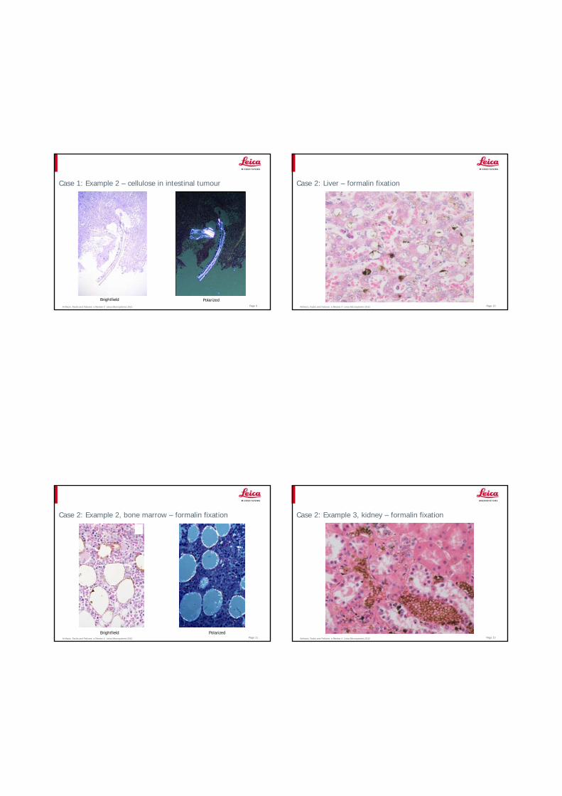
Case 1: Example 2 – cellulose in intestinal tumour
Brightfield PolarizedArtifacts, Faults and Failures: a Review © Leica Microsystems 2012 Page 10
Case 2: Liver – formalin fixation
Artifacts, Faults and Failures: a Review © Leica Microsystems 2012 Page 11
Case 2: Example 2, bone marrow – formalin fixation
Brightfield PolarizedArtifacts, Faults and Failures: a Review © Leica Microsystems 2012 Page 12
Case 2: Example 3, kidney – formalin fixation
Artifacts, Faults and Failures: a Review © Leica Microsystems 2012 Page 13
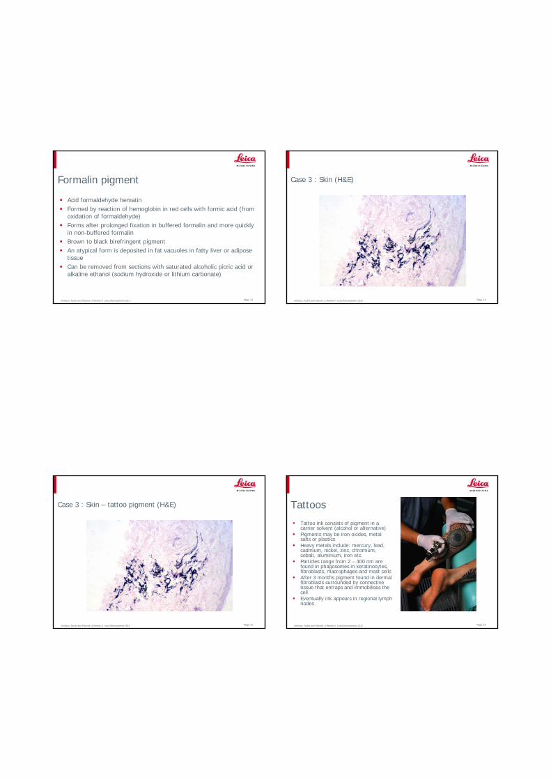
Formalin pigment
Acid formaldehyde hematinFormed by reaction of hemoglobin in red cells with formic acid (from oxidation of formaldehyde)Forms after prolonged fixation in buffered formalin and more quickly in non-buffered formalinBrown to black birefringent pigmentAn atypical form is deposited in fat vacuoles in fatty liver or adipose tissueCan be removed from sections with saturated alcoholic picric acid or alkaline ethanol (sodium hydroxide or lithium carbonate)
Artifacts, Faults and Failures: a Review © Leica Microsystems 2012 Page 14
Case 3 : Skin (H&E)
Artifacts, Faults and Failures: a Review © Leica Microsystems 2012 Page 15
Case 3 : Skin – tattoo pigment (H&E)
Artifacts, Faults and Failures: a Review © Leica Microsystems 2012 Page 16
Tattoos
Tattoo ink consists of pigment in a carrier solvent (alcohol or alternative)Pigments may be iron oxides, metal salts or plasticsHeavy metals include: mercury, lead, cadmium, nickel, zinc, chromium, cobalt, aluminium, iron etc.Particles range from 2 – 400 nm are found in phagosomes in keratinocytes, fibroblasts, macrophages and mast cellsAfter 3 months pigment found in dermal fibroblasts surrounded by connective tissue that entraps and immobilises the cellEventually ink appears in regional lymph nodes
Artifacts, Faults and Failures: a Review © Leica Microsystems 2012 Page 17
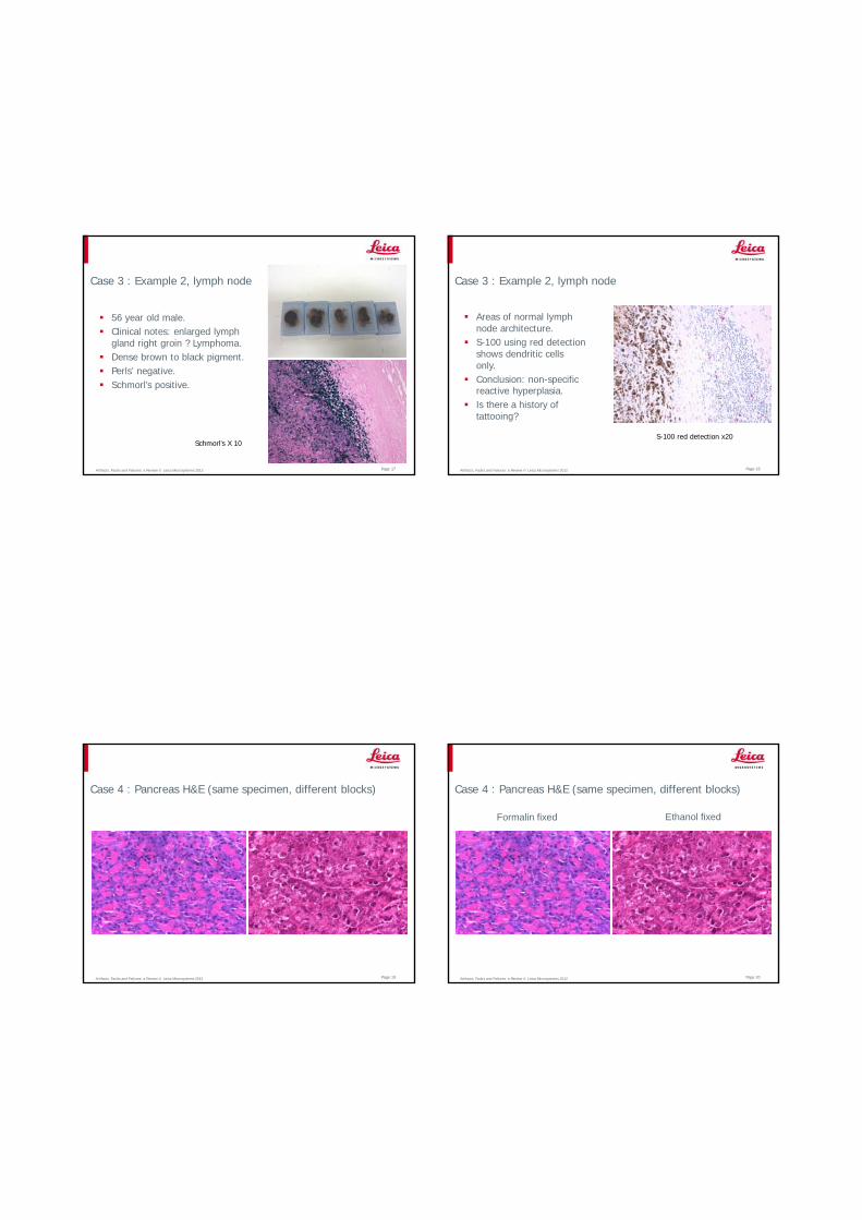
Case 3 : Example 2, lymph node
56 year old male.Clinical notes: enlarged lymph gland right groin ? Lymphoma.Dense brown to black pigment.Perls’ negative.Schmorl’s positive.
Schmorl’s X 10
Artifacts, Faults and Failures: a Review © Leica Microsystems 2012 Page 18
Case 3 : Example 2, lymph node
Areas of normal lymph node architecture.S-100 using red detection shows dendritic cells only.Conclusion: non-specific reactive hyperplasia.Is there a history of tattooing?
S-100 red detection x20
Artifacts, Faults and Failures: a Review © Leica Microsystems 2012 Page 19
Case 4 : Pancreas H&E (same specimen, different blocks)
Artifacts, Faults and Failures: a Review © Leica Microsystems 2012 Page 20
Case 4 : Pancreas H&E (same specimen, different blocks)
Ethanol fixedFormalin fixed

Artifacts, Faults and Failures: a Review © Leica Microsystems 2012 Page 21
Case 5 : Endoscopic biopsy (H&E)
Artifacts, Faults and Failures: a Review © Leica Microsystems 2012 Page 22
Case 5 : Endoscopic biopsy (H&E)
H&E PAS
Artifacts, Faults and Failures: a Review © Leica Microsystems 2012 Page 23
Case 5 : Endoscopic biopsy (with contaminating sesame seed)
Sesame seeds from gut contents
Artifacts, Faults and Failures: a Review © Leica Microsystems 2012 Page 24
Case 6 : Lung (H&E)
Artifacts, Faults and Failures: a Review © Leica Microsystems 2012 Page 25
Case 6: Lung-mechanical specimen compression (H&E)
With forceps while tissue is fresh (during removal)Local compression (cassette bars)
Artifacts, Faults and Failures: a Review © Leica Microsystems 2012 Page 26
Case 7: Splenic capsule (H&E)
Artifacts, Faults and Failures: a Review © Leica Microsystems 2012 Page 27
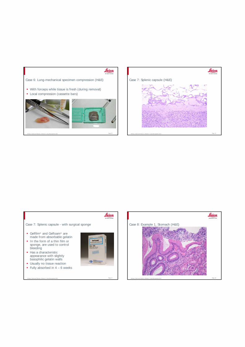
Case 7: Splenic capsule - with surgical sponge
Gelfilm® and Gelfoam® are made from absorbable gelatinIn the form of a thin film or sponge, are used to control bleedingHas a characteristic appearance with slightly basophilic gelatin wallsUsually no tissue reactionFully absorbed in 4 – 6 weeks
Artifacts, Faults and Failures: a Review © Leica Microsystems 2012 Page 28
Case 8: Example 1, Stomach (H&E)
Artifacts, Faults and Failures: a Review © Leica Microsystems 2012 Page 29
Case 8: Example 2, Cardiac muscle (H&E)
Artifacts, Faults and Failures: a Review © Leica Microsystems 2012 Page 30
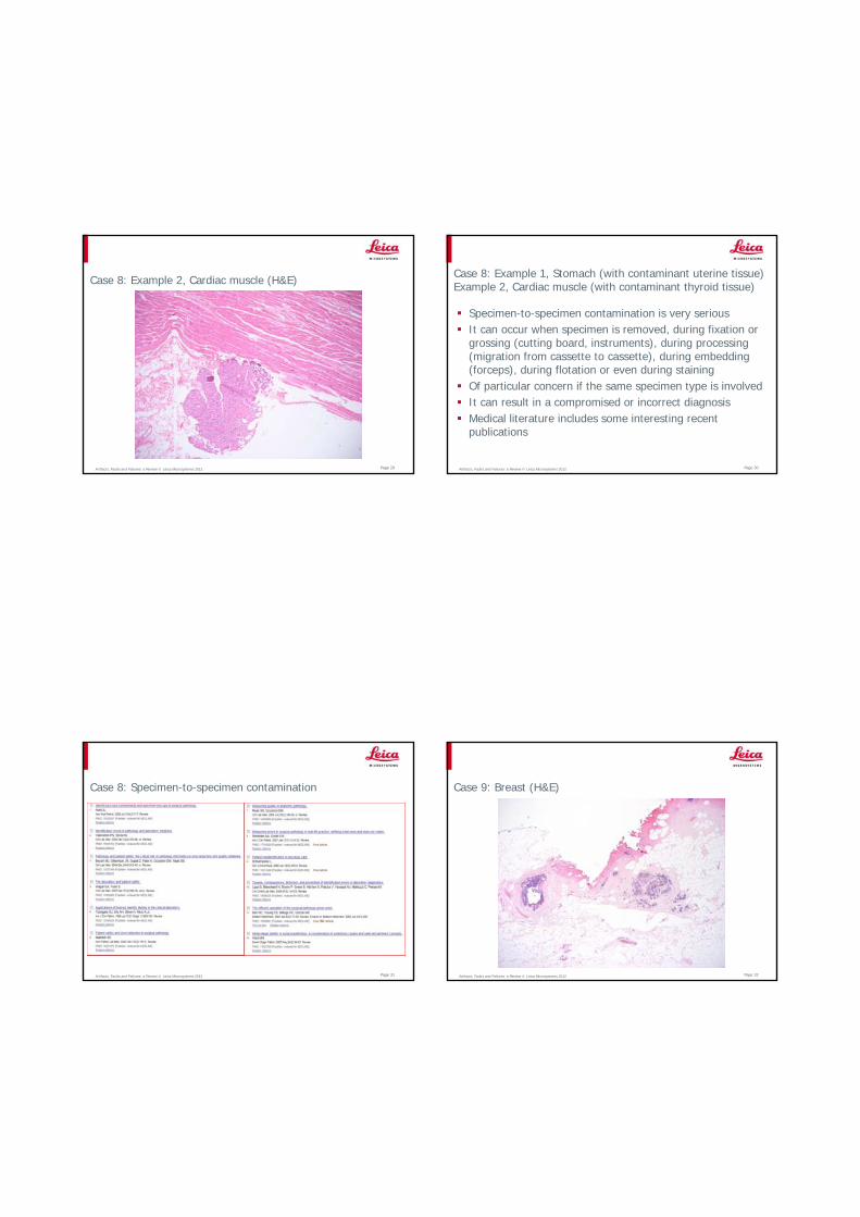
Case 8: Example 1, Stomach (with contaminant uterine tissue) Example 2, Cardiac muscle (with contaminant thyroid tissue)
Specimen-to-specimen contamination is very seriousIt can occur when specimen is removed, during fixation or grossing (cutting board, instruments), during processing (migration from cassette to cassette), during embedding (forceps), during flotation or even during stainingOf particular concern if the same specimen type is involvedIt can result in a compromised or incorrect diagnosisMedical literature includes some interesting recent publications
Artifacts, Faults and Failures: a Review © Leica Microsystems 2012 Page 31
Case 8: Specimen-to-specimen contamination
Artifacts, Faults and Failures: a Review © Leica Microsystems 2012 Page 32
Case 9: Breast (H&E)
Artifacts, Faults and Failures: a Review © Leica Microsystems 2012 Page 33
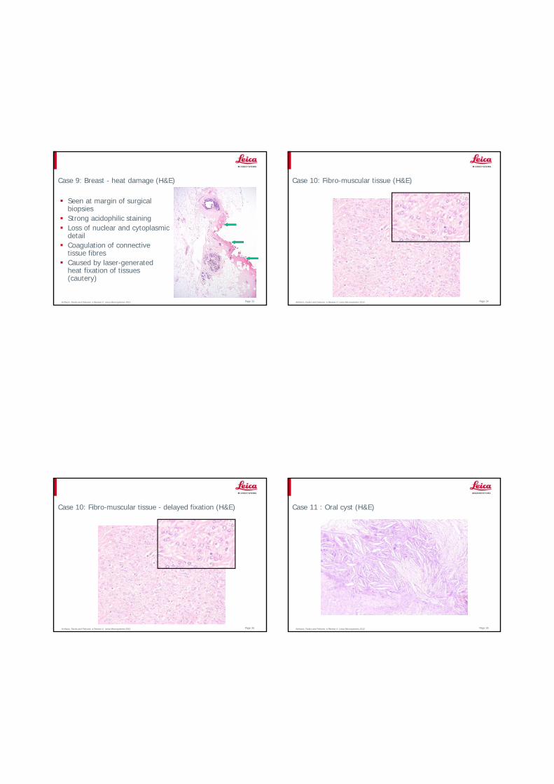
Case 9: Breast - heat damage (H&E)
Seen at margin of surgical biopsiesStrong acidophilic stainingLoss of nuclear and cytoplasmic detailCoagulation of connective tissue fibresCaused by laser-generated heat fixation of tissues (cautery)
Artifacts, Faults and Failures: a Review © Leica Microsystems 2012 Page 34
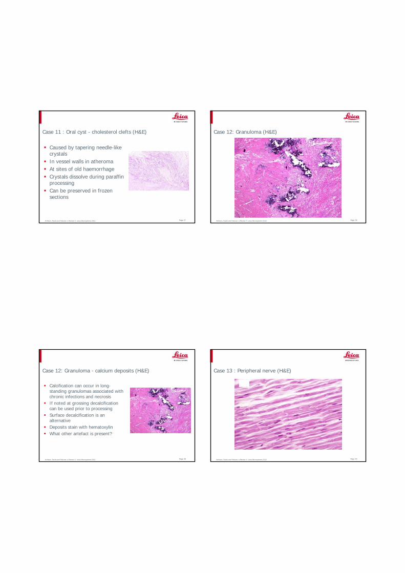
Case 10: Fibro-muscular tissue (H&E)
Artifacts, Faults and Failures: a Review © Leica Microsystems 2012 Page 35
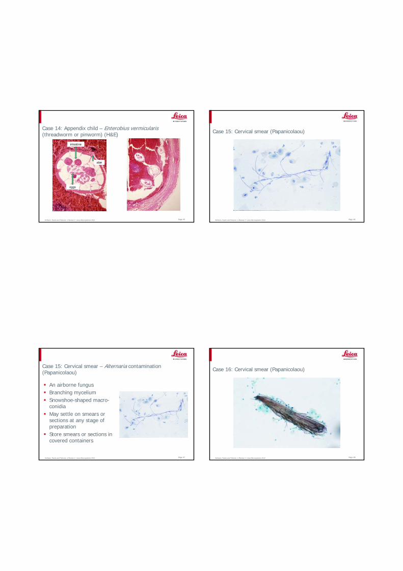
Case 10: Fibro-muscular tissue - delayed fixation (H&E)
Artifacts, Faults and Failures: a Review © Leica Microsystems 2012 Page 36
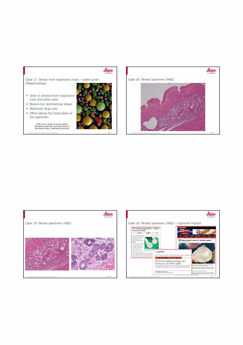
Case 11 : Oral cyst (H&E)
Artifacts, Faults and Failures: a Review © Leica Microsystems 2012 Page 37
Case 11 : Oral cyst - cholesterol clefts (H&E)
Caused by tapering needle-like crystalsIn vessel walls in atheromaAt sites of old haemorrhageCrystals dissolve during paraffin processingCan be preserved in frozen sections
Artifacts, Faults and Failures: a Review © Leica Microsystems 2012 Page 38
Case 12: Granuloma (H&E)
Artifacts, Faults and Failures: a Review © Leica Microsystems 2012 Page 39
Case 12: Granuloma - calcium deposits (H&E)
Calcification can occur in long-standing granulomas associated with chronic infections and necrosisIf noted at grossing decalcification can be used prior to processingSurface decalcification is an alternative Deposits stain with hematoxylinWhat other artefact is present?
Artifacts, Faults and Failures: a Review © Leica Microsystems 2012 Page 40
Case 13 : Peripheral nerve (H&E)

Artifacts, Faults and Failures: a Review © Leica Microsystems 2012 Page 41
Case 13 : Peripheral nerve (H&E)
Artifacts, Faults and Failures: a Review © Leica Microsystems 2012 Page 42
Case 13 : Peripheral nerve - Schmidt-Lanterman incisures
Artifacts, Faults and Failures: a Review © Leica Microsystems 2012
Case 13 : Peripheral nerve
Artifacts, Faults and Failures: a Review © Leica Microsystems 2012 Page 44
Case 14: Appendix child (H&E)
Artifacts, Faults and Failures: a Review © Leica Microsystems 2012 Page 45
Case 14: Appendix child – Enterobius vermicularis(threadworm or pinworm) (H&E)
intestine
alae
eggs
Artifacts, Faults and Failures: a Review © Leica Microsystems 2012 Page 46
Case 15: Cervical smear (Papanicolaou)
Artifacts, Faults and Failures: a Review © Leica Microsystems 2012 Page 47
Case 15: Cervical smear – Alternaria contamination (Papanicolaou)
An airborne fungusBranching myceliumSnowshoe-shaped macro-conidiaMay settle on smears or sections at any stage of preparationStore smears or sections in covered containers
Artifacts, Faults and Failures: a Review © Leica Microsystems 2012 Page 48
Case 16: Cervical smear (Papanicolaou)

Artifacts, Faults and Failures: a Review © Leica Microsystems 2012 Page 49
Case 16: Cervical smear – contaminant insect material? (Papanicolaou)
Two smears collected on the same occasionContain contaminant insect material from an unidentified source
Artifacts, Faults and Failures: a Review © Leica Microsystems 2012
Case 16: Example 2, cardiac muscle – insect contaminant (H&E)
Page 50
Artifacts, Faults and Failures: a Review © Leica Microsystems 2012 Page 51
Case 17: Smear from respiratory tract (Papanicoloau)
Artifacts, Faults and Failures: a Review © Leica Microsystems 2012 Page 52
Case 17: Smear from respiratory tract – pollen grain (Papanicoloau)
Seen in smears from respiratory tract and other sitesBizarre but symmetrical shapeRelatively large sizeOften above the focal plane of the specimen
Artifacts, Faults and Failures: a Review © Leica Microsystems 2012 Page 53
Case 17: Smear from respiratory tract – pollen grain (Papanicoloau)
Seen in smears from respiratory tract and other sitesBizarre but symmetrical shapeRelatively large sizeOften above the focal plane of the specimen
Pollen from a variety of common plantsMicrograph (colorized): Dartmouth Electron Microscope Facility (Wikimedia Commons)
Artifacts, Faults and Failures: a Review © Leica Microsystems 2012 Page 54
Case 18: Breast specimen (H&E)
Artifacts, Faults and Failures: a Review © Leica Microsystems 2012 Page 55
Case 18: Breast specimen (H&E)
Artifacts, Faults and Failures: a Review © Leica Microsystems 2012 Page 56
Case 18: Breast specimen (H&E) – ruptured implant
Artifacts, Faults and Failures: a Review © Leica Microsystems 2012 Page 57
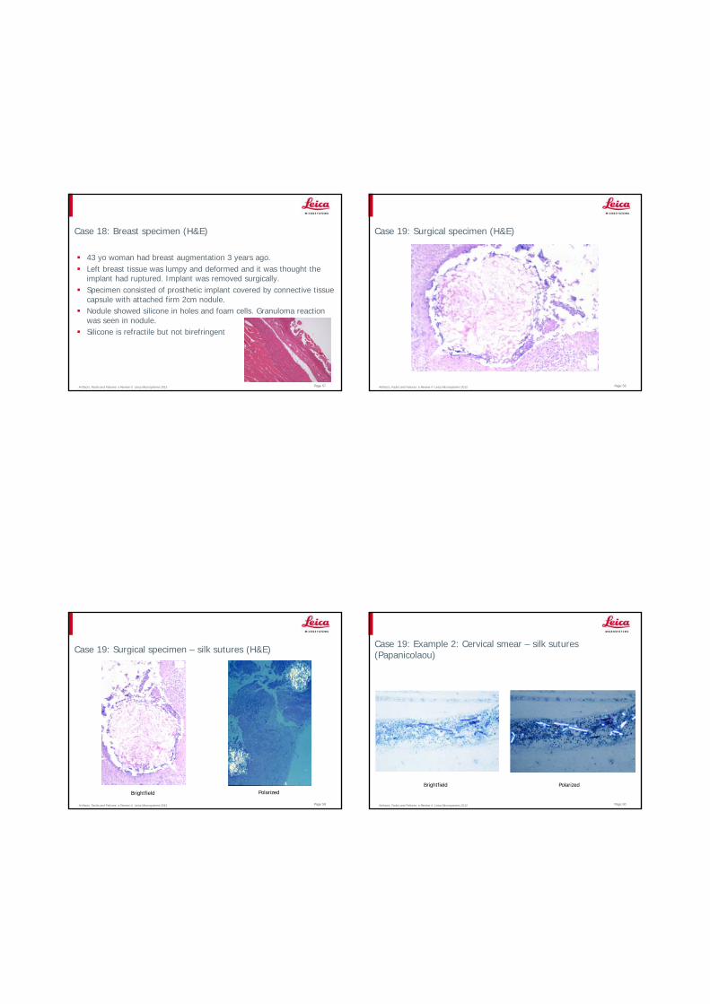
Case 18: Breast specimen (H&E)
43 yo woman had breast augmentation 3 years ago.Left breast tissue was lumpy and deformed and it was thought the implant had ruptured. Implant was removed surgically.Specimen consisted of prosthetic implant covered by connective tissue capsule with attached firm 2cm nodule.Nodule showed silicone in holes and foam cells. Granuloma reaction was seen in nodule.Silicone is refractile but not birefringent
Artifacts, Faults and Failures: a Review © Leica Microsystems 2012 Page 58
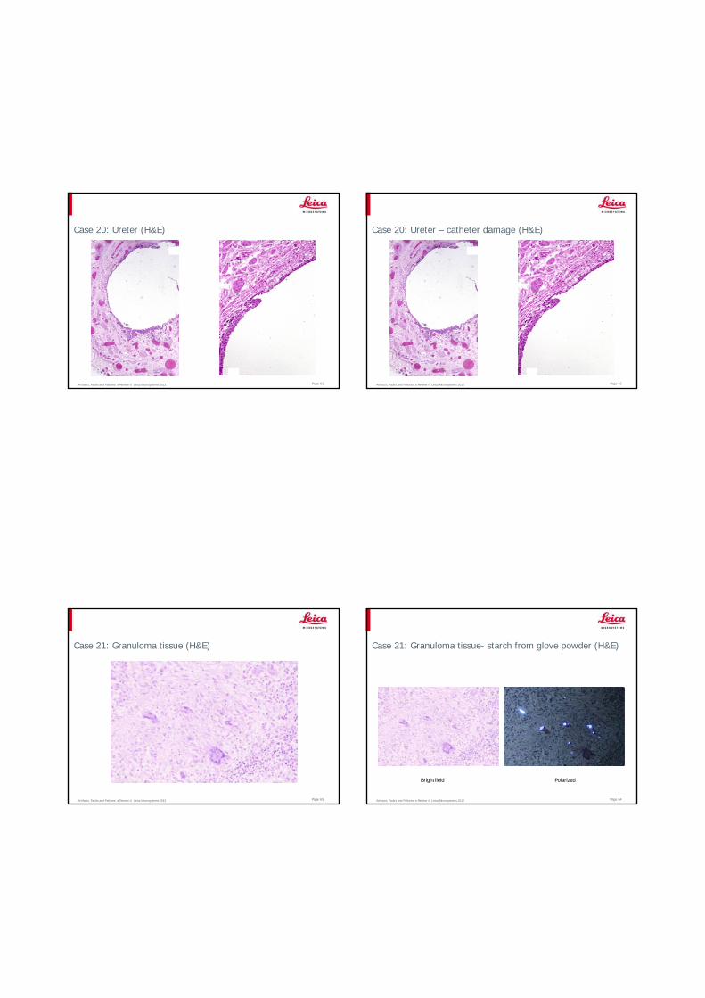
Case 19: Surgical specimen (H&E)
Artifacts, Faults and Failures: a Review © Leica Microsystems 2012 Page 59
Case 19: Surgical specimen – silk sutures (H&E)
Brightfield Polarized
Artifacts, Faults and Failures: a Review © Leica Microsystems 2012 Page 60
Case 19: Example 2: Cervical smear – silk sutures (Papanicolaou)
Brightfield Polarized
Artifacts, Faults and Failures: a Review © Leica Microsystems 2012 Page 61
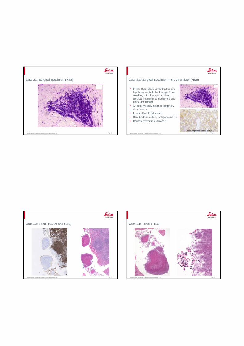
Case 20: Ureter (H&E)
Artifacts, Faults and Failures: a Review © Leica Microsystems 2012 Page 62
Case 20: Ureter – catheter damage (H&E)
Artifacts, Faults and Failures: a Review © Leica Microsystems 2012 Page 63
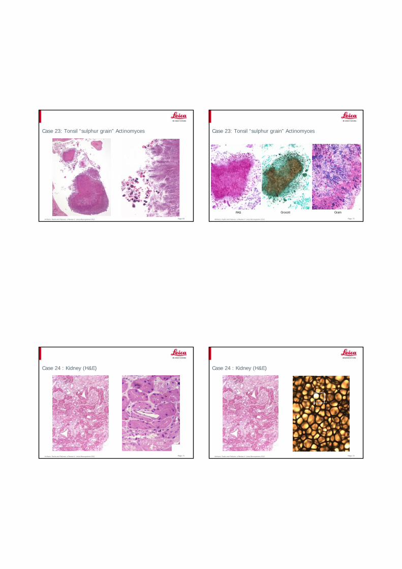
Case 21: Granuloma tissue (H&E)
Artifacts, Faults and Failures: a Review © Leica Microsystems 2012 Page 64
Case 21: Granuloma tissue- starch from glove powder (H&E)
Brightfield Polarized
Artifacts, Faults and Failures: a Review © Leica Microsystems 2012 Page 65
Case 22: Surgical specimen (H&E)
Artifacts, Faults and Failures: a Review © Leica Microsystems 2012 Page 66
Case 22: Surgical specimen – crush artifact (H&E)
In the fresh state some tissues are highly susceptible to damage from crushing with forceps or other surgical instruments (lymphoid and glandular tissue)Artifact typically seen at periphery of specimenIn small localized areasCan displace cellular antigens in IHCCauses irreversible damage
B cell lymphoma stained for L26
Artifacts, Faults and Failures: a Review © Leica Microsystems 2012 Page 67
Case 23: Tonsil (CD20 and H&E)
Artifacts, Faults and Failures: a Review © Leica Microsystems 2012 Page 68
Case 23: Tonsil (H&E)
Artifacts, Faults and Failures: a Review © Leica Microsystems 2012 Page 69
Case 23: Tonsil “sulphur grain” Actinomyces
Artifacts, Faults and Failures: a Review © Leica Microsystems 2012 Page 70
Case 23: Tonsil “sulphur grain” Actinomyces
PAS Grocott Gram
Artifacts, Faults and Failures: a Review © Leica Microsystems 2012 Page 71
Case 24 : Kidney (H&E)
Artifacts, Faults and Failures: a Review © Leica Microsystems 2012 Page 72
Case 24 : Kidney (H&E)
Artifacts, Faults and Failures: a Review © Leica Microsystems 2012 Page 73
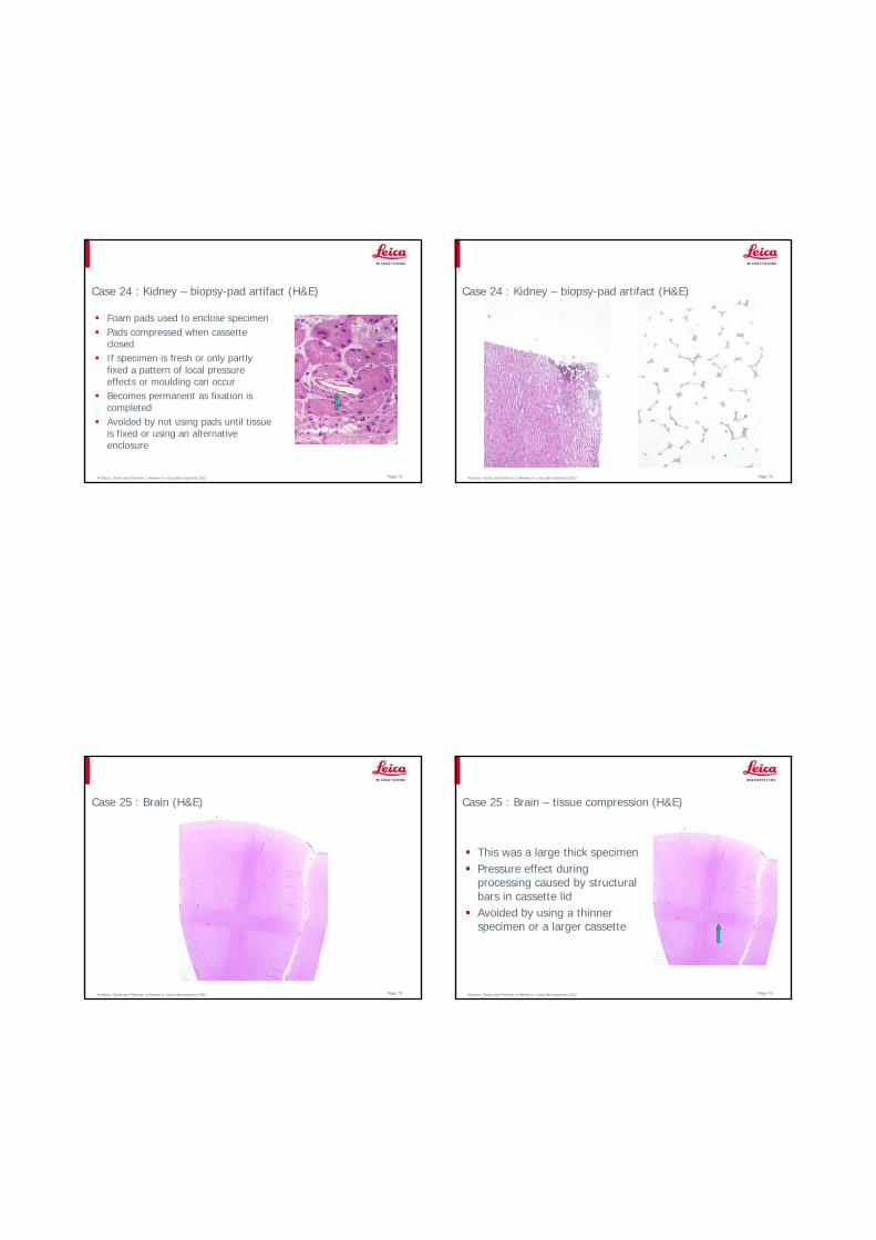
Case 24 : Kidney – biopsy-pad artifact (H&E)
Foam pads used to enclose specimenPads compressed when cassette closedIf specimen is fresh or only partly fixed a pattern of local pressure effects or moulding can occurBecomes permanent as fixation is completedAvoided by not using pads until tissue is fixed or using an alternative enclosure
Artifacts, Faults and Failures: a Review © Leica Microsystems 2012 Page 74
Case 24 : Kidney – biopsy-pad artifact (H&E)
Artifacts, Faults and Failures: a Review © Leica Microsystems 2012 Page 75
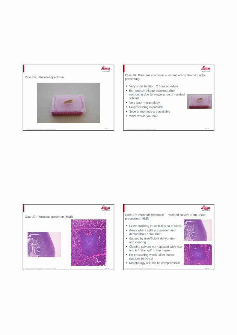
Case 25 : Brain (H&E)
Artifacts, Faults and Failures: a Review © Leica Microsystems 2012 Page 76
Case 25 : Brain – tissue compression (H&E)
This was a large thick specimenPressure effect during processing caused by structural bars in cassette lidAvoided by using a thinner specimen or a larger cassette
Artifacts, Faults and Failures: a Review © Leica Microsystems 2012 Page 77
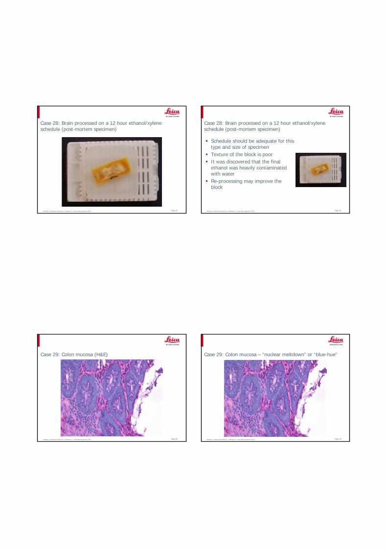
Case 26: Pancreas specimen
Artifacts, Faults and Failures: a Review © Leica Microsystems 2012 Page 78
Case 26: Pancreas specimen – incomplete fixation & under-processing
Very short fixation, 2 hour scheduleExtreme shrinkage occurred after sectioning due to evaporation of residual solventVery poor morphologyRe-processing is possibleSeveral methods are availableWhat would you do?
Artifacts, Faults and Failures: a Review © Leica Microsystems 2012 Page 79
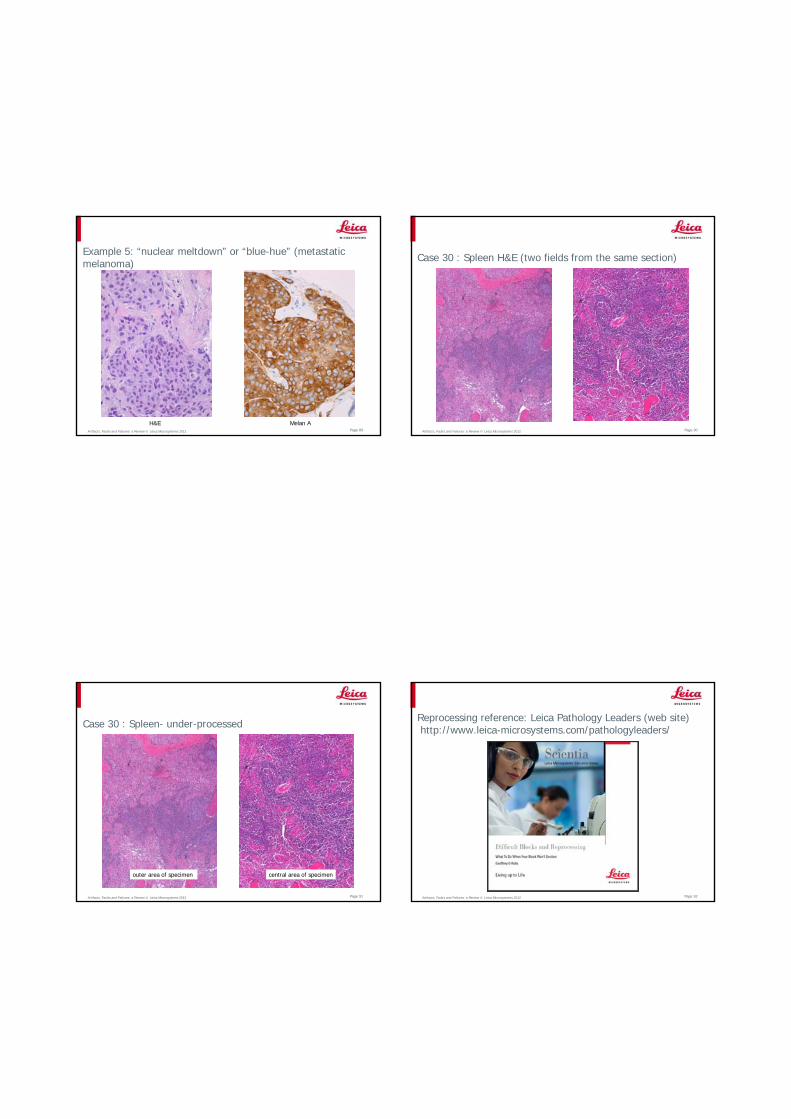
Case 27: Pancreas specimen (H&E)
Artifacts, Faults and Failures: a Review © Leica Microsystems 2012 Page 80
Case 27: Pancreas specimen – retained solvent from under-processing (H&E)
Gross cracking in central area of blockAreas where cells are swollen and demonstrate “blue hue” Caused by insufficient dehydration and clearingClearing solvent not replaced with wax and is “retained” in the tissueRe-processing would allow better sections to be cutMorphology will still be compromised
Artifacts, Faults and Failures: a Review © Leica Microsystems 2012 Page 81
Case 28: Brain processed on a 12 hour ethanol/xylene schedule (post-mortem specimen)
Artifacts, Faults and Failures: a Review © Leica Microsystems 2012 Page 82
Case 28: Brain processed on a 12 hour ethanol/xylene schedule (post-mortem specimen)
Schedule should be adequate for this type and size of specimenTexture of the block is poorIt was discovered that the final ethanol was heavily contaminated with waterRe-processing may improve the block
Artifacts, Faults and Failures: a Review © Leica Microsystems 2012 Page 83
Case 29: Colon mucosa (H&E)
Artifacts, Faults and Failures: a Review © Leica Microsystems 2012 Page 84
Case 29: Colon mucosa – “nuclear meltdown” or “blue-hue”

Artifacts, Faults and Failures: a Review © Leica Microsystems 2012
Some causes of “nuclear meltdown” or “blue-hue”
Allowing a specimen to dry out before fixation Using xylene that has been contaminated with water for clearingUsing wax contaminated with formalin or formalin and ethanol during processingFailing to completely replace solvent with wax (retained solvent) – schedule too short, expired reagents, processor faultOver-heating the section when dryingIneffective de-waxing during staining (“pink disease”)
Page 85 Artifacts, Faults and Failures: a Review © Leica Microsystems 2012
Avoiding “nuclear meltdown” or “blue-hue”
Avoid improper handling of fresh specimensFix properlyProcess thoroughly with un-contaminated reagentsAvoid excessive heat when drying sectionsMake sure de-waxing is complete prior to stainingNote that re-processing can help, staining may be improved, but morphology generally remains compromised
Page 86
Artifacts, Faults and Failures: a Review © Leica Microsystems 2012 Page 87
Example 2: Kidney – “nuclear meltdown” or “blue-hue”
Artifacts, Faults and Failures: a Review © Leica Microsystems 2012 Page 88
Examples 3 & 4: “nuclear meltdown” or “blue-hue”
Spleen Gut -muscularis
Artifacts, Faults and Failures: a Review © Leica Microsystems 2012 Page 89
Example 5: “nuclear meltdown” or “blue-hue” (metastatic melanoma)
H&E Melan AArtifacts, Faults and Failures: a Review © Leica Microsystems 2012 Page 90
Case 30 : Spleen H&E (two fields from the same section)
Artifacts, Faults and Failures: a Review © Leica Microsystems 2012 Page 91
Case 30 : Spleen- under-processed
outer area of specimen central area of specimen
Artifacts, Faults and Failures: a Review © Leica Microsystems 2012 Page 92
Reprocessing reference: Leica Pathology Leaders (web site)http://www.leica-microsystems.com/pathologyleaders/
Artifacts, Faults and Failures: a Review © Leica Microsystems 2012 Page 93
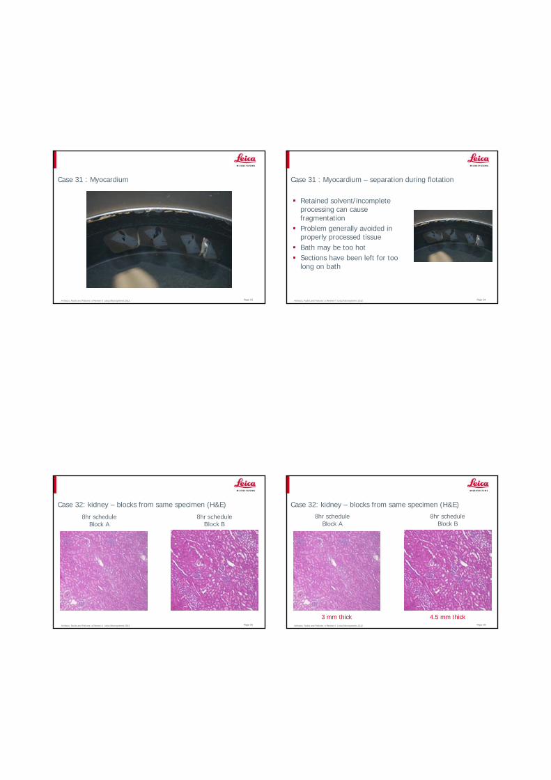
Case 31 : Myocardium
Artifacts, Faults and Failures: a Review © Leica Microsystems 2012 Page 94
Case 31 : Myocardium – separation during flotation
Retained solvent/incomplete processing can cause fragmentationProblem generally avoided in properly processed tissueBath may be too hotSections have been left for too long on bath
Artifacts, Faults and Failures: a Review © Leica Microsystems 2012 Page 95
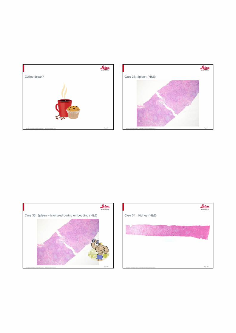
Case 32: kidney – blocks from same specimen (H&E)
8hr schedule Block A
8hr schedule Block B
Artifacts, Faults and Failures: a Review © Leica Microsystems 2012 Page 96
Case 32: kidney – blocks from same specimen (H&E)
8hr schedule Block A
8hr schedule Block B
3 mm thick 4.5 mm thick
Artifacts, Faults and Failures: a Review © Leica Microsystems 2012 Page 97
Coffee Break?
Artifacts, Faults and Failures: a Review © Leica Microsystems 2012 Page 98
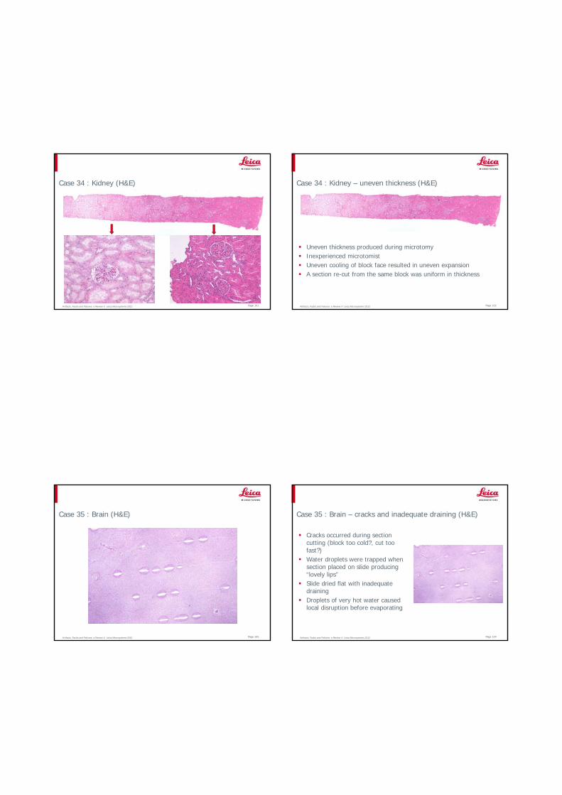
Case 33: Spleen (H&E)
Artifacts, Faults and Failures: a Review © Leica Microsystems 2012 Page 99
Case 33: Spleen – fractured during embedding (H&E)
Artifacts, Faults and Failures: a Review © Leica Microsystems 2012 Page 100
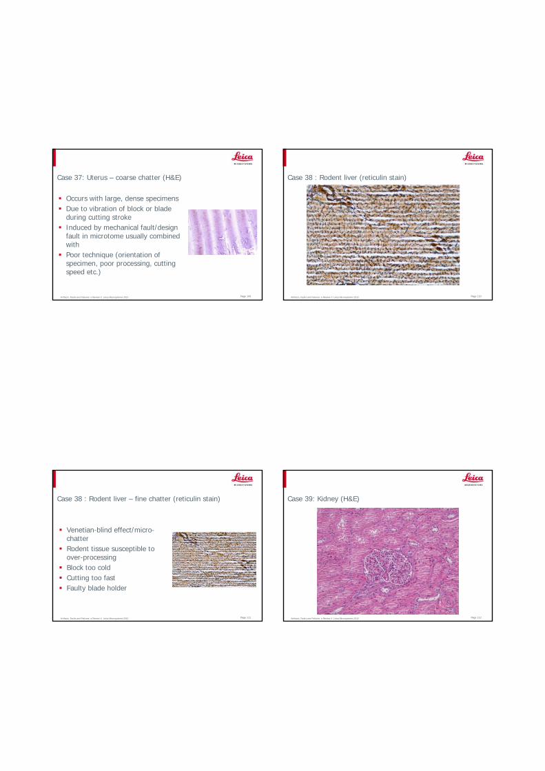
Case 34 : Kidney (H&E)
Artifacts, Faults and Failures: a Review © Leica Microsystems 2012 Page 101
Case 34 : Kidney (H&E)
Artifacts, Faults and Failures: a Review © Leica Microsystems 2012 Page 102
Case 34 : Kidney – uneven thickness (H&E)
Uneven thickness produced during microtomyInexperienced microtomistUneven cooling of block face resulted in uneven expansionA section re-cut from the same block was uniform in thickness
Artifacts, Faults and Failures: a Review © Leica Microsystems 2012 Page 103
Case 35 : Brain (H&E)
Artifacts, Faults and Failures: a Review © Leica Microsystems 2012 Page 104
Case 35 : Brain – cracks and inadequate draining (H&E)
Cracks occurred during section cutting (block too cold?, cut too fast?)Water droplets were trapped when section placed on slide producing “lovely lips”Slide dried flat with inadequate drainingDroplets of very hot water caused local disruption before evaporating

Artifacts, Faults and Failures: a Review © Leica Microsystems 2012 Page 105
Case 36: Submucosa colon (H&E) POC (H&E)
Artifacts, Faults and Failures: a Review © Leica Microsystems 2012 Page 106
Case 36: Submucosa colon - severe blade defect (H&E)
In these cases microtomist was a learnerForceps were dropped onto bladeDamage to the section was visible in ribbon and during flotationSection should never have been picked up
Artifacts, Faults and Failures: a Review © Leica Microsystems 2012 Page 107
Case 36: Blade defect (visible at flotation)
Artifacts, Faults and Failures: a Review © Leica Microsystems 2012 Page 108
Case 37: Uterus (H&E)
Artifacts, Faults and Failures: a Review © Leica Microsystems 2012 Page 109
Case 37: Uterus – coarse chatter (H&E)
Occurs with large, dense specimensDue to vibration of block or blade during cutting strokeInduced by mechanical fault/design fault in microtome usually combined withPoor technique (orientation of specimen, poor processing, cutting speed etc.)
Artifacts, Faults and Failures: a Review © Leica Microsystems 2012 Page 110
Case 38 : Rodent liver (reticulin stain)
Artifacts, Faults and Failures: a Review © Leica Microsystems 2012 Page 111
Case 38 : Rodent liver – fine chatter (reticulin stain)
Venetian-blind effect/micro-chatterRodent tissue susceptible to over-processingBlock too coldCutting too fastFaulty blade holder
Artifacts, Faults and Failures: a Review © Leica Microsystems 2012 Page 112
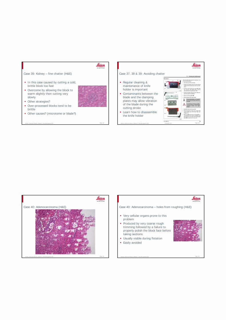
Case 39: Kidney (H&E)
Artifacts, Faults and Failures: a Review © Leica Microsystems 2012 Page 113
Case 39: Kidney – fine chatter (H&E)
In this case caused by cutting a cold, brittle block too fastOvercome by allowing the block to warm slightly then cutting very slowlyOther strategies?Over-processed blocks tend to be brittleOther causes? (microtome or blade?)
Artifacts, Faults and Failures: a Review © Leica Microsystems 2012 Page 114
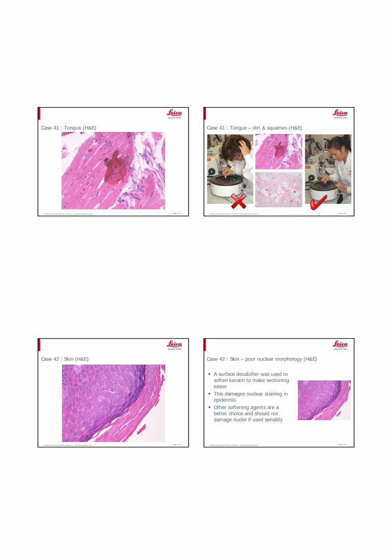
Case 37, 38 & 39: Avoiding chatter
15 October 2012
Regular cleaning & maintenance of knife holder is importantContaminants between the blade and the clamping plates may allow vibration of the blade during the cutting strokeLearn how to disassemble the knife holder
Artifacts, Faults and Failures: a Review © Leica Microsystems 2012 Page 115
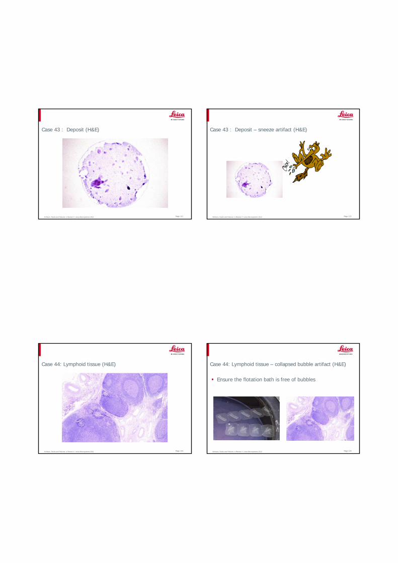
Case 40: Adenocarcinoma (H&E)
Artifacts, Faults and Failures: a Review © Leica Microsystems 2012 Page 116
Case 40: Adenocarcinoma – holes from roughing (H&E)
Very cellular organs prone to this problemProduced by very coarse rough trimming followed by a failure to properly polish the block face before taking sectionsUsually visible during flotationEasily avoided
Artifacts, Faults and Failures: a Review © Leica Microsystems 2012 Page 117
Case 41 : Tongue (H&E)
Artifacts, Faults and Failures: a Review © Leica Microsystems 2012 Page 118
Case 41 : Tongue – dirt & squames (H&E)
Artifacts, Faults and Failures: a Review © Leica Microsystems 2012 Page 119
Case 42 : Skin (H&E)
Artifacts, Faults and Failures: a Review © Leica Microsystems 2012 Page 120
Case 42 : Skin – poor nuclear morphology (H&E)
A surface decalcifier was used to soften keratin to make sectioning easierThis damages nuclear staining in epidermisOther softening agents are a better choice and should not damage nuclei if used sensibly
Artifacts, Faults and Failures: a Review © Leica Microsystems 2012 Page 121
Case 43 : Deposit (H&E)
Artifacts, Faults and Failures: a Review © Leica Microsystems 2012 Page 122
Case 43 : Deposit – sneeze artifact (H&E)
Artifacts, Faults and Failures: a Review © Leica Microsystems 2012 Page 123
Case 44: Lymphoid tissue (H&E)
Artifacts, Faults and Failures: a Review © Leica Microsystems 2012 Page 124
Case 44: Lymphoid tissue – collapsed bubble artifact (H&E)
Ensure the flotation bath is free of bubbles

Artifacts, Faults and Failures: a Review © Leica Microsystems 2012 Page 125
Case 45: Spleen (H&E)
Artifacts, Faults and Failures: a Review © Leica Microsystems 2012 Page 126
Case 45: Spleen – heat damage during drying (H&E)
Slide dried flat on a hotplateUneven surface temperature produced hot-spotsProduced cracking and some cell shrinkage
Artifacts, Faults and Failures: a Review © Leica Microsystems 2012 Page 127
Case 46: Vagina (H&E)
Artifacts, Faults and Failures: a Review © Leica Microsystems 2012 Page 128
Case 46: Vagina – contaminant hair (H&E)

Artifacts, Faults and Failures: a Review © Leica Microsystems 2012 Page 129
Case 47 : Mucosa (H&E)
Artifacts, Faults and Failures: a Review © Leica Microsystems 2012 Page 130
Case 47 : Mucosa (H&E)
Artifacts, Faults and Failures: a Review © Leica Microsystems 2012 Page 131
Case 47 : Mucosa – eosin staining fault (H&E)
Only eosinophils and red cells have stained with eosinIn this case the water wash that follows the blueing alkali was skippedEosin will not bind under alkaline conditions
Artifacts, Faults and Failures: a Review © Leica Microsystems 2012 Page 132
Case 48: Liver after hematoxylin stain (incomplete H&E)
Artifacts, Faults and Failures: a Review © Leica Microsystems 2012 Page 133
Case 48: Liver after hematoxylin stain (incomplete H&E)
Incorrect setting of microscope condenser diaphragm when examining wet section (no cover slip)
Partially closed Fully open
Artifacts, Faults and Failures: a Review © Leica Microsystems 2012 Page 134
Case 49: Endoscopic biopsy (H&E)
Artifacts, Faults and Failures: a Review © Leica Microsystems 2012 Page 135
Case 49 : Endoscopic biopsy – residual wax (H&E)
two sections mounted on the same slide
Artifacts, Faults and Failures: a Review © Leica Microsystems 2012 Page 136
Case 50: Skin (H&E)
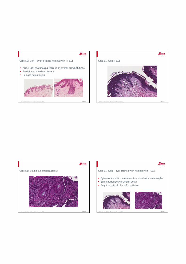
Artifacts, Faults and Failures: a Review © Leica Microsystems 2012 Page 137
Case 50: Skin – over-oxidized hematoxylin (H&E)
Nuclei lack sharpness & there is an overall brownish tingePrecipitated mordant presentReplace hematoxylin
Artifacts, Faults and Failures: a Review © Leica Microsystems 2012 Page 138
Case 51: Skin (H&E)
Artifacts, Faults and Failures: a Review © Leica Microsystems 2012 Page 139
Case 51: Example 2, mucosa (H&E)
Artifacts, Faults and Failures: a Review © Leica Microsystems 2012 Page 140
Case 51: Skin – over-stained with hematoxylin (H&E)
Cytoplasm and fibrous elements stained with hematoxylinSome nuclei lack chromatin detailRequires acid alcohol differentiation

Artifacts, Faults and Failures: a Review © Leica Microsystems 2012 Page 141
Case 52: Appendix (H&E)
Artifacts, Faults and Failures: a Review © Leica Microsystems 2012 Page 142
Case 52: Appendix – needle crystals present (H&E)
Crystals present on top of sectionProbably precipitated mordant from hematoxylinCrystallization may occur because of solvent evaporation and concentration of salts or dyes in the staining bathEvaporation occurs during manual staining Filter reagent or replace
Artifacts, Faults and Failures: a Review © Leica Microsystems 2012 Page 143
Case 52: Example 2, Masson Trichrome
Artifacts, Faults and Failures: a Review © Leica Microsystems 2012 Page 144
Case 53: Bone marrow (PAS)
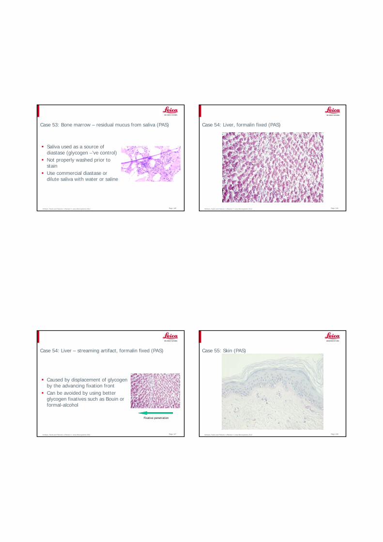
Artifacts, Faults and Failures: a Review © Leica Microsystems 2012 Page 145
Case 53: Bone marrow – residual mucus from saliva (PAS)
Saliva used as a source of diastase (glycogen –’ve control)Not properly washed prior to stainUse commercial diastase or dilute saliva with water or saline
Artifacts, Faults and Failures: a Review © Leica Microsystems 2012 Page 146
Case 54: Liver, formalin fixed (PAS)
Artifacts, Faults and Failures: a Review © Leica Microsystems 2012 Page 147
Case 54: Liver – streaming artifact, formalin fixed (PAS)
Caused by displacement of glycogen by the advancing fixation frontCan be avoided by using better glycogen fixatives such as Bouin or formal-alcohol
Fixative penetration
Artifacts, Faults and Failures: a Review © Leica Microsystems 2012 Page 148
Case 55: Skin (PAS)
Artifacts, Faults and Failures: a Review © Leica Microsystems 2012 Page 149
Case 55: Skin – under-oxidized (PAS)
Insufficient oxidation with periodic acidNote staining of basement membrane
30 sec 5 min
Artifacts, Faults and Failures: a Review © Leica Microsystems 2012 Page 150
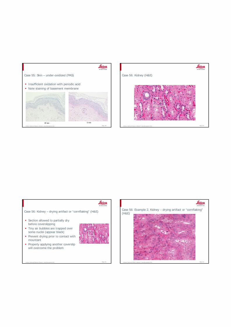
Case 56: Kidney (H&E)
Artifacts, Faults and Failures: a Review © Leica Microsystems 2012 Page 151
Case 56: Kidney – drying artifact or “cornflaking” (H&E)
Section allowed to partially dry before coverslippingTiny air bubbles are trapped over some nuclei (appear black)Prevent drying prior to contact with mountantProperly applying another coverslip will overcome the problem
Artifacts, Faults and Failures: a Review © Leica Microsystems 2012 Page 152
Case 56: Example 2, Kidney – drying artifact or “cornflaking” (H&E)
Artifacts, Faults and Failures: a Review © Leica Microsystems 2012 Page 153
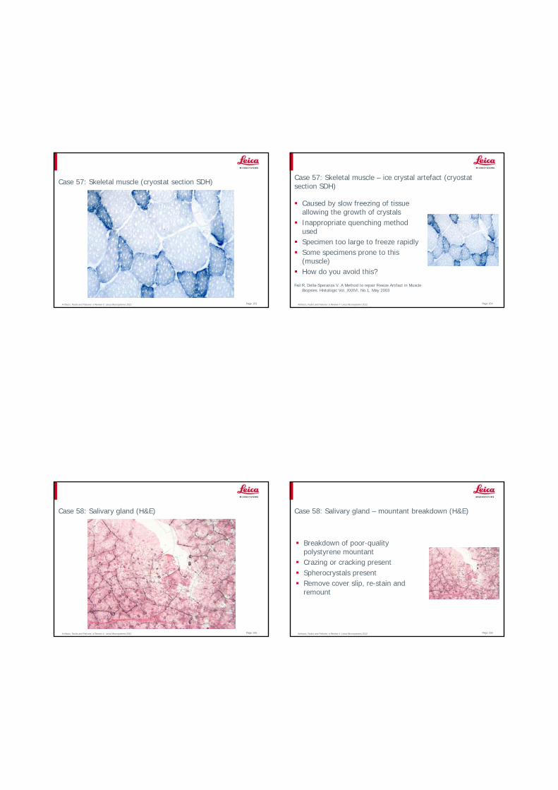
Case 57: Skeletal muscle (cryostat section SDH)
Artifacts, Faults and Failures: a Review © Leica Microsystems 2012 Page 154
Case 57: Skeletal muscle – ice crystal artefact (cryostat section SDH)
Caused by slow freezing of tissue allowing the growth of crystalsInappropriate quenching method usedSpecimen too large to freeze rapidlySome specimens prone to this (muscle)How do you avoid this?
Fail R, Della-Speranza V. A Method to repair Freeze Artifact in Muscle Biopsies. Histologic Vol. XXXVI, No.1, May 2003
Artifacts, Faults and Failures: a Review © Leica Microsystems 2012 Page 155
Case 58: Salivary gland (H&E)
Artifacts, Faults and Failures: a Review © Leica Microsystems 2012 Page 156
Case 58: Salivary gland – mountant breakdown (H&E)
Breakdown of poor-quality polystyrene mountantCrazing or cracking presentSpherocrystals presentRemove cover slip, re-stain and remount
Artifacts, Faults and Failures: a Review © Leica Microsystems 2012 Page 157
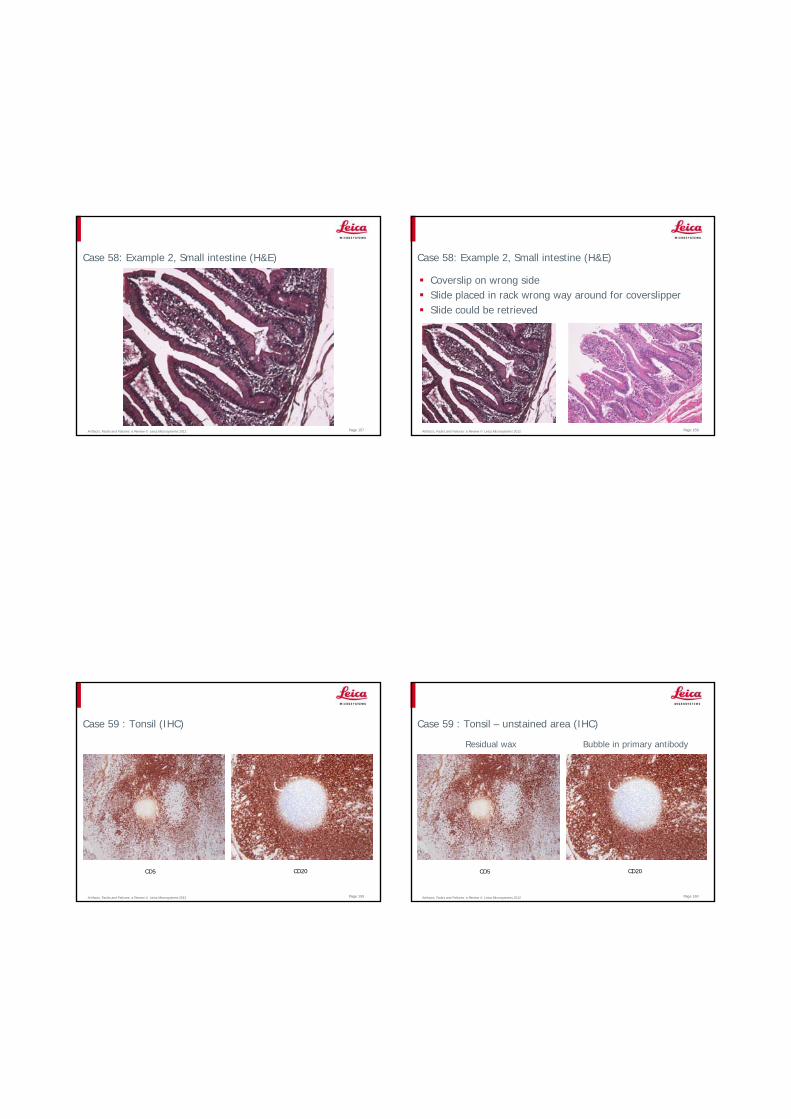
Case 58: Example 2, Small intestine (H&E)
Artifacts, Faults and Failures: a Review © Leica Microsystems 2012 Page 158
Case 58: Example 2, Small intestine (H&E)
Coverslip on wrong sideSlide placed in rack wrong way around for coverslipperSlide could be retrieved
Artifacts, Faults and Failures: a Review © Leica Microsystems 2012 Page 159
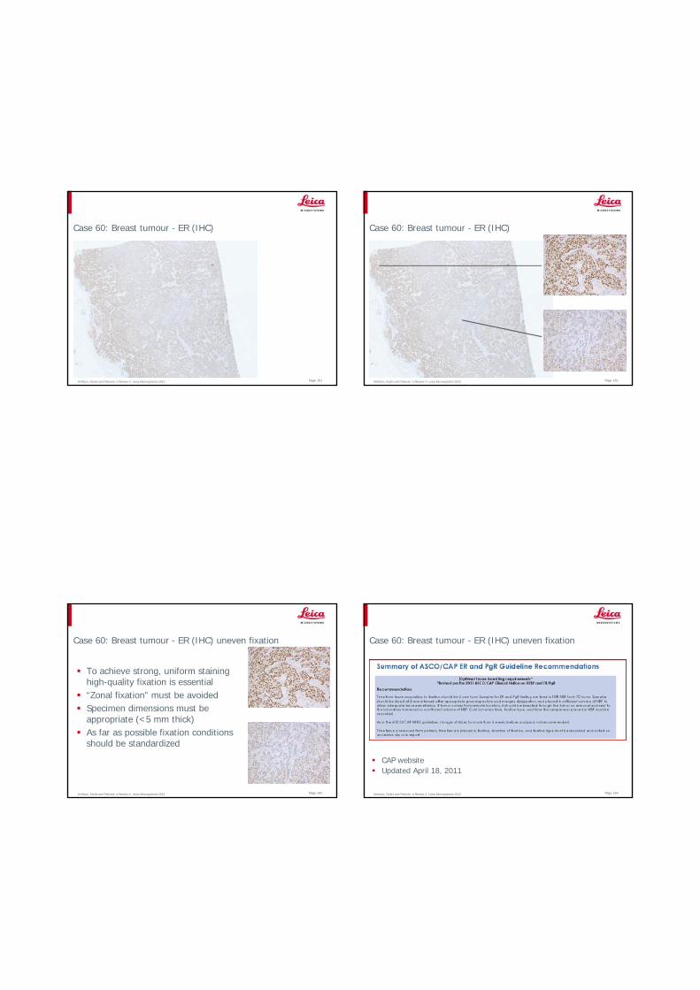
Case 59 : Tonsil (IHC)
CD5 CD20
Artifacts, Faults and Failures: a Review © Leica Microsystems 2012 Page 160
Case 59 : Tonsil – unstained area (IHC)
Residual wax Bubble in primary antibody
CD5 CD20
Artifacts, Faults and Failures: a Review © Leica Microsystems 2012 Page 161
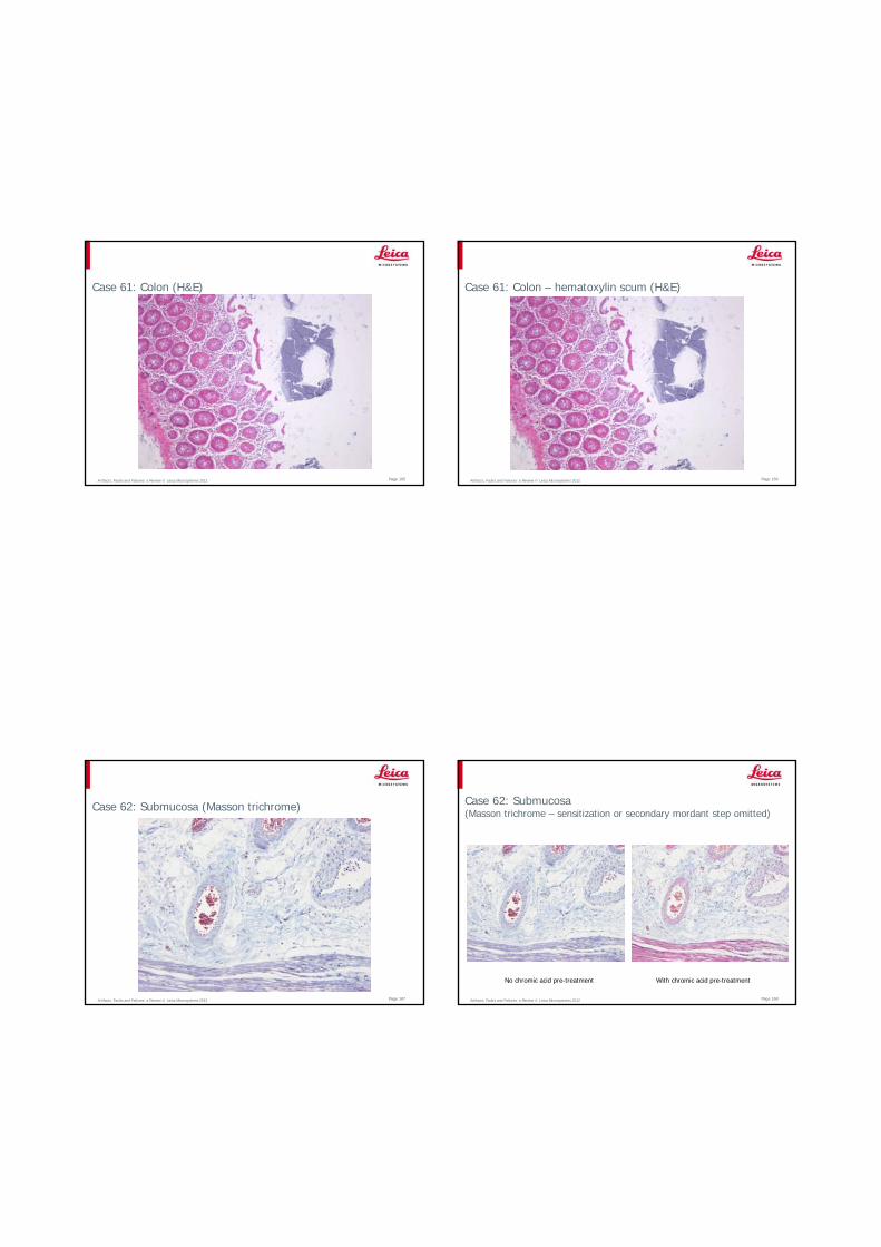
Case 60: Breast tumour - ER (IHC)
Artifacts, Faults and Failures: a Review © Leica Microsystems 2012 Page 162
Case 60: Breast tumour - ER (IHC)
Artifacts, Faults and Failures: a Review © Leica Microsystems 2012 Page 163
Case 60: Breast tumour - ER (IHC) uneven fixation
To achieve strong, uniform staining high-quality fixation is essential“Zonal fixation” must be avoidedSpecimen dimensions must be appropriate (<5 mm thick)As far as possible fixation conditions should be standardized
Artifacts, Faults and Failures: a Review © Leica Microsystems 2012 Page 164
Case 60: Breast tumour - ER (IHC) uneven fixation
CAP websiteUpdated April 18, 2011
Artifacts, Faults and Failures: a Review © Leica Microsystems 2012
Case 61: Colon (H&E)
Page 165 Artifacts, Faults and Failures: a Review © Leica Microsystems 2012
Case 61: Colon – hematoxylin scum (H&E)
Page 166
Artifacts, Faults and Failures: a Review © Leica Microsystems 2012 Page 167
Case 62: Submucosa (Masson trichrome)
Artifacts, Faults and Failures: a Review © Leica Microsystems 2012 Page 168
Case 62: Submucosa(Masson trichrome – sensitization or secondary mordant step omitted)
No chromic acid pre-treatment With chromic acid pre-treatment
Artifacts, Faults and Failures: a Review © Leica Microsystems 2012 Page 169
Case 63 : Gut muscularis externa (H&E)
Artifacts, Faults and Failures: a Review © Leica Microsystems 2012 Page 170
Case 63 : Gut muscularis externa – bleaching from micro-projector (H&E)
Artifacts, Faults and Failures: a Review © Leica Microsystems 2012 Page 171
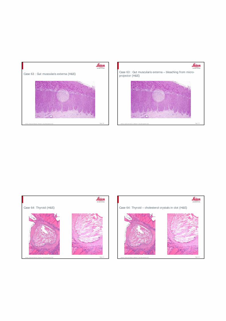
Case 64: Thyroid (H&E)
Artifacts, Faults and Failures: a Review © Leica Microsystems 2012 Page 172
Case 64: Thyroid – cholesterol crystals in clot (H&E)
Artifacts, Faults and Failures: a Review © Leica Microsystems 2012 Page 173
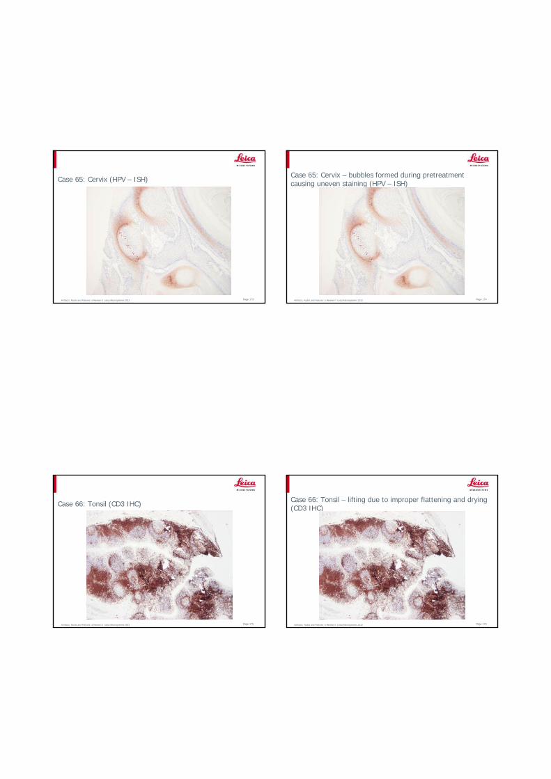
Case 65: Cervix (HPV – ISH)
Artifacts, Faults and Failures: a Review © Leica Microsystems 2012 Page 174
Case 65: Cervix – bubbles formed during pretreatment causing uneven staining (HPV – ISH)
Artifacts, Faults and Failures: a Review © Leica Microsystems 2012 Page 175
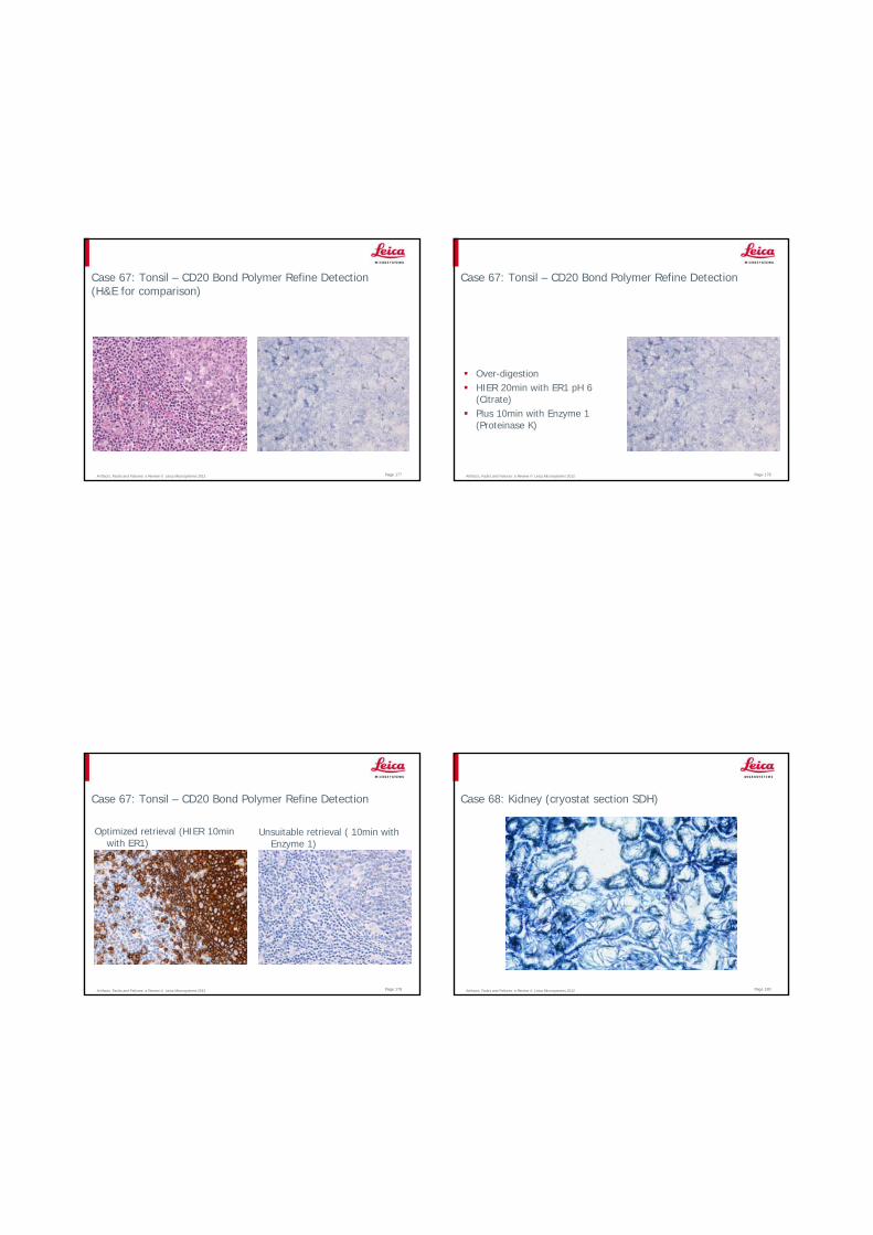
Case 66: Tonsil (CD3 IHC)
Artifacts, Faults and Failures: a Review © Leica Microsystems 2012 Page 176
Case 66: Tonsil – lifting due to improper flattening and drying (CD3 IHC)
Artifacts, Faults and Failures: a Review © Leica Microsystems 2012 Page 177
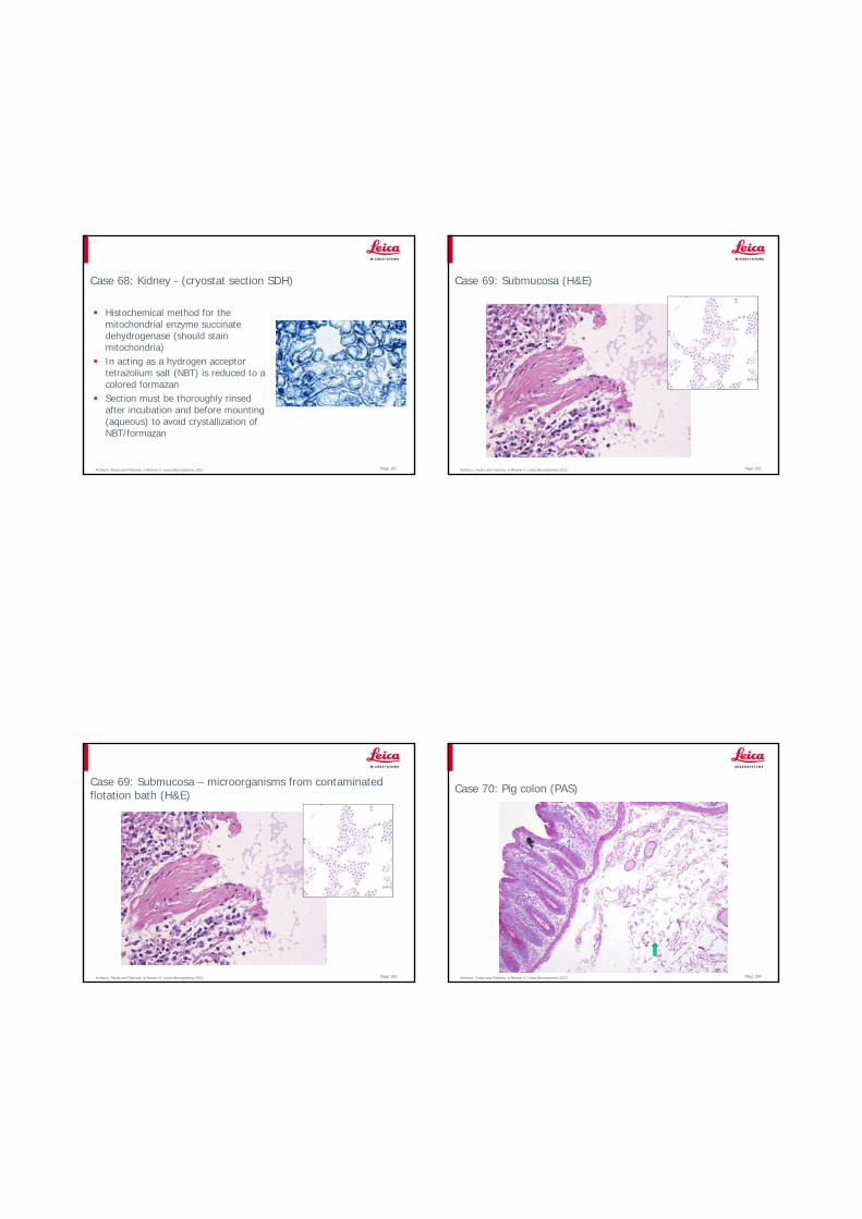
Case 67: Tonsil – CD20 Bond Polymer Refine Detection (H&E for comparison)
Artifacts, Faults and Failures: a Review © Leica Microsystems 2012 Page 178
Case 67: Tonsil – CD20 Bond Polymer Refine Detection
Over-digestion HIER 20min with ER1 pH 6 (Citrate)Plus 10min with Enzyme 1 (Proteinase K)
Artifacts, Faults and Failures: a Review © Leica Microsystems 2012 Page 179
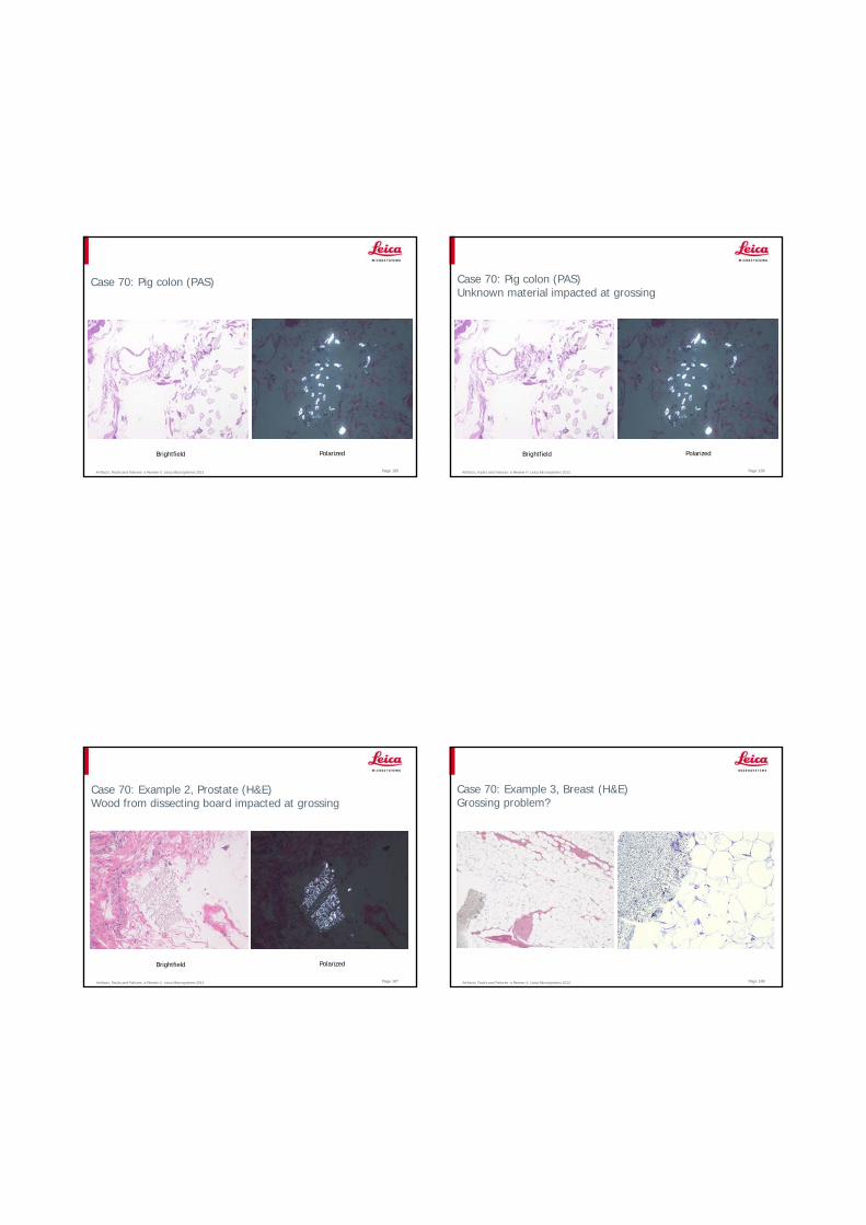
Case 67: Tonsil – CD20 Bond Polymer Refine Detection
Optimized retrieval (HIER 10min with ER1)
Unsuitable retrieval ( 10min with Enzyme 1)
Artifacts, Faults and Failures: a Review © Leica Microsystems 2012 Page 180
Case 68: Kidney (cryostat section SDH)
Artifacts, Faults and Failures: a Review © Leica Microsystems 2012 Page 181
Case 68: Kidney - (cryostat section SDH)
Histochemical method for the mitochondrial enzyme succinate dehydrogenase (should stain mitochondria)In acting as a hydrogen acceptor tetrazolium salt (NBT) is reduced to a colored formazanSection must be thoroughly rinsed after incubation and before mounting (aqueous) to avoid crystallization of NBT/formazan
Artifacts, Faults and Failures: a Review © Leica Microsystems 2012
Case 69: Submucosa (H&E)
Page 182
Artifacts, Faults and Failures: a Review © Leica Microsystems 2012
Case 69: Submucosa – microorganisms from contaminated flotation bath (H&E)
Page 183 Artifacts, Faults and Failures: a Review © Leica Microsystems 2012
Case 70: Pig colon (PAS)
Page 184
Artifacts, Faults and Failures: a Review © Leica Microsystems 2012
Case 70: Pig colon (PAS)
Brightfield Polarized
Page 185 Artifacts, Faults and Failures: a Review © Leica Microsystems 2012
Case 70: Pig colon (PAS)Unknown material impacted at grossing
Brightfield Polarized
Page 186
Artifacts, Faults and Failures: a Review © Leica Microsystems 2012
Case 70: Example 2, Prostate (H&E)Wood from dissecting board impacted at grossing
Brightfield Polarized
Page 187 Artifacts, Faults and Failures: a Review © Leica Microsystems 2012
Case 70: Example 3, Breast (H&E)Grossing problem?
Page 188
Artifacts, Faults and Failures: a Review © Leica Microsystems 2012
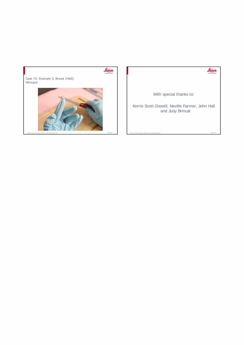
Case 70: Example 3, Breast (H&E)Whoops!
Page 189 Artifacts, Faults and Failures: a Review © Leica Microsystems 2012 Page 190
With special thanks to:
Kerrie Scott-Dowell, Neville Farmer, John Hall and Judy Brincat