Embed Size (px)
Citation preview

ARVO 2016 Annual Meeting Abstracts
These abstracts are licensed under a Creative Commons Attribution-NonCommercial-No Derivatives 4.0 International License. Go to http://iovs.arvojournals.org/ to access the versions of record.
227 Glaucoma: New IdeasMonday, May 02, 2016 11:00 AM–12:45 PM6A Paper SessionProgram #/Board # Range: 1791–1797Organizing Section: Glaucoma
Program Number: 1791Presentation Time: 11:00 AM–11:15 AMRestoration of trabecular meshwork cellularity and function following iPSC transplantation in vivoMarkus H. Kuehn1, 2, Wei Zhu1, 2, Oliver W. Gramlich1, 2, Lauren Laboissonniere3, Jeffrey Trimarchi3, Budd A. Tucker1. 1Ophthalmology and Visual Sciences, University of Iowa, Iowa City, IA; 2Center for Prevention and Treatment of Visual Loss, Veterans Affairs, Iowa City, IA; 3Genetics, Development and Cell Biology, Iowa State University, Ames, IA.Purpose: Primary open angle glaucoma is frequently associated with loss and dysfunction of trabecular meshwork (TM) cells. The purpose of this study was to determine if restoration of TM cellularity, using TM cells derived from induced pluripotent stem cells (iPSC), can lead to lasting functional rescue of the TM in a genetic mouse model of the disease.Methods: Mouse iPSC were differentiated into TM-like cells (iPSC-TM) and injected into the anterior chamber of MYOCY437H
mice (N=22) as described previously. Controls included MYOCY437H
mice receiving sham injections (N=16) or fibroblasts (N=8) as well as age matched wild type mice (N=20). Intraocular pressure (IOP) was measured by rebound tonometry and aqueous humor outflow facility by direct cannulation. 12 weeks after transplantation retinal ganglion cell (RGC) and TM cellularity was assessed by immunohistochemistry and gene expression patterns in the TM was determined using gene expression arrays.Results: When compared to sham injected MYOCY437H mice, animals having received iPSC-TM display significantly lower IOP (16.4±3.1 vs 12.2±2.8 mmHg, P=0.0006) and higher aqueous humor outflow facility (0.016±0.005 vs. 0.027±0.01 µl/min/mmHg, P=0.04). Treated mice also display higher RGC density (2222.1±250.4 vs. 1706.9±491.3 RGC/mm2) and higher TM cellularity than control mice (25.7±7.3 vs. 54.9±7.2 TM/section, P=6.83E-06). However, surviving iPSC-TM are rarely observed in the TM 12 weeks after transplantation. These values observed in iPSC-TM recipients MYOCY437H mice resemble those of wild type mice. In contrast, sham injected eyes or those having received a transplantation of unrelated cells, do not differ significantly from those of age mated untreated MYOCY437H animals. Gene expression analyses demonstrate that a large number of cell cycle associated genes were expressed at higher levels in the TM of eyes having received transplanted iPSC-TM.Conclusions: These data demonstrate that functional restoration of glaucomatous TM, resulting decreased IOP and RGC rescue, is possible using iPSC-TM. Unexpectedly, the effect of iPSC-TM transplantation does not appear to be a functional replacement of lost TM cells. Rather it appears that transplantation induces a proliferative response of the remaining endogenous TM cells that results in normal TM cellularity and restoration of IOP control.Commercial Relationships: Markus H. Kuehn, None; Wei Zhu, None; Oliver W. Gramlich, None; Lauren Laboissonniere, None; Jeffrey Trimarchi, None; Budd A. Tucker, NoneSupport: VA I01 RX001163
Program Number: 1792Presentation Time: 11:15 AM–11:30 AMYoung bone marrow Sca-1+ stem cells home to aged retina and protect retina from acute glaucoma injuryZhengbo Shao1, 2, Jie Wu3, 2, Guoqing Du1, 2, Huifang Song2, Shu-Hong Li2, Sheng He2, Jiao Li2, Jun Wu2, Richard D. Weisel2, Huiping Yuan1, Ren-Ke Li2. 1The Second Affiliated Hospital of Harbin Medical University, Harbin, China; 2Surgery, Toronto General Research Institute, University Health Network, Toronto, ON, Canada; 3Basic Medical Science Institute of Harbin Medical University, Harbin, China.Purpose: Glaucoma is a major clinical ocular disorder, especially prevalent in the aged population. Retinal ganglion cell (RGC) apoptosis and optic nerve degeneration in aged glaucoma patients typically result in blindness. Recently, we demonstrated that young bone marrow (BM) stem cells can regenerate aged tissue and enhance tissue repair. This study investigated whether BM-derived stem cell homing to the retina increased regeneration/repair mechanisms in the retina using a mouse model of this neurodegenerative disease.Methods: Young BM stem cell antigen 1 (Sca-1+) and Sca-1- cells from GFP+ mice (2-3 months old) were isolated by immunomagnetic cell sorting and transplanted into lethally-irradiated aged recipient mice (20-22 months old) to generate Sca-1+ [Y(Sca1+)-O] and Sca-1- [Y(Sca1-)-O] chimeras. Homing efficiency of Sca-1 cells in chimeric aged mice was evaluated by immunostaining. Acute ocular hypertensive glaucoma was induced 8 weeks after BM reconstitution and vision function was evaluated by the light/dark exploration and optomoter tests. Western blot and RT-qPCR were performed for mechanistic analyses.Results: The light/dark exploration and optomoter tests showed better preservation of visual function in Sca-1+ than Sca-1- chimeras (p<0.05, n=10/group) 7 days after induction of acute ocular hypertension. Sca-1+ cells had greater capability to home to the retina (p<0.01, n=4-6/group) than Sca-1- cells. There was more cell differentiation into glia, microglia and neural cells in the Sca-1+ than Sca-1- group (p<0.05, n=4) after retinal injury. Furthermore, Sca-1+ cells in the retina prevented cellular apoptosis of host retinal cells (p<0.01, n=6/group). Exploration of potential mechanisms revealed that fibroblast growth factor 2 (FGF2) expression increased significantly in Sca-1+ chimeras compared to Sca-1- chimeras after acute glaucoma injury. In vitro study also demonstrated that Sca-1+ cells had higher expression levels of FGF2 (both mRNA and protein) than Sca-1- cells (p<0.01). After application of an FGF2-neutralizing antibody, activation of the Akt pathway was blocked, and the beneficial effects of Sca-1+ cells were lost.Conclusions: Homed BM Sca-1 cells in the retina prevented cellular apoptosis after acute glaucoma injury in aged mice. FGF2 and its signal pathway may play an important role in BM Sca-1 cell-mediated retinal neuroprotection.Commercial Relationships: Zhengbo Shao, None; Jie Wu, None; Guoqing Du; Huifang Song, None; Shu-Hong Li, None; Sheng He, None; Jiao Li, None; Jun Wu, None; Richard D. Weisel, None; Huiping Yuan, None; Ren-Ke Li, NoneSupport: The Canadian Institutes of Health Research Foundation grant # 143323 to Ren-Ke Li; National Natural Science Foundation of China #81500713 to Zhengbo Shao; National Natural Science Foundation of China #81271000 and #81470634 to Huiping Yuan.

ARVO 2016 Annual Meeting Abstracts
These abstracts are licensed under a Creative Commons Attribution-NonCommercial-No Derivatives 4.0 International License. Go to http://iovs.arvojournals.org/ to access the versions of record.
Program Number: 1793Presentation Time: 11:30 AM–11:45 AMOptic nerve and retinal ganglion cell degeneration after two weeks of intracranial pressure (ICP) elevation in miceGuofu Shen, Derek Nusbaum, Benjamin J. Frankfort. Ophthalmology, Baylor College of Medicine, Houston, TX.Purpose: To establish normal ranges and diurnal curves of ICP in freely moving C57Bl/6 mice. To confirm that ICP can be elevated experimentally for up to 2 weeks.Methods: 12-week old, male, C57Bl/6 mice were included in this study and maintained according to a standard 12 hour light-dark cycle. A radiofrequency transmitter for pressure monitoring and a custom cannula for the instillation of ACSF were placed in the subarachnoid space (Nusbaum et. al. 2015). Mice were allowed to range freely within a custom caging system. Normal ICP was continuously monitored for 1-2 week after surgery. ICP data were collected through wireless receivers for later offline analysis. For ICP elevation experiments, the infusion of ACSF was driven by gravity and the level of ICP adjusted manually. After maintaining ICP at approximately 20 mmHg for 2 weeks, the visual function of 4 mice was evaluated through OKR and whole field bilateral ERGs, and the results were compared with the data collected from control mice exposed to ICP surgery but without ICP elevation. Structural changes, such as RGC count and axon number within the optic nerve were also compared using immunofluorescence with Tuj1 staining and transmission electron microscopy, respectively.Results: Control mice showed a higher ICP level and more ICP variance at night, consistent with the nocturnal habitat of mice. Elevation of ICP to approximately 20 mmHg for two weeks caused a significant decrease of RGC number. In the central retina, the RGC density decreased from 4226±255 (n=20 regions from 3 normal ICP mice) to 2926±149 (n=44 regions from 6 elevated ICP mice). In the surrounding retina, similar changes were observed. Increased axonal degeneration (blebbing, distortion and rupture of myelin sheaths, vacuolization of axons) and significant axon loss was observed in the optic nerve nerves of mice exposed to elevated ICP. Analysis of OKR and ERG are still ongoing. The IOP of the animals was not changed by the ICP increase.Conclusions: We successfully validated a novel experimental system for the monitoring and manipulation of ICP in active, awake mice for up to 2 weeks. With this animal model, a close correlation between the ICP elevation and function/structure deterioration was observed. This model can be used to study both the effects of ICP change and alteration of the ICP-IOP gradient in a variety of ophthalmologic and neurologic conditions.Commercial Relationships: Guofu Shen; Derek Nusbaum, None; Benjamin J. Frankfort, NoneSupport: NIH R01 EY025601 and K08 EY021479 to BJF
Program Number: 1794Presentation Time: 11:45 AM–12:00 PMHigh-resolution mapping of in-vivo stretch and compression of the lamina cribrosa in response to acute changes in intraocular and/or intracranial pressuresIan A. Sigal1, 2, Alexandra Judisch1, Huong Tran1, 2, Bo Wang1, 2, Matthew A. Smith1, 2, Larry Kagemann1, 2, Hiroshi Ishikawa1, 2, Elizabeth Tyler-Kabara1, Joel S. Schuman1, 2, Gadi Wollstein1, 2. 1Department of Ophthalmology, University of Pittsburgh, Pittsburgh, PA; 2Department of Bioengineering, University of Pittsburgh, Pittsburgh, PA.Purpose: To map with high level of detail the in-vivo stretch and compression of the lamina cribrosa (LC) in response to acute changes in intraocular and/or intracranial pressures (IOP and ICP).
Methods: Three eyes of 3 macaque monkeys were imaged in-vivo with SD-OCT (Bioptigen, Durham, NC) while IOP and ICP were controlled through cannulas in the anterior chamber and the lateral ventricle, respectively. IOP and ICP were set to all combinations of IOPs (15 - baseline, 5, 30 and 50mmHg) and ICPs (10 – baseline, 5, 25 and 40mmHg), and allowed to equilibrate for 10min. A recently developed tracking algorithm based on image registration was adapted to OCT and used to extract the pressure-induced tissue displacements, stretch and compression.Results: Pressure variations from the baseline caused substantial LC stretch and compression, sometimes exceeding 30%. Effects differed between LC regions and animals, and depended on whether the pressures were increased or decreased (Figure). There were strong interactions between the two pressures.Conclusions: The effects of IOP and ICP on the LC are detectable in-vivo. These effects are highly nonlinear and interact strongly. Translaminar pressure difference (IOP-ICP) is too simplistic to describe LC mechanics. Alterations in IOP and ICP affected different regions of the LC differentially. Our methods open a new way to investigate the interplay between IOP and ICP.
C-mode views through the LC of Monkey 1 color coded by stretch and compression resulting from pressure changes from the baseline (B). Increases in IOP caused LC stretch, larger at elevated ICP. Increases in ICP caused LC compression, larger at baseline IOP. Decreases in IOP or ICP caused smaller LC stretch and compression. Simultaneous changes in IOP and ICP that maintained the translaminar pressure difference, also produced small but significant stretch and compression.Commercial Relationships: Ian A. Sigal, None; Alexandra Judisch, None; Huong Tran, None; Bo Wang, None; Matthew A. Smith, None; Larry Kagemann, None; Hiroshi Ishikawa, None; Elizabeth Tyler-Kabara, None; Joel S. Schuman, Zeiss (P); Gadi Wollstein, NoneSupport: NIH grants R01-EY023966, R01-EY025011, R01EY011289, P30-EY008098, T32-EY017271, Eye and Ear Foundation (Pittsburgh, PA), Glaucoma Research Foundation Shaffer Grant

ARVO 2016 Annual Meeting Abstracts
These abstracts are licensed under a Creative Commons Attribution-NonCommercial-No Derivatives 4.0 International License. Go to http://iovs.arvojournals.org/ to access the versions of record.
Program Number: 1795Presentation Time: 12:00 PM–12:15 PMFinite element analysis predicts high optic nerve head strains during horizontal eye movementsXiaofei Wang1, Mani Baskaran2, Helmut Rumpel3, Shamira Perera2, Winston E. Lim3, Monisha E. Nongpiur2, Tin Aung2, Dan Milea2, Michael J. Girard1, 2. 1Ophthalmic Engineering & Innovation Laboratory, Department of Biomedical Engineering, National University of Singapore, Singapore, Singapore; 2Singapore Eye Research Institute, Singapore National Eye Centre, Singapore, Singapore; 3Department of Diagnostic Radiology, Singapore General Hospital, Singapore, Singapore.Purpose: (1) To combine finite element (FE) analysis and dynamic magnetic resonance imaging (MRI) to estimate optic nerve head (ONH) strains (deformations) during eye movements, and identify factors influencing such strains; (2) To compare ONH strains induced by eye movements with those induced by intraocular pressure (IOP) which are critical in glaucoma.Methods: The eyes and orbital tissues of a healthy subject were visualized during visually-guided horizontal eye movements, using dynamic MRI. A baseline FE model of the left eye was reconstructed in the primary gaze position and included details from the orbital tissues (visualized with MRI; Figure 1A) and from the ONH tissues (using measurements from the literature). The effect of a lateral eye movement of 13° was then simulated, based on the MRI findings. ONH strains due to eye movements were compared with those resulting from an IOP of 50 mmHg. Finally, a sensitivity study was performed, in which we varied the stiffness of all connective tissues to understand their influence on the ONH during eye movements.Results: The baseline FE model matched the MRI scan well (Figure 1A). Our models predicted that, during eye movements, the retrobulbar portion of optic nerve had a pulling action on the ONH, which resulted in large strains within the ONH tissues. Specifically, the strains generated within the prelamina (mean: 0.027), the lamina cribrosa (0.018), and the retrolamina (0.041) following an eye movement were higher than those resulting from an IOP of 50 mmHg (Figure 1B). These results held true even when considering variations in connective tissue stiffness. In addition, we found that stiff scleras reduced lamina cribrosa and prelamina strains during eye movements, but stiff dura and pia maters significantly increased those strains.Conclusions: This study is the first to combine FE with MRI to estimate ONH strains during eye movements. Our models predict high ONH strains during eye movements, which were aggravated with stiffer optic nerve sheaths. Further studies are needed to explore a possible link between ONH strains induced by eye movements and axonal loss in glaucoma.
Figure 1. (A) FE model in primary and left gaze positions of a left globe (yellow outlines) superimposed on the MRI scan indicating a good match. (B) Predicted ONH strains for various loading scenarios.Commercial Relationships: Xiaofei Wang, None; Mani Baskaran, None; Helmut Rumpel, None; Shamira Perera, None; Winston E. Lim, None; Monisha E. Nongpiur, None; Tin Aung, None; Dan Milea, None; Michael J. Girard, NoneSupport: NUS Young Investigator Award (NUSYIA_FY13_P03, R-397-000-174-133); Ministry of Education, Academic Research Funds, Tier 1 (R-397-000-140-133; R-397-000-181-112); Singapore Eye Research Institute Pilot Grant
Program Number: 1796Presentation Time: 12:15 PM–12:30 PMTime-lapse retinal ganglion cell dendritic field degeneration imaged in organotypic retinal explant cultureThomas V. Johnson, Ericka N. Oglesby, Matthew R. Steinhart, Elizabeth Cone-Kimball, Joan Jefferys, Harry A. Quigley. Wilmer Eye Institute, Johns Hopkins University, Baltimore, MD.Purpose: To develop an ex vivo organotypic retinal explant culture system suitable for multiple time-point imaging of living retinal ganglion cell (RGC) dendritic arbors over a period of 1 week, and capable of detecting dendrite neuroprotection conferred by experimental treatments.Methods: Thy1-YFP mouse retinas were explanted and maintained in organotypic culture. RGC dendritic arbors were imaged repeatedly using confocal laser scanning microscopy. Maximal projection z-stacks were traced by two masked investigators and dendritic fields were analyzed for characteristics including branch number, size, and complexity. One group of explants was treated with BDNF+CNTF added to the culture media. Changes in individual dendritic fields over time were detected using pair-wise comparison testing.Results: RGCs in mouse retinal explant culture began to degenerate after 3 days with 52.4% surviving at 7 days. Dendritic field parameters showed minimal change over 8 hours in culture. Intra-observer and inter-observer measurements of dendrite characteristics were strongly correlated (Spearman rank correlations consistently >0.80). Statistically significant (p<0.001) dendritic tree degeneration was detected following seven days in culture including: 40-50%
ARVO 2016 Annual Meeting Abstracts
These abstracts are licensed under a Creative Commons Attribution-NonCommercial-No Derivatives 4.0 International License. Go to http://iovs.arvojournals.org/ to access the versions of record.
decreases in number of branch segments, number of junctions, number of terminal branches, and total branch length. Scholl analyses similarly demonstrated a significant decrease in dendritic field complexity. Treatment of explants with BDNF + CNTF significantly attenuated dendritic field degeneration.Conclusions: Retinal explant culture of Thy1-YFP tissue provides a useful model for time-lapse imaging of RGC dendritic field degeneration over a course of several days, and is capable of detecting neuroprotective amelioration of dendritic pruning.Commercial Relationships: Thomas V. Johnson, None; Ericka N. Oglesby, None; Matthew R. Steinhart, None; Elizabeth Cone-Kimball, None; Joan Jefferys, None; Harry A. Quigley, NoneSupport: NIH Core Grant EY-O1765
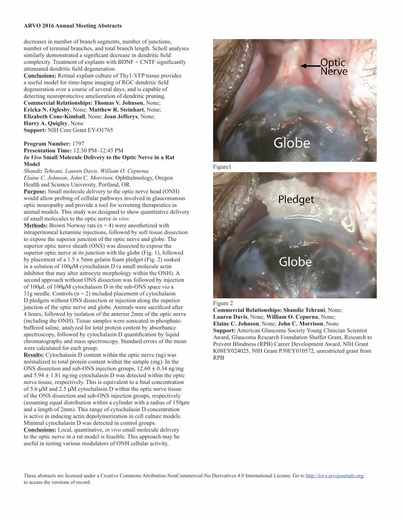
Program Number: 1797Presentation Time: 12:30 PM–12:45 PMIn Vivo Small Molecule Delivery to the Optic Nerve in a Rat ModelShandiz Tehrani, Lauren Davis, William O. Cepurna, Elaine C. Johnson, John C. Morrison. Ophthalmology, Oregon Health and Science University, Portland, OR.Purpose: Small molecule delivery to the optic nerve head (ONH) would allow probing of cellular pathways involved in glaucomatous optic neuropathy and provide a tool for screening therapeutics in animal models. This study was designed to show quantitative delivery of small molecules to the optic nerve in vivo.Methods: Brown Norway rats (n = 4) were anesthetized with intraperitoneal ketamine injections, followed by soft tissue dissection to expose the superior junction of the optic nerve and globe. The superior optic nerve sheath (ONS) was dissected to expose the superior optic nerve at its junction with the globe (Fig. 1), followed by placement of a 1.5 x 5mm gelatin foam pledget (Fig. 2) soaked in a solution of 100µM cytochalasin D (a small molecule actin inhibitor that may alter astrocyte morphology within the ONH). A second approach without ONS dissection was followed by injection of 100µL of 100µM cytochalasin D in the sub-ONS space via a 31g needle. Controls (n = 2) included placement of cytochalasin D pledgets without ONS dissection or injection along the superior junction of the optic nerve and globe. Animals were sacrificed after 4 hours, followed by isolation of the anterior 2mm of the optic nerve (including the ONH). Tissue samples were sonicated in phosphate-buffered saline, analyzed for total protein content by absorbance spectroscopy, followed by cytochalasin D quantification by liquid chromatography and mass spectroscopy. Standard errors of the mean were calculated for each group.Results: Cytochalasin D content within the optic nerve (ng) was normalized to total protein content within the sample (mg). In the ONS dissection and sub-ONS injection groups, 12.60 ± 0.34 ng/mg and 5.94 ± 1.81 ng/mg cytochalasin D was detected within the optic nerve tissue, respectively. This is equivalent to a final concentration of 5.6 µM and 2.5 µM cytochalasin D within the optic nerve tissue of the ONS dissection and sub-ONS injection groups, respectively (assuming equal distribution within a cylinder with a radius of 150µm and a length of 2mm). This range of cytochalasin D concentration is active in inducing actin depolymerization in cell culture models. Minimal cytochalasin D was detected in control groups.Conclusions: Local, quantitative, in vivo small molecule delivery to the optic nerve in a rat model is feasible. This approach may be useful in testing various modulators of ONH cellular activity.
Figure1
Figure 2Commercial Relationships: Shandiz Tehrani, None; Lauren Davis, None; William O. Cepurna, None; Elaine C. Johnson, None; John C. Morrison, NoneSupport: American Glaucoma Society Young Clinician Scientist Award, Glaucoma Research Foundation Shaffer Grant, Research to Prevent Blindness (RPB) Career Development Award, NIH Grant K08EY024025, NIH Grant P30EY010572, unrestricted grant from RPB