Embed Size (px)
Citation preview
www.rsc.org/chemcommRegistered Charity Number 207890
As featured in:
See Jonathan P. Hill et al., Chem. Commun., 2012, 48, 3933.
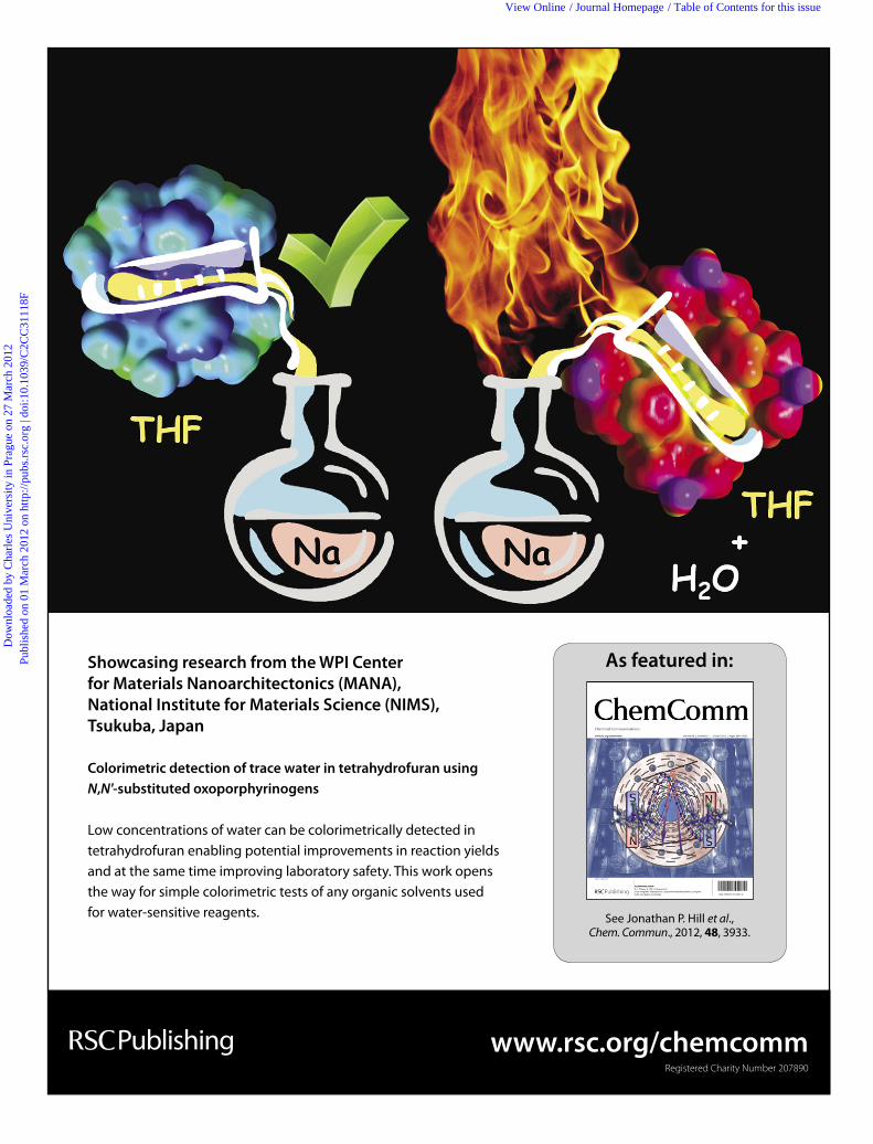
Showcasing research from the WPI Center
for Materials Nanoarchitectonics (MANA),
National Institute for Materials Science (NIMS),
Tsukuba, Japan
Colorimetric detection of trace water in tetrahydrofuran using
N,N'-substituted oxoporphyrinogens
Low concentrations of water can be colorimetrically detected in
tetrahydrofuran enabling potential improvements in reaction yields
and at the same time improving laboratory safety. This work opens
the way for simple colorimetric tests of any organic solvents used
for water-sensitive reagents.
Dow
nloa
ded
by C
harl
es U
nive
rsity
in P
ragu
e on
27
Mar
ch 2
012
Publ
ishe
d on
01
Mar
ch 2
012
on h
ttp://
pubs
.rsc
.org
| do
i:10.
1039
/C2C
C31
118F
View Online / Journal Homepage / Table of Contents for this issue

This article is part of the
Porphyrins & Phthalocyanines
web themed issue
Guest editors: Jonathan Sessler, Penny Brothers and Chang-Hee Lee
All articles in this issue will be gathered together
online at www.rsc.org/porphyrins
Dow
nloa
ded
by C
harl
es U
nive
rsity
in P
ragu
e on
27
Mar
ch 2
012
Publ
ishe
d on
01
Mar
ch 2
012
on h
ttp://
pubs
.rsc
.org
| do
i:10.
1039
/C2C
C31
118F
View Online
This journal is c The Royal Society of Chemistry 2012 Chem. Commun., 2012, 48, 3933–3935 3933
Cite this: Chem. Commun., 2012, 48, 3933–3935
Colorimetric detection of trace water in tetrahydrofuran using
N,N0-substituted oxoporphyrinogenswzShinsuke Ishihara,
aJan Labuta,
aTomas Sikorsky,
bcJaroslav V. Burda,
dNaoko Okamoto,
a
Hideki Abe,eKatsuhiko Ariga
afand Jonathan P. Hill*
af
Received 15th February 2012, Accepted 28th February 2012
DOI: 10.1039/c2cc31118f
Oxoporphyrinogens (OxPs) bind water molecules at pyrrolic
NH and quinonoid carbonyl groups leading to visible colour
changes due to variation in the p-electronic structure of OxPs.
Introduction of hydrophilic substituents at two pyrrole NH
groups improves sensitivity to H2O, and one OxP derivative is
a colorimetric indicator of trace H2O (B50 ppm) in THF.
Quantitative analysis of H2O in organic solvents is important
in fundamental and industrial applications1 especially in polar
aprotic solvents in which water is miscible and which are used
as solvents for water-flammable reagents (e.g., alkali metals,
organolithiums, Grignard reagents).2 The Karl-Fischer method3
permits detection of H2O down to theB1 ppm level but it can be
inconvenient due to instrumental requirements and toxic reagents
(i.e., methanol, I2 and SO2). Alternatively, use of a dye
molecule as an H2O-indicator would faciltate H2O detection
in organic solvents. Fluorescence of some dyes is quenched (or
emission spectra change) in response to variations in H2O
concentration4 with one such molecule enabling detection of
B20 ppmH2O in some solvents,4bwhich approaches Karl-Fischer
sensitivity. However, the accuracy of fluorescence analysis can
be detrimentally influenced by many factors including other
quenchers, photobleaching, concentration, temperature, etc.
In contrast, colorimetric analyses of H2O using solvatochromic
dyes is generally more stable although sensitivities may be lower
(B1000 ppm H2O) as colour changes depend on the variation
of total solvent polarity.5
Here we show that oxoporphyrinogens (Fig. 1), a class of
porphyrinoid, are available as colorimetric indicators for trace
contaminating H2O in tetrahydrofuran (THF) to a level of
B50 ppm by means of UV-Vis spectroscopy. Investigations
conducted using 1H-NMR spectroscopy and theoretical
calculations reveal that OxPs bind H2O molecules at
both pyrrolic NH and quinonoid CQO groups, varying the
p-electronic structure and leading to visible colour changes.
Even though the detection limit of H2O by these OxP deriva-
tives is somewhat low compared to the best fluorescent probe,
the use of OxP has great advantages of (i) the UV-Vis
spectrum being easy to obtain and more stable than the
fluorescence spectrum; (ii) the sensitivity of OxP to H2O is
variable by synthetic modifications; (iii) OxP can be reused
without chemical change or photobleaching.
OxP belongs to the calixpyrrole family6 and contains a
cyclic tetrapyrrole conjugated with quinonoid moieties at its
meso-positions. OxP binds a variety of guest molecules at its
pyrrolic NH’s as well as at the quinonoid CQO groups. In
contrast to typical calixpyrroles, OxPs have a strong absorp-
tion in the visible light region due to p-conjugation between
tetrapyrrole and quinonoid substituents. This conjugation is
sensitive to binding of guests to OxPs and these compounds
have been reported to behave as probes for anions or
solvents,7 and enantiomeric excess.8 For solvents, it was
thought that major interactions between OxP derivatives and
solvents occurred with pyrrolic NH groups acting as H-bond
donors7a,9 and that tert-butyl groups largely prevented any
strong intermolecular interations involving the CQO
groups.10
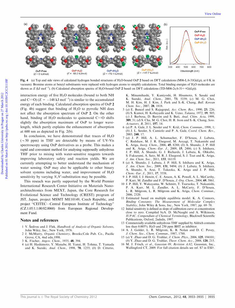
Fig. 1 Chemical structures of oxoporphyrinogens (OxPs) 1–6 used
for colorimetric detection of H2O in THF (Symbols denote the
assignment of 1H-NMR peaks of OxP 2 shown in Fig. 3.).
a International Center for Materials Nanoarchitectonics (MANA),National Institute for Materials Science (NIMS), Namiki 1-1,Tsukuba, Ibaraki 305-0044, Japan.E-mail: [email protected]
bDepartment of Macromolecular Physics, Charles University,V Holesovickach 2, 180 00 Prague 8, Czech Republic
c CEITEC—Central European Institute of Technology,Masaryk University, CZ-62500 Brno, Czech Republic
dDepartment of Chemical Physics and Optics, Charles University,Ke Karlovu 3, 121 16 Prague 2, Czech Republic
e Research Unit for Environmental Remediation Materials, NIMS,Namiki 1-1, Tsukuba, Ibaraki 305-0044, Japan
f JST-CREST, Namiki 1-1, Tsukuba, Ibaraki 305-0044, Japanw Electronic supplementary information (ESI) available: Details onsynthesis and characterization of materials, experimental methods,binding study, and DFT calculations. See DOI: 10.1039/c2cc31118fz This article is part of the ChemComm ’Porphyrins and phthalocya-nines’ web themed issue.
ChemComm Dynamic Article Links
www.rsc.org/chemcomm COMMUNICATION
Dow
nloa
ded
by C
harl
es U
nive
rsity
in P
ragu
e on
27
Mar
ch 2
012
Publ
ishe
d on
01
Mar
ch 2
012
on h
ttp://
pubs
.rsc
.org
| do
i:10.
1039
/C2C
C31
118F
View Online
3934 Chem. Commun., 2012, 48, 3933–3935 This journal is c The Royal Society of Chemistry 2012
OxP derivatives 1–6 (Fig. 1) were synthesized according to
reportedmethods9 and investigated as colorimetric H2O indicators
in THF. OxPs 1–5 demonstrate visible colour changes from
red-purple to blue-purple by hydration of THF (Fig. 2a). On
the other hand, OxP 6, which lacks pyrrolic NH due to full
N-substitution, remained red-purple even after addition of a
large excess of H2O. As shown in Fig. 2(b), addition of H2O to an
anhydrous solution of OxP in THF attenuates the absorption at
507 nm while increasing the absorption intensity at 600 nm and
750 nm. On the basis of the variation of absorption at 507 nm and
600 nm (Fig. 2c and d), the binding constants (K1, K2, K3)11 and
sensitivity12 to H2O are evaluated by a fitting method and are
summarized in Table 1. As a result, it was found that OxP 5
substituted with hydrophilic carboxylate groups possesses the
highest K1 and sensitivity to H2O. It is likely that carboxylate
groups attract water molecules increasing their availability in the
vicinity of OxP or that carboxylate moieties stabilize the
H2O-bound state of OxP. Differences in H2O sensitivity
arising from variation of N-substitution is a unique feature
since it implies possible improvements in peformance by synthesis
of more H2O-sensitive OxPs. Importantly, OxP 5 is responsive to
37 ppm H2O in THF (Fig. 2b, inset), so that it is technically
possible to distinguish between freshly-purchased anhydrous THF
(H2O o 50 ppm)13 and older wet THF (H2O 4 50 ppm) simply
by recording a UV-Vis spectrum of a solution of OxP of known
concentration (which can be determined from the isosbestic point
at 535 nm). Moreover, a map of calibration curves for OxP 5
can be constructed and used for fast determination of water
concentration over a broad range (see ESIw). In addition, it should
be noted that OxP is reusable (see ESIw), and that H2O-sensitivity
of OxP 5 is hardly affected by the inhibitor, 2,6-di-tert-butyl-4-
cresol (BHT, 600 ppm).13
1H-NMR spectra of OxP 2 in THF-d8 suggest that OxP
bindsH2Omolecules both at pyrrolic NH and at quinonoid CQO.
The peak at 9.8 ppm due to pyrrolic NH protons shifts downfield
as H2O is added (Fig. 3a), indicating occurrence of hydrogen
bonding between pyrrolic NH and H2O. Concurrently,1H-NMR
peaks of tert-butyl groups at around 1.3 ppm become further
divided (Fig. 3c). Since tert-butyl groups cannot form hydrogen
bonds with H2O, it appears that CQO groups close to the tert-
butyl groups interact with H2O molecules similar to the case of
unsubstituted OxP in the solid state.14 Also, 1H-NMR peaks
at around 6.6–7.7 ppm corresponding to pyrrole b-H and
hemiquinonoid methine-H undergo downfield or upfield shifts
(Fig. 3b) suggesting that binding of H2O molecules at pyrrolic
NH and quinonoid CQO causes changes in the p-electronicconjugation of OxP. Consequently, the absorption spectrum
(i.e., colour) of OxP should be changed. Binding of H2O
molecules at pyrrolic NH as well as quinonoid CQO is also
supported by some X-ray crystal structures of OxPs.9,14
Optical properties of OxP were investigated using time-
dependent density functional theory (TD-DFT).15,16 These
calculations were applied in order to estimate at which position
(i.e., NH or CQO) interactions with water have a greater
impact on the variations in the absorption spectra of OxP. As
shown in Fig. 4(a), OxP 2 binds one H2O molecule at pyrrolic
NH and up to four H2O molecules at quinonoid CQO. The
interaction energy of H2O with OxP implies that the NH
position (E = �68 kJ mol�1) is more energetically favourable
than quinonoid CQO (E = �20 kJ mol�1 per H2O molecule).
However, binding of H2O at NH and quinonoid groups is
apparently neither competitive nor cooperative since the total
Fig. 2 (a) Colour variation of OxP 5 in anhydrous and hydrated
THF. (b) Variation in absorption spectra of OxP 5 in response to H2O
concentration (THF, [5] = 3.5 � 10�6 M, 1 cm cell). Inset: magnified
spectra around 500 nm. (c) and (d) Variation of absorption at 507 nm
and 600 nm for OxP 5 in response to H2O concentration in THF. H2O
concentration is classified into three regions in (d); green (anhydrous:
H2O o 50 ppm), yellow (normal: 50 ppm o H2O o 500 ppm), and
red (hydrated: 500 ppm o H2O) according to the guaranteed H2O
level of commercially available THF.
Table 1 Binding constantsa (K1, K2, and K3) and sensitivities of OxPs1–6 to H2O in THF
Compound K1/M�1 K2/M
�1 K3/M�1
Sensitivity UV-Vis(10�3 a.u. per ppm)
1 10.3 (�2.1)c 2.36 0.00 0.050 (�0.005)2 22.9 (�4.6) 0.42 0.19 0.024 (�0.006)3 16.4 (�3.3)c 1.28 0.07 0.032 (�0.006)4 26.9 (�5.4) 0.78 0.03 0.049 (�0.016)5 89.8 (�13.5) 5.53 0.01 0.102 (�0.003)5+BHTb 42.0 (�8.4) 1.49 0.14 0.087 (�0.009)6 69.0 (�55.2)d 0.00 0.00 0.003 (�0.003)a Binding constants (K1, K2, and K3) are determined by the curve
fitting method from Abs. at 507 nm and 600 nm. Binding models are
shown in ESI. b 2,6-Di-tert-butyl-4-cresol (BHT, 600 ppm) was added.c Determined only from Abs. at 600 nm. d Compound 6 generally
shows only a weak response to the presence of water (see ESI).
Fig. 31H-NMR spectra of OxP 2 in THF-d8 ([2] = 7.0 � 10�4 M)
showing (a) pyrrolic NH region, (b) b-pyrrole and quinonoid moiety
peaks, and (c) tert-butyl peaks. Assignments of peaks obtained by
COSY and NOESY are shown in Fig. 1 (* indicates solvent impurity).
Dow
nloa
ded
by C
harl
es U
nive
rsity
in P
ragu
e on
27
Mar
ch 2
012
Publ
ishe
d on
01
Mar
ch 2
012
on h
ttp://
pubs
.rsc
.org
| do
i:10.
1039
/C2C
C31
118F
View Online
This journal is c The Royal Society of Chemistry 2012 Chem. Commun., 2012, 48, 3933–3935 3935
interaction energy of five H2O molecules (bound to both NH
and CQO) (E= �148 kJ mol�1) is similar to the accumulated
energy of each binding. Calculated absorption spectra of OxP 2
(Fig. 4b) suggest that binding of H2O to pyrrolic NH does
not affect the absorption spectrum of OxP 2. On the other
hand, binding of H2O molecules to quinonoid CQO shifts
slightly the absorption maximum of OxP to longer wave-
length, which partly explains the enhancement of absorption
at 600 nm as depicted in Fig. 2(b).
In conclusion, we have demonstrated that traces of H2O
(B50 ppm) in THF are detectable by means of UV-Vis
spectroscopy using OxP derivatives as a probe. This makes a
rapid and convenient method for analysing supposedly anhydrous
THF prior to mixing with water-sensitive reagents towards
improving laboratory safety and reaction yields. We are
currently attempting to better understand the mechanism of
water sensing. OxP may also be applicable to other binary
solvent systems including water, and improvement of H2O
sensitivity by varying N,N0-substitution may be possible.
This reseach was partly supported by the World Premier
International Research Center Initiative on Materials Nano-
architechtonics from MEXT, Japan, the Core Research for
Evolutional Science and Technology (CREST) program of
JST, Japan, project MSMT ME10149, Czech Republic, and
project ‘‘CEITEC—Central European Institute of Technology’’
(CZ.1.05/1.1.00/02.0068) from European Regional Develop-
ment Fund.
Notes and references
1 V. Sedivec and J. Flek, Handbook of Analysis of Organic Solvents,John Wiley, Inc., New York, 1976.
2 J. McMurry, Organic Chemistry, Brooks/Cole Pub. Co., PacificGrove, CA, 4nd edn,1998.
3 K. Fischer, Angew. Chem., 1935, 48, 394.4 (a) H. Hashimoto, Y. Manabe, H. Yanai, H. Tohma, T. Yamadaand K. Suzuki, Anal. Chem., 1998, 70, 1255; (b) D. Citterio,
K. Minamihashi, Y. Kuniyoshi, H. Hisamoto, S. Sasaki andK. Suzuki, Anal. Chem., 2001, 73, 5339; (c) M. G. Choi,M. H. Kim, H. J. Kim, J. Park and S.-K. Chang, Bull. KoreanChem. Soc., 2007, 28, 1818.
5 (a) E. Buncel and S. Rajagopal, Acc. Chem. Res., 1990, 23, 226;(b) S. Kumoi, H. Kobayashi and K. Ueno, Talanta, 1972, 19, 505;(c) J. Barbosa, D. Barron and S. Butı, Anal. Chim. Acta, 1999,389, 31; (d) S. Cha, M. G. Choi, H. R. Jeon and S.-K. Chang, Sens.Actuators, B, 2011, 157, 14.
6 (a) P. A. Gale, J. L. Sessler and V. Kral, Chem. Commun., 1998, 1;(b) J. L. Sessler, S. Camiolo and P. A. Gale, Coord. Chem. Rev.,2003, 240, 17.
7 (a) J. P. Hill, A. L. Schumacher, F. D’Souza, J. Labuta,C. Redshaw, M. J. R. Elsegood, M. Aoyagi, T. Nakanishi andK. Ariga, Inorg. Chem., 2006, 45, 8288; (b) A. Shundo, J. P. Hilland K. Ariga, Chem.–Eur. J., 2009, 15, 2486; (c) S. Ishihara,J. P. Hill, A. Shundo, G. J. Richards, J. Labuta, K. Ohkubo,S. Fukuzumi, A. Sato, M. R. J. Elsegood, S. J. Teat and K. Ariga,J. Am. Chem. Soc., 2011, 133, 16119.
8 (a) A. Shundo, J. Labuta, J. P. Hill, S. Ishihara and K. Ariga,J. Am. Chem. Soc., 2009, 131, 9494; (b) J. Labuta, S. Ishihara,A. Shundo, S. Arai, T. Takeoka, K. Ariga and J. P. Hill,Chem.–Eur. J., 2011, 17, 3558.
9 J. P. Hill, I. J. Hewitt, C. E. Anson, A. K. Powell, A. L. McCarthy,P. Karr, M. Zandler and F. D’Souza, J. Org. Chem., 2004, 69, 5861.
10 J. P. Hill, Y. Wakayama, W. Schmitt, T. Tsuruoka, T. Nakanishi,P. A. Karr, M. L. Zandler, A. L. McCarty, F. D’Souza,L. R. Milgrom, L. R. Milgrom and K. Ariga, Chem. Commun.,2006, 2320.
11 Estimated based on multiple equilibria model. K. A. Connors,Binding Constants: The Measurement of Molecular ComplexStability, John Wiley & Sons, Inc., New York, 1987, pp. 69–70.
12 Initial sensitivity is defined as slope of calibration curve at concentrationclose to zero. Compiled byA. D. McNaught and A. Wilkinson,IUPAC. Compendium of Chemical Terminology, Blackwell ScientificPublications, Oxford, 2ndedn, 1997.
13 Commercially available anhydrous THF supplied by Aldrich containsless than 0.005% H2O and 250 ppm BHT as inhibitor.
14 A. J. Golder, L. R. Milgrom, K. B. Nolan and D. C. Povey,J. Chem. Soc., Chem. Commun., 1987, 1788.
15 (a) Y. Zhao and D. G. Truhlar, J. Chem. Phys., 2006, 125, 194101;(b) Y. Zhao and D. G. Truhlar, Theor. Chem. Acc., 2008, 120, 215.
16 M. J. Frisch, et al., Gaussian 09, Revision A.02, Gaussian, Inc.,Wallingford CT, 2009. For full citation details see ref. S7 in ESIw.
Fig. 4 (a) Top and side views of calculated hydrogen bonded structures of H2O-bound OxP 2 based on DFT calculations (M06-L/6-31G(d,p), at 0 K in
vacuum). Bromine atoms at benzyl substituents were replaced with hydrogen atoms to simplify calculations. Total binding energies of H2O molecules are
shown as E (kJ mol�1). (b) Calculated absorption spectra of H2O-bound OxP 2 based on DFT calculations (TD-M06-2x/6-31++G(d,p)).
Dow
nloa
ded
by C
harl
es U
nive
rsity
in P
ragu
e on
27
Mar
ch 2
012
Publ
ishe
d on
01
Mar
ch 2
012
on h
ttp://
pubs
.rsc
.org
| do
i:10.
1039
/C2C
C31
118F
View Online