Embed Size (px)
Citation preview
Michael Fritsch MD, PhD
Aspen 2014 Perinatal - Lecture 3
Objectives: Brief discussion of two remaining placental topics:
Review timing of common placental pathologies. Review the associations between poor perinatal outcomes and placental pathologies.
Perinatal Pathology Update:
Karyotype versus microarray analysis in perinatal medicine.
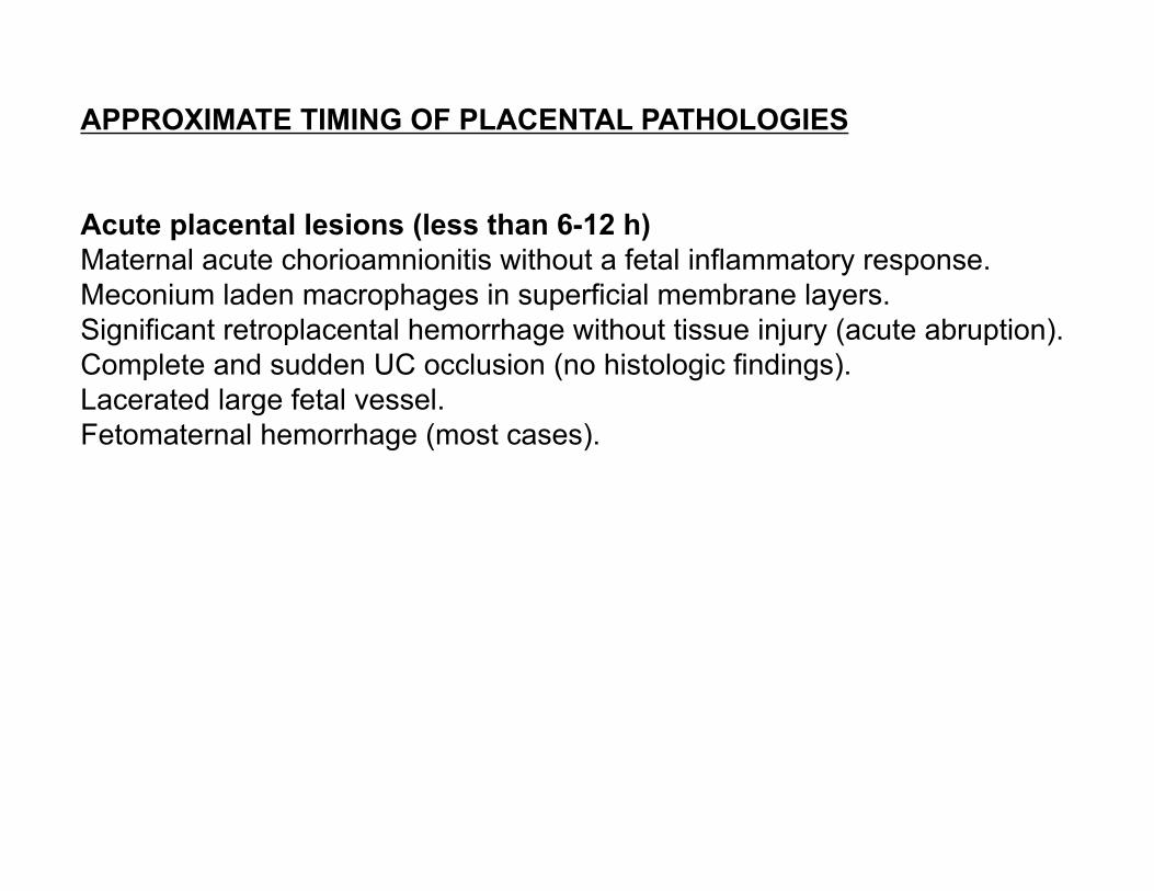
APPROXIMATE TIMING OF PLACENTAL PATHOLOGIES Acute placental lesions (less than 6-12 h) Maternal acute chorioamnionitis without a fetal inflammatory response. Meconium laden macrophages in superficial membrane layers. Significant retroplacental hemorrhage without tissue injury (acute abruption). Complete and sudden UC occlusion (no histologic findings). Lacerated large fetal vessel. Fetomaternal hemorrhage (most cases).
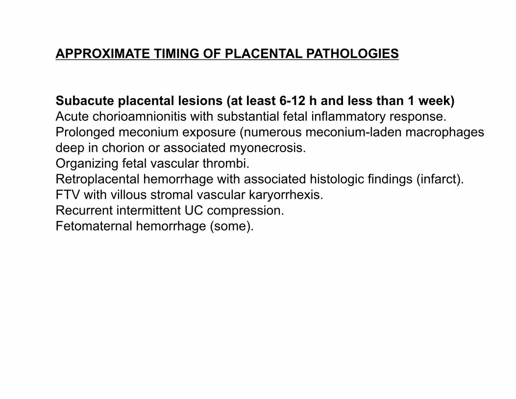
APPROXIMATE TIMING OF PLACENTAL PATHOLOGIES Subacute placental lesions (at least 6-12 h and less than 1 week) Acute chorioamnionitis with substantial fetal inflammatory response. Prolonged meconium exposure (numerous meconium-laden macrophages deep in chorion or associated myonecrosis. Organizing fetal vascular thrombi. Retroplacental hemorrhage with associated histologic findings (infarct). FTV with villous stromal vascular karyorrhexis. Recurrent intermittent UC compression. Fetomaternal hemorrhage (some).
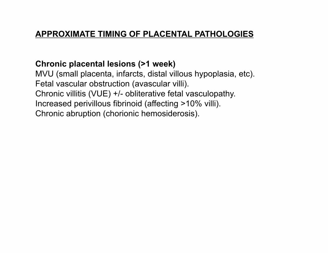
APPROXIMATE TIMING OF PLACENTAL PATHOLOGIES Chronic placental lesions (>1 week) MVU (small placenta, infarcts, distal villous hypoplasia, etc). Fetal vascular obstruction (avascular villi). Chronic villitis (VUE) +/- obliterative fetal vasculopathy. Increased perivillous fibrinoid (affecting >10% villi). Chronic abruption (chorionic hemosiderosis).
PLACENTAL LESIONS ASSOCIATED ADVERSE OUTCOMES
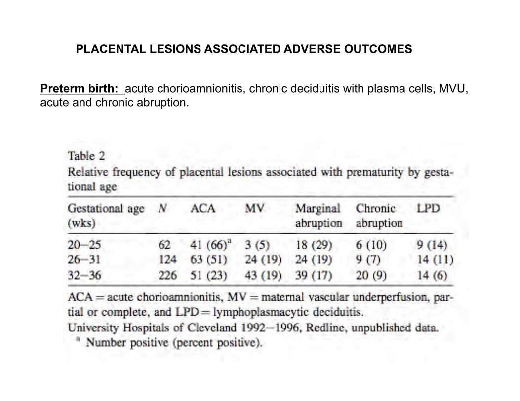
Preterm birth: acute chorioamnionitis, chronic deciduitis with plasma cells, MVU, acute and chronic abruption.
PLACENTAL LESIONS ASSOCIATED ADVERSE OUTCOMES
PLACENTAL LESIONS ASSOCIATED ADVERSE OUTCOMES
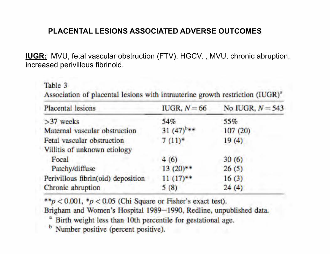
IUGR: MVU, fetal vascular obstruction (FTV), HGCV, , MVU, chronic abruption, increased perivillous fibrinoid.
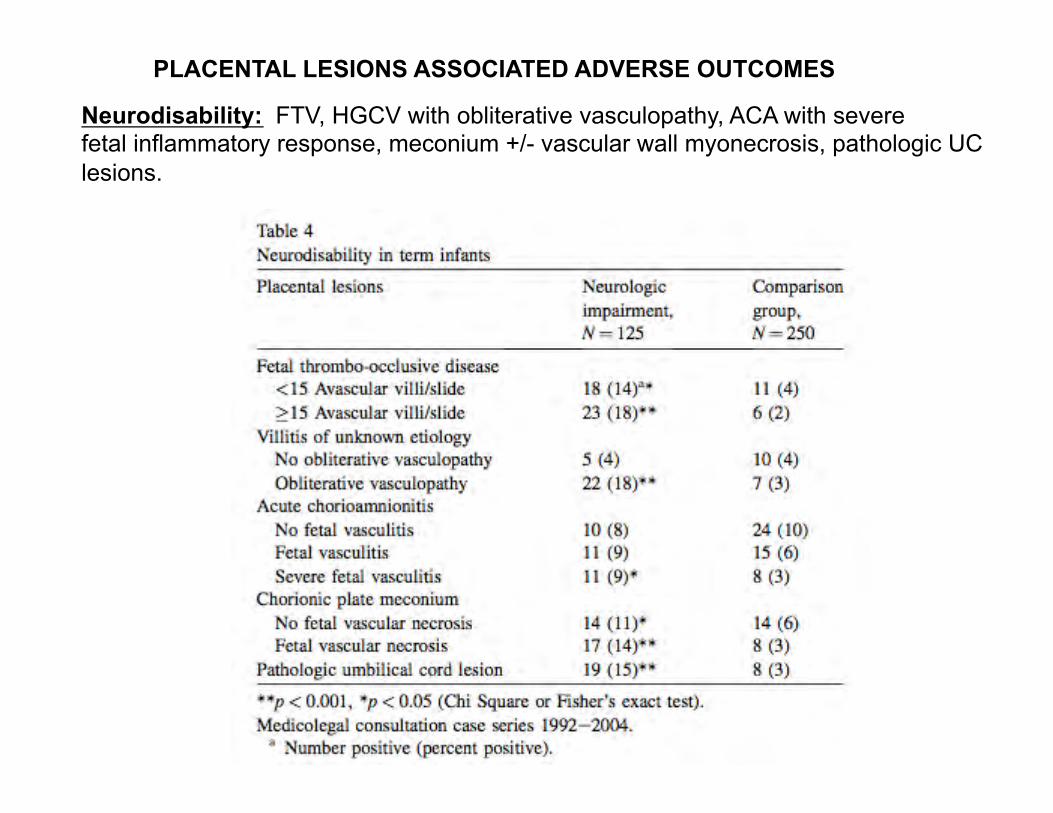
Neurodisability: FTV, HGCV with obliterative vasculopathy, ACA with severe fetal inflammatory response, meconium +/- vascular wall myonecrosis, pathologic UC lesions.
PLACENTAL LESIONS ASSOCIATED ADVERSE OUTCOMES
PLACENTAL LESIONS ASSOCIATED ADVERSE OUTCOMES
Maternal recurrent pregnancy loss: MVU, chronic deciduitis with plasma cells, CV (VUE), MPVFD, chronic histiocytic intervillositis.
PLACENTAL LESIONS ASSOCIATED ADVERSE OUTCOMES
Redline RW. Correlation of placental pathology with perinatal brain injury. In: Surgical Pathology Clinics Placental Pathology. Baergen RN & Goldblum JR eds. Elsevier, Philadelphia, 2013. Birth asphyxia defined by low cord pH and elevated base excess with resulting neonatal encephalopathy (NE).
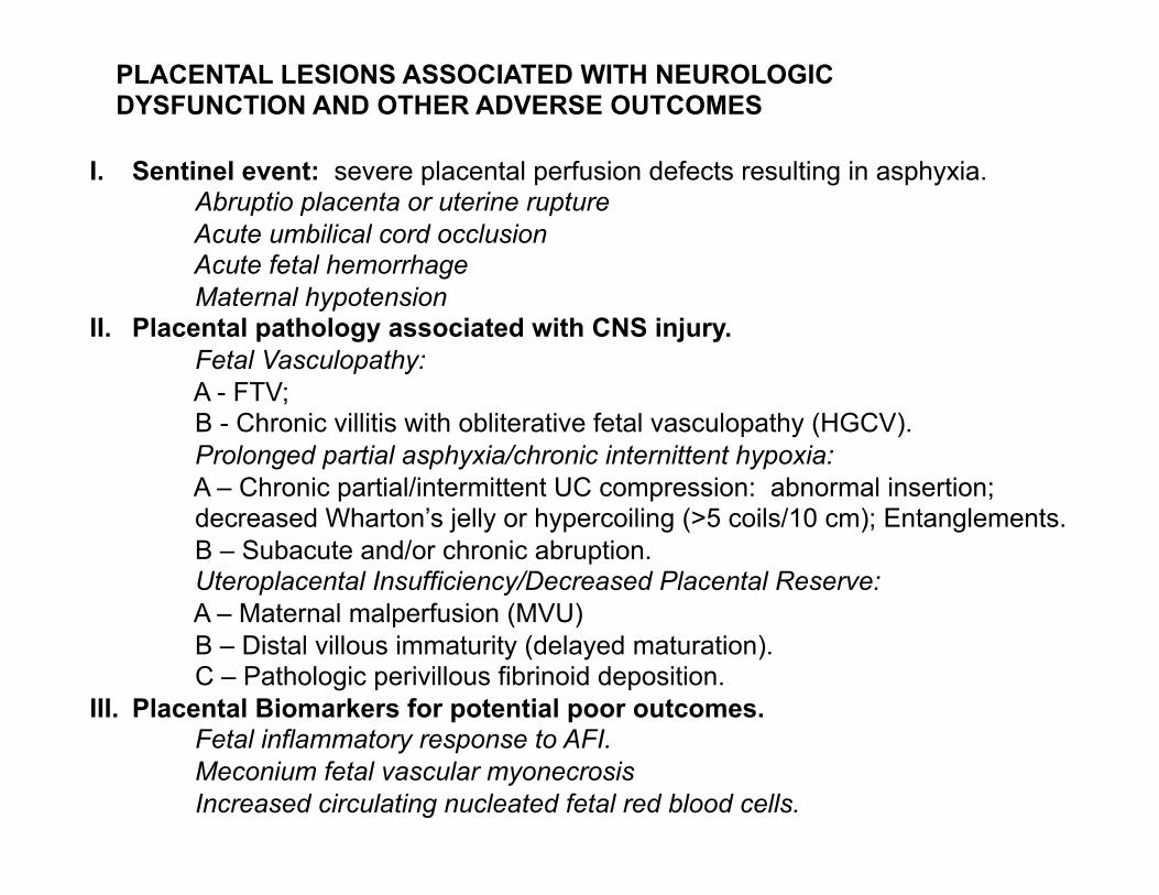
PLACENTAL LESIONS ASSOCIATED WITH NEUROLOGIC DYSFUNCTION AND OTHER ADVERSE OUTCOMES
I. Sentinel event: severe placental perfusion defects resulting in asphyxia. Abruptio placenta or uterine rupture Acute umbilical cord occlusion Acute fetal hemorrhage Maternal hypotension
II. Placental pathology associated with CNS injury. Fetal Vasculopathy: A - FTV; B - Chronic villitis with obliterative fetal vasculopathy (HGCV). Prolonged partial asphyxia/chronic internittent hypoxia: A – Chronic partial/intermittent UC compression: abnormal insertion; decreased Wharton’s jelly or hypercoiling (>5 coils/10 cm); Entanglements. B – Subacute and/or chronic abruption. Uteroplacental Insufficiency/Decreased Placental Reserve: A – Maternal malperfusion (MVU) B – Distal villous immaturity (delayed maturation). C – Pathologic perivillous fibrinoid deposition.
III. Placental Biomarkers for potential poor outcomes. Fetal inflammatory response to AFI. Meconium fetal vascular myonecrosis Increased circulating nucleated fetal red blood cells.
Ona M Faye-Petersen MD Course Director Linda M Ernst MD MPH Assistant Course Director
PERINATAL PATHOLOGY ANNOUNCEMENTS
--------------------------------------------------------------------------------------------------------------------------
Useful Pediatric Pathology App for your cell phone. Go to Apps store and type in PedsPath. This will bring up Peds Path Measurements (Free) by Adrian Arva. Very helpful to have handy organ weights and growth parameters.
POSSIBLE PERINATAL TOPICS FOR DISCUSSION I. The Perinatal Autopsy Perinatal pathology: practice suggestions for limited-resource settings. Roberts DJ. Arch Pathol Lab Med. 2013 Jun;137(6):775-81. doi: 10.5858/arpa.2011-0560-SA. PMID: 23721272 [PubMed - indexed for MEDLINE] Ernst LM. A pathologist's perspective on the pernatal autopsy. Am J Perinatol In Press.
II. Stillbirth Classification schemes for the causes of stillbirth (INCODE).
III. **Karyotyping versus Microarray**
THE UTILITY OF KARYOTYPE VS. MICROARRAY IN ASSESSING FOR GENETIC
ABNORMALITIES.
A CASE Clinical History: • 32-34 week live born male neonate delivered via C-section at an outside institution. • 28 year old G5P4 mother with no prenatal care and a history of drug use. • Mother presented 12 hours prior to delivery with bleeding and foul smelling discharge. • Ultrasound revealed a large abdominal wall defect and agenesis of the lower spine in fetus. • At delivery Apgars were 7 and 8. • Intubated/pressors and transferred to Lurie Children’s Hospital. • Mother decided to pursue palliative measures and infant expired at 24 hours of age. • A full autopsy was requested.
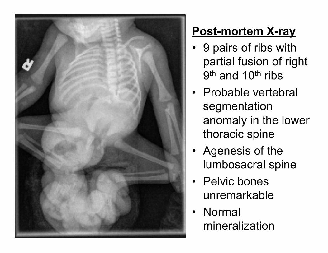
Post-mortem X-ray • 9 pairs of ribs with
partial fusion of right 9th and 10th ribs
• Probable vertebral segmentation anomaly in the lower thoracic spine
• Agenesis of the lumbosacral spine
• Pelvic bones unremarkable
• Normal mineralization
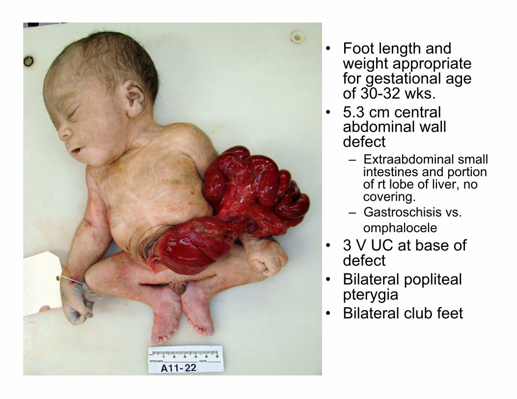
• Foot length and weight appropriate for gestational age of 30-32 wks.
• 5.3 cm central abdominal wall defect – Extraabdominal small
intestines and portion of rt lobe of liver, no covering.
– Gastroschisis vs. omphalocele
• 3 V UC at base of defect
• Bilateral popliteal pterygia
• Bilateral club feet
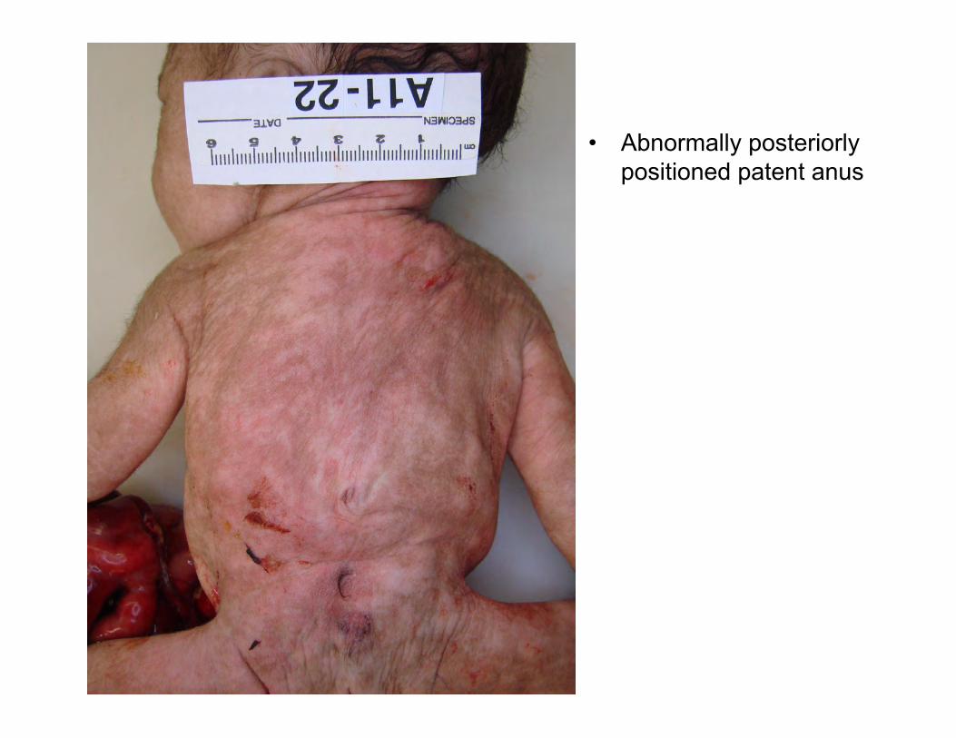
• Abnormally posteriorly positioned patent anus
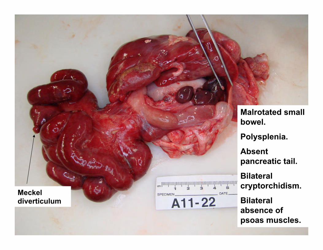
Malrotated small bowel.
Polysplenia.
Absent pancreatic tail.
Bilateral cryptorchidism.
Bilateral absence of psoas muscles.
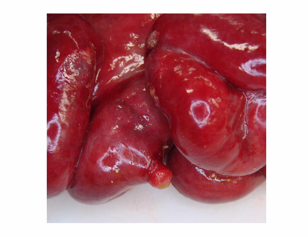
Meckel diverticulum
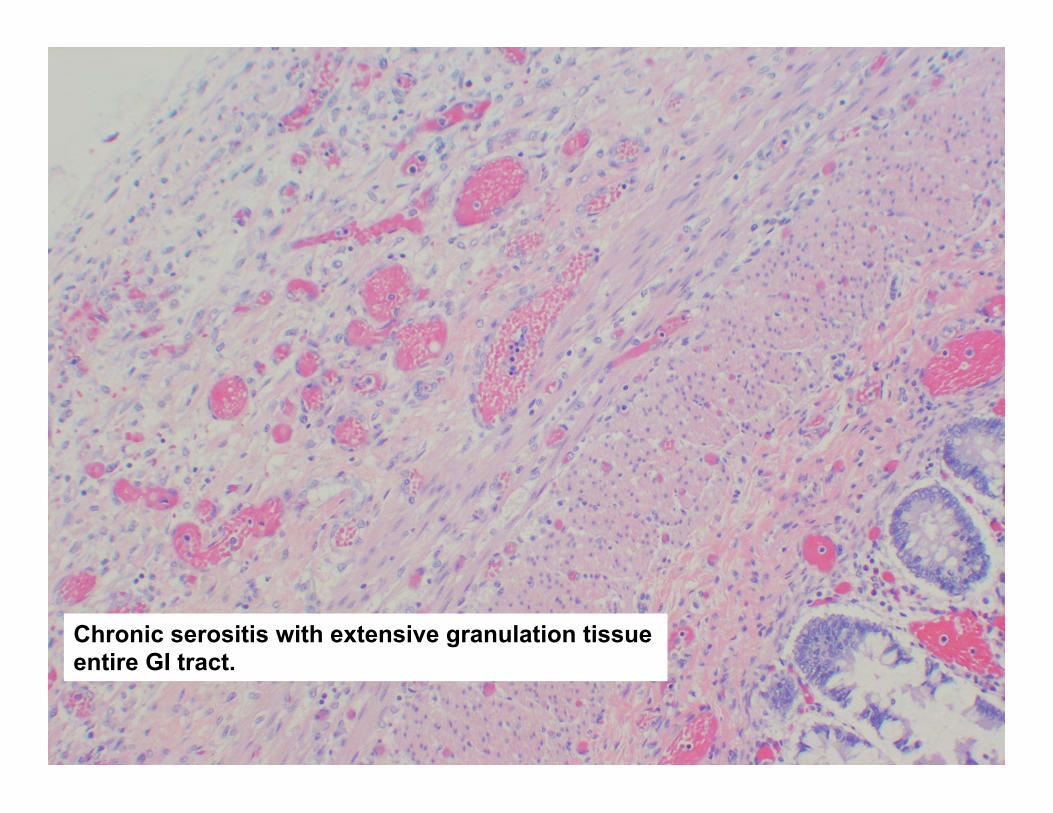
Chronic serositis with extensive granulation tissue entire GI tract.
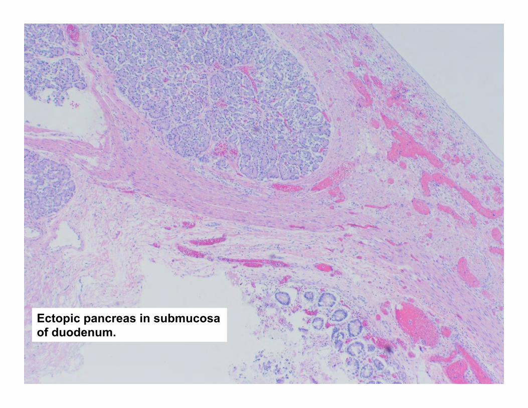
Ectopic pancreas in submucosa of duodenum.
Kidney
• Single horseshoe kidney with cystic change.
• Bilateral hydroureter with focal areas of stricture.
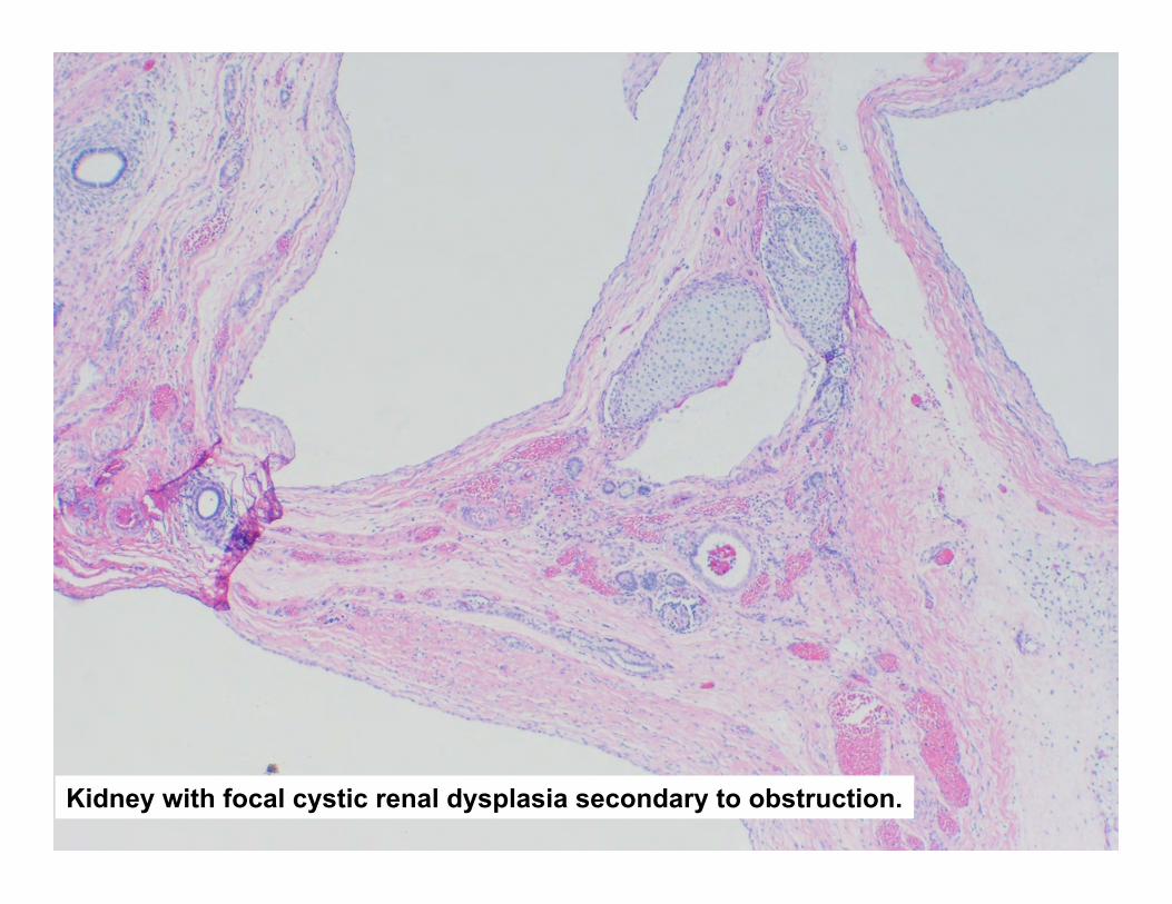
Kidney with focal cystic renal dysplasia secondary to obstruction.
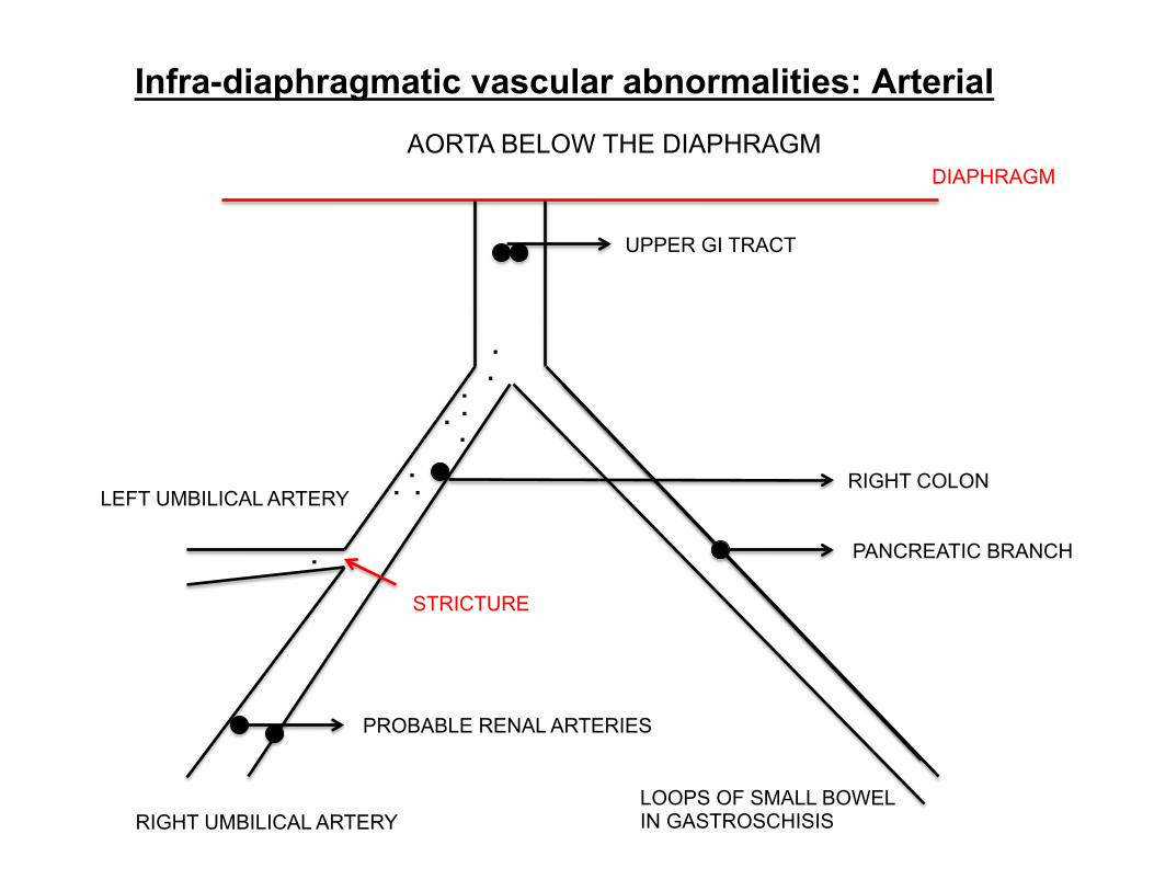
Infra-diaphragmatic vascular abnormalities: Arterial AORTA BELOW THE DIAPHRAGM
DIAPHRAGM
UPPER GI TRACT
RIGHT COLON
PANCREATIC BRANCH
LOOPS OF SMALL BOWEL IN GASTROSCHISIS
LEFT UMBILICAL ARTERY
RIGHT UMBILICAL ARTERY
PROBABLE RENAL ARTERIES
STRICTURE
.
. . . .
. .
.
.
.
Infra-diaphragmatic vascular abnormalities: Venous
• No definitive inferior vena cava. • A confluence of veins from kidneys, GI,
adrenals formed a vein that passed separately and centrally through the diaphragm.
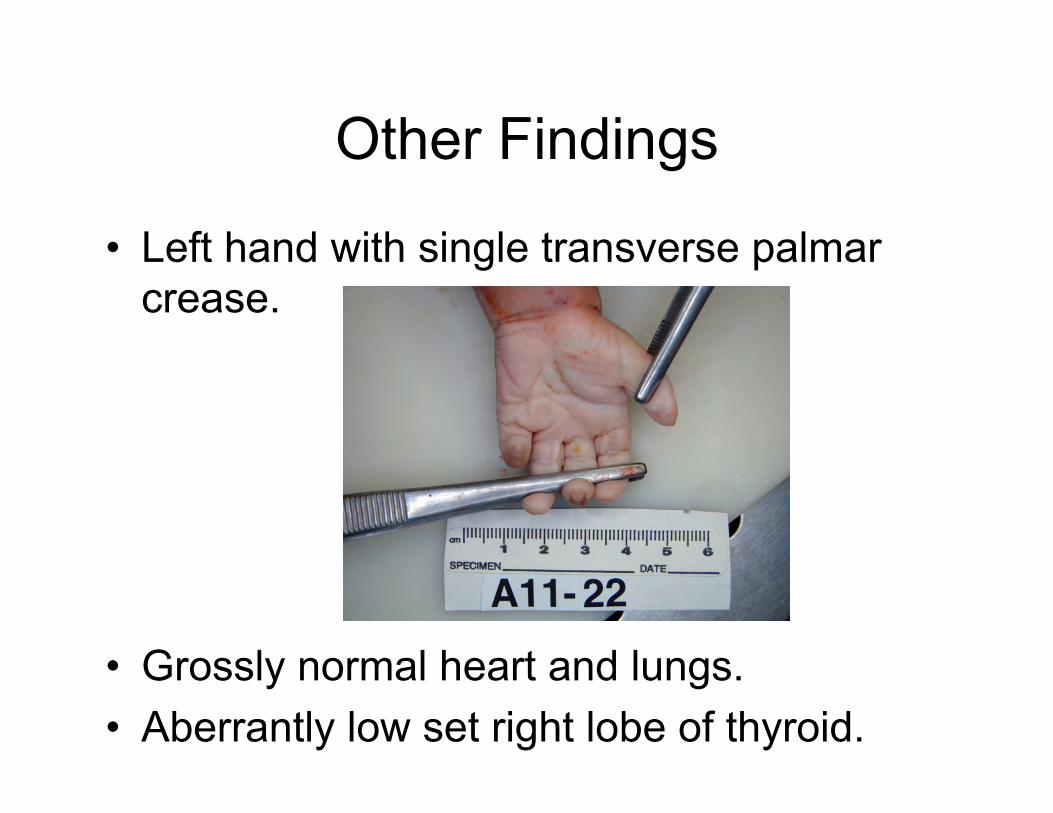
Other Findings
• Left hand with single transverse palmar crease.
• Grossly normal heart and lungs. • Aberrantly low set right lobe of thyroid.
CNS
• Cortical dysplasia. • Partial fusion of thalami. • Spinal cord with lumbosacral fusion of
grey matter with dysplasia – consistent with sacral agenesis.
• Mild hypoxic ischemic injury (ponto-subiculum neuronal necrosis with periventricular leukomalacia).
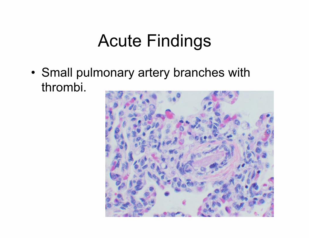
Acute Findings
• Small pulmonary artery branches with thrombi.
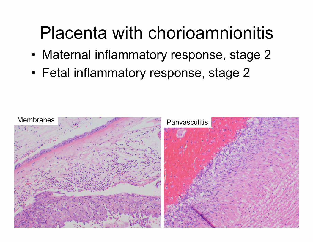
Placenta with chorioamnionitis • Maternal inflammatory response, stage 2 • Fetal inflammatory response, stage 2
Membranes Panvasculitis
Summary • Multiple congenital anomalies • Evidence of acute chorioamnionitis in placenta • Evidence of neonatal DIC

Differential Diagnosis?
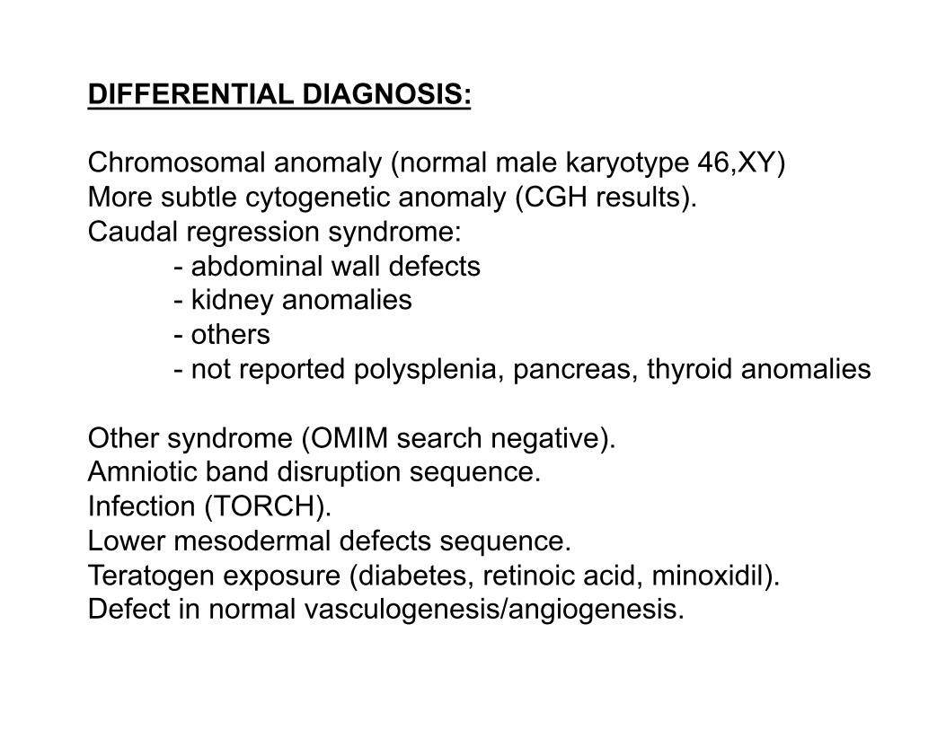
DIFFERENTIAL DIAGNOSIS: Chromosomal anomaly (normal male karyotype 46,XY) More subtle cytogenetic anomaly (CGH results). Caudal regression syndrome:
- abdominal wall defects - kidney anomalies - others - not reported polysplenia, pancreas, thyroid anomalies
Other syndrome (OMIM search negative). Amniotic band disruption sequence. Infection (TORCH). Lower mesodermal defects sequence. Teratogen exposure (diabetes, retinoic acid, minoxidil). Defect in normal vasculogenesis/angiogenesis.
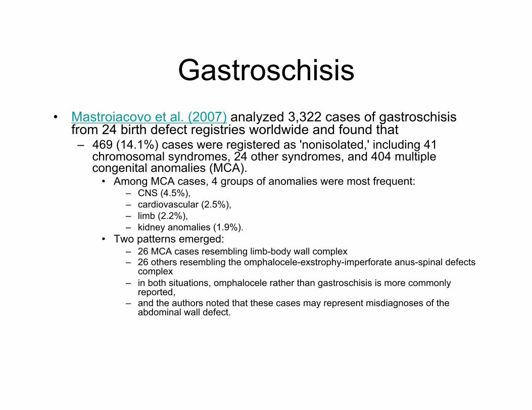
Gastroschisis • Mastroiacovo et al. (2007) analyzed 3,322 cases of gastroschisis
from 24 birth defect registries worldwide and found that – 469 (14.1%) cases were registered as 'nonisolated,' including 41
chromosomal syndromes, 24 other syndromes, and 404 multiple congenital anomalies (MCA).
• Among MCA cases, 4 groups of anomalies were most frequent: – CNS (4.5%), – cardiovascular (2.5%), – limb (2.2%), – kidney anomalies (1.9%).
• Two patterns emerged: – 26 MCA cases resembling limb-body wall complex – 26 others resembling the omphalocele-exstrophy-imperforate anus-spinal defects
complex – in both situations, omphalocele rather than gastroschisis is more commonly
reported, – and the authors noted that these cases may represent misdiagnoses of the
abdominal wall defect.
Additional Molecular Workup by:
Lawrence Jennings MD, PhD Katrin Carlson Leuer PhD
This was a very unusual case. Initially we did chromosome analysis on peripheral blood that showed a normal male karytoype 46,XY in 20 cells. Then the CGH microarray results came back with an abnormality dup(3)(q23q29). Phenotype is not the same as seen in chromosome 3 duplication q/ deletion p syndrome (craniofacial and cardiac defects). (Fineman RM et al. Pediatrics 61:611-619, 1978 and Faas BHW et al. Clin Genet 62:315-320, 2002)
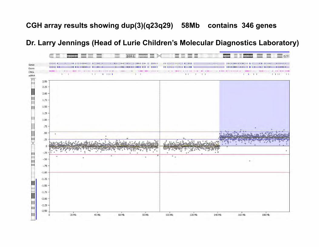
CGH array results showing dup(3)(q23q29) 58Mb contains 346 genes Dr. Larry Jennings (Head of Lurie Children’s Molecular Diagnostics Laboratory)
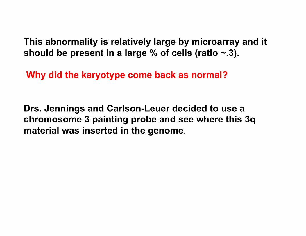
This abnormality is relatively large by microarray and it should be present in a large % of cells (ratio ~.3). Why did the karyotype come back as normal? Drs. Jennings and Carlson-Leuer decided to use a chromosome 3 painting probe and see where this 3q material was inserted in the genome.
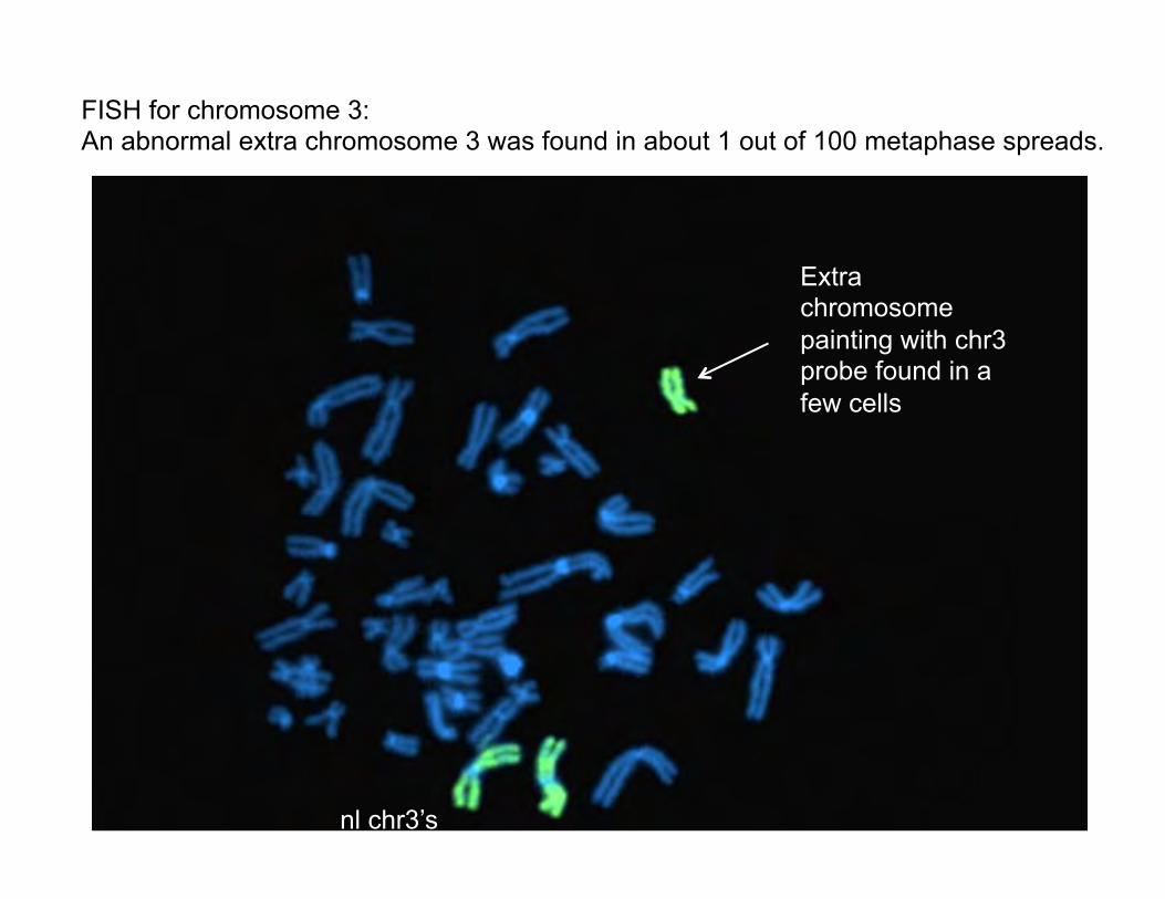
The FISH paint showed that most of the metaphase spreads were normal, none had insertion of this material anywhere, but a few had an additional chromosome that completely painted with chr 3.
nl chr3’s
Extra chromosome painting with chr3 probe found in a few cells
FISH for chromosome 3: An abnormal extra chromosome 3 was found in about 1 out of 100 metaphase spreads.
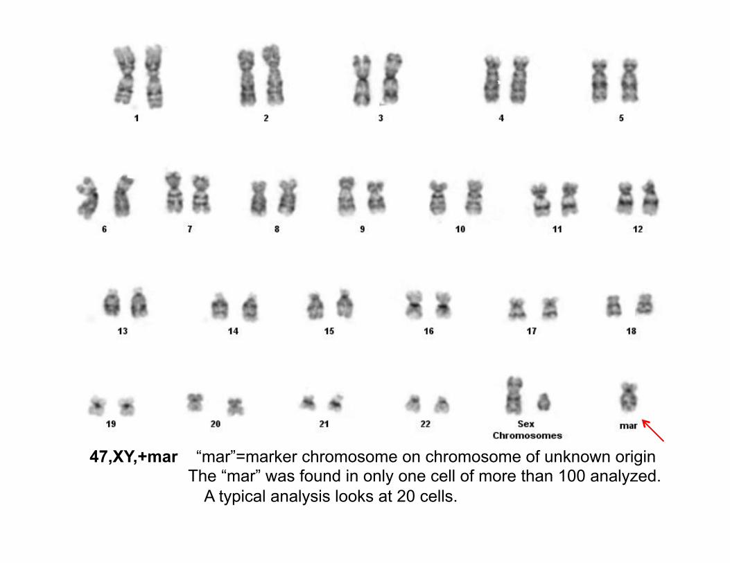
47,XY,+mar “mar”=marker chromosome on chromosome of unknown origin The “mar” was found in only one cell of more than 100 analyzed.
A typical analysis looks at 20 cells.
There were two confusing issues: 1) The marker chromosome was much larger than the
duplicated segment identified by microarray would have predicted it to be.
2) Only 1 in 100 metaphase spreads showed the marker chromosome and this does not seem to account for the quantity seen by CGH.
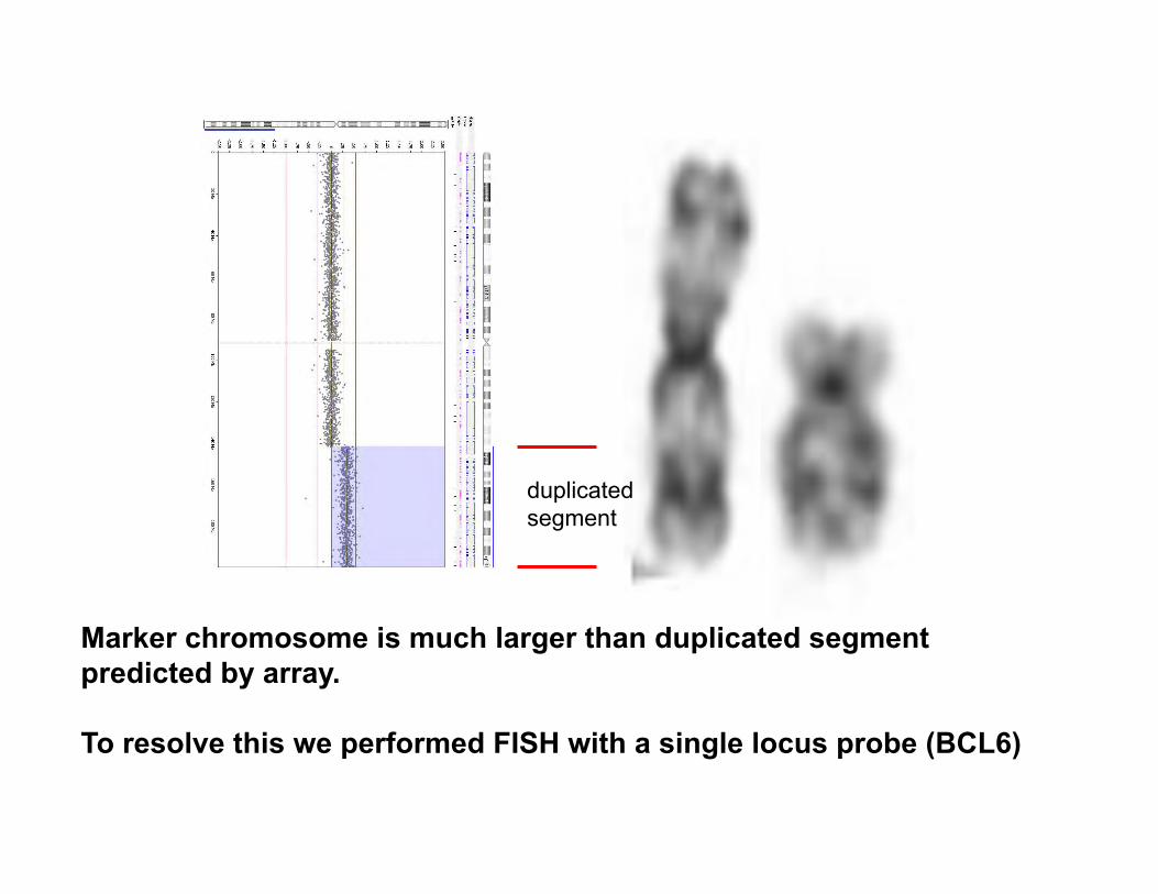
duplicated segment
Marker chromosome is much larger than duplicated segment predicted by array. To resolve this we performed FISH with a single locus probe (BCL6)
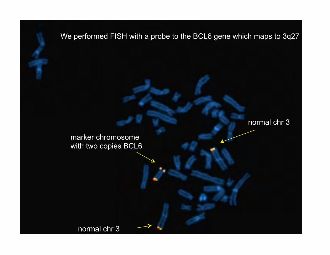
We performed FISH with a probe to the BCL6 gene which maps to 3q27
normal chr 3
normal chr 3
marker chromosome with two copies BCL6
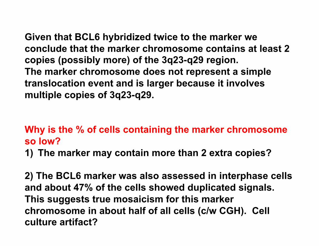
Given that BCL6 hybridized twice to the marker we conclude that the marker chromosome contains at least 2 copies (possibly more) of the 3q23-q29 region. The marker chromosome does not represent a simple translocation event and is larger because it involves multiple copies of 3q23-q29. Why is the % of cells containing the marker chromosome so low? 1) The marker may contain more than 2 extra copies? 2) The BCL6 marker was also assessed in interphase cells and about 47% of the cells showed duplicated signals. This suggests true mosaicism for this marker chromosome in about half of all cells (c/w CGH). Cell culture artifact?
FINAL DIAGNOSES: Multiple congenital anomalies due to mosaicism for an extra chromosome containing duplications of part of chromosome 3q. (male karyotype 46,XY + mar) Preterm premature delivery due to acute chorioamnionitis. Diffuse microthrombi in pulmonary artery branches.
KARYOTYPE VS MICROARRAY When do you perform karyotype vs. microarray? What are the advantages and disadvantages of microarray? What are the current recommendations for microarraay? How do the recommendations affect pediatric pathologists and the perinatal autopsy?
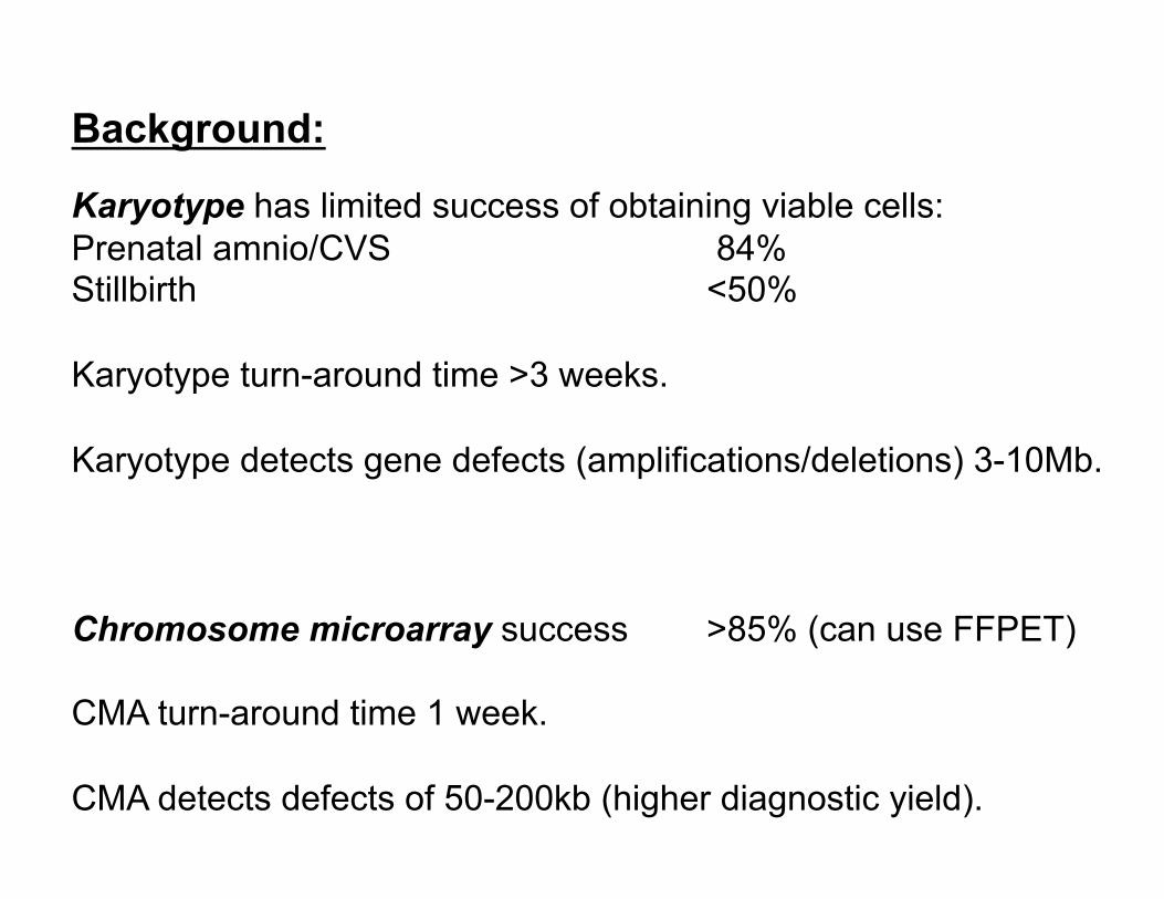
Background: Karyotype has limited success of obtaining viable cells: Prenatal amnio/CVS 84% Stillbirth <50% Karyotype turn-around time >3 weeks. Karyotype detects gene defects (amplifications/deletions) 3-10Mb. Chromosome microarray success >85% (can use FFPET) CMA turn-around time 1 week. CMA detects defects of 50-200kb (higher diagnostic yield).
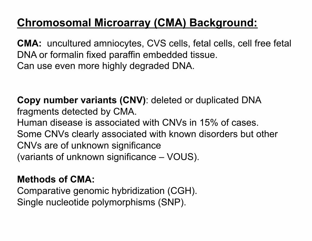
Chromosomal Microarray (CMA) Background: CMA: uncultured amniocytes, CVS cells, fetal cells, cell free fetal DNA or formalin fixed paraffin embedded tissue. Can use even more highly degraded DNA. Copy number variants (CNV): deleted or duplicated DNA fragments detected by CMA. Human disease is associated with CNVs in 15% of cases. Some CNVs clearly associated with known disorders but other CNVs are of unknown significance (variants of unknown significance – VOUS). Methods of CMA: Comparative genomic hybridization (CGH). Single nucleotide polymorphisms (SNP).
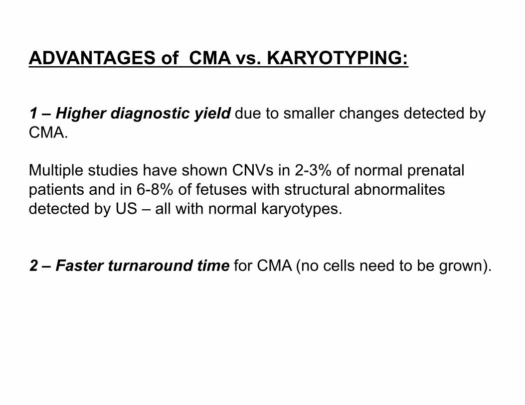
ADVANTAGES of CMA vs. KARYOTYPING: 1 – Higher diagnostic yield due to smaller changes detected by CMA. Multiple studies have shown CNVs in 2-3% of normal prenatal patients and in 6-8% of fetuses with structural abnormalites detected by US – all with normal karyotypes. 2 – Faster turnaround time for CMA (no cells need to be grown).
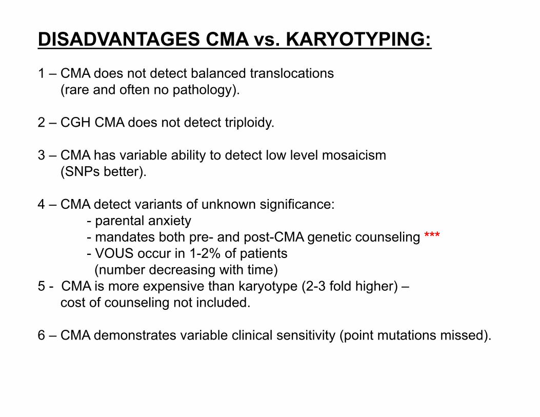
DISADVANTAGES CMA vs. KARYOTYPING: 1 – CMA does not detect balanced translocations (rare and often no pathology). 2 – CGH CMA does not detect triploidy. 3 – CMA has variable ability to detect low level mosaicism (SNPs better). 4 – CMA detect variants of unknown significance:
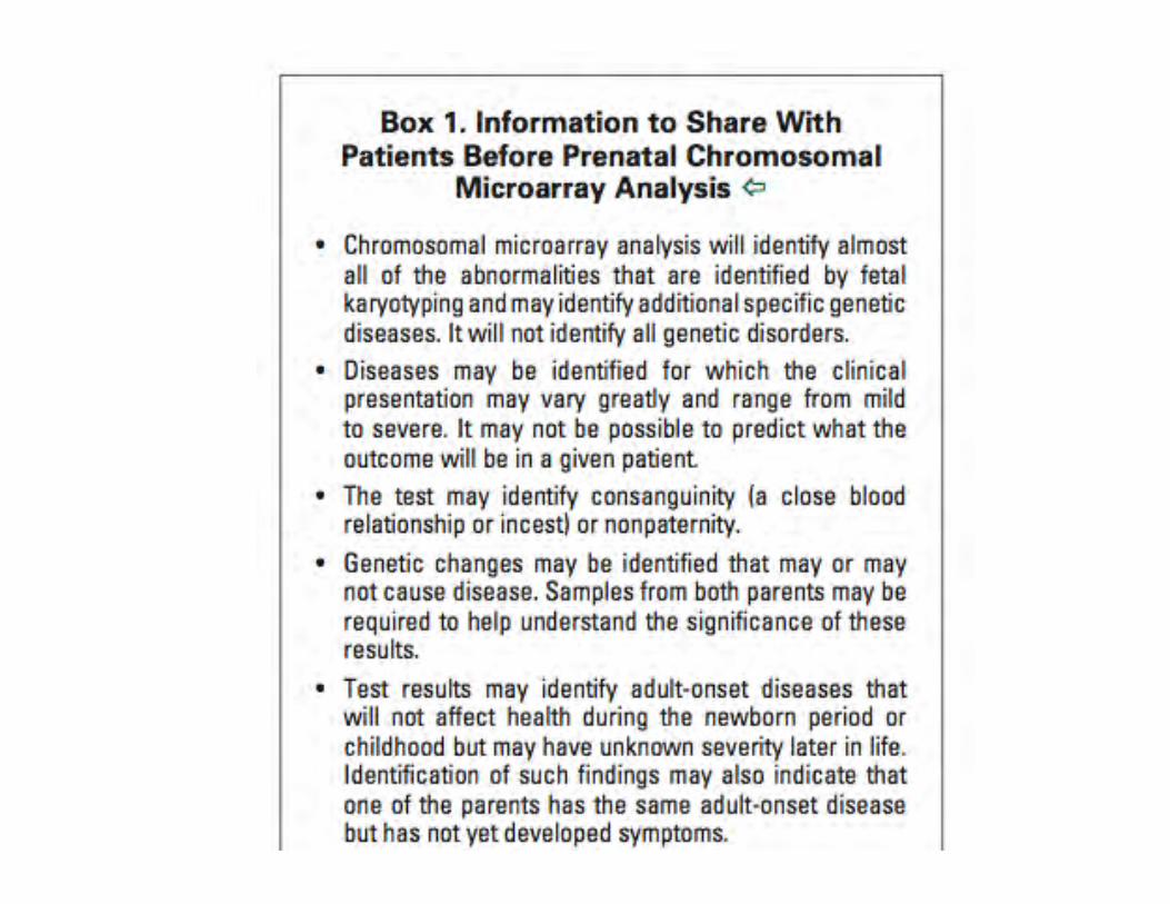
- parental anxiety - mandates both pre- and post-CMA genetic counseling *** - VOUS occur in 1-2% of patients (number decreasing with time)
5 - CMA is more expensive than karyotype (2-3 fold higher) – cost of counseling not included. 6 – CMA demonstrates variable clinical sensitivity (point mutations missed).
Reddy UM et al. Karyotype versus microarray testing for genetic abnormalities after stillbirth. N Engl J Med 367:2185-2193, 2012.
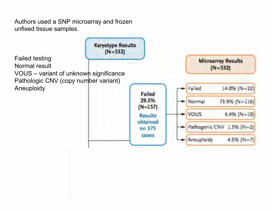
Authors used a SNP microarray and frozen unfixed tissue samples. Failed testing Normal result VOUS – variant of unknown significance Pathologic CNV (copy number variant) Aneuploidy
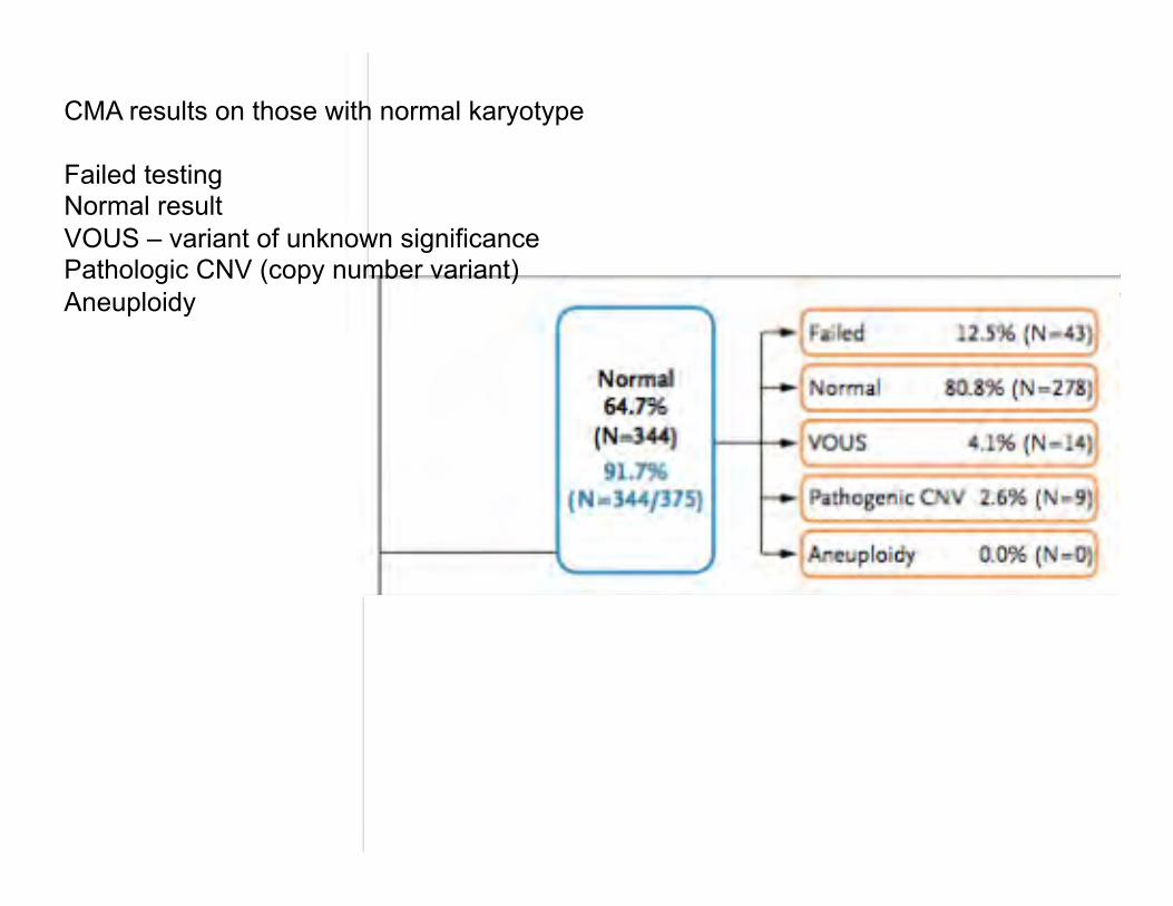
CMA results on those with normal karyotype Failed testing Normal result VOUS – variant of unknown significance Pathologic CNV (copy number variant) Aneuploidy
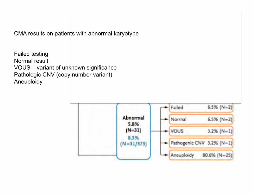
CMA results on patients with abnormal karyotype Failed testing Normal result VOUS – variant of unknown significance Pathologic CNV (copy number variant) Aneuploidy
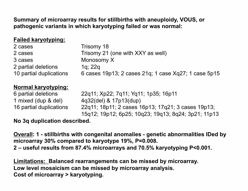
Summary of microarray results for stillbirths with aneuploidy, VOUS, or pathogenic variants in which karyotyping failed or was normal: Failed karyotyping: 2 cases Trisomy 18 2 cases Trisomy 21 (one with XXY as well) 3 cases Monosomy X 2 partial deletions 1q; 22q 10 partial duplications 6 cases 19p13; 2 cases 21q; 1 case Xq27; 1 case 5p15 Normal karyotyping: 6 partial deletions 22q11; Xp22; 7q11; Yq11; 1p35; 16p11 1 mixed (dup & del) 4q32(del) & 17p13(dup) 16 partial duplications 22q11; 18p11; 2 cases 16p13; 17q21; 3 cases 19p13;
15q12; 19p12; 6p25; 10q23; 19q13; 8q24; 3p21; 11p13 No 3q duplication described. Overall: 1 - stillbirths with congenital anomalies - genetic abnormalities IDed by microarray 30% compared to karyotype 19%, P=0.008. 2 – useful results from 87.4% microarrays and 70.5% karyotyping P<0.001. Limitations: Balanced rearrangements can be missed by microarray. Low level mosaicism can be missed by microarray analysis. Cost of microarray > karyotyping.
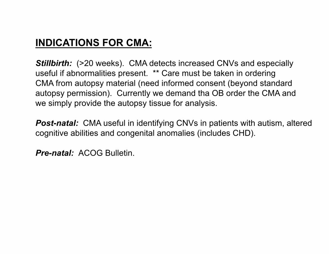
INDICATIONS FOR CMA: Stillbirth: (>20 weeks). CMA detects increased CNVs and especially useful if abnormalities present. ** Care must be taken in ordering CMA from autopsy material (need informed consent (beyond standard autopsy permission). Currently we demand tha OB order the CMA and we simply provide the autopsy tissue for analysis. Post-natal: CMA useful in identifying CNVs in patients with autism, altered cognitive abilities and congenital anomalies (includes CHD). Pre-natal: ACOG Bulletin.
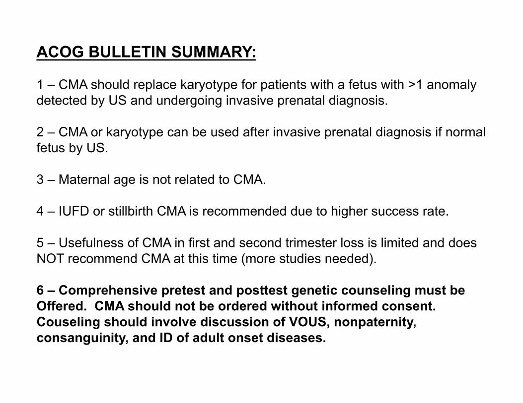
ACOG BULLETIN SUMMARY: 1 – CMA should replace karyotype for patients with a fetus with >1 anomaly detected by US and undergoing invasive prenatal diagnosis. 2 – CMA or karyotype can be used after invasive prenatal diagnosis if normal fetus by US. 3 – Maternal age is not related to CMA. 4 – IUFD or stillbirth CMA is recommended due to higher success rate. 5 – Usefulness of CMA in first and second trimester loss is limited and does NOT recommend CMA at this time (more studies needed). 6 – Comprehensive pretest and posttest genetic counseling must be Offered. CMA should not be ordered without informed consent. Couseling should involve discussion of VOUS, nonpaternity, consanguinity, and ID of adult onset diseases.
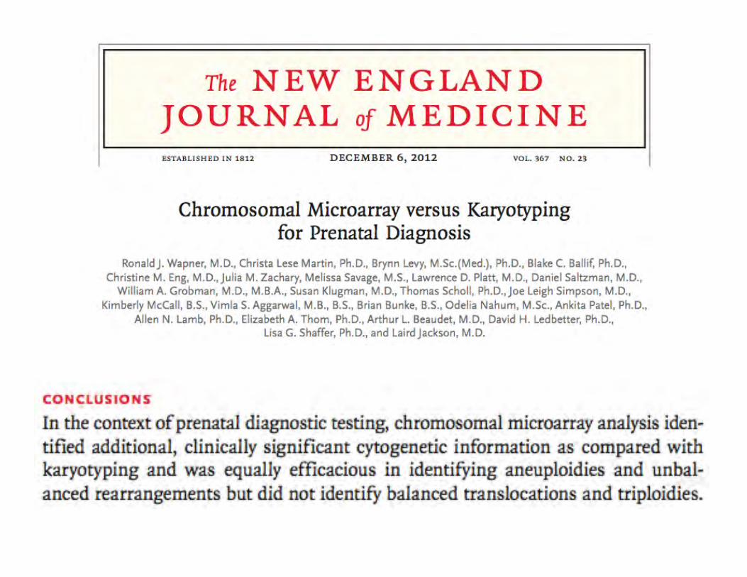
CONCLUSION AND TAKEHOME MESSAGE Microarray analysis is more likely than karyotype analysis to provide a genetic diagnosis, especially in the setting of multiple congenital anomalies.
REFERENCES: 1. Reddy Um et al. Karyotype versus microarray testing for genetic abnormalities after stillbirth. N Engl J Med 367:2185—2193, 2012. 2. Fineman RM et al. Chromosome 3 duplication q/deletion p syndrome. Pediatrics 61:611-618, 1978. 3. Faas BHW et al. A new case of dup(3q) syndrome due to a pure duplication of 3qter. Clin Genet 62:315-320, 2002. 4. Chen CP. Chromosomal abnormalities associated with omphalocele. Taiwanese J Obstet Gynecol 46:1-8, 2007.