Embed Size (px)
Citation preview
VOLUME 10 DECEMBER 1950 NUMBER 12
Assay of Frozen Mouse Mammary Carcinoma for theMammary Tumor Milk Agent*
JOHN J. BITTNER, PH.D., AND DAVID T IMAGAWA, PH.D.
(From the Diris-ion of Cancer Biology, Department of Physiology, and Department of Bacteriology and Immunology,University of Minnesota Medical School, Minneapolis 14, Minn.)
cause and that this, in mammals as in birds, is avirus.―
Mann (33—35)injected frozen spontaneous andtransplanted mammary cancers and explained thedevelopment of tumors on the basis that the mammary tumor milk agent-virus was liberated in itsactive form following freezing, since mammary tumors arose at the site of injection of the suspendedfrozen tumor-mince. The tumors developed asreadily in males as in female mice, but only whenthe frozen tissue was injected into the mammarygland region, because of what the author called“selective infectivity.― She found that, withinlimits, the longer the tissue was frozen, the moretumors developed, and the interval between injection and the appearance of the tumors was relatedto their rate of growth. Fast growing cancers produced growths within 10 days, while slow growingones might be delayed for 7—1@weeks following theinoculation of their frozen suspensions. Mann (35)also demonstrated that mammary tumors could beproduced by the injection of tumors which hadbeen frozen and dried in vacuo (15), provided nothawing occurred before the material was dry.
Gye, Begg, Mann, and Craigie (@9) reportedthat some tumors could withstand a succession offreezing and thawing, which they claimed completely excluded the persistence of tumor cells, butMann (34) observed a progessive deterioration ofactivity of mammary tumors after a second thawing. Individual tumors differed, but in no instancedid a tumor inactive after a second thawing produce tumors after a third.
The exact age of the animals at the time of injection was not stated except that “Although all came
739
ThiB One
I@II@IhII@III@III1 111HUI@I1OLD-CZW-ZGFT
In a series of publications during the past yearGye and his associates (28, @29,33—35)interpretedtheir data to imply that when various types ofcancers from mice were maintained at —79°C.,the malignant cells “arereadily killed by extremecold, but the dead malignant cells are neverthelesscapable of starting a new, strictly new, tumour invirtue of the intrinsic virus which they contain―(@8). It was suggested that the active form of thevirus, changed from a latent form because offreezing, was capable of altering the connectivetissue of the injected host to sarcoma or the normalmammary gland to mammary carcinoma, depending upon the type of cancer being tested. Normaland frozen embryonic tissue served as controls forthe cancer studies (Mann, 3@).
Active dried tissue was obtained by Gye (@28)from fresh sporadic tumors which had developedspontaneously in mice of the R3 and C3H stocks.However, positive results with dried tumors weresecured only when the tissue was minced (14) anddried in a Craigie desiccator (15). When the tissueswere subjected to the Knox method of drying, onlynegative results were obtained (@29).
Gye (@8) postulated that the negative resultsobtained by other investigators were due to imperfect technic and stated that their experimentaldata indicated “that cancer has a continuing
* Assisted by grants from the National Cancer Institute of
the National Institute of Health, Public Health Service, theAmerican Cancer Society upon recommendation of the Committee on Growth of the National Research Council, and theGraduate School Cancer Research Fund of the University ofMinnesota.
Received for publication August @,1950.
CANCER RESEARCH
on June 10, 2019. © 1950 American Association for Cancer Research. cancerres.aacrjournals.org Downloaded from
740 Cancer Re$earch
from high cancer lines, they were used before theywere old enough to develop sporadic tumours, andin all cases the tumours obtained were at the siteof inoculation― (33). The tumor suspension was“inoculated in the right mammary region of miceof the same strain as the tumours― (33).
The studies to be reported here are concernedwith the propagation of mammary tumors following the injection of suspensions of frozen mammary carcinoma and the comparative activity oftransplanted fresh tissue of the same tumors. Assays were made of the fresh and frozen mammarytumors for the mammary tumor milk agent-virusby the injection of cell-free extracts using differentroutes of administration. A tumor that resultedafter the inoculation of a frozen suspension of tumor-mince was also tested for the agent.
MATERIALS AND METHODS
Mice of several inbred stocks and one group ofF1 hybrids were used in the various experiments.All the animals were susceptible for the development of spontaneous mammary cancer, and allpossessed the mammary tumor milk agent exceptthe fostered lines of the Z (C3H) and A strains andtheir hybrids. These are referred to as the Zb andAx lines; their reciprocal hybrids as either ZbAxF1(Zb 9 X Axd―)or AxZbF1 (Ax@ X Zbd').
The spontaneous mammary tumor No. 8415Aarose in an AZF1 breeder produced by mating anA female with a Z male. This particular tumor wastransplanted by the trocar method in F1 hybridswithout the milk agent. In eight passages, a totalof 65 mice were inoculated, and the grafts grewprogressively in all. During the first three passagesthe tumor was transferred every 4 weeks; afterthat, because of the increased growth of the transplants, inoculation was made approximately every@2weeks. Within 6 days some growths attained asize of 0.75 cm. in diameter.
One of the tumors which arose, following the injection of the frozen tumor-mince of AZF1 No.8415A, was continued as AZF1 No. 8415B. Thistumor was maintained for three passages in eitherAxZbF1 or ZbAxF1 hybrids, and none of the @23mice which were inoculated was resistant to thegrafts.
A mammary tumor from a breeding Z (C3H) female was designated as tumor No. 8044 and wastransplanted in F1 hybrids made by reciprocalmatings of A and Z mice without the milk agent.This transplant has been continued for 40 passages, and @238mice have been inoculated. All haveshown progressively growing tumors, and the miceusually succumb within 3—4weeks after they have
been inoculated. The tumors may attain the size of1 cm. in diameter within 1 week.
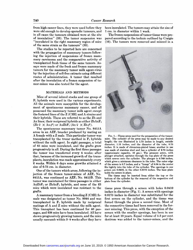
The frozen suspensions of tumor tissue were prepared according to the technic outlined by Craigie(16). The tumors were removed and minced in a
FIG. 1 .—Tissue press used for the preparation of the tumormice. The cylinder of the press may be made to any desiredlength; the one illustrated is 1.75 inches in length, outsidediameter, 1.13 inches, and the diameter of the tube, 0.75inches. It is made of chromium-plated brass; another in usewas made of stainless steel and has a cylinder of @.75inches(approximate capacity, 10 gin.). The pressure screw, withplunger attached, operates through the top plate of the presswhich screws onto the cylinder. The plunger is 0.749 inches,which gives a minimum clearance in the tube. The outer edgeof the screen is 0.7 inches, and a “hump―of 0.04 in the centerfits tightly into the tube of the cylinder. The openings in onescreen are 0.0410, in the other 0.0175 inches. The base plateholds the screen in place.
The tissue may be inserted from either the top or thebottom of the cylinder by the removal of the respective endplate from the cylinder.
tissue press through a screen with holes 0.0410inches in diameter (Fig. 1). A screen with openings0.0175 inches in diameter was substituted for thefirst screen on the cylinder, and the tissue wasforced through the press a second time. Most ofthe connective tissue had been removed in the former mincing. This press, with the exception of thescreen with the smaller openings, has been in usefor at least 10 years. Equal volume of 5.3 per centdextrose was added to the tumor-mince, and the
on June 10, 2019. © 1950 American Association for Cancer Research. cancerres.aacrjournals.org Downloaded from
BITTNER AND IMAGAWA—M?l/C Agent in Frozen Mouse Cancer 741
suspension was stored in glass ampoules,@ cc. pervial, which were sealed. The ampoules were frozenin cellosolve and dry ice and stored in a thermosjug with dry ice at a constant temperature of—79°C.
For testing the ability of the frozen tumormince to produce tumors, the ampoules were transferred to a water bath kept at 37°C. until the tissue had thawed. Six cc. of saline was added to thecontents of each tube, and the suspension was injected so that each animal received an amountequal to 0.05 gm. of the original tumor-mince. Thesite of injection is given for each experiment.
In addition to testing the frozen tumor-mince,cell-free extracts were also employed. Details will
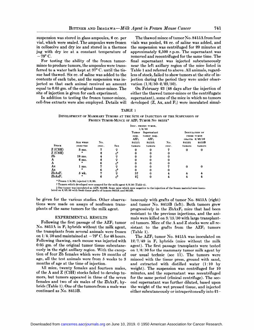
The thawed mince of tumor No. 8415A from fourvials was pooled, @4cc. of saline was added, andthe suspension was centrifuged for @20minutes atapproximately @2,500r.p.m. The supernatant wasremoved and recentrifuged for the same time. Thefinal supernatant was injected subcutaneouslynear the left axillary region of the mice listed inTable 1 and referred to above. All animals, regardless of stock, failed to show tumors at the site of injection during the period they were under observation (1/6/50—@2/@23/50).
On February @23(48 days after the injection ofeither the thawed tumor-mince or the centrifugatesupernatant), some of the mice in which no tumorsdeveloped (Z, Ax, and F1) were inoculated simul
TABLE 1
DEVELOPMENT OF MAMMARY TUMORS AT THE SITE OF INJECTION OF THE SUSPENSION OF
FROZEN TUMOR-MINCE OF AZF1 TUMOR No 8415A5
AGE WHEN
INJECTED
3 mo.
18 mo.@ mo.
1 mo.I'
5 wk.
INOC. FROZENTUMOR,1/6/50Tumor
SupernatantINOCULATIONOFsusp.
tumor susp.FRESHTUMORAZF1
AZF1GRAFTS1/13/50No.8415A
8415ANo.8415A8415BINOC.SEXtumors
tumorsINOC.tumorstumors790
0700d'000490
0090006d'000790
07006S'00079St
04446ci―0444
STOCK
Z (CSH)Z (C3H)ZbAAAxAxZbAxF1ZbAxF1
* Frozen 1/4/50, injected 1/6/50.
Tumors which developed were assayed for the milk agent 1/9/50 (Table 4).
@ One tumor was inoculated as AZF, 8415B. Some mice which were negative to the injection of the frozen material were inoculated on 1/13/50 with fresh tissue grafts of tumors 8415A and 8415B.
be given for the various studies. Other observations were made on assays of nonfrozen transplants of the same tumors for the milk agent.
EXPERIMENTAL RESULTSFollowing the first passage of the AZF1 tumor
No. 84l5A in F1 hybrids without the milk agent,the transplants from several animals were frozenon 1,/4@'50 and maintained at —79°C. for 48 hours.Following thawing, each mouse was injected with0.05 gm. of the original tumor tissue. subcutaneously in the right axillary region. With the excep
tion of four Zb females which were 18 months ofage, all the test animals were from 5 weeks to 3months of age at the time of injection.
All mice, twenty females and fourteen males,of the A and Z (C3H) stocks failed to develop tumors, but tumors appeared in three of the sevenfemales and two of six males of the ZbAxF1 hybrids (Table 1). One of the tumors from a male wascontinued as No. 8415B.
taneously with grafts of tumor No. 8415A (right)and tumor No. 8415B (left). Both tumors grewprogressively in the ZbAxF1 mice that had beenresistant to the previous injections, and the animals were killed on 3/15/50 with large transplanted tumors. Mice of the A and Z stocks were all resistant to the grafts from the AZF1 tumors(Table 1).
The AZF1 tumor No. 8415A was inoculated onU2/7/49 in F1 hybrids (mice without the milkagent). The first passage transplants were testedon 1/6/50 for the mammary tumor milk agent byour usual technic (see 11). The tumors wereminced with the tissue press, ground with sand,and extracted with distilled water (1: 10 byweight). The suspension was centrifuged for 10minutes, and the supernatant was recentrifugedfor the same period (clinical centrifuge). The second supernatant was further diluted, based uponthe weight of the wet pressed tissue, and injectedeither subcutaneously or intraperitoneally into @21—
on June 10, 2019. © 1950 American Association for Cancer Research. cancerres.aacrjournals.org Downloaded from
74@2 Cancer Research
@25-day-old ZBC females. These are susceptibleanimals which do not possess the milk agent; theincidence of tumors in controls is less than @2percent (see 11). No mammary tumors have developed in any of the injected mice after 185 days(Table @).
The frozen tumor-mince of the AZF1 tumor No.8415A, frozen on 1/4/50, was assayed on 1/11/50
TABLE@
ASSAY OF THE FIRST PASSAGE TRANSPLANTS
(FRESH TISSUE) OF THE AZF1 TUMOR No.8415A FOR THE MAMMARY TUMOR MILKAGENT*
tissue. Two fractions of the second supernatant.
were used by different routes of administration.One animal that received the material intraperitoneally developed a tumor after 138 days (Table4, Section A), and another in the same series hada mammary tumor at a later age.
The AZF1 tumor 8415B, derived from the frozentissue of No. 8415A, was carried for three passages
and assayed for the milk agent (Table 4, SectionB). All the test animals have remained free of tumors for 66 days.
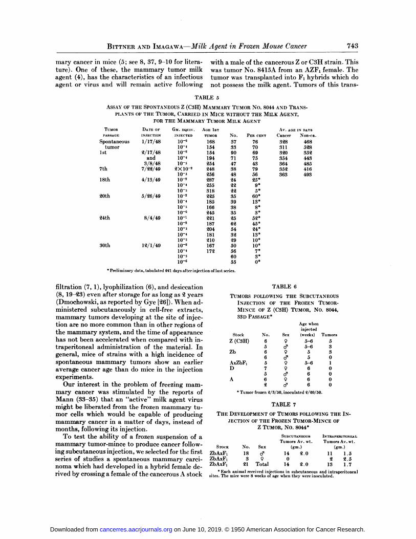
The Z (C3H) tumor No. 8044 has been found topossess the mammary tumor milk agent after it
has been transplanted for 30 passages in eitherZbAxF1 or AxZbF1 hybrids—mice which themselves did not carry the agent. These data are tab
ulated in Table 5. Only preliminary observationsmay be given for many of the groups. The earliesttumor appeared in a test animal (ZBC) which was154 days of age, and the average cancer ages forcompleted groups ranged from 31 1 to 364 days ofage.
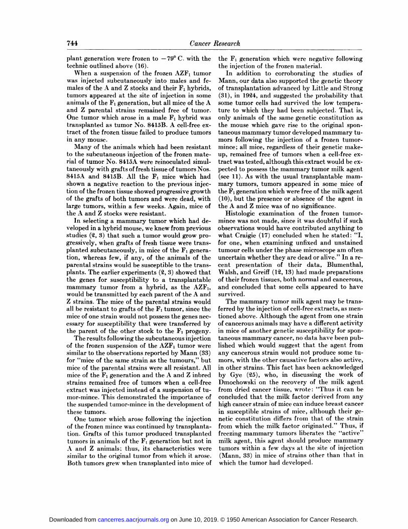
Transplants of the 33d passage of the Z tumorNo. 8044 were minced and frozen in dextrose on@2/3/50. The tissue was maintained at —79°C.until @/@0/50, when the contents of several ampoules were tested by injecting mice subcutaneously with 0.05 gm. of the original material.
Some animals of the Z and Zb lines and theAxZbF1 hybrids developed tumors at the site ofinjection, while all mice of the A and D stocks remained free of tumor (Table 6).
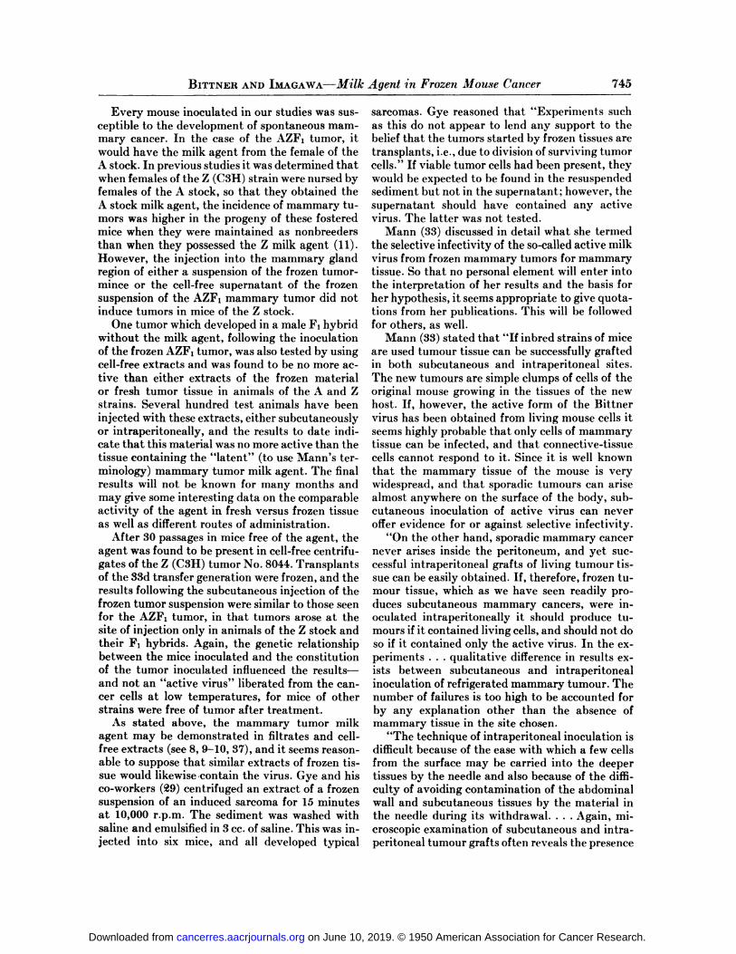
Other vials of tumor No. 8044, frozen on @/3/5O,were used on 3/31/50. In this study, mice of theZbAxF1 generation, when approximately 8 weeksof age, received both subcutaneous and intraperitoneal injections of 0.05 gm. of the originaltumor-mince. As seen from the data (Table 7),there was no significant variation in the number oftumors to be observed at the two sites of inoculation. One male with a large subcutaneous tumorcould not undergo autopsy to determine if it mightalso have an internal tumor.
Weights were taken of twelve of the subcutaneous and all the intraperitoneal tumors. Althoughthe subcutaneous tumors were much more hemorrhagic than the abdominal growths, the fluid wasextruded before the tumors were weighed. The tumors from the subcutaneous area varied between0.9 and 3.6 gm.—average, @.0gm.; the range forthe internal tumors was 0. 15—3.3gm.—average, 1.5gm. Two animals had several internal masses.
DISCUSSION
At least three causative factors have been foundto play a role in the genesis of spontaneous mam
GM.SUBCUTANEOUSLNTRAPERITONEALEQUIV.INJECTIONINJECTIONINJECTEDNo.
TumorsNo.Tumors@X10—233
00l0@35045 0
* Data tabulated after 185 days.
TABLE 3
ASSAY OF THE FROZEN FIRST PASSAGE TRANSPLANTS OF THE AZF1 TUMOR No. 8415A FOR
THE MAMMARY TUMOR MILK AGENT*
GM. SUBCUTANEOUS INTRAPERITONEAL
EQUIV. INJECTION @JECTION
INJECTED No. Tumors No. Tumors
@X102 33 0 35@10@ 33 0 34 0
frozen 1/4/50; tested 1/11/50. Data tabulatedafter 180 days.
TABLE 4
ASSAY OF TUMOR FOR THE MILK AGENT
SUBCUTANEOUS INTRAPERITONEAL
INJECTION INJECTION
No. Tumors No. Tumors
for the so-called “active―(Mann, 3@) milk agent.The second supernatant was injected subcutaneously into 66 and intraperitoneally into 69 youngZBC females (Table 3). To date, after 180 days,two tumors have developed in the animals following the intraperitoneal administration of the extract.
Two tumors which developed following the subcutaneous injection on 1/6/50 of the frozen tumormince of the AZF1 tumor No. 8415A were testedon @/9/50 for the milk agent by using the fresh
GM. EQUIV.INJECTED
Section A@X102 33 0 @8 0
10@ @8 0 30@Section B
@X10@ 16 0 53 01O@ 50 0 3@ 0
Section A: Assay of tumor for the milk agent which developedfollowing the injection of the frozen tissue of theAZF1 tumor 8415A. Frozen tissue injected 1/6/50,tumors tested 1/9/50. The data were tabulated 161days later, first tumor after 138 days.
Section B: Assay for the agent in the third passage transplantsof tumor 8514B. Data recorded after 76 days.
on June 10, 2019. © 1950 American Association for Cancer Research. cancerres.aacrjournals.org Downloaded from
BITTNER AND IIs@u'@GAwA—Milk Agent in Frozen Mouse Cancer 743
mary cancer in mice (5; see 8, 37, 9—10for literature). One of these, the mammary tumor milkagent (4), has the characteristics of an infectious
agent or virus and will remain active following
with a male of the cancerous Z or C3H strain. Thiswas tumor No. 8415A from an AZF1 female. Thetumor was transplanted into F1 hybrids which donot possess the milk agent. Tumors of this trans
TABLE 5
ASSAY OF THE SPONTANEOUS Z (C3H) MAMMARY TUMOR No.8044 AND TRANS
PLANTS OF THE TUMOR, CARRIED IN MICE WITHOUT THE MILK AGENT,FOR THE MAMMARY TUMOR MILK AGENT
TUMORDATE OFGM. EQUIV.AGE 1STAv. AGEINDAYSPASSAGEINJECTIONINJECTEDTUMORNo.PER
CENTCancerNon-ca.Spontaneous1/17/4810@1683776328468tumor10415433703115281st2/17/48
and3/8/4810@
104105154
19425490
714769
7543320
354364352
4434857th7/22/492X1O@
103248 25638 4879 56352 36341649318th4/13/4910@
104105287
25531824
222225@
9*5*20th5/26/4910@
10410—5106225
18516624535
39383560*
13*8*
3*24th8/4/4910—1
10@10310—410@221
18720418121025
6254322952*
45*24*13*
10*30th12/1/49l0@
10@10@10—6167
17250 56605510*
7*3*0*
5Preliminary data, tabulated 111 days after injection of last series.
filtration (7, 1), lyophilization (6), and desiccation(8,19—@3)evenafterstorageforaslongas @2years(Dmochowski, as reported by Gye [p26]).When administered subcutaneously in cell-free extracts,mammary tumors developing at the site of injection are no more common than in other regions ofthe mammary system, and the time of appearancehas not been accelerated when compared with intraperitoneal administration of the material. Ingeneral, mice of strains with a high incidence ofspontaneous mammary tumors show an earlier
average cancer age than do mice in the injectionexperiments.
Our interest in the problem of freezing mammary cancer was stimulated by the reports ofMann (33—35)that an “active―milk agent virusmight be liberated from the frozen mammary tumor cells which would be capable of producingmammary cancer in a matter of days, instead ofmonths, following its injection.
To test the ability of a frozen suspension of amammary tumor-mince to produce cancer following subcutaneous injection, we selected for the firstseries of studies a spontaneous mammary carci
noma which had developed in a hybrid female derived by crossing a female of the cancerous A stock
TABLE 6
TUMORS FOLLOWING THE SUBCUTANEOUS
INJECTION OF THE FROZEN TUMORMINCE OF Z (C3H) TUMOR, No. 8044,
33D PASSAGE*
Age when
injectedStock No. Sex (weeks) Tumors
Z(C3H) 6 9 5—6 55 ci― 5—fl 3
Zb 6 9 5 36 ci― 5 0
AxZbF1 5 9 5—6 1D 7 9 6 0
5 c3' 6 0A 6 9 6 0
@ ci― 6 0
5Tumor frozen 1/3/50, inoculated 1/10/50.
TABLE 7
THE DEVELOPMENT OF TUMORS FOLLOWING THE INJECTION OF THE FROZEN TUMOR-MINCE OF
Z TUMOR, No. 8044*
STOCK No. SEX
ZbAxF1 18 ci― 14 11ZbAxF1 3 9 0 2ZbAxF1 21 Total 14 2.0 13
* Each animal received injections in subcutaneous and intraperitoneal
sites.The micewere8 weeksofagewhen theywereinoculated.
SUBCUTANEOUS
Tumors Av. wt.
(gm.)
2.0
INTRAPERITONEAL
Tumors Av. wt.
(gm.)
1.52.51.7
on June 10, 2019. © 1950 American Association for Cancer Research. cancerres.aacrjournals.org Downloaded from
744 Cancer Research
plant generation were frozen to —79°C . with thetechnic outlined above (16).
When a suspension of the frozen AZF1 tumorwas injected subcutaneously into males and females of the A and Z stocks and their F1 hybrids,tumors appeared at the site of injection in someanimals of the F1 generation, but all mice of the Aand Z parental strains remained free of tumor.One tumor which arose in a male F1 hybrid wastransplanted as tumor No. 8415B. A cell-free extract of the frozen tissue failed to produce tumorsin any mouse.
Many of the animals which had been resistantto the subcutaneous injection of the frozen material of tumor No. 8415A were reinoculated simultaneously with grafts of fresh tissue of tumors Nos.8415A and 8415B. All the F1 mice which hadshown a negative reaction to the previous injection of the frozen tissue showed progressive growthof the grafts of both tumors and were dead, withlarge tumors, within a few weeks. Again, mice ofthe A and Z stocks were resistant.
In selecting a mammary tumor which had developed in a hybrid mouse, we knew from previousstudies (@2,3) that such a tumor would grow progressively, when grafts of fresh tissue were transplanted subcutaneously, in mice of the F1 generation, whereas few, if any, of the animals of theparental strains would be susceptible to the transplants. The earlier experiments (p2,3) showed thatthe genes for susceptibility to a transplantablemammary tumor from a hybrid, as the AZF1,would be transmitted by each parent of the A andZ strains. The mice of the parental strains wouldall be resistant to grafts of the F1 tumor, since themice of one strain would not possess the genes necessary for susceptibility that were transferred bythe parent of the other stock to the F1 progeny.
The results following the subcutaneous injectionof the frozen suspension of the AZF1 tumor weresimilar to the observations reported by Mann (33)for “miceof the same strain as the tumours,― butmice of the parental strains were all resistant. Allmice of the F1 generation and the A and Z inbredstrains remained free of tumors when a cell-freeextract was injected instead of a suspension of tumor-mince. This demonstrated the importance ofthe suspended tumor-mince in the development ofthese tumors.
One tumor which arose following the injectionof the frozen mince was continued by transplantation. Grafts of this tumor produced transplantedtumors in animals of the F1 generation but not inA and Z animals; thus, its characteristics weresimilar to the original tumor from which it arose.Both tumors grew when transplanted into mice of
the F1 generation which were negative followingthe injection of the frozen material.
In addition to corroborating the studies ofMann, our data also supported the genetic theoryof transplantation advanced by Little and Strong(31), in 19@24,and suggested the probability thatsome tumor cells had survived the low temperature to which they had been subjected. That is,only animals of the same genetic constitution asthe mouse which gave rise to the original spontaneous mammary tumor developed mammary tumors following the injection of a frozen tumormince ; all mice, regardless of their genetic makeup, remained free of tumors when a cell-free cxtract was tested, although this extract would be cxpected to possess the mammary tumor milk agent(see 11). As with the usual transplantable mammary tumors, tumors appeared in some mice ofthe F1 generation which were free of the milk agent(10),butthepresenceorabsenceofthe agentinthe A and Z mice was of no significance.
Histologic examination of the frozen tumormince was not made, since it was doubtful if suchobservations would have contributed anything towhat Craigie (17) concluded when he stated : “I,for one, when examining unfixed and unstainedtumour cells under the phase microscope am oftenuncertain whether they are dead or alive.―In a recent presentation of their data, Blumenthal,Walsh, and Greiff (U2, 13) had made preparationsof their frozen tissues, both normal and cancerous,and concluded that some cells appeared to havesurvived.
The mammary tumor milk agent may be transferred by the injection of cell-free extracts, as mentioned above. Although the agent from one strainof cancerous animals may have a different activityin mice of another genetic susceptibility for spontaneous mammary cancer, no data have been published which would suggest that the agent fromany cancerous strain would not produce some tumors, with the other causative factors also active,in other strains. This fact has been acknowledgedby Gye (@25),who, in discussing the work ofDmochowski on the recovery of the milk agentfrom dried cancer tissue, wrote: “Thusit can beconcluded that the milk factor derived from anyhigh cancer strain of mice can induce breast cancerin susceptible strains of mice, although their genetic constitution differs from that of the strainfrom which the milk factor originated.― Thus, iffreezing mammary tumors liberates the “active―milk agent, this agent should produce mammarytumors within a few days at the site of injection(Mann, 33) in mice of strains other than that inwhich the tumor had developed.
on June 10, 2019. © 1950 American Association for Cancer Research. cancerres.aacrjournals.org Downloaded from
BITTNER AND IMAGAWA—Milk Agent in Frozen Mouse Cancer 745
Every mouse inoculated in our studies was susceptible to the development of spontaneous mammary cancer. In the case of the AZF1 tumor, itwould have the milk agent from the female of theA stock. In previous studies it was determined thatwhen females of the Z (CSH) strain were nursed byfemales of the A stock, so that they obtained theA stock milk agent, the incidence of mammary tumors was higher in the progeny of these fosteredmice when they were maintained as nonbreedersthan when they possessed the Z milk agent (11).However, the injection into the mammary glandregion of either a suspension of the frozen tumormince or the cell-free supernatant of the frozensuspension of the AZF1 mammary tumor did notinduce tumors in mice of the Z stock.
One tumor which developed in a male F1 hybridwithout the milk agent, following the inoculationof the frozen AZF1 tumor, was also tested by usingcell-free extracts and was found to be no more active than either extracts of the frozen materialor fresh tumor tissue in animals of the A and Zstrains. Several hundred test animals have beeninjected with these extracts, either subcutaneouslyor intraperitoneally, and the results to date mdicate that this material was no more active than thetissue containing the “latent―(to use Mann's terminology) mammary tumor milk agent. The finalresults will not be known for many months andmay give some interesting data on the comparableactivity of the agent in fresh versus frozen tissueas well as different routes of administration.
After 30 passages in mice free of the agent, theagent was found to be present in cell-free centrifugates of the Z (C3H) tumor No. 8044. Transplantsof the 33d transfer generation were frozen, and theresults following the subcutaneous injection of thefrozen tumor suspension were similar to those seenfor the AZF1 tumor, in that tumors arose at thesite of injection only in animals of the Z stock andtheir F1 hybrids. Again, the genetic relationshipbetween the mice inoculated and the constitutionof the tumor inoculated influenced the resultsand not an “activevirus― liberated from the cancer cells at low temperatures, for mice of otherstrains were free of tumor after treatment.
As stated above, the mammary tumor milkagent may be demonstrated in filtrates and cellfree extracts (see 8, 9—10,37), and it seems reasonable to suppose that similar extracts of frozen tissue would likewise contain the virus. Gye and hisco-workers (@9) centrifuged an extract of a frozensuspension of an induced sarcoma for 15 minutesat 10,000 r.p.m. The sediment was washed withsaline and emulsified in 3 cc. of saline. This was injected into six mice, and all developed typical
sarcomas. Gye reasoned that “Experiments suchas this do not appear to lend any support to thebelief that the tumors started by frozen tissues aretransplants, i.e., due to division of surviving tumorcells.―If viable tumor cells had been present, theywould be expected to be found in the resuspendedsediment but not in the supernatant; however, thesupernatant should have contained any activevirus. The latter was not tested.
Mann (33) discussed in detail what she termedthe selective infectivity of the so-called active milkvirus from frozen mammary tumors for mammarytissue. So that no personal element will enter intothe interpretation of her results and the basis forher hypothesis, it seems appropriate to give quotations from her publications. This will be followedfor others, as well.
Mann (33) stated that “Ifinbred strains of miceare used tumour tissue can be successfully graftedin both subcutaneous and intraperitoneal sites.The new tumours are simple clumps of cells of theoriginal mouse growing in the tissues of the newhost. If, however, the active form of the Bittnervirus has been obtained from living mouse cells itseems highly probable that only cells of mammarytissue can be infected, and that connective-tissuecells cannot respond to it. Since it is well knownthat the mammary tissue of the mouse is verywidespread, and that sporadic tumours can arisealmost anywhere on the surface of the body, subcutaneous inoculation of active virus can neveroffer evidence for or against selective infectivity.
“Onthe other hand, sporadic mammary cancernever arises inside the peritoneum, and yet suecessful intraperitoneal grafts of living tumour tissue can be easily obtained. If, therefore, frozen tumour tissue, which as we have seen readily produces subcutaneous mammary cancers, were inoculated intraperitoneally it should produce tumours if it contained living cells, and should not doso if it contained only the active virus. In the experiments . . . qualitative difference in results exists between subcutaneous and intraperitonealinoculation of refrigerated mammary tumour. Thenumber of failures is too high to be accounted forby any explanation other than the absence ofmammary tissue in the site chosen.
“Thetechnique of intraperitoneal inoculation isdifficult because of the ease with which a few cellsfrom the surface may be carried into the deepertissues by the needle and also because of the difficulty of avoiding contamination of the abdominalwall and subcutaneous tissues by the material inthe needle during its withdrawal.. . . Again, microscopic examination of subcutaneous and intraperitoneal tumour grafts often reveals the presence
on June 10, 2019. © 1950 American Association for Cancer Research. cancerres.aacrjournals.org Downloaded from
746 Cancer Research
of hair follicles which have been forced in by thegrafting-needle. A further source of error is aslightly lateral inoculation. Unless the skin and theabdominal wall are pierced exactly in the midlinethere is always the risk of a mammary tubule beinginvolved in the puncture.
“. . . In any case a failure to obtain growths in
90 per cent of the intraperitoneal experiments asagainst a success in 75 per cent of subcutaneousinoculations (where the chances of the virus encountering a mammary tubule are high but noteven there 100 per cent) is strongly in favour ofselective infectivity of the virus as against cellsurvival.―
Thus, while “thetechnique of intraperitonealinoculation is difficult, . . . the chances of the virusencountering a mammary tubule are high but noteven 100 per cent― following subcutaneous inoculation.
Regarding the distribution of the mammaryglands and the development of mammary cancerin males, Mann (33) stated : “Inthe male mouse,mammary tubules, imperfectly developed andwithout secreting acini, are present over largeareas of the subcutaneous tissue, but even in highcancer strains male mice do not develop mammarycancer unless they are subjected to prolongedtreatment with oestrin, when tumours occur as inthe females—i.e., the virus changes from the latentto the active form.―
These observations on the relative size of themammary glands in male mice are not supportedby details from other published reports. Gardner(p24), in 1935, found that “Themammary glands ofthe normal male mouse persist as rudimentaryducts in the subcutaneous tissue throughout life.They undergo little, if any, growth after the weaning age.―When male mice of various strains werefrom 6 to 9 weeks of age, Richardson and Cloudman (38) made whole mounts of their third glands.In males of the strains which showed the most extensive development, the mammary gland did notinvolve an area much greater than 1 X 1 cm. inarea. Also, a gross dissection of a lactating femaleshowed that intraperitoneal injection could bemade without coming in contact with the mammary glands (11).
Twenty-one animals, eighteen males and threefemales, were tested by subcutaneous and intraperitoneal injections of a frozen suspension of thetumor-mince. Fourteen tumors were found in thesubcutaneous region as against thirteen internaltumors. Although only a limited number of micewas tested, no difference was noted based upon theroute of administration of the frozen tissue. Inmost experiments, Mann used only six animals,
and her figures represent many series with severaldifferent tumors.
In 1949, Mann (33) tested the transplantablecarcinoma “63―and determined that the inoculation of a frozen suspension was active in 19 (44 percent of the mice developed tumors) of @2@2experiments, while negative results were obtained in 3experiments because of technical errors. The yearpreviously, Dmochowski (@2) assayed the carcinoma “63―for the mammary tumor milk agentand was unable to demonstrate the presence of theagent in desiccated tissue of tumors from the samelaboratory. The tumors were from the 4@28th passage. No mention was made by Mann of these negative results by an accepted method of testing forthe agent.
The usual method of reporting scientific data isto refer in the manuscript to any previous resultsother investigators have observed in that particular field. As this was not followed by either Gye(@28)or Mann (33—35),it seemsprobable that theyproposed that the reader should accept their theories as being original with them.
The interpretation of the experimental resultsfollowing the use of either frozen and/or dried tumor tissue is dependent upon whether or not somecells may survive. In reporting their preliminarydata and advancing the viral theory, Gye (@28)made no direct reference to the previous studies ofothers, but, however, concluded that “Wethinkwe are justified in making the contention that thenegative results of the past are merely negativeand have the value of negative experiment whichis the result of imperfect technique.―
In a recent publication, Blumenthal and Walsh(U2) referred to fifteen manuscripts, published previously to 1949, on the successful transfer of tumorand normal tissues following storage at low temperatures for as long as@ years. The subject hasalso been reviewed by Hirschberg and Rusch (30).Only a few of these experiments, selected as pertinent to the discussion, will be considered here.
In 1939, Mider and Morton (36) froze mousesarcoma and rat carcinoma to a minimum temperature of —74°C. by using approximately the sametechnic as the British workers. When the tissuewas frozen en masse, tumors developed following
the subcutaneous inoculation of the thawed tissue.Repeated freezing and thawing reduced the number of tumors induced by the material. Among thethree tumors they tested, sarcoma was more resistent to cold when frozen en masse than carcinoma, whereas, when frozen in a saline suspension,the opposite was found. If rat skin was maintainedfor @4hours at a temperature of —74°C. andtransplanted subcutaneously, 1@Iider and Morton
on June 10, 2019. © 1950 American Association for Cancer Research. cancerres.aacrjournals.org Downloaded from
BITTNER AND IMAGA WA—Milk Agent in Frozen Mouse Cancer 747
oral administration of an extract of lyophilizedmammary cancer (frozen to —7@°C . and dried invacuo) (6).
Gye (@8) reported that when fresh sporadic tumors from animals of the R3 and C3H stocks weredried, with the Craigie desiccator (15), active drytissue was obtained. The type of tumor which theyused was not specified, but these strains have ahigh incidence of spontaneous mammary cancer.However, until this new apparatus was employed,Gye and his associates (@29)had been unsuccessful,in many experiments, in the propagation of driedtumor tissue, or “Along experience of negative results with a different and simpler drying method―(18).
Craigie (17) discussed the survival of cancerouscells following drying in their new apparatus asfollows : “Testsof dried material obtained at vanous times during the development of the dryingequipment (Craigie [15']) were negative,althoughone positive result was obtained with C3H sarcoma and others were obtained by Gye, Begg, Mann,and Craigie (p491)―In a later report (18) he coneluded : “Itwould therefore seem to be reasonableto accept the possibility that a few cells (about 1in 1,000,000) survived the drying process.― Presumably, the number of cells to survive dryingwith their new equipment was sufficient to producetransplantable tumors at the site of inoculation. Ifthis new desiccator did not completely destroy allthe tumor cells, the tumors which arose at the siteof inoculation of the partially dried material wouldbe due to the propagation of these viable cells andnot due to the action of any virus.
In view of the many experiments of othersshowing that tissues would survive freezing, it isinteresting to cite from the 1947—48report of Gye(@7) as Director of the Imperial Cancer Research
Fund: “Forty years ago Salvin-Moore successfully transplanted mouse carcinomata and sarcomata after they had been exposed to the temperature of liquid air. Cramer grew sarcomata afterfreezing them with CO2 snow, but felt that his results were attributable to the survival of a fewcells. Gye and Purdy (1931) studied the matterfurther and concluded that a few cells were able toremain viable though exposed to freezing temperatures. The subsequent work of Breedis and Furth(1938) and others has produced additional evidence that such survival may occur. Breedis andFurth drew attention to the value of freezing andstoring on CO2 snow as a method of preserving tumouns, but although this method has been used bysome it has not been generally adopted. Dr.Craigie is studying certain aspects of the problem
1 References numbered as in this paper.
(36) found that when it was removed 10 days laterthere was histologic evidence of growth, as demonstrated by mitotic figures, characteristic stainingof the connective tissue, etc.
Blumenthal and his associates (1@, 13) concluded that normal tissue (thyroid and parathyroid ofthe guinea pig) and mouse Sarcoma 37 would survive freezing to either —70°C. or —190°C. andcould later be successfully transplanted. The mostaccurate indicator of viability, mitotic activity,was noted in the transplants. In referring to theconclusions of Gye et al., they stated that their“findingsdo not disprove the possibility of a viraltransmission of neoplastic disease but rather provethat the criteria used for determining whether ornot this mode of transmission obtains must bemore strictly defined.―
Craigie (16) reviewed the literature in a later report, the first paper in this series to do so, andstated: “Much of the experimental work reportedon the production of tumours with frozen andthawed material is concerned with the interpretation of the survival of activity, and it is clear thatunder certain conditions malignant cells and normal skin . . . will survive freezing temperatures forsome time.― He tested various solutions to determine the ability of the cells to survive and concluded : “Thedegree of survival of activity in dcxtrose preparations at a low temperature is adequate for experimental work requiring the use oftransplantable tumours and for some purposesmay offer a more convenient source of materialthan fresh tumour tissue.― Elsewhere, Craigie (17)wrote: “Itis evident from the results of this experiment that the degree of survival of the differenttumours varied considerably. Some lots producedtumours at all sites injected, others only someComplete failure to survive freezing has been observed so far only with two benzpyrene tumoursmentioned and an undifferentiated tumour...arising near the point of needle entry of intraperitoneal injection of methyicholanthrene.―
Mider and Morton (36) dried at least one tumor, while it was in the frozen state, in a lyophileapparatus and determined that the subcutaneousinjection of the dried material did not produce tumors at the site of inoculation. Although themammary tumor milk agent will remain activefollowing desiccation (8, 19—@3),the subcutaneousinjection of fresh, dried tumor tissue has not “induced― mammary cancer at the site of injectionwithin a matter of days or weeks; but after severalmonths the typical spontaneous mammary tumorsdevelop. These tumors appear with the usual random distribution seen for this type of cancer inmice. Similar results have been obtained following
on June 10, 2019. © 1950 American Association for Cancer Research. cancerres.aacrjournals.org Downloaded from
748 Cancer Research
that do not seem to have been explored, with theimmediate objective of further improving this potentially valuable method of preserving certaintransplantable tumours.―
Let us now refer to the papers published bySalvin-Moore and his associates (39—40)in 1908which were mentioned by Gye (@27)in 1947—48.Salvin-Moore exposed mouse tumors to the actionof liquid air for periods of from @20minutes to@hour. “Theywere then at once introduced intohealthy mice beneath the skin, the presumptionbeing that in these circumstances the tumour cellswould be destroyed by the action of the liquid airand consequently that they would multiply nofurther.― However, in some cases new tumorsarose at the site of inoculation of the frozen tissue.
Salvin-Moore and Walker (40) discussed theirresults as follows : “Fromthese observations it isrendered clear that exposure to liquid air at a ternperature of about —195°does not necessarily destroy the potentiality of the substance of a mousetumour to produce fresh turnours of the same kindin mice into which such frozen tumour substancehas been grafted.
“Thesefacts in themselves are somewhat surprising, and they immediately raise a number ofquestions which it will be desirable to have elucidated in the interests of research concerning thenature of cancer. In the first place, it is renderedclear that exposure to liquid air for a certain penod of time does not destroy the principle uponwhich the vitality of mouse cancer depends. If, asmay be the case, the cells composing the mass ofthe tumour, and constituting the grafts, are killedby exposure to liquid air, then the development ofmouse cancer after such exposure indicates notmerely that the growth of similar tumours is dependent on the integrity of the ‘cancercells,' butalso that the new tissues are not necessarilyformed from the implanted cells at all, and mayarise from the cells of the new host in response tosome stimulus introduced along with the frozenmaterial, and quite independent of the integrity ofthe so-called ‘cancercells.'―
The authors (40) furthermore stated that theirobservations suggested “thatthe production ofnew tumours in the hosts into which the frozencancer tissue has been introduced may possiblynot be dependent upon the introduction of the‘cancercells' at all, but upon the action of a viruswhich is independent of these cells, and retains itsactivity after being subjected to the temperatureof liquid air.― Salvin-Moore and Walker recognized the possibility that “itis not certainthat the cells from the tumour introduced into anew individual are killed by half an hour's expo
sure to the temperature of liquid air, particularlyas the seeds of some plants and trypanosomes aresaid to survive this temperature.―
Earlier in this discussion we quoted a statementby Gye (@8) “thatthe negative results of the pastare merely negative and have the value of negativeexperiment which is the result of imperfect technique.― These positive results were probably obtamed, as demonstrated by Craigie (18), becauseof imperfect technic for the dehydration of cancerous tissue, either fresh or frozen. The tumorsproduced following the inoculation of suspensionsof frozen tumors were likewise the result of thepropagation of viable tumor cells. These data wereincorrectly interpreted by the authors regardless ofthe published reports of many workers to the contrary.
Mider and Morton (36) commented upon thevirus hypothesis of Salvin-Moore as follows:“This,however, has no bearing on the moot pointconcerning the possible relationship to transplantable tumors.― There is little doubt but what thisremark is as valid today for the inoculation offrozen tumor tissue as when it was made in 1939,and it applies equally well to the 1949 theory ofGye and Mann, since their explanation is practically identical with the one advanced by SalvinMoore in 1908.
It has often been said that many theories areadvanced only for the stimulus they may give toothers for further research. Perhaps in some casesthis may result in an increase in our knowledge ofthe problem being investigated, while in other instances it may actually retard progress until thehypothesis has either been refuted or confirmed.This may be necessary, especially when the experi
. ments are completed without adequate controls.
SUMMARYSalvin-Moore, in 1908, determined that tumors
would develop in mice following the subcutaneousimplantation of neoplasms which had been exposed to the temperature of liquid air (—195°C.).He proposed a virus theory to explain the results,although he considered the possibility that somecells had remained viable.
In 1949, Gye and his associates submitted asimilar theory based upon observations obtainedfollowing the freezing and drying of mouse cancers. Furthermore, Mann stated that when mammary cancers (both transplanted and spontaneous) were frozen, the milk agent virus was liberated in its “active―form and when injected subcutaneously, mammary tumors would be producedat the site of injection within a short interval ofdays or weeks. Few mammary tumors arose at
on June 10, 2019. © 1950 American Association for Cancer Research. cancerres.aacrjournals.org Downloaded from
BITTNER AND IMAGAWA—Milk Agent in Frozen Mouse Cancer 749
other sites of inoculation because of the “selectiveinfectivity― of the virus for mammary tissue.
To test for the active mammary tumor milkagent virus, transplanted mammary tumors wereminced, suspended in dextrose, and frozen to—79°C . The thawed tumor-mince produced mammary tumors at the site of injection only in mice ofstocks which would be susceptible to fresh tissuegrafts of the tumor being tested. Negative resultswere obtained with mice of other strains, althoughthey were susceptible to the development of spontaneous mammary cancer. Intraperitoneal inoculation of the frozen material was as effective assubcutaneous injection in the production of mammary tumors.
Whereas cell-free centnifugates have been foundto contain the mammary tumor milk agent, cellfree extracts of the frozen mammary tumor-mincefailed to induce tumors, within the specified interval, in any animal regardless of its genetic constitution.
Preliminary data indicate that the milk agentwas no more active in cell-free extracts of thefrozen than the fresh tissue of the same tumor, regardless of the route of administration. The finalresults of these tests will not be apparent formonths, but the earliest tumors to be observed developed in mice which had received intrapenitonealinjections.
The revival of this viral theory for mouse cancerby Gye et al. and Mann disregarded the previousfinding of many investigators that mouse tissues(normalandcancerous)survivedfreezingto lowtemperatures. Evidence was also cited that thenewly designed desiccator, used for their studies,did not destroy all cells. The fact that other meth- -ods of dehydration, including their own studies,had given negative results which would refute thevirus hypothesis was disregarded as due to imperfect technic.
The experimental data submitted by Gye et a!.and Mann were, without a doubt, based upon observations which they either incorrectly interpreted, as in the case of the studies with frozentissues, or secured by using new equipment beforeit had been adequately tested and which resultedin imperfect technic.
REFERENCES1. ANDERVONT, H. H., and BRYAN, W. R. Properties of the
Mouse Mammary Tumor Milk Agent. J. Nat. CancerInst., 5: 143—49,1944.
2. BIrrNER, J. J. A Genetic Study of the Transplantation ofTumors Arising in Hybrid Mice. Am. J. Cancer, 15:2202—47,1931.
3. . Genetic Studies on the Transplantation of Tumors.VII. Comparative Study of Tumors 19308 A, B, and C.Ibid.,17:724—34,1933.
4. . Some Possible Effects of Nursing on the Mainmary Gland Tumor Incidence in Mice. Science, 24: 162,1936.
5. . “Influences―of Breast-Cancer Development inMice. Pub. Health Rep., 54: 1590—97,1939.
6. . The Preservation by Freezing and Drying inVacuo of the Milk-Influence for the Development ofBreast Cancer in Mice. Science, 93:527—28, 1941.
7. . The Milk Influence of Breast Tumors in Mice.Ibid., 95:462—63, 1942.
8. —@. Inciting Influences in the Etiology of MammaryCancer in Mice. A.A.A.S. Research Conference on Cancer,pp. 63—96.1945.
9. ———.The Causes and Control of Mammary Cancer inMice. Harvey Lectures. 42:221—46, 1946—47.
10. . The Transplantability of Mammary Cancer inMice Associated with the Source of the Mammary TumorMilk Agent. Cancer Research, 7:741—45, 1947.
1 1. . Some Enigmas Associated with the Genesis ofMammary Cancer in Mice. Ibid., 8: 625—39,1948.
12. BLUMENTHAL,H. T., and WALSH, L. B. Survival of GuineaPig Thyroid and Parathyroid Autotranspiants PreviouslySubjected to Extremely Low Temperatures. Proc. Soc.Exper. Biol. & Med., 73:62—67, 190.
13. BLUMENTHAL, H. T. ; WALSH, L. B. ; and GREIFF, D.Studies on the Effect of Low Temperatures on the Traissplantability of Normal and Neoplastic Tissue. Cancer Research,10:205,1950.
14. (i@RAIGIE,J. A Pressure Mincer for the Preparation ofTumour Suspensions. Brit. J. Cancer, 3:249—50, 1949.
15. . A Drying Apparatus for the Study of TumourTransmission. Ibid., pp. 250—55, 1949.
16. ———.The Preservation of Suspensions of Tumour Cellsiii Dextrose at Low Temperatures. Ibid., pp. 268—74, 1949.
17. ——-—.A Quantitative Approach to the Study of Tramisplantable Tumours. Brit. M. J., 2: 1485—91,1949.
18. ——. Director's report. 47th Annual Report, ImperialCancer Research Fund, pp. 5—18.1949—50.
19. DMocHowsKI, L. Mammary Tumour-inducing Factorand Genetic Constitution. Brit. J. Exper. Path., 25: 138—40, 1944.
20. —@—. Comparative Potency of the Mammary Tumour
Agent of Mice of Different Genetic Constitutions. Ibid.,26:267—69,1945.
21. ——--—.Age and Dosage in the Induction of Breast Cancerin Mice by the Mouse Mammary Tumour Agent. Ibid.,pp. 192—97,1945.
22. ——. Mammary Tumour-inducing Factor and Genetic(‘onstitution. Brit. J. Cancer, 2:94—102, 1948.
23. ————.Survival of the Milk Factor in a TransplantableBreast Tumour in Mice. Ibid., 3:246—48, 1949.
24. GARDNER, W. U. The Effect of Ovarian Hormones andOvarian Grafts upon the Mammary Glands of Male Mice.Endocrinology, 19: 656—67,1947.
25. GYE, W. E. Director's report. 42d Annual Report, Impenal Cancer Research Fund, pp. 5—10.1944—45.
26. . Director's report. 43d Annual Report, ImperialCancer Research Fund, pp. 7—13.1945—46.
27.@. Director's report. 45th Annual Report, ImperialCancer Research Fund, pp. 5—13. 1947—48.
28. . The Propagation of Mouse Tumours by Means ofDried Tissue.Brit.M. J.,1:511—15,1949.
29. GYE, W. E.; BEGG, A. M.; MANN, I.; and CRAIGIE, J. TheSurvival of Activity of Mouse Sarcoma Tissue after Freezing and Drying. Brit. J. Cancer, 3:259-67, 1949.
30. HIRSCHBERG, E., and RUSCH, H. P. Comments @nRecentExperimentswith Frozen and Dried Tissueas Evidenceforthe VirusEtiologyof Tumors. Cancer Research,10:335—38, 1950.
on June 10, 2019. © 1950 American Association for Cancer Research. cancerres.aacrjournals.org Downloaded from
750 Cancer Research
31. LITTLE, C. C., and STRONG, L. C. Genetic Studies on theTransplantation of Two Adenocarcinomata. J. Exper.Zoöl.,41:93—114,1924.
32. MANN, I. A Study of Cell Survival in Embryonic Tissue(;rafts in Inbred Strains under Various Conditions. Brit. J.Cancer, 3:255—59, 1949.
33. . Effect of Low Temperatures on the Bittner Virusof Mouse Carcinoma. Brit. M. J., 2:251—53, 1949.
34. . Effect of Repeated Freezing and Thawing onMouse Carcinoma Tissue. Ibid., pp. 253—55,1949.
35. MANN, I., and DUNN, W. J. Propagation of MouseCarcinoma by Dried Tumour Tissue. Brit. M. J., 2:255—57,1949.
36. MIDER, G. B., and MORTON, J. J. The Effect of Freezingin Vitroon Some TransplantableMammalian Tumors andon Normal Rat Skin.Am. J.Cancer,35:502—9,1939.
37. NATIONAL CANCER INSTITUTE, STAFF. A Symposium onMammary Tumors in Mice. A. A. A. S. 1945.
38. RICHARDSON, F. L.,and CLOrDMAN, A. M. The Mammary
(;land Development in Male Mice at Nine Weeks of Age.Anat. Rec., 97:223—37, 1947.
39. SALVIN-MOORE, J. E., and BARRATT, J. 0. W. Note uponthe Effect of Liquid Air upon the Graftable Cancer ofMice. Lancet, 1:227, 1908.
40. SALVIN-M00RE, J. E., and WALKER, C. E. On the Relationship of Cancer Cells to the Development of Cancer.Lancet, 1:226—27, 1908.
on June 10, 2019. © 1950 American Association for Cancer Research. cancerres.aacrjournals.org Downloaded from
1950;10:739-750. Cancer Res John J. Bittner and David T. Imagawa Tumor Milk AgentAssay of Frozen Mouse Mammary Carcinoma for the Mammary
Updated version
http://cancerres.aacrjournals.org/content/10/12/739
Access the most recent version of this article at:
E-mail alerts related to this article or journal.Sign up to receive free email-alerts
Subscriptions
Reprints and
To order reprints of this article or to subscribe to the journal, contact the AACR Publications
Permissions
Rightslink site. Click on "Request Permissions" which will take you to the Copyright Clearance Center's (CCC)
.http://cancerres.aacrjournals.org/content/10/12/739To request permission to re-use all or part of this article, use this link
on June 10, 2019. © 1950 American Association for Cancer Research. cancerres.aacrjournals.org Downloaded from