Embed Size (px)
Citation preview
Assembly of Dimeric Variants of Coumermycins by Tandem Action of the FourBiosynthetic Enzymes CouL, CouM, CouP, and NovN†
Caren L. Freel Meyers,‡ Markus Oberthu¨r,§ Lutz Heide,| Daniel Kahne,§ and Christopher T. Walsh*,‡
Department of Biological Chemistry and Molecular Pharmacology, HarVard Medical School, Boston, Massachusetts 02115,Department of Chemistry, Princeton UniVersity, Princeton, New Jersey 08544, and Pharmaceutical Institute, Tuebingen
UniVersity, 72076 Tuebingen, Germany
ReceiVed July 19, 2004; ReVised Manuscript ReceiVed September 20, 2004
ABSTRACT: Coumermycin A1 is a member of the aminocoumarin family of antibiotics. Unlike its structuralrelatives, novobiocin and clorobiocin, coumermycin A1 is a dimer built on a 3-methyl-2,4-dicarboxypyrrolescaffold and bears two decorated noviose sugar components which are the putative target binding motifsfor DNA gyrase. Starting with this scaffold, we have utilized the ligase CouL for mono- and bisamideformation with aminocoumarins to provide substrates for the glycosyltransferase CouM. CouM wassubsequently shown to catalyze mono- and bisnoviosylation of the resulting CouL products. CouP wasshown to possess 4′-O-methyltransferase activity on products from tandem CouL, CouM assays. A fourthenzyme, NovN, the 3′-O-carbamoyltransferase from the novobiocin operon, was then able to carbamoylateeither or both arms of the CouP product. The tandem action of CouL, CouM, CouP, and NovN thusgenerates a biscarbamoyl analogue of the pseudodimer coumermycin A1. Starting from alternative dicarboxyscaffolds, these four enzymes can be utilized in tandem to create additional variants of dimericaminocoumarin antibiotics.
The bacterial type II topoisomerases, DNA gyrase andtopoisomerase IV, are required for DNA replication andcatalyze the biochemical reactions that control the topologyof DNA including steps in DNA decatenation and topoisomerrelaxation number control (1). Both DNA gyrase andtopoisomerase IV (topo IV)1 are antibiotic targets in bacterialpathogens. Among the known inhibitors of gyrase and topoIV are the synthetic fluoroquinolone class of antibioticswhich target the A subunits (2-4) and the natural productaminocoumarins which target the B subunits of bothenzymes, respectively (5-9). The fluoroquinolines cipro-floxacin and levofloxacin are potent inhibitors of gyrase andtopo IV and have experienced widespread clinical use asantibiotics. However, the aminocoumarin family of antibiot-ics including novobiocin, clorobiocin, and coumermycin A1
has found limited use in the clinic because of problems
associated with low solubility, poor pharmacokinetics, andpoor cell wall penetration (10-12). Novobiocin has, how-ever, received renewed attention as a result of its potentactivity against methicillin-resistantStaphylococcus aureus(MRSA) bacterial strains (13). The increasing prevalence ofantibiotic resistance to the synthetic antibacterial fluoro-quinolones in addition to the recent sequencing of thebiosynthetic gene clusters for novobiocin (14), clorobiocin(15), and the pseudodimer coumermycin A1 (16) hasprompted an interest in the optimization of aminocoumarinantibiotics by structural variation.
A comparison of the biosynthetic gene clusters fornovobiocin, clorobiocin, and coumermycin A1 reveals asimilar logic for the biosynthetic assembly of all three familymembers. The components of the tripartite scaffolds ofnovobiocin and clorobiocin are constructed by enzymesencoded in subclusters ofnoV andclo genes. The prenylated4-hydroxybenzoate component is derived from 4-hydroxy-phenylpyruvate (17) while the 7-hydroxy-3-aminocoumarinand theL-hexose substrate TDP-L-noviose are derived fromtyrosine andD-glucose, respectively (18-20). The planarscaffolds in novobiocin and clorobiocin are formed followingenzymatic ligation of the prenylated benzoate and aminocou-marin by the ligases NovL and CloL (15, 21). Attachmentof the noviose moiety to the planar scaffold is catalyzed bythe dedicated glycosyltransferases NovM and CloM (15, 22).Antibiotic activity is only attained following two subsequentenzymatic tailoring reactions on the noviose ring. 4′-O-Methylation is catalyzed by methyltransferases NovP (23)and CloP (15), and 3′-O-carbamoylation (novobiocin) or 3′-O-acylation (clorobiocin) is catalyzed by the carbamoyl-transferase NovN (23) and acyltransferase CloN2 (24). The
† We gratefully acknowledge support from National Institutes ofHealth Grants 49338 (to C.T.W.), 66174 (to D.K.), and AI0-54007-01(to C.L.F.M.).
* To whom correspondence should be addressed. E-mail:[email protected]. Phone: (617) 432-1715. Fax:(617) 432-0438.
‡ Harvard Medical School.§ Princeton University.| Tuebingen University.1 Abbreviations: topo IV, topoisomerase IV; MRSA, methicillin-
resistantStaphylococcus aureus; TDP, thymidine diphosphate; ATP,adenosine triphosphate; TFA, trifluoroacetic acid; THF, tetrahydrofuran;DMF, dimethylformamide; PyBOB, benzotriazol-1-yloxypyrrolidino-phosphonium hexafluorophosphate; DMSO, dimethyl sulfoxide; IPTG,isopropylâ-D-thiogalactopyranoside; EDTA, ethylenediaminetetraaceticacid; TCEP, tris(2-carboxyethyl)phosphine; BSA, bovine serum albu-min; MES, 2-morpholinoethanesulfonic acid; SAM,S-adenosylme-thionine; MIC, minimum inhibitory concentration; DBU, 1,8-diazabicyclo-[5.4.0]undec-7-ene; HATU, 2-(7-azabenzotriazol-1-yl)-1,1,3,3-tetramethyl-uronium hexafluorophosphate; DIPEA, diisopropylethylamine.
15022 Biochemistry2004,43, 15022-15036
10.1021/bi048457z CCC: $27.50 © 2004 American Chemical SocietyPublished on Web 11/04/2004
aminocoumarins target DNA gyrase by inhibiting ATPhydrolysis in the B subunit. The 3′-substituent on the noviosering, carbamoyl in novobiocin and methylpyrrolyl in cloro-biocin, is presented by the planar aminocoumarin scaffoldto the ATP binding site of the GyrB subunit (5, 6).
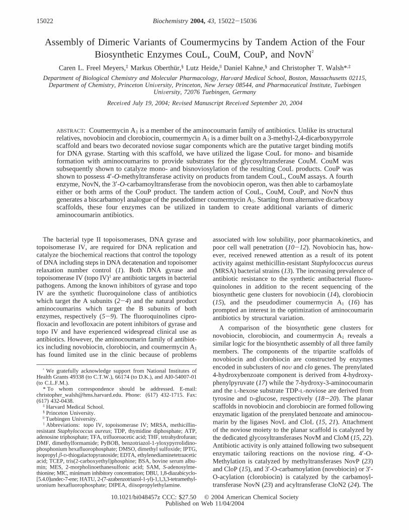
Coumermycin A1 bears resemblance to the tripartiteaminocoumarin antibiotic novobiocin in the aminocoumarincomponent and to clorobiocin in the noviose sugar compo-nent (Figure 1). The most striking feature of coumermycinA1, however, is that it is pseudodimeric, containing fivestructural elements consisting of two decorated noviosyl-aminocoumarin elements linked through a dicarboxypyrrolescaffold. Furthermore, coumermycin A1 shows the highestantibiotic potency in the aminocoumarin family (25). Thefour enzymes that catalyze the five-part assembly andtailoring in coumermycin biosynthesis are the ligase CouL,the glycosyltransferase CouM, the methyltransferase CouP,and the acyltransferase CouN2. In principle, the five-partenzymatic assembly could be achieved using purifiedenzymes. This would establish a foundation for variation ofone or more of the elements of the scaffold and pharma-cophores. CouL has previously been reported by Heide andco-workers to catalyze the double ligation of 3-methyl-2,4-dicarboxylic acid with two aminocoumarin components (26).
We report herein the overproduction and purification ofboth CouM and CouP fromEscherichia coliand confirmthe catalytic functions of each enzyme for single and doubleglycosylation and 4′-O-methylation, respectively. A fullreconstitution of late stage coumermycin A1 biosynthesis hasnot been possible because the methylpyrrolyl transferaseCouN2 is not soluble when heterologously overproduced inE. coli. Alternatively, we have used the novobiocin carbam-oyltransferase NovN to effect biscarbamoylation on thecoumermycin scaffold. Thus, the four-enzyme sequenceCouL, CouM, CouP, NovN has been employed to achieve atotal of eight enzymatic transformations to create variantsof the dimeric aminocoumarin coumermycin A1. With thetandem action of the four enzymes we demonstrate that it ispossible to vary all five structural elements in the coumer-mycin scaffold to generate novel coumermycin analogues.
MATERIALS AND METHODS
Bacterial Strains, Plasmids, Materials, and Instrumenta-tion. Chemically competentE. coli TOP10 and BL21(DE3)cell strains were purchased from Invitrogen. Restrictionendonucleases and T4 DNA ligase were purchased from NewEngland BioLabs. The pET37b overexpression vector waspurchased from Novagen.Pfu DNA polymerase was pur-chased from Stratagene. DNA primers for PCR amplificationwere purchased from Integrated DNA Technologies. HPLCanalysis of crude enzymatic reaction mixtures was carriedout using a Beckman Gold Nouveau System Gold with aVydac small-pore C18 column (250× 4.6 mm). Enzymaticreaction products were confirmed by LCMS using a Shi-madzu LCMS-QP8000R.
All chemicals were purchased from Aldrich or Sigma andused without further purification. Solvents were of reagentgrade and were further dried when necessary. Analytical thin-layer chromatography was performed on glass plates pre-coated with silica gel (250µm, Sorbent Technologies), withdetection by UV and/or spraying with H2SO4 (50%). Flashchromatography was carried out on silica gel (60 Å, 32-63µm), purchased from Sorbent Technologies. AnalyticalHPLC of reaction mixtures was performed on a Hewlett-Packard 1100 series instrument using a Phenomenex Luna5 µm C18 column (250 mm× 4.6 mm). Reactions weremonitored by HPLC using a linear gradient from 20% CH3-CN in H2O/0.1% TFA to 100% CH3CN/0.1% TFA over 20min. Preparative HPLC was performed on a Varian ProStarinstrument (flow rate 45 mL/min) using a Phenomenex Luna10 µm C18 column (250× 50 mm) or on a Hitachi L6200instrument (flow rate 7.5 mL/min) using a Phenomenex Luna5 µm C18 column (250× 21.2 mm). NMR spectra wererecorded on Varian Mercury 300 MHz and Inova 400 or500 MHz spectrometers. Designation of NMR peaks: H-5,H-6, H-8, or 8-Me (coumarin); H-1′, H-2′, H-3′, ... (noviose).Mass spectra (ESI) were obtained at the Mass SpectroscopyFacility at the Department of Chemistry, Princeton Univer-sity.
TDP-L-noviose (22) and 8-demethylaminocoumarin33(27) were prepared according to previously reported proce-dures. CouL was overproduced and purified as previouslyreported (26).
Preparation of Aglycon Substrates. (A) 3-Amino-4,7-dihydroxy-8-methylcoumarin(1) was prepared according toa previously reported method (28) with modifications in theisolation procedure. Novobiocin sodium salt (Sigma, 5.0 g)was heated in a mixture of pyridine and acetic anhydride(5:1, 120 mL) under reflux for 4 h. After being cooled toroom temperature, the mixture was then carefully acidifiedwith 5 N HCl while the temperature of the reaction mixturewas kept below 25°C, upon which a brown gum precipitated.After the mixture was allowed to settle, the aqueous phasewas decanted. Precipitation of the crude aminocoumarincleavage product was induced by the addition of EtOAc (250mL). Following filtration, the gray precipitate was refluxedin EtOAc (125 mL) for 1 h, cooled to room temperature,filtered, and dried, which afforded the 3,4-oxazole derivativeof 7-O-(2′-O-acetyl-3′-O-carbamoyl-4′-O-methyl-R-L-novio-syl)-3-amino-4,7-dihydroxy-8-methylcoumarin (3.3 g) as alight brown amorphous powder:Rf ) 0.13, petroleum ether/EtOAc, 1:2. The oxazole derivative (1.4 g) was suspended
FIGURE 1: Aminocoumarin family of antibiotics.
Combinatorial Aminocoumarin Biosynthesis Biochemistry, Vol. 43, No. 47, 200415023
in anhydrous MeOH (20 mL) and 10% HCl/MeOH (70 mL)and was refluxed for 2 h. The clear solution was evaporateduntil precipitation started (ca. 15 mL final volume) and keptat 4 °C overnight. The precipitate was filtered and washedwith ice-cold MeOH to give a first crop of aminocoumarin1. Evaporation of the mother liquor to a small volume toinduce precipitation was repeated twice, which afforded atotal of 510 mg of1.
(B) 3-Methylpyrrole-2,4-dicarboxylic acid diethyl ester(34) was prepared in analogy to a previously publishedprocedure (29). A mixture of ethyl isocyanoacetate (4.4 mL,40 mmol) and DBU (6.0 mL, 40 mmol) in THF (60 mL)was heated to 50°C. A solution of acetaldehyde (2.8 mL,25 mmol) in THF (20 mL) was added dropwise over 15 min,and stirring of the mixture was continued for 1 h at thistemperature. The solution was cooled to room temperature,neutralized with AcOH, and evaporated. The residue wasdissolved in EtOAc (300 mL) and washed with 1 N HCl,saturated aqueous NaHCO3, and brine. The organic phasewas dried (MgSO4), filtered, and evaporated, and the residuewas purified by flash chromatography using silica gel(petroleum ether/EtOAc, 6:1 to 4:1). Evaporation of theproduct-containing fractions gave the pyrrole diethyl ester(3.2 g, 70%) as a light yellow solid:Rf ) 0.20, petroleumether/EtOAc, 6:1.1H NMR (300 MHz, CDCl3): δ 9.65 (brs, 1 H, NH), 7.48 (d, 1 H,J5,NH ) 3.5 Hz, H-5), 4.34, 4.28(2 q, each 2 H,J ) 7.1 Hz, 2 CH2CH3), 2.59 (s, 3 H, 3-Me),1.37 and 1.34 (2 t, each 3 H,J ) 7.1 Hz, 2 CH2CH3). 13CNMR (75 MHz, CDCl3): δ 165.0, 162.0, 129.9, 127.3, 121.0,117.0, 60.7, 59.9, 14.6, 11.5. MS(ESI) for C11H15NO4
(225.24): 226 [M+ H]+, 248 [M + Na]+.(C) 3-Methylpyrrole-2,4-dicarboxylic Acid (2). 3-Meth-
ylpyrrole-2,4-dicarboxylic acid diethyl ester (34) (1.0 g, 4.44mmol) and NaOH (1.76 g, 44 mmol) were heated to refluxin a H2O/EtOH mixture (1:1, 60 mL) overnight. After beingcooled to room temperature, H2O (30 mL) was added, andthe solution was acidified with concentrated HCl (pH) 2)to precipitate the acid. The mixture was kept at 4°C for 4h and then filtered, and the residue was washed with coldH2O and cold acetone and dried under high vacuum to yield2 (0.6 g, 80%) as a yellow powder.1H NMR (500 MHz,CD3OD): δ 7.43 (s, 1 H, H-5), 2.58 (s, 3 H, 3-Me);13CNMR (125 MHz, CDCl3): δ 167.3, 163.2, 129.9, 127.8,121.1, 116.0, 10.3. MS(ESI) for C7H7NO4 (169.14): 168 [M- H]-.
(D) 3-Methylpyrrole-2,4-dicarboxylic Acid 4-Ethyl Ester(35). Diethyl ester34 (500 mg, 2.22 mmol) and KOH (148mg, 2.64 mmol) were heated to reflux in a H2O/EtOHmixture (1:4, 9 mL) for 2.5 h. After being cooled to roomtemperature, the yellow solution was poured into ice-water(60 mL) and acidified with 1 N HCl (pH) 3). The aqueousphase containing a white precipitate was extracted withEtOAc (3× 150 mL), and the combined organic phases werewashed with brine (2× 100 mL), dried (MgSO4), filtered,and evaporated. The residue, which contained small impuri-ties of starting material (Rf ) 0.85, CH2Cl2/MeOH, 9:1) and2-ethyl ester (Rf ) 0.28, CH2Cl2/MeOH 9:1), was purifiedby flash chromatography using silica gel (CH2Cl2/MeOH,15:1) to afford the monoester35 as a colorless powder (290mg, 66%): Rf ) 0.21, CH2Cl2/MeOH, 9:1.1H NMR (300MHz, DMSO-d6): δ 12.00 (br s, 2 H, COOH, NH), 7.39 (d,1 H, J5,NH ) 3.6 Hz, H-5), 4.16 (q, 2 H,J ) 7.1 Hz, CH2-
CH3), 2.48 (s, 3 H, 3-Me), 1.24 (t, 3 H,J ) 7.1 Hz, CH2CH3);13C NMR (75 MHz, DMSO-d6): δ 164.0, 162.3, 128.1,127.1, 121.1, 115.2, 59.0, 14.3, 11.1. MS(ESI) for C9H11-NO4 (197.19): 196 [M- H]-.
(E) 3-Methylpyrrole-2,4-dicarboxylic Acid 2-(3-Amino-4,7-dihydroxy-8-methylcoumarin Amide) 4-Ethyl Ester (7). Toa solution of aminocoumarin1 (24 mg, 0.1 mmol) andmonoester35 (20 mg, 0.1 mmol) in DMF (1.5 mL) wasaddedN-methylmorpholine (33µL, 0.3 mmol) and PyBOP(52 mg, 0.1 mmol) under argon. After being stirred at roomtemperature for 40 h, the mixture was purified by preparativereversed-phase HPLC (20% CH3CN in H2O/0.1% TFA to100% CH3CN/0.1% TFA over 40 min) to the give 4-ethylester36 (30 mg, 78%).1H NMR (300 MHz, DMSO-d6): δ12.00 (br s, 2 H, OH, NH), 10.50, 8.72 (2 s, each 1 H, NH,OH), 7.58 (d, 1 H,J5,NH ) 3.4 Hz, H-5pyrr), 7.57 (d, 1 H,J5,6
) 8.7 Hz, H-5), 6.90 (d, 1 H,J5,6 ) 8.7 Hz, H-6), 4.19 (q,2 H, J ) 7.2 Hz, CH2CH3), 2.48 (s, 3 H, 3-Mepyrr), 2.17 (s,3 H, 8-Me), 1.28 (t, 3 H,J ) 7.2 Hz, CH2CH3).
To this compound was added concentrated H2SO4 (0.4mL), and the mixture was stirred at 40°C for 2 h. Thesolution was carefully diluted with DMF/H2O (1:1, 1.5 mL)and purified by reversed-phase HPLC to give monomer7(8 mg, 66%) as a powder.1H NMR (500 MHz, DMSO-d6):δ 12.00 (br s, 2 H, OH, NH), 10.45, 8.65 (2 s, each 1 H,NH, OH), 7.59 (d, 1 H,J5,6 ) 8.6 Hz, H-5), 7.55 (d, 1 H,J5,NH ) 3.2 Hz, H-5pyrr), 6.89 (d, 1 H,J5,6 ) 8.6 Hz, H-6),2.57 (s, 3 H, 3-Mepyrr), 2.18 (s, 3 H, 8-Me).
Preparation of pCouM-pET37b and pCouP-pET28a OVer-expression Constructs.The gene encoding CouM wasamplified from Streptomyces rishiriensis(DSM 40489)genomic DNA. Amplification was accomplished using thefoward primer 5′-AATTCACATATGAGAGTGCTGTTCAC-GAGC-3′ and the reverse primer 5′-TGCGGCCGCAAG-CTTTTACTGTCGACCGTGCGA-3′. The forward primerintroduced anNdeI restriction site (restriction site underlinedabove), and the reverse primer introduced aHindIII restric-tion site. PCR reactions were carried out usingPfu DNApolymerase as described by Stratagene. The amplified genewas inserted into the linearized pET37b vector following arestriction digest withNdeI/HindIII. Expression of pCouM-pET37b was accomplished following transformation intoE.coli TOP10 competent cells. The gene sequence wasconfirmed by comparison to theS. rishiriensissequencereported by Heide et al. (Accession Number AF235050).
Generation of the expression construct pCouP-pET28a wasaccomplished as described for pCouM-pET37b. The forwardprimer used for PCR amplification was 5′-GCGTAT-CATATGGAGGTGGCACCTATCGTAAGC-3′ and intro-duced anNdeI restriction site. The reverse primer used was5′-ACGGAAAAGCTTTCACTCGGTCTGCCAGTA-3′ andintroduced aHindIII restriction site.
OVerproduction and Purification of CouM.Purified pCouM-pET37b plasmid was transformed into BL21(DE3) competentE. coli cells. Transformants harboring the pCouM-pET37bconstruct were grown in LB medium supplemented withkanamycin (50µg/mL). The cells were grown at 25°C toan OD of∼0.6 and induced with 60µM IPTG. Shaking wascontinued overnight at 25°C. Cells were harvested bycentrifugation (20 min at 6000g) and frozen at-20 °C. Cellswere thawed and resuspended in 75 mL of buffer A (25 mMTris-HCl, pH 8.0, 400 mM NaCl, 2 mM imidazole, 10%
15024 Biochemistry, Vol. 43, No. 47, 2004 Freel Meyers et al.

glycerol). Resuspended cells were lysed by French press (twopassages at 15000 psi), and the cell debris was removed bycentrifugation (30 min at 9500g). The supernatant wasincubated with 2 mL of superflow Ni(II) affinity resin(Qiagen) at 4°C for 1.5 h. The resin was loaded onto acolumn and washed with 10 mL of buffer B (25 mM Tris-HCl, pH 8.0, 400 mM NaCl, 5 mM imidazole, 10% glycerol).CouM was eluted from the column in a stepwise imidazolegradient (20-200 mM imidazole). Fractions containing pureCouM (as determined by SDS-PAGE) were combined anddialyzed against 1 L of buffer C (50 mM Tris-HCl, pH 8.0,100 mM NaCl, 1 mM EDTA, 10% glycerol) overnight at 4°C. A second dialysis was carried out in 1 L of buffer D (50mM Tris-HCl, pH 8.0, 100 mM NaCl, 1 mM TCEP, 10%glycerol). The protein was flash frozen in liquid nitrogenand stored at-80 °C. The concentration of purified CouMwas measured spectrophotometrically at 280 nm using thecalculated extinction coefficient 38820 M-1 cm-1. Large-scale overproduction afforded an overall yield of>2 mg/LCouM. CouM could not be concentrated above 0.9 mg/mL.
OVerproduction and Purification of CouP.Purified pCouP-pET28a plasmid was transformed into BL21(DE3) competentE. coli cells. Transformants harboring the pCouP-pET28aconstruct were grown in LB medium supplemented withkanamycin (50µg/mL). The cells were grown at 25°C toan OD of∼0.6 and induced with 60µM IPTG. Shaking wascontinued overnight at 25°C. Cell lysis and CouP purifica-tion were carried out as described for CouM above. Large-scale overproduction afforded an overall yield of>23 mg/LCouP.
Characterization of CouM.Initial characterization ofCouM was carried out in a tandem CouL, CouM reaction(360µL) at ambient temperature and contained 75 mM Tris-HCl (pH 7.5), 10 mM MnCl2, 5 mM ATP, 1 mg/mL BSA,5% DMSO, 100µM aminocoumarin1, 100µM dicarboxylicacid2, 200µM TDP-L-noviose, 500 nM CouL, and 500 nMCouM. Aliquots (50µL) were quenched at specified timepoints (5 min, 10 min, 30 min, 60 min, and 2 h) in 100µLof cold methanol. Quenched aliquots were incubated at 4°C for 20 min, and the supernatant was analyzed by reversed-phase HPLC [gradient 15:85 CH3CN/H2O (0.1% TFA) to100% CH3CN over 20 min]. The bisnoviosyl CouM product4 was confirmed by LCMS (ESI for C41H45N3O18: calcd,867.3; obsd, 866.4 [M- H]-).
The distributive action of CouM for mononoviosylationfollowed by bisnoviosylation was confirmed in a reaction(370 µL) carried out at ambient temperature containing 75mM Tris-HCl (pH 7.5), 10 mM MnCl2, 1 mg/mL BSA, 5%DMSO, 100µM diamide 3, 200 µM TDP-L-noviose, and200 nM CouM. Aliquots (50µL) were quenched at 1, 5, 10,30, and 60 min and analyzed as described above. Theformation of the monoglycosylated intermediates5 and 6was observed by reversed-phase HPLC [gradient 15:85 CH3-CN/H2O (0.1% TFA) to 100% CH3CN over 20 min]followed by formation of the bisglycosylated CouM product4 characterized above. The mixture of monoglycosylatedintermediates5 and 6 was confirmed by LCMS (ESI forC34H33N3O14: calcd, 707.2; obsd, 706.0 [M- H]-). Sub-sequent optimization of reaction conditions revealed anoptimal buffer and pH of MES, pH 6.
The glycosylation of monoamide7 was carried out atambient temperature in a reaction mixture (370µL) contain-
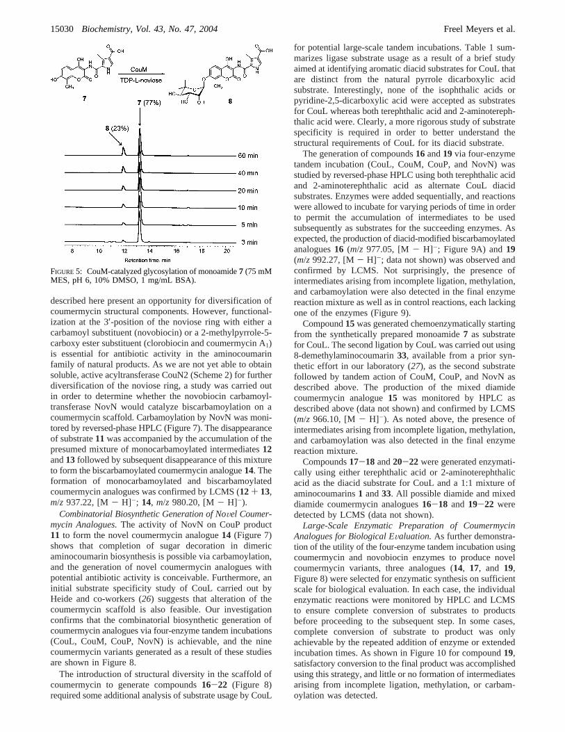
ing 75 mM Tris-HCl (pH 7.5), 10 mM MnCl2, 1 mg/mLBSA, 5% DMSO, 100µM monoamide7, 100 µM TDP-L-noviose, and 200 nM CouM. Aliquots (50µL) werequenched at the time points specified above and analyzedas previously described. The formation of glycosylatedmonoamide8 was monitored by reversed-phase HPLC[gradient 15:85 CH3CN/H2O (0.1% TFA) to 100% CH3CNover 20 min] and confirmed by LCMS (ESI for C24H26N2O11:calcd, 518.1; obsd, 569.0 [M- H]-).
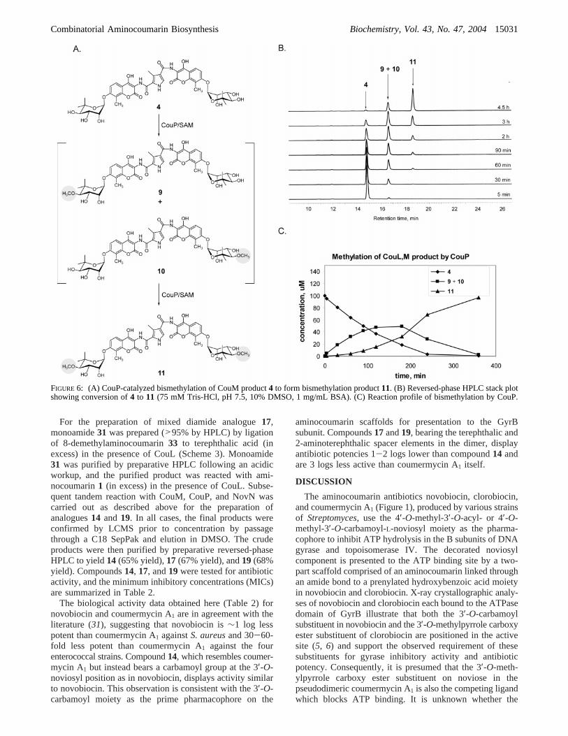
Characterization of CouP.Methyltransferase activity ofCouP was confirmed in a reaction (410µL) containing 75mM Tris-HCl (pH 7.5), 1 mg/mL BSA, 10% DMSO, 100µM CouM product4 (obtained from a tandem enzymaticincubation of diamide3 with TDP-L-noviose and CouM),500µM S-adenosylmethionine (SAM), and 1µM CouP. Thedistributive action of CouP to catalyze monomethylation of4 followed by bismethylation to form CouP product11 wasmonitored by reversed-phase HPLC [gradient 15:85 CH3-CN/H2O (0.1% TFA) to 100% CH3CN over 20 min].Aliquots (50µL) were quenched at 5 min, 30 min, 60 min,90 min, 2 h, and 3 h. The formation of the monomethylatedintermediates9 and 10 was observed by reversed-phaseHPLC and confirmed by LCMS (ESI for C42H47N3O18:calcd, 881.3; obsd, 880.3 [M- H]-). The formation of CouPproduct11 was confirmed by LCMS (ESI for C43H49N3O18:calcd, 895.3; obsd, 894.3 [M- H]-).
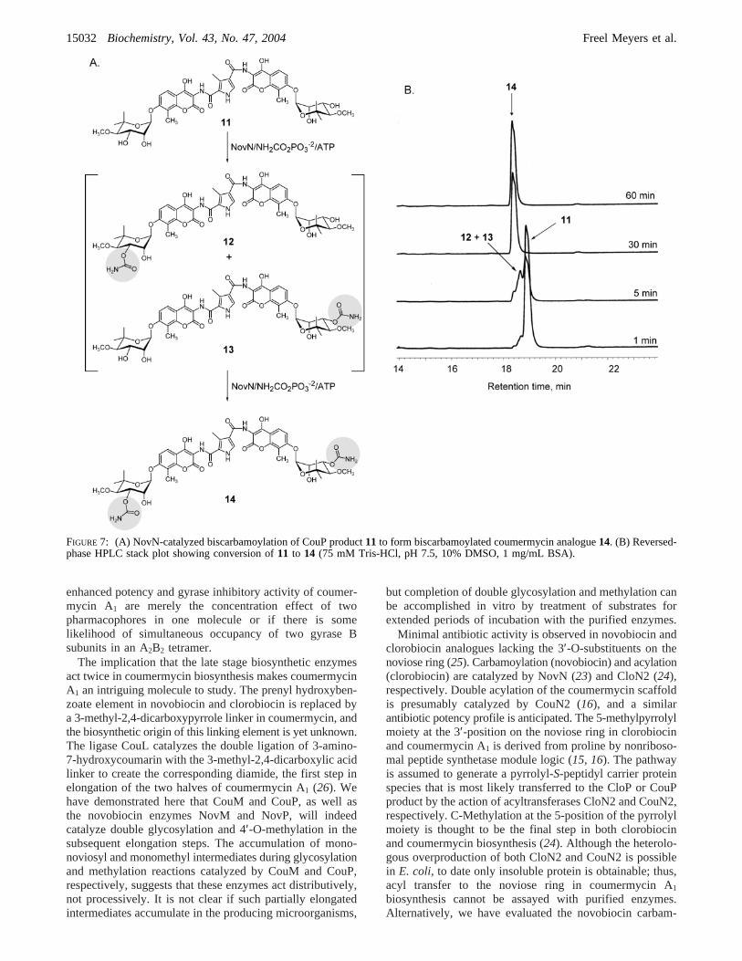
Carbamoylation of the Coumermycin Scaffold by NoVN.The ability of NovN to catalyze carbamoylation on acoumermycin scaffold was confirmed in a reaction (210µL)containing 75 mM Tris-HCl (pH 7.5), 1 mg/mL BSA, 10%DMSO, 100µM CouP product11 (obtained from a tandemenzymatic incubation of diamide3 with TDP-L-noviose/CouM and SAM/CouP), 500µM carbamoyl phosphate, and1 µM NovN. The distributive action of CouN to catalyzemonocarbamoylation of11 followed by biscarbamoylationto form NovN product14 was monitored by reversed-phaseHPLC [gradient 15:85 CH3CN/H2O (0.1% TFA) to 100%CH3CN over 20 min]. Aliquots (50µL) were quenched at1, 5, 30, and 60 min. The formation of the monocarbamoy-lated intermediates12 and 13 was observed by reversed-phase HPLC and confirmed by LCMS (ESI for C44H50N4O19:calcd, 938.3; obsd, 937.2 [M- H]-). The formation of NovNproduct14 was confirmed by LCMS (ESI for C45H51N5O20:calcd, 981.3; obsd, 980.2 [M- H]-).
Small-Scale Generation of Coumermycin VariantsViaFour Enzyme Tandem Incubations.A general procedure isdescribed for the generation of coumermycin variants16-22 on a small scale via four enzyme tandem incubations.For the generation of compounds16 and 19, the reactionmixture (100µL) contained 75 mM Tris-HCl (pH 7.5), 10mM MnCl2, 4 mM ATP, 1 mg/mL BSA, 10% DMSO, 150µM diacid substrate, and 200µM aminocoumarin1. For thegeneration of compounds17, 18, and20-22, a 1:1 mixtureof aminocoumarin1 (200 µM) and 8-demethylaminocou-marin33 (200µM) was added to the reaction mixture. CouLwas added to a final concentration of 1µM, and the reactionmixture was incubated at room temperature for 4 h. TDP-L-noviose (final concentration) 150µM) and CouM (finalconcentration) 1 µM) were added, and the reaction mixturewas incubated for an additional 4 h at room temperature.S-Adenosylmethionine (SAM) (final concentration) 1 mM)and CouP (final concentration) 1 µM) were added, and
Combinatorial Aminocoumarin Biosynthesis Biochemistry, Vol. 43, No. 47, 200415025
the reaction mixture was incubated overnight at roomtemperature. Finally, carbamoyl phosphate (final concentra-tion ) 500 µM) and NovN (1 µM) were added, and thereaction mixture was incubated for 4 h atroom temperature.The reaction was then quenched with methanol (200µL)and incubated at 4°C for 20 min. The supernatant wasanalyzed by reversed-phase HPLC and LCMS.
For the generation of mixed diamide coumermycin ana-logue15, the reaction mixture (200µL) contained 75 mMTris-HCl (pH 7.5), 10 mM MnCl2, 4 mM ATP, 1 mg/mLBSA, 10% DMSO, 50µM monoamide substrate7, and 100µM 8-demethylaminocoumarin33. CouL (final concentration) 1 µM) was added, and the four-enzyme tandem incubationsequence was carried out as described for compounds16-22 above. The formation of compound15 was monitoredby reversed-phase HPLC and confirmed by LCMS.
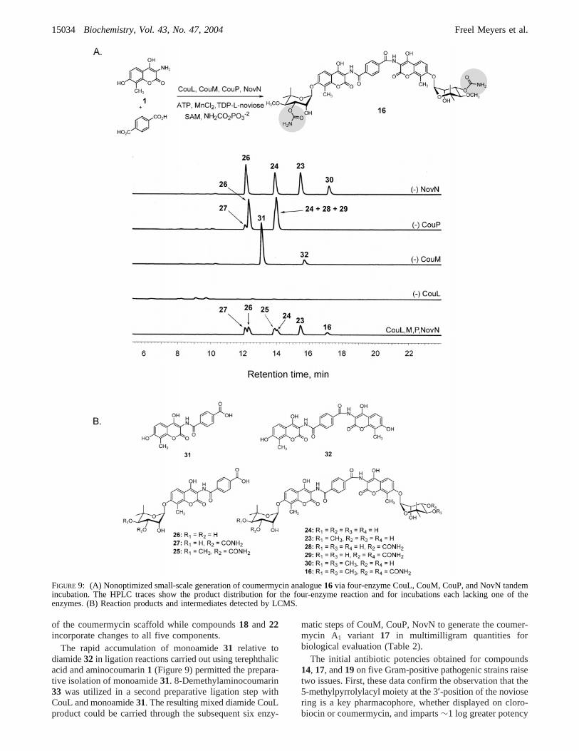
LCMS Characterization of Analogues15-22. The forma-tion of the coumermycin analogues described above wasconfirmed by LCMS. It is assumed that a mixture of2-aminoterephthalic acid mixed diamide analogues20 and21 is formed as a result of adding a 1:1 mixture ofaminocoumarins1 and33 to the reaction mixture. Furtheranalysis of the fragmentation patterns of these analogues isnecessary in order to confirm this assumption: mixeddiamide analogue15 (ESI for C44H49N5O20: calcd, 967.3;obsd, 966.1 [M- H]-); terephthalic acid analogue16 (ESIfor C46H50N4O20: calcd, 978.3; obsd, 977.1 [M- H]-);terephthalic acid analogue17 (ESI for C45H48N4O20: calcd,964.3; obsd, 963.4 [M- H]-); terephthalic acid analogue18 (ESI for C44H46N4O20: calcd, 950.3; obsd, 950.2 [M-H]-); 2-aminoterephthalic acid analogue19 (ESI forC46H51N5O20: calcd, 993.3; obsd, 992.3 [M- H]-); 2-ami-noterephthalic acid analogues20and21 (ESI for C45H49N5O20:calcd, 979.3; obsd, 978.1 [M- H]-); 2-aminoterephthalicacid analogue22 (ESI for C44H47N5O20: calcd, 965.3; obsd,964.2 [M - H]-).
LCMS Characterization of Intermediates23-30. Inter-mediates arising from incomplete ligation, methylation, andcarbamoylation in the tandem incubation to produce analogue16 (Figure 9) were characterized by LCMS: intermediate23 (ESI for C43H46N2O18: calcd, 878.3; obsd, 877.4 [M-H]-); intermediate24 (ESI for C42H44N2O18: calcd, 864.3;obsd, 863.4 [M- H]-); intermediate25 (ESI for C27H28N2O12:calcd, 572.2; obsd, 571.2 [M- H]-); intermediate26 (ESIfor C25H25NO11: calcd, 515.1; obsd, 514.2 [M- H]-);intermediate27 (ESI for C26H26N2O12: calcd, 558.2; obsd,557.2 [M - H]-); intermediate28 (ESI for C43H45N3O19:calcd, 907.3; obsd, 906.3 [M- H]-); intermediate29 (ESIfor C44H46N4O20: calcd, 950.3; obsd, 949.2 [M- H]-);intermediate30 (ESI for C44H48N2O18: calcd, 892.3; obsd,891.4 [M - H]-).
Large-Scale Preparation of Coumermycin Analogues. (A)Coumermycin Analogue14. For the preparation of thebiscarbamoyl coumermycin analogue14, aminocoumarin1(9.3 mg, 38.3µmol) and dicarboxylic acid2 (2.6 mg, 15.3µmol) were each dissolved in DMSO (0.50 mL) and addedto a MES-buffered solution (131 mL, pH 7) containingDMSO (14.3 mL, 10% v/v). ATP and MgCl2 were added toa final concentration of 4 and 10 mM, respectively, followedby the addition of BSA (153 mg, final concentration) 1mg/mL). CouL was added to a final concentration of 500nM (1.6 mL of 48.6 µM stock), and the reaction was
incubated at ambient temperature for 24 h. The reaction wasfollowed by reversed-phase HPLC [gradient 15:85 CH3CN/H2O (0.1% TFA) to 100% CH3CN over 20 min]. AdditionalCouL (1.6 mL of 48.6µM stock) was added two times overthe next 48 h to ensure complete conversion of the diacidand aminocoumarin substrates to the CouL diamide product3. When ligation was>90% complete (as determined byHPLC), TDP-L-noviose (21.4 mg, 38.3µmol) was addedfollowed by the addition of CouM (0.74 mL of 20.8µMstock) to a final concentration of 100 nM. The reactionmixture was incubated overnight at room temperature.Reversed-phase HPLC analysis indicated that glycosylationwas complete after 10 h.
The reaction mixture was diluted by the addition of 50mL of 10% DMSO in 75 mM MES, pH 7, since CouPoperates more efficiently at low substrate concentration.CouP was added to a final concentration of 1µM (3.3 mLof 60.2 µM stock) followed by the addition of SAM (33.3mg, 76.6µmol). The reaction was monitored by reversed-phase HPLC. Additional CouP (3.3 mL of 60.2µM stock)was added after 24 h, and monitoring was continued byHPLC. Slow but complete conversion of CouM product4to the bismethyl CouP product was observed over 4 days,indicating that CouP retains activity for an extended periodof time at ambient temperature.
Carbamoyl phosphate (11.6 mg, 76.6µmol) was addedfollowed by the addition of NovN (2.2 mL of 45.5µM stock)to a final concentration of 500 nM. The reaction wasincubated at room temperature and monitored by HPLC andLCMS. Very slow conversion of the CouP product11 wasobserved under these conditions. It was necessary to addNovN and carbamoyl phosphate three additional times overthe next 72 h in order to ensure complete conversion to thebiscarbamoyl NovN product14.
The crude reaction mixture was adjusted to pH 6 by theaddition of concentrated HCl and desalted and concentratedby passage over a C18 SepPak (900 mg bed) conditionedwith 75 mM MES, pH 6. Following loading of the crudereaction product, the SepPak was washed with 75 mM MES,pH 6 (10 mL), followed by water (10 mL). The crudereaction product was eluted from the SepPak using DMSO(5 mL). Fractions containing product14 (as determined byHPLC) were combined, and the product was purified bypreparative reversed-phase HPLC [gradient 15:85 CH3CN/H2O (0.1% TFA) to 100% CH3CN over 20 min]. Lyo-philization of the product-containing fractions affordedcoumermycin analogue14 (9.6 mg, 65% yield) as a whitepowder.1H NMR (500 MHz, DMSO-d6): δ 12.45-12.25(br s, 2 H, OH, NH), 11.93, 9.00, 8.69 (3 s, 3 H, NH, OH),7.82 (br s, 1 H, H-5pyrr), 7.76 (2 d, 2 H,J5,6 ) 8.7 Hz, H-5),7.17 (2 d, 2 H,J5,6 ) 8.7 Hz, H-6), 6.73, 6.52 (2 br s, 4 H,CONH2), 5.62 (d, 2 H,J1,2 ) 5.1 Hz, H-1′), 5.54 (s, 2 H,2′-OH), 5.15 (dd, 2 H,J2,3 ) 3.1 Hz,J3,4 ) 9.9 Hz, H-3′),4.08-4.01 (m, 2 H, H-2′), 3.48 (d, 2 H,J3,4 ) 9.9 Hz, H-4′),3.47 (s, 6 H, 4′-OMe), 2.62 (s, 3 H, 3-Mepyrr), 2.23 (s, 6 H,8-Me), 1.26, 1.05 (2 s, 12 H, 6′-CH3).
(B) Coumermycin Analogue19. Analogue19was preparedas described above starting with 2-aminoterephthalic acid(2.8 mg, 15.3µmol) and aminocoumarin1 (9.3 mg, 38.3µmol). Following lyophilization,19 (10.4 mg, 68% yield)was obtained as a bright yellow solid.1H NMR (500 MHz,DMSO-d6): δ 7.90 (d, 1 H, H-6terepht), 7.76 (d, 2 H,J5,6 )
15026 Biochemistry, Vol. 43, No. 47, 2004 Freel Meyers et al.
8.8 Hz, H-5), 7.35 (s, 1 H, H-3terepht), 7.22-7.14 (m, 3 H,H-6, H-5terepht), 6.78-6.60 (br s, 4 H, CONH2), 5.62 (d, 2H, J1,2 ) 5.2 Hz, H-1′), 5.54 (s, 2 H, 2′-OH), 5.18 (dd, 2 H,J2,3 ) 3.2 Hz,J3,4 ) 9.8 Hz, H-3′), 4.10-4.00 (m, 2 H, H-2′),3.48 (d, 2 H,J3,4 ) 9.9 Hz, H-4′), 3.48 (s, 6 H, 4′-OMe),2.21 (s, 6 H, 8-Me), 1.26, 1.06 (2 s, 12 H, 6′-CH3).
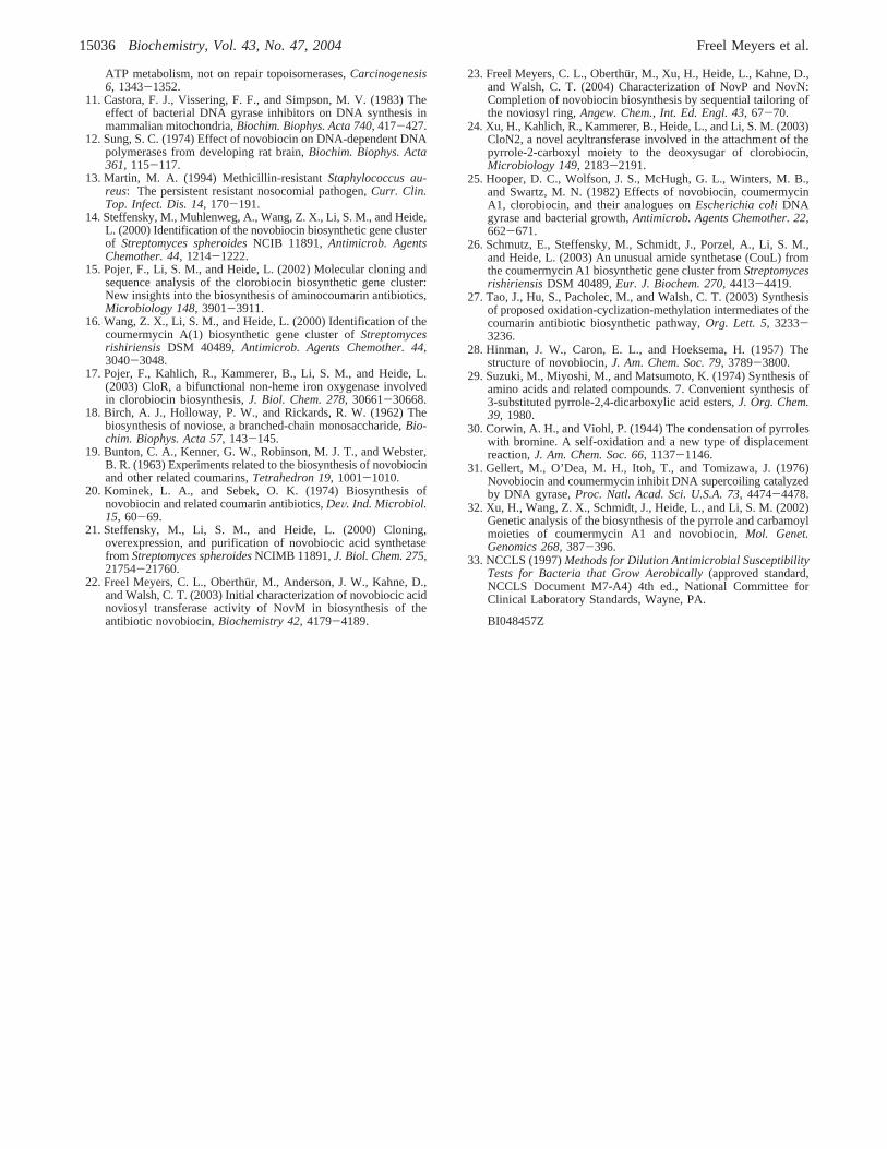
(C) Mixed Diamide Coumermycin Analogue17. For thepreparation of mixed diamide analogue17, 8-demethylami-nocoumarin33 (3 mg, 13.1µmol) and terephthalic acid (21.8mg, 131µmol) were each dissolved in DMSO (0.50 mL)and added to a MES-buffered solution (56 mL, pH 7)containing DMSO (9 mL). ATP and MgCl2 were added toa final concentration of 4 and 10 mM, respectively, followedby the addition of BSA (100 mg, final concentration) 1mg/mL). CouL was added to a final concentration of 1µM(3.2 mL of 30.9µM stock), and the reaction was incubatedat ambient temperature for 1 h. The reaction was analyzedby HPLC as described above; the mixture contained>95%monoamide31 and<5% diamide.
The reaction mixture was adjusted to pH 3 and extractedthree times with ethyl acetate (50 mL). The organic extrac-tions were combined and concentrated under reduced pres-sure. The crude reaction product was purified by preparativereversed-phase HPLC and lyophilized to afford monoamide31 as a bright yellow solid (3.2 mg, 72% yield).
Monoamide31 (2.5 mg, 7.33µmol) and aminocoumarin1 (8.9 mg, 36.6µmol) were dissolved in DMSO (0.50 mL)and added to a MES-buffered solution (60 mL, pH 7)containing DMSO (6.9 mL). ATP and MgCl2 were addedto a final concentration of 4 and 10 mM, respectively,followed by the addition of BSA (100 mg, final concentration) 1 mg/mL). CouL was added to a final concentration of 1µM (3.2 mL of 30.9µM stock), and the four-enzyme tandemincubation and purification were carried out as described forcompounds14 and19 above. Lyophilization of the purifiedproduct afforded mixed diamide coumermycin analogue17(4.7 mg, 67% yield) as a yellow solid.1H NMR (500 MHz,DMSO-d6): δ 12.10-11.90 (br s, 2 H, OH), 9.65 (br s, 2H, NH), 8.12 (s, 4 H, H-2,3,5,6terepht), 7.85, 7.76 (2 d, 2 H,J5,6 ) 8.6 Hz, H-5), 7.17 (d, 1 H,J5,6 ) 8.6 Hz, H-6), 7.02-7.00 (m, 2 H, H-6, H-8), 6.80-6.50 (br s, 4 H, CONH2),5.64-5.62 (m, 2 H, H-1′), 5.59, 5.55 (2 d, 2 H,J2-OH ) 1.7Hz, 2′-OH), 5.16, 5.07 (2 dd, 2 H,J2,3 ) 3.1 Hz,J3,4 ) 9.9Hz, H-3′), 4.10-4.05 (m, 2 H, H-2′), 3.48-3.44 (m, 8 H,H-4′, 4′-OMe), 2.33 (s, 3 H, 8-Me), 1.27, 1.23, 1.08, 1.06(4 s, 12 H, 6′-CH3).
Determination of Minimum Inhibitory Concentrations forAnalogues14, 17, and 19. Twenty-two hour minimuminhibitory concentrations (MICs) were determined againststrains grown in brain-heart infusion broth in a microdilutionformat according to NCCLS guidelines (33). The followingstrains were used for the MIC determination:Enterococcusfaecium, 49624; E. faecium, resistant (VanA), CL4931;Enterococcus faecalis, 29212;E. faecalis, resistant (VanB),CL4877;S. aureus, 29213.
RESULTS
OVerproduction and Purification of S. rishiriensis CouMand CouP in E. coli.CouM is the putative glycosyltransferasein coumermycin A1 biosynthesis as it bears significantsequence homology to the family of glycosyltransferases
including the novobiocin glycosyltransferase NovM (16, 22).CouM presumably catalyzes the transfer of two noviosylmoieties from TDP-L-noviose to the diamide aglycon3(Scheme 2) to form the bisnoviosyl aminocoumarin diamide4. The 42 kDa enzyme was overproduced heterologously andpurified fromE. coli BL21(DE3) cells harboring the CouMexpression construct. Cells were grown at 25°C to an ODof ∼0.6 followed by induction with IPTG (60µM). TheC-terminal His8 fusion protein CouM was then purified tohomogeneity by Ni(II) affinity chromatography (Figure 2).
CouP bears homology to the family ofO-methyltrans-ferases including the novobiocinO-methyltransferase NovP(16, 23). CouP presumably catalyzes the first step in sugardecoration by O-methylation at the 4′-position of eachnoviose component (Figure 6A) to form the precursor tocoumermycin A1. The 31 kDa enzyme was overproducedheterologously and purified fromE. coli BL21(DE3) cellsharboring the CouP expression construct as described abovefor the overproduction and purification of CouM. TheN-terminal His6 fusion protein CouP was purified to homo-geneity by Ni(II) affinity chromatography (Figure 2).
Preparation of Aglycon Substrates.Aminocoumarin1 wasobtained from novobiocin through a two-step degradationprocedure (step 1, Ac2O/pyridine; step 2, HCl/MeOH) (28).3-Methylpyrrole-2,4-dicarboxylic acid (2) was obtained byDBU-catalyzed condensation of acetaldehyde with ethylisocyanoacetate (Scheme 1) (29), which afforded diethyl ester34 (70%). Both ester groups could be cleaved by heating34and sodium hydroxide (10 equiv) in a water/ethanol mixtureunder reflux for 16 h to give diacid2 (80%). Monoethylester35 was required for the synthesis of monomer7 andcould be prepared from diester34 based on the higherreactivity of the 2-carboxyethyl group toward base (30).Thus, saponification using 1.2 equiv of potassium hydroxidein an ethanol/water mixture under reflux for 2.5 h affordedthe 2-carboxylic acid35 in 66% yield. Coupling of ami-nocoumarin1 with acid 35 using HATU as the activatingagent proceeded in unsatisfying yields (only 10% of protectedmonomer36could be isolated). When the corresponding acidchloride (obtained from35using oxalyl chloride/DMF) wascoupled with1 in DMF in the presence of DIPEA, the yieldof 36 was slightly improved (40%). Finally, it was foundthat activation of acid35 with PyBOP proceeded smoothlyand afforded36 in 76% yield. Deprotection of the remainingcarboxylic ester under acidic conditions then gave monomer7 in 66% yield.
FIGURE 2: Heterologous overproduction of CouL, CouM, and CouPfrom E. coli.
Combinatorial Aminocoumarin Biosynthesis Biochemistry, Vol. 43, No. 47, 200415027
Characterization of CouM Glycosyltransferase ActiVity.Heide and co-workers recently demonstrated that the ligaseCouL catalyzes two amide bond forming reactions (26), thefirst between the aminocoumarin1 and 3-methylpyrrole-2,4-dicarboxylic acid (2) to form a monoamide which thenundergoes subsequent ligation with a second molecule ofaminocoumarin1 to form diamide3. Glycosylation by CouMin coumermycin A1 biosynthesis presumably follows amidebond formation (Scheme 2), and CouM is expected to processthe distinct scaffolds required for the first and secondglycosylation reactions in the same way that CouL processestwo distinct carboxylic acid substrates.
Initial confirmation of glycosyltransferase activity wasaccomplished in a tandem CouL, CouM enzyme incubation(Figure 3). The appearance of the bisnoviosyl product4 wasmonitored by reversed-phase HPLC, and the CouM productwas confirmed by LCMS (m/z 866.40 [M - H]-). FurtherHPLC analysis of CouM activity was carried out using puri-fied diamide scaffold3 as substrate for CouM (Figure 4).Disappearance of substrate3 in the presence of CouM (100
nM) and TDP-L-noviose was accompanied by the accumula-tion of an intermediate product peak, presumably corre-sponding to a mixture of mononoviosyl intermediates5 and6 (m/z706.00 [M- H]-) and the bisnoviosyl CouM product4 (m/z 866.45 [M - H]-). Under these conditions, greaterthan 70% of diamide3 underwent glycosylation over 60 min(Figure 4C). In contrast, less than 25% of monoamide7underwent glycosylation to afford the corresponding glyco-sylated monoamide8 (m/z517.00, [M- H]-) under the sameconditions (Figure 5), suggesting that while CouM will acceptvariant aminocoumarin scaffolds as substrates, diamide3 ismost likely the preferred initial aglycon substrate for CouM.
The general pattern of partial accumulation of a monogly-cosylated intermediate that then undergoes bisglycosylationby tandem action of CouM is evident from Figure 4.However, a detailed kinetic analysis of both initial monogly-cosylation and tandem bisglycosylation has not yet beencarried out to obtain kinetic parameterskcat and KM. It isassumed that a mixture of regioisomers resulting fromglycosylation at either 7-hydroxy group of the aglyconscaffold is formed during the initial sugar transfer from TDP-L-noviose (5 and6, Figure 4A). Until each regioisomer hasbeen prepared and characterized separately, and conditionshave been determined for the chromatographic separationof 5 and6, the efficiency of CouM in the formation of eitherisomer5 or 6 and the extent to which CouM prefers eachintermediate for bisglycosylation cannot be accurately de-termined. However, it is apparent from the studies carriedout during the course of this work that CouM will effectivelyprocess a diamide scaffold to the corresponding bisglyco-sylated product, and efforts to utilize CouM as a glycosyl-transferase for the large-scale preparation of variant coumer-mycin analogues are described below.
Characterization of CouP Methyltransferase ActiVity. Dec-oration of the noviose sugar components in coumermycinA1 biosynthesis is expected to follow the same biosyntheticlogic as that confirmed for sugar tailoring in novobiocin bio-synthesis (22). Thus, it is anticipated that the putative meth-yltransferase CouP catalyzes bis-O-methylation at the 4′-posi-tion of each noviose ring following bisglycosylation by CouM.
The methyltransferase activity of CouP was confirmedusing as substrate the CouM product4 obtained viaenzymatic glycosylation of synthetic scaffold3. Not surpris-ingly, CouP exhibits a reaction profile similar to thatobserved during glycosylation by CouM (Figure 6C). Thedisappearance of4 in the presence of CouP (1µM) andS-adenosylmethionine (SAM) was monitored by reversed-phase HPLC (Figure 6B), and the formation of the presumedmixture of monomethyl intermediates9 and10 (m/z880.30,[M - H]-) followed by formation of CouP product11 (m/z894.30, [M - H]-) was confirmed by LCMS. As notedabove, a detailed kinetic analysis of a reaction profileinvolving the probable formation of a mixture of regioiso-mers is not feasible unless the reaction intermediates areprepared and studied separately. Therefore, a comprehensivekinetic analysis has not yet been carried out for the reactionscatalyzed by CouP.
It is interesting to note that the glycosyltransferase NovMand the methyltransferase NovP from the novobiocin bio-synthetic cluster (22, 23) catalyze comparable transformationson the coumermycin scaffold with similar relative efficienciesto CouM and CouP (data not shown). This suggests there is
Scheme 1a
a Reagents and conditions: (a) DBU, 70%; (b) NaOH, 80%; (c)KOH, 66%; (d) PyBOP, NMM, 78%; (e) H2SO4, 66%.
Scheme 2: Coumermycin A1 Biosynthesis
15028 Biochemistry, Vol. 43, No. 47, 2004 Freel Meyers et al.
little deviation of the fundamental enzyme mechanisms forglycosylation and methylation by CouM and CouP comparedto NovM and NovP.
Carbamoyltransferase ActiVity on a Coumermycin Scaf-fold. The characterization of the ligase CouL (26) andpreliminary confirmation of CouM and CouP functions
FIGURE 3: Confirmation of CouM activity in a tandem CouL, CouM incubation of substrates1 and2.
FIGURE 4: (A) CouM-catalyzed bisglycosylation of diamide substrate3 to form CouM product4. (B) Reversed-phase HPLC stack plotshowing conversion of3 to 4 (75 mM MES, pH 6, 10% DMSO, 1 mg/mL BSA). (C) Reaction profile of bisglycosylation by CouM.
Combinatorial Aminocoumarin Biosynthesis Biochemistry, Vol. 43, No. 47, 200415029
described here present an opportunity for diversification ofcoumermycin structural components. However, functional-ization at the 3′-position of the noviose ring with either acarbamoyl substituent (novobiocin) or a 2-methylpyrrole-5-carboxy ester substituent (clorobiocin and coumermycin A1)is essential for antibiotic activity in the aminocoumarinfamily of natural products. As we are not yet able to obtainsoluble, active acyltransferase CouN2 (Scheme 2) for furtherdiversification of the noviose ring, a study was carried outin order to determine whether the novobiocin carbamoyl-transferase NovN would catalyze biscarbamoylation on acoumermycin scaffold. Carbamoylation by NovN was moni-tored by reversed-phase HPLC (Figure 7). The disappearanceof substrate11was accompanied by the accumulation of thepresumed mixture of monocarbamoylated intermediates12and13 followed by subsequent disappearance of this mixtureto form the biscarbamoylated coumermycin analogue14. Theformation of monocarbamoylated and biscarbamoylatedcoumermycin analogues was confirmed by LCMS (12+ 13,m/z 937.22, [M- H]-; 14, m/z 980.20, [M- H]-).
Combinatorial Biosynthetic Generation of NoVel Coumer-mycin Analogues.The activity of NovN on CouP product11 to form the novel coumermycin analogue14 (Figure 7)shows that completion of sugar decoration in dimericaminocoumarin biosynthesis is possible via carbamoylation,and the generation of novel coumermycin analogues withpotential antibiotic activity is conceivable. Furthermore, aninitial substrate specificity study of CouL carried out byHeide and co-workers (26) suggests that alteration of thecoumermycin scaffold is also feasible. Our investigationconfirms that the combinatorial biosynthetic generation ofcoumermycin analogues via four-enzyme tandem incubations(CouL, CouM, CouP, NovN) is achievable, and the ninecoumermycin variants generated as a result of these studiesare shown in Figure 8.
The introduction of structural diversity in the scaffold ofcoumermycin to generate compounds16-22 (Figure 8)required some additional analysis of substrate usage by CouL
for potential large-scale tandem incubations. Table 1 sum-marizes ligase substrate usage as a result of a brief studyaimed at identifying aromatic diacid substrates for CouL thatare distinct from the natural pyrrole dicarboxylic acidsubstrate. Interestingly, none of the isophthalic acids orpyridine-2,5-dicarboxylic acid were accepted as substratesfor CouL whereas both terephthalic acid and 2-aminotereph-thalic acid were. Clearly, a more rigorous study of substratespecificity is required in order to better understand thestructural requirements of CouL for its diacid substrate.
The generation of compounds16 and19 via four-enzymetandem incubation (CouL, CouM, CouP, and NovN) wasstudied by reversed-phase HPLC using both terephthalic acidand 2-aminoterephthalic acid as alternate CouL diacidsubstrates. Enzymes were added sequentially, and reactionswere allowed to incubate for varying periods of time in orderto permit the accumulation of intermediates to be usedsubsequently as substrates for the succeeding enzymes. Asexpected, the production of diacid-modified biscarbamoylatedanalogues16 (m/z 977.05, [M - H]-; Figure 9A) and19(m/z 992.27, [M- H]-; data not shown) was observed andconfirmed by LCMS. Not surprisingly, the presence ofintermediates arising from incomplete ligation, methylation,and carbamoylation were also detected in the final enzymereaction mixture as well as in control reactions, each lackingone of the enzymes (Figure 9).
Compound15was generated chemoenzymatically startingfrom the synthetically prepared monoamide7 as substratefor CouL. The second ligation by CouL was carried out using8-demethylaminocoumarin33, available from a prior syn-thetic effort in our laboratory (27), as the second substratefollowed by tandem action of CouM, CouP, and NovN asdescribed above. The production of the mixed diamidecoumermycin analogue15 was monitored by HPLC asdescribed above (data not shown) and confirmed by LCMS(m/z 966.10, [M - H]-). As noted above, the presence ofintermediates arising from incomplete ligation, methylation,and carbamoylation was also detected in the final enzymereaction mixture.
Compounds17-18 and20-22 were generated enzymati-cally using either terephthalic acid or 2-aminoterephthalicacid as the diacid substrate for CouL and a 1:1 mixture ofaminocoumarins1 and33. All possible diamide and mixeddiamide coumermycin analogues16-18 and 19-22 weredetected by LCMS (data not shown).
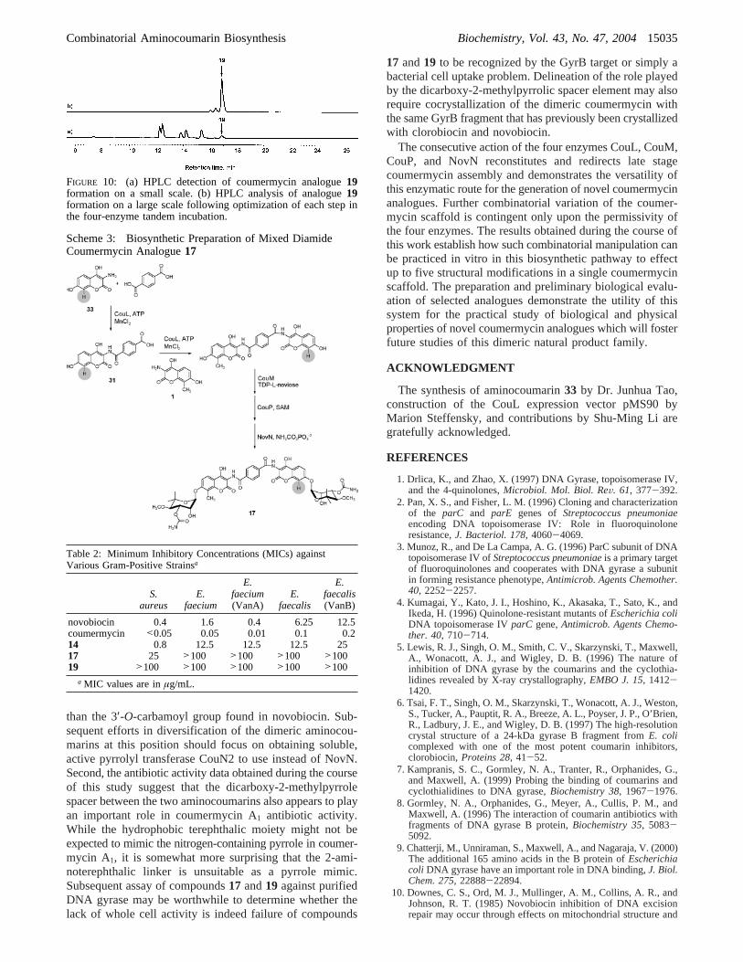
Large-Scale Enzymatic Preparation of CoumermycinAnalogues for Biological EValuation.As further demonstra-tion of the utility of the four-enzyme tandem incubation usingcoumermycin and novobiocin enzymes to produce novelcoumermycin variants, three analogues (14, 17, and 19,Figure 8) were selected for enzymatic synthesis on sufficientscale for biological evaluation. In each case, the individualenzymatic reactions were monitored by HPLC and LCMSto ensure complete conversion of substrates to productsbefore proceeding to the subsequent step. In some cases,complete conversion of substrate to product was onlyachievable by the repeated addition of enzyme or extendedincubation times. As shown in Figure 10 for compound19,satisfactory conversion to the final product was accomplishedusing this strategy, and little or no formation of intermediatesarising from incomplete ligation, methylation, or carbam-oylation was detected.
FIGURE 5: CouM-catalyzed glycosylation of monoamide7 (75 mMMES, pH 6, 10% DMSO, 1 mg/mL BSA).
15030 Biochemistry, Vol. 43, No. 47, 2004 Freel Meyers et al.
For the preparation of mixed diamide analogue17,monoamide31 was prepared (>95% by HPLC) by ligationof 8-demethylaminocoumarin33 to terephthalic acid (inexcess) in the presence of CouL (Scheme 3). Monoamide31 was purified by preparative HPLC following an acidicworkup, and the purified product was reacted with ami-nocoumarin1 (in excess) in the presence of CouL. Subse-quent tandem reaction with CouM, CouP, and NovN wascarried out as described above for the preparation ofanalogues14 and 19. In all cases, the final products wereconfirmed by LCMS prior to concentration by passagethrough a C18 SepPak and elution in DMSO. The crudeproducts were then purified by preparative reversed-phaseHPLC to yield14 (65% yield),17 (67% yield), and19 (68%yield). Compounds14, 17, and19 were tested for antibioticactivity, and the minimum inhibitory concentrations (MICs)are summarized in Table 2.
The biological activity data obtained here (Table 2) fornovobiocin and coumermycin A1 are in agreement with theliterature (31), suggesting that novobiocin is∼1 log lesspotent than coumermycin A1 againstS. aureusand 30-60-fold less potent than coumermycin A1 against the fourenterococcal strains. Compound14, which resembles coumer-mycin A1 but instead bears a carbamoyl group at the 3′-O-noviosyl position as in novobiocin, displays activity similarto novobiocin. This observation is consistent with the 3′-O-carbamoyl moiety as the prime pharmacophore on the
aminocoumarin scaffolds for presentation to the GyrBsubunit. Compounds17and19, bearing the terephthalic and2-aminoterephthalic spacer elements in the dimer, displayantibiotic potencies 1-2 logs lower than compound14 andare 3 logs less active than coumermycin A1 itself.
DISCUSSION
The aminocoumarin antibiotics novobiocin, clorobiocin,and coumermycin A1 (Figure 1), produced by various strainsof Streptomyces, use the 4′-O-methyl-3′-O-acyl- or 4′-O-methyl-3′-O-carbamoyl-L-noviosyl moiety as the pharma-cophore to inhibit ATP hydrolysis in the B subunits of DNAgyrase and topoisomerase IV. The decorated noviosylcomponent is presented to the ATP binding site by a two-part scaffold comprised of an aminocoumarin linked throughan amide bond to a prenylated hydroxybenzoic acid moietyin novobiocin and clorobiocin. X-ray crystallographic analy-ses of novobiocin and clorobiocin each bound to the ATPasedomain of GyrB illustrate that both the 3′-O-carbamoylsubstituent in novobiocin and the 3′-O-methylpyrrole carboxyester substituent of clorobiocin are positioned in the activesite (5, 6) and support the observed requirement of thesesubstituents for gyrase inhibitory activity and antibioticpotency. Consequently, it is presumed that the 3′-O-meth-ylpyrrole carboxy ester substituent on noviose in thepseudodimeric coumermycin A1 is also the competing ligandwhich blocks ATP binding. It is unknown whether the
FIGURE 6: (A) CouP-catalyzed bismethylation of CouM product4 to form bismethylation product11. (B) Reversed-phase HPLC stack plotshowing conversion of4 to 11 (75 mM Tris-HCl, pH 7.5, 10% DMSO, 1 mg/mL BSA). (C) Reaction profile of bismethylation by CouP.
Combinatorial Aminocoumarin Biosynthesis Biochemistry, Vol. 43, No. 47, 200415031
enhanced potency and gyrase inhibitory activity of coumer-mycin A1 are merely the concentration effect of twopharmacophores in one molecule or if there is somelikelihood of simultaneous occupancy of two gyrase Bsubunits in an A2B2 tetramer.
The implication that the late stage biosynthetic enzymesact twice in coumermycin biosynthesis makes coumermycinA1 an intriguing molecule to study. The prenyl hydroxyben-zoate element in novobiocin and clorobiocin is replaced bya 3-methyl-2,4-dicarboxypyrrole linker in coumermycin, andthe biosynthetic origin of this linking element is yet unknown.The ligase CouL catalyzes the double ligation of 3-amino-7-hydroxycoumarin with the 3-methyl-2,4-dicarboxylic acidlinker to create the corresponding diamide, the first step inelongation of the two halves of coumermycin A1 (26). Wehave demonstrated here that CouM and CouP, as well asthe novobiocin enzymes NovM and NovP, will indeedcatalyze double glycosylation and 4′-O-methylation in thesubsequent elongation steps. The accumulation of mono-noviosyl and monomethyl intermediates during glycosylationand methylation reactions catalyzed by CouM and CouP,respectively, suggests that these enzymes act distributively,not processively. It is not clear if such partially elongatedintermediates accumulate in the producing microorganisms,
but completion of double glycosylation and methylation canbe accomplished in vitro by treatment of substrates forextended periods of incubation with the purified enzymes.
Minimal antibiotic activity is observed in novobiocin andclorobiocin analogues lacking the 3′-O-substituents on thenoviose ring (25). Carbamoylation (novobiocin) and acylation(clorobiocin) are catalyzed by NovN (23) and CloN2 (24),respectively. Double acylation of the coumermycin scaffoldis presumably catalyzed by CouN2 (16), and a similarantibiotic potency profile is anticipated. The 5-methylpyrrolylmoiety at the 3′-position on the noviose ring in clorobiocinand coumermycin A1 is derived from proline by nonriboso-mal peptide synthetase module logic (15, 16). The pathwayis assumed to generate a pyrrolyl-S-peptidyl carrier proteinspecies that is most likely transferred to the CloP or CouPproduct by the action of acyltransferases CloN2 and CouN2,respectively. C-Methylation at the 5-position of the pyrrolylmoiety is thought to be the final step in both clorobiocinand coumermycin biosynthesis (24). Although the heterolo-gous overproduction of both CloN2 and CouN2 is possiblein E. coli, to date only insoluble protein is obtainable; thus,acyl transfer to the noviose ring in coumermycin A1
biosynthesis cannot be assayed with purified enzymes.Alternatively, we have evaluated the novobiocin carbam-
FIGURE 7: (A) NovN-catalyzed biscarbamoylation of CouP product11 to form biscarbamoylated coumermycin analogue14. (B) Reversed-phase HPLC stack plot showing conversion of11 to 14 (75 mM Tris-HCl, pH 7.5, 10% DMSO, 1 mg/mL BSA).
15032 Biochemistry, Vol. 43, No. 47, 2004 Freel Meyers et al.

oyltransferase NovN as a catalyst for biscarbamoylation ofthe CouL, CouM, CouP enzymatic product and haveobserved mono- and biscarbamoylation to create the biscar-bamoyl analogue14, an analogue previously obtained in lowyield in vivo (32). Furthermore, we were able to carry outthe tandem four-enzyme incubation and facilitate the com-plete conversion of intermediates in all eight enzymatic stepsto produce multimilligram quantities of14 for biologicalevaluation against the authentic natural product coumermycinA1 (Table 2).
The in vitro assembly of the coumermycin A1 scaffoldand alternate carbamoyl substitution at the 3′-position ofnoviose by NovN led to additional studies aimed at introduc-ing diversity into the planar scaffold of coumermycin A1.Heide and co-workers recently demonstrated that the ligaseCouL will accept alternative dicarboxylic acids as substrates(26). We have extended this study to include a few additionalcommercially available diacids, and the usage of these diacidsas substrates for CouL is summarized in Table 1. Of the sixaromatic diacids tested, terephthalic acid and 2-aminotereph-thalic acid underwent ligation to aminocoumarin1 in thepresence of CouL to form the corresponding mono- anddiamides. The resulting diamides were then shown toundergo glycosylation, methylation, and carbamoylation inthe six subsequent steps to generate coumermycin analogues
16 and19 (Figure 8) exemplifying modification of three ofthe five components in the coumermycin scaffold. Introduc-tion of the 2-aminoterephthalic acid moiety in analogue19significantly improves the aqueous solubility propertiesrelative to coumermycin A1 and coumermycin analogue16and could, in principle, be further modified at the 2-aminoposition. As for compound14, we are able to facilitate thecomplete conversion of intermediates in all eight enzymaticsteps to produce multimilligram quantities of19 for biologi-cal evaluation against the authentic natural product coumer-mycin A1 (Table 2).
Further study of the ligation by CouL revealed that amixture of aminocoumarin1 and 8-demethylaminocoumarin33 easily undergoes ligation in the presence of terephthalicacid or 2-aminoterephthalic acid to give the correspondingmixtures of diamides. The subsequent tandem action ofCouM, CouP, and NovN resulted in the formation of amixture of compounds16-18 and 19-22, which includeexamples of mixed diamide coumermycin analogues. Like-wise, treatment of synthetic monoamide7 with 8-demethyl-aminocoumarin33 in the presence of CouL afforded thecorresponding mixed diamide. Tandem action of CouM,CouP, and NovN provided coumermycin analogue15,illustrating the modification of three components in thecoumermycin scaffold. Compounds17, 20, and21 demon-strate structural modification to four of the five components
FIGURE 8: Coumermycin analogues generated via four-enzymeCouL, CouM, CouP, and NovN tandem incubation.
Table 1: Diacid Substrate Usage by CouL
Combinatorial Aminocoumarin Biosynthesis Biochemistry, Vol. 43, No. 47, 200415033
of the coumermycin scaffold while compounds18 and 22incorporate changes to all five components.
The rapid accumulation of monoamide31 relative todiamide32 in ligation reactions carried out using terephthalicacid and aminocoumarin1 (Figure 9) permitted the prepara-tive isolation of monoamide31. 8-Demethylaminocoumarin33 was utilized in a second preparative ligation step withCouL and monoamide31. The resulting mixed diamide CouLproduct could be carried through the subsequent six enzy-
matic steps of CouM, CouP, NovN to generate the coumer-mycin A1 variant 17 in multimilligram quantities forbiological evaluation (Table 2).
The initial antibiotic potencies obtained for compounds14, 17, and19on five Gram-positive pathogenic strains raisetwo issues. First, these data confirm the observation that the5-methylpyrrolylacyl moiety at the 3′-position of the noviosering is a key pharmacophore, whether displayed on cloro-biocin or coumermycin, and imparts∼1 log greater potency
FIGURE 9: (A) Nonoptimized small-scale generation of coumermycin analogue16 via four-enzyme CouL, CouM, CouP, and NovN tandemincubation. The HPLC traces show the product distribution for the four-enzyme reaction and for incubations each lacking one of theenzymes. (B) Reaction products and intermediates detected by LCMS.
15034 Biochemistry, Vol. 43, No. 47, 2004 Freel Meyers et al.
than the 3′-O-carbamoyl group found in novobiocin. Sub-sequent efforts in diversification of the dimeric aminocou-marins at this position should focus on obtaining soluble,active pyrrolyl transferase CouN2 to use instead of NovN.Second, the antibiotic activity data obtained during the courseof this study suggest that the dicarboxy-2-methylpyrrolespacer between the two aminocoumarins also appears to playan important role in coumermycin A1 antibiotic activity.While the hydrophobic terephthalic moiety might not beexpected to mimic the nitrogen-containing pyrrole in coumer-mycin A1, it is somewhat more surprising that the 2-ami-noterephthalic linker is unsuitable as a pyrrole mimic.Subsequent assay of compounds17 and19 against purifiedDNA gyrase may be worthwhile to determine whether thelack of whole cell activity is indeed failure of compounds
17 and19 to be recognized by the GyrB target or simply abacterial cell uptake problem. Delineation of the role playedby the dicarboxy-2-methylpyrrolic spacer element may alsorequire cocrystallization of the dimeric coumermycin withthe same GyrB fragment that has previously been crystallizedwith clorobiocin and novobiocin.
The consecutive action of the four enzymes CouL, CouM,CouP, and NovN reconstitutes and redirects late stagecoumermycin assembly and demonstrates the versatility ofthis enzymatic route for the generation of novel coumermycinanalogues. Further combinatorial variation of the coumer-mycin scaffold is contingent only upon the permissivity ofthe four enzymes. The results obtained during the course ofthis work establish how such combinatorial manipulation canbe practiced in vitro in this biosynthetic pathway to effectup to five structural modifications in a single coumermycinscaffold. The preparation and preliminary biological evalu-ation of selected analogues demonstrate the utility of thissystem for the practical study of biological and physicalproperties of novel coumermycin analogues which will fosterfuture studies of this dimeric natural product family.
ACKNOWLEDGMENT
The synthesis of aminocoumarin33 by Dr. Junhua Tao,construction of the CouL expression vector pMS90 byMarion Steffensky, and contributions by Shu-Ming Li aregratefully acknowledged.
REFERENCES
1. Drlica, K., and Zhao, X. (1997) DNA Gyrase, topoisomerase IV,and the 4-quinolones,Microbiol. Mol. Biol. ReV. 61, 377-392.
2. Pan, X. S., and Fisher, L. M. (1996) Cloning and characterizationof the parC and parE genes of Streptococcus pneumoniaeencoding DNA topoisomerase IV: Role in fluoroquinoloneresistance,J. Bacteriol. 178, 4060-4069.
3. Munoz, R., and De La Campa, A. G. (1996) ParC subunit of DNAtopoisomerase IV ofStreptococcus pneumoniaeis a primary targetof fluoroquinolones and cooperates with DNA gyrase a subunitin forming resistance phenotype,Antimicrob. Agents Chemother.40, 2252-2257.
4. Kumagai, Y., Kato, J. I., Hoshino, K., Akasaka, T., Sato, K., andIkeda, H. (1996) Quinolone-resistant mutants ofEscherichia coliDNA topoisomerase IVparC gene,Antimicrob. Agents Chemo-ther. 40, 710-714.
5. Lewis, R. J., Singh, O. M., Smith, C. V., Skarzynski, T., Maxwell,A., Wonacott, A. J., and Wigley, D. B. (1996) The nature ofinhibition of DNA gyrase by the coumarins and the cyclothia-lidines revealed by X-ray crystallography,EMBO J. 15, 1412-1420.
6. Tsai, F. T., Singh, O. M., Skarzynski, T., Wonacott, A. J., Weston,S., Tucker, A., Pauptit, R. A., Breeze, A. L., Poyser, J. P., O’Brien,R., Ladbury, J. E., and Wigley, D. B. (1997) The high-resolutioncrystal structure of a 24-kDa gyrase B fragment fromE. colicomplexed with one of the most potent coumarin inhibitors,clorobiocin,Proteins 28, 41-52.
7. Kampranis, S. C., Gormley, N. A., Tranter, R., Orphanides, G.,and Maxwell, A. (1999) Probing the binding of coumarins andcyclothialidines to DNA gyrase,Biochemistry 38, 1967-1976.
8. Gormley, N. A., Orphanides, G., Meyer, A., Cullis, P. M., andMaxwell, A. (1996) The interaction of coumarin antibiotics withfragments of DNA gyrase B protein,Biochemistry 35, 5083-5092.
9. Chatterji, M., Unniraman, S., Maxwell, A., and Nagaraja, V. (2000)The additional 165 amino acids in the B protein ofEscherichiacoli DNA gyrase have an important role in DNA binding,J. Biol.Chem. 275, 22888-22894.
10. Downes, C. S., Ord, M. J., Mullinger, A. M., Collins, A. R., andJohnson, R. T. (1985) Novobiocin inhibition of DNA excisionrepair may occur through effects on mitochondrial structure and
FIGURE 10: (a) HPLC detection of coumermycin analogue19formation on a small scale. (b) HPLC analysis of analogue19formation on a large scale following optimization of each step inthe four-enzyme tandem incubation.
Scheme 3: Biosynthetic Preparation of Mixed DiamideCoumermycin Analogue17
Table 2: Minimum Inhibitory Concentrations (MICs) againstVarious Gram-Positive Strainsa
S.aureus
E.faecium
E.faecium(VanA)
E.faecalis
E.faecalis(VanB)
novobiocin 0.4 1.6 0.4 6.25 12.5coumermycin <0.05 0.05 0.01 0.1 0.214 0.8 12.5 12.5 12.5 2517 25 >100 >100 >100 >10019 >100 >100 >100 >100 >100
a MIC values are inµg/mL.
Combinatorial Aminocoumarin Biosynthesis Biochemistry, Vol. 43, No. 47, 200415035
ATP metabolism, not on repair topoisomerases,Carcinogenesis6, 1343-1352.
11. Castora, F. J., Vissering, F. F., and Simpson, M. V. (1983) Theeffect of bacterial DNA gyrase inhibitors on DNA synthesis inmammalian mitochondria,Biochim. Biophys. Acta 740, 417-427.
12. Sung, S. C. (1974) Effect of novobiocin on DNA-dependent DNApolymerases from developing rat brain,Biochim. Biophys. Acta361, 115-117.
13. Martin, M. A. (1994) Methicillin-resistantStaphylococcus au-reus: The persistent resistant nosocomial pathogen,Curr. Clin.Top. Infect. Dis. 14, 170-191.
14. Steffensky, M., Muhlenweg, A., Wang, Z. X., Li, S. M., and Heide,L. (2000) Identification of the novobiocin biosynthetic gene clusterof Streptomyces spheroidesNCIB 11891, Antimicrob. AgentsChemother. 44, 1214-1222.
15. Pojer, F., Li, S. M., and Heide, L. (2002) Molecular cloning andsequence analysis of the clorobiocin biosynthetic gene cluster:New insights into the biosynthesis of aminocoumarin antibiotics,Microbiology 148, 3901-3911.
16. Wang, Z. X., Li, S. M., and Heide, L. (2000) Identification of thecoumermycin A(1) biosynthetic gene cluster ofStreptomycesrishiriensis DSM 40489, Antimicrob. Agents Chemother. 44,3040-3048.
17. Pojer, F., Kahlich, R., Kammerer, B., Li, S. M., and Heide, L.(2003) CloR, a bifunctional non-heme iron oxygenase involvedin clorobiocin biosynthesis,J. Biol. Chem. 278, 30661-30668.
18. Birch, A. J., Holloway, P. W., and Rickards, R. W. (1962) Thebiosynthesis of noviose, a branched-chain monosaccharide,Bio-chim. Biophys. Acta 57, 143-145.
19. Bunton, C. A., Kenner, G. W., Robinson, M. J. T., and Webster,B. R. (1963) Experiments related to the biosynthesis of novobiocinand other related coumarins,Tetrahedron 19, 1001-1010.
20. Kominek, L. A., and Sebek, O. K. (1974) Biosynthesis ofnovobiocin and related coumarin antibiotics,DeV. Ind. Microbiol.15, 60-69.
21. Steffensky, M., Li, S. M., and Heide, L. (2000) Cloning,overexpression, and purification of novobiocic acid synthetasefrom Streptomyces spheroidesNCIMB 11891,J. Biol. Chem. 275,21754-21760.
22. Freel Meyers, C. L., Oberthu¨r, M., Anderson, J. W., Kahne, D.,and Walsh, C. T. (2003) Initial characterization of novobiocic acidnoviosyl transferase activity of NovM in biosynthesis of theantibiotic novobiocin,Biochemistry 42, 4179-4189.
23. Freel Meyers, C. L., Oberthu¨r, M., Xu, H., Heide, L., Kahne, D.,and Walsh, C. T. (2004) Characterization of NovP and NovN:Completion of novobiocin biosynthesis by sequential tailoring ofthe noviosyl ring,Angew. Chem., Int. Ed. Engl. 43, 67-70.
24. Xu, H., Kahlich, R., Kammerer, B., Heide, L., and Li, S. M. (2003)CloN2, a novel acyltransferase involved in the attachment of thepyrrole-2-carboxyl moiety to the deoxysugar of clorobiocin,Microbiology 149, 2183-2191.
25. Hooper, D. C., Wolfson, J. S., McHugh, G. L., Winters, M. B.,and Swartz, M. N. (1982) Effects of novobiocin, coumermycinA1, clorobiocin, and their analogues onEscherichia coliDNAgyrase and bacterial growth,Antimicrob. Agents Chemother. 22,662-671.
26. Schmutz, E., Steffensky, M., Schmidt, J., Porzel, A., Li, S. M.,and Heide, L. (2003) An unusual amide synthetase (CouL) fromthe coumermycin A1 biosynthetic gene cluster fromStreptomycesrishiriensisDSM 40489,Eur. J. Biochem. 270, 4413-4419.
27. Tao, J., Hu, S., Pacholec, M., and Walsh, C. T. (2003) Synthesisof proposed oxidation-cyclization-methylation intermediates of thecoumarin antibiotic biosynthetic pathway,Org. Lett. 5, 3233-3236.
28. Hinman, J. W., Caron, E. L., and Hoeksema, H. (1957) Thestructure of novobiocin,J. Am. Chem. Soc. 79, 3789-3800.
29. Suzuki, M., Miyoshi, M., and Matsumoto, K. (1974) Synthesis ofamino acids and related compounds. 7. Convenient synthesis of3-substituted pyrrole-2,4-dicarboxylic acid esters,J. Org. Chem.39, 1980.
30. Corwin, A. H., and Viohl, P. (1944) The condensation of pyrroleswith bromine. A self-oxidation and a new type of displacementreaction,J. Am. Chem. Soc. 66, 1137-1146.
31. Gellert, M., O’Dea, M. H., Itoh, T., and Tomizawa, J. (1976)Novobiocin and coumermycin inhibit DNA supercoiling catalyzedby DNA gyrase,Proc. Natl. Acad. Sci. U.S.A. 73, 4474-4478.
32. Xu, H., Wang, Z. X., Schmidt, J., Heide, L., and Li, S. M. (2002)Genetic analysis of the biosynthesis of the pyrrole and carbamoylmoieties of coumermycin A1 and novobiocin,Mol. Genet.Genomics 268, 387-396.
33. NCCLS (1997)Methods for Dilution Antimicrobial SusceptibilityTests for Bacteria that Grow Aerobically(approved standard,NCCLS Document M7-A4) 4th ed., National Committee forClinical Laboratory Standards, Wayne, PA.
BI048457Z
15036 Biochemistry, Vol. 43, No. 47, 2004 Freel Meyers et al.


![Designing Dimeric Lanthanide(III)-Containing Ionic liquids › ws › files › 158240242 › ...COMMUNICATION Designing Dimeric Lanthanide(III)-Containing Ionic liquids Éadaoin McCourt,[a]](https://img.pdfslide.net/doc/110x75/60b904bbc8cfbf6cfb110109/designing-dimeric-lanthanideiii-containing-ionic-liquids-a-ws-a-files-a.jpg)