Embed Size (px)
Citation preview

1
Assess Patient’s Overall Cardiopulmonary Status by Palpation, Percussion, and Auscultation Robert Harwood, MSA, RRT-NPS
Objectives At the end of this presentation the student should be able to:
• Assess patient’s overall cardiopulmonary status by palpation
• Assess patient’s overall cardiopulmonary status by percussion
• Assess patient’s overall cardiopulmonary status by auscultation
2
Palpation • Pulse
Heart rate−normal vs. tachycardia, bradycardia Rhythm − regular vs. irregular due to arrhythmia Strength – weak, thready or bounding
• Pulsus alternans −arterial pulse waveform showing alternating strong and weak beats, left ventricular systolic impairment
• Pulsus paradoxus − an exaggeration (more than 10 torr) of the normal variation during the inspiratory phase of respiration, in which the blood pressure declines as one inhales and increases as one exhales. Cause: obstructive lung disease (e.g. asthma, COPD).
• Common sites: radial pulse but also precordium, carotid, brachial, femoral. • Best site for non-emergency measurement of pulse: radial.
Palpation • Point of Maximal Impulse (PMI) or Apex Beat
Apex beat can be palpated in the precordium left 5th intercostal space, at the point of intersection with the left midclavicular line Lateral and/or inferior displacement
• Cardiomegaly • Pleural or pulmonary diseases • Deformities of the chest wall or the thoracic vertebra
A forceful impulse – hypertension • Tracheal Position
Determine shifting from atelectasis, pneumothorax, pleural effusion, massive hemothorax

3
Palpation • Vocal Frenitus
Vibrations created by vocal cords during phonation Felt through palms of hands-tactile fremitus Transmitted down tracheobronchial tree = alveoli = chest wall
Assess by having patient say “ninety-nine” Increased:
• Consolidation from pneumonia, tumor, atelectasis Decreased:
• Unilateral: Bronchial obstruction, pneumothorax, pleural effusion • Diffuse: COPD (hyperinflated lung), obese chest wall
• Secretion fremitus Vibrations produced by passage of air through thick secretions. May clear after cough When felt on chest wall: tactile fremitus
Palpation Normal chest wall expands symmetrically during deep inspiration • Abnormal expansion
Bilateral: COPD, neuromuscular disease Unilateral: lobar consolidation, atelectasis, pleural effusion, pneumothorax
• Subcutaneous emphysema Cause: positive pressure ventilation Air pockets palpable on skin when air leak from the lung is present Palpation crepitus or crackling feeling
• Tenderness in abdominal area Enlarged liver, hepatomegaly Problems with breathing due to restriction of diaphragm
4
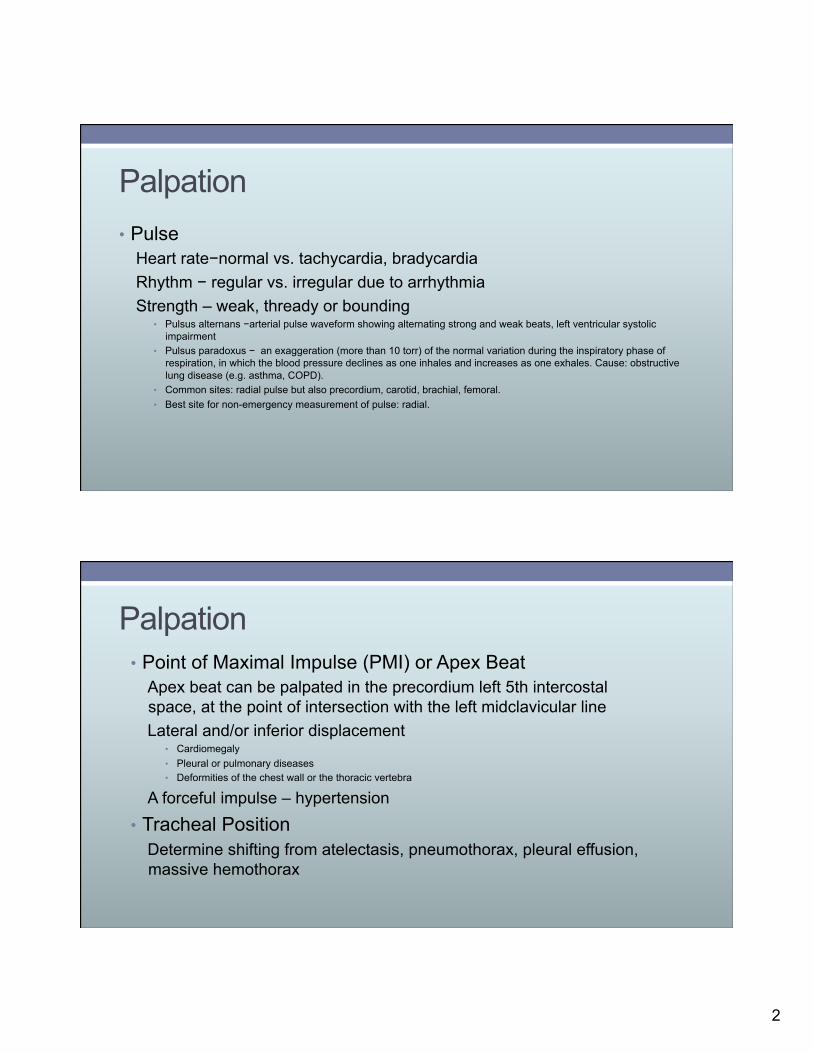
Percussion Percussion determines whether the underlying tissues are filled with air, fluid, or solid material • Sounds
Normal-resonant sound is low pitched, hollow Flat or dull- heard over bones, fluid from pleural effusion Hyperresonant-louder and low pitched-hyperinflated: COPD Tympanic-hollow and high pitched from excessive air-pneumothorax Diaphragm position-normal to dull sound. Can detect excursion by percussing during inspiration then expiration
Normal excursion is 5-7 cm in adults
Sample Question A 67-year-old patient is in the ED complaining of pain and shortness of breath following a coughing episode. Percussion of the chest reveals a tympanic sound emitted from the left lung and a resonant sound emitted from the right lung. Which of the following is present based on these findings? a. Left-sided pneumothorax b. Right-sided pleurisy c. Right-sided pleural effusion d. Left-sided atelectasis

5
Sample Question Answer a. Correct: Left-sided pneumothorax-tympanic percussion sound emitted from the left lung would be most
associated with pneumothorax. Also, the patient experienced distress following a cough that had pain, this would also be suggestive of a pneumothorax.
Auscultation-Breath Sounds
Normal Breath Sounds
Tracheal and Bronchial Over trachea and mainstem bronchi
Bronchovesicular Between scapulae and sternum (upper part)
Vesicular Peripheral lung

6
Auscultation – Breath Sounds • Descriptors of Breath Sounds
• Diminished or reduced-decreased intensity-atelectasis, pneumothorax • Absent-no breath sounds • Harsh-inspiratory and expiratory component equal similar to bronchial
but not in the bronchial area-inflammation • Adventitious-abnormal breath sounds
• Wheeze - continuous high-pitched sounds from the chest-asthma • Stridor - high pitched sound that may be heard without the aid of a
stethoscope from the neck area and upper airway area- obstruction upper airway
• Pleural friction rub-loud, harsh continuous sound located midaxillary area of lung. Complaint of pain during breathing
Breath Sounds−Discontinuous
Early Crackles
COPD
Chronic Bronchitis
Emphysema
Asthma

7
Breath Sounds − Discontinuous
Late Crackles
Pulmonary Edema
Pneumonia Atelectasis
Auscultation − Heart Sounds • S1
First heart sound − closure of mitral and tricuspid valves-systole • S2
Second heart sound – closing of pulmonary and aortic valves-end of systole Splitting of S2: normal during breathing and increased during inspiration
• S3 Third heart sound – heard during diastole from rapid filling of ventricles following systole
• S4 Fourth heart sound − occurs just before S1. Produce in the same way as S3
May indicate heart problems

8
Auscultation −Heart Sounds
Abnormal heart sounds
Gallop Rhythm Heart Disease
Loud P2
Pulmonary Hypertension
Murmur Incompetent Heart
Valve/Stenosis
Auscultation − Voice Sounds • Bronchophony
• Ask the patient to repeat a word several times while auscultating symmetrical areas of each lung. The number “99" is commonly used
• Normal breath sounds-muffled “99” • Pneumonia causes consolidation-clear “99” • Increased lung tissue density • Bronchophony-increase in intensity or clarity of vocal resonance due to
increased lung tissue density • Associated with:
Bronchial breath sounds, increased vocal fremitus and dull percussion note

9
Auscultation − Voice Sounds • Egophony-spoken voice increases in intensity
• Normal lung will have the “E” sound • Egophony will have an “A” sound over area of lung that has
consolidation • Also compressed lung from pleural effusion • Chart this as “E to A” sound indicating consolidation and identify where
it is located • Whispered Pectoriloquy
• Patient whispers 1-2-3 • Normal sound-muffled low-pitched and unable to identify the 1-2-3 • Consolidation-high pitched sounds over affected area; sound of 1-2-3
much clearer
Auscultation − Voice Sounds • Patient whispers 1-2-3 • Normal sound-muffled low-pitched and unable to identify the
1-2-3 • Consolidation-high pitched sounds over affected area; sound
of 1-2-3 much clearer

10
•
Blood Pressure
Overview of Physical Exam Findings ABNORMALITY INSPECTION AUSCULATION PALPATION PERCUSSION
Tension Pneumothorax Progressive cyanosis, distress, accessory muscle use
Distant breath sounds Tracheal displacement away from affected side
Hollow, high pitched
Pleural Effusion Distress, accessory muscle use, asymmetrical chest expansion
Distant to absent breath sounds “E to A” voice sound in lung above effusion
Reduced expansion on affected side
Flat to dull
Atelectasis (pneumonia) Respiratory distress, accessory muscle use
Audible wheezes/ crackles. Clear “99” sound “E to A” voice sound
Asymmetrical unilateral expansion or bilateral expansion. Increased vocal fremitus
Dull
COPD Accessory muscles inspiration/ expiration, tachypnea, tripod position
Early crackles, expiratory wheezing, Muffled “99” voice sound
Decreased vocal fremitus Increased resonance to hyperresonant

11
Overview of Physical Exam Findings
ABNORMALITY INSPECTION AUSCULATION PALPATION PERCUSSION
Flail Chest Distress, accessory muscle use, paradoxical respirations
Absent or distant breath sounds
Crepitus on affected side Dullness
Asthma Accessory muscle use, tachypnea
Wheezing, early crackles
Symmetrical chest movement
Increased resonance
Hemothorax Respiratory distress, accessory muscle use, paradoxical respirations
Absent or distant breath sounds
Crepitus, tracheal shift away from affected side
Dullness
Cardiac Tamponade Accessory muscles use, JVD
Late crackles Paradoxical pulse Resonant
Sample Question While assessing a patient admitted to ICU for status asthmaticus, you notice on the heart monitor the patient’s blood pressure drops during inspiration and increases during expiration. This is referred to as: a. Pulsus alternans b. Pulsus bigeminus c. Pulsus paradoxus d. Pulsus bisferiens

12
Sample Question Answer c. Correct: pulsus paradoxus. a. Incorrect: pulsus alternans − arterial pulse waveform
showing alternating strong and weak beats left ventricular systolic impairment
b. Incorrect: pulsus bigeminus − groups of two heartbeats close together followed by a longer pause
d. Incorrect: pulsus bisferiens − a double peak per one cardiac cycle
Sample Question
A 76-year-old male has been admitted to the ICU with a diagnosis of pulmonary edema from CHF. Which of the following breath sounds would you expect to hear during auscultation? a. Early crackles b. Stridor c. Bronchial d. Late crackles

13
Sample Question Answer
d. Correct: Late crackles − also with pneumonia and atelectasis
Sample Question
Upon reviewing a patient’s chart prior to administering therapy, you notice an entry that states that the patient has a loud P2 heart sound. This would indicate the presence of which of the following? a. Mitral valve stenosis b. Aortic valve stenosis c. Tricuspid valve stenosis d. Pulmonary hypertension

14
Sample Question Answer
d. Correct: Pulmonary hypertension
Sample Question
During physical examination using palpation, an abnormal bilateral expansion of the chest wall is assessed. This would indicate the presence of which of the following? a. Right-sided atelectasis b. Left-sided pleural effusion c. Right-sided lobar consolidation d. COPD

15
Sample Question Answer
d. Correct: COPD - from reduced bilateral expansion
Sample Question Which of the following physical assessment findings would be associated with consolidation from pneumonia? I. Bronchial breath sounds II. Dull percussion note III. Muffled “99” voice sound IV. Increased vocal fremitus
a. I, II only b. II, IV only c. I, II, IV only d. I, II, III, IV

16
Sample Question Answer
c. Correct: Bronchial breath sounds, dull percussion note, increased vocal fremitus
References Clinical Assessment in Respiratory Care, Sixth Edition, Robert L. Wilkins,
James R. Dexter, Albert J. Heuer, (2009), The Mosby/Elsevier Company, St. Louis, MO.
Egan’s Fundamentals of Respiratory Care, Ninth Edition, Robert L. Wilkins, James K. Stoller, Robert M. Kacmarek, (2009), Mosby/Elsevier Company, St. Louis, MO.
Respiratory Care Principles and Practice, Second Edition, Dean R. Hess, Neil R. MacIntyre, Shelley C. Mishoe, William F. Galvin, Alexander B. Adams, (2011), Jones and Bartlett Learning, Sudbury, MA.
The Essentials of Respiratory Care. Robert M. Kacmarek, Steven Dimas, Craig W. Mack (2005). Fourth Edition. Mosby/Elsevier, St. Louis, MO.