Embed Size (px)
Citation preview
Assessing the Nature of Lipid Raft MembranesPerttu S. Niemela1,2*, Samuli Ollila1,2, Marja T. Hyvonen1,2,3, Mikko Karttunen4, Ilpo Vattulainen1,5,6,7
1 Laboratory of Physics, Helsinki University of Technology, Helsinki, Finland, 2 Helsinki Institute of Physics, Helsinki University of Technology, Helsinki, Finland, 3 Wihuri
Research Institute, Helsinki, Finland, 4 Department of Applied Mathematics, The University of Western Ontario, London, Ontario, Canada, 5 Memphys Center for
Biomembrane Physics, University of Southern Denmark, Odense, Denmark, 6 Physics Department, University of Southern Denmark, Odense, Denmark, 7 Institute of Physics,
Tampere University of Technology, Tampere, Finland
The paradigm of biological membranes has recently gone through a major update. Instead of being fluid andhomogeneous, recent studies suggest that membranes are characterized by transient domains with varying fluidity. Inparticular, a number of experimental studies have revealed the existence of highly ordered lateral domains rich insphingomyelin and cholesterol (CHOL). These domains, called functional lipid rafts, have been suggested to take partin a variety of dynamic cellular processes such as membrane trafficking, signal transduction, and regulation of theactivity of membrane proteins. However, despite the proposed importance of these domains, their properties, andeven the precise nature of the lipid phases, have remained open issues mainly because the associated short time andlength scales have posed a major challenge to experiments. In this work, we employ extensive atom-scale simulationsto elucidate the properties of ternary raft mixtures with CHOL, palmitoylsphingomyelin (PSM), and palmitoyloleoyl-phosphatidylcholine. We simulate two bilayers of 1,024 lipids for 100 ns in the liquid-ordered phase and one system ofthe same size in the liquid-disordered phase. The studies provide evidence that the presence of PSM and CHOL in raft-like membranes leads to strongly packed and rigid bilayers. We also find that the simulated raft bilayers arecharacterized by nanoscale lateral heterogeneity, though the slow lateral diffusion renders the interpretation of theobserved lateral heterogeneity more difficult. The findings reveal aspects of the role of favored (specific) lipid–lipidinteractions within rafts and clarify the prominent role of CHOL in altering the properties of the membrane locally in itsneighborhood. Also, we show that the presence of PSM and CHOL in rafts leads to intriguing lateral pressure profilesthat are distinctly different from corresponding profiles in nonraft-like membranes. The results propose that thefunctioning of certain classes of membrane proteins is regulated by changes in the lateral pressure profile, which canbe altered by a change in lipid content.
Citation: Niemela PS, Ollila S, Hyvonen MT, Karttunen M, Vattulainen I (2007) Assessing the nature of lipid raft membranes. PLoS Comput Biol 3(2): e34. doi:10.1371/journal.pcbi.0030034
Introduction
The understanding of lipid membrane structures and theirrole in cellular functions has developed significantly since theintroduction of the classical fluid-mosaic model by Singerand Nicolson [1]. The fluid-mosaic model predicted thatcellular membranes are fluid and characterized by randomdistribution of molecular components in the membrane,resulting in lateral and rotational freedom. The more recentpicture is considerably more elaborate, however. A largenumber of experimental results converge toward the ideathat lateral domains enriched in sphingomyelin (SM) andcholesterol (CHOL) exist in biological membranes. Thesenanosized domains, called functional lipid rafts, have beensuggested to take part in various dynamic cellular processessuch as membrane trafficking, signal transduction, andregulation of the activity of membrane proteins [2–4]. Theexistence of stable lipid rafts in biological membranes isunder intense scrutiny, and their existence is actually underdebate since the lipid rafts, if they do exist, are probably toosmall to be resolved by techniques such as fluorescencemicroscopy [5]. Direct evidence of rafts in vivo is mainlybased on monitoring the motions of membrane proteins [6]or on differential partitioning of fluorescent probes inmembrane environments [7]. It is, however, difficult toperform experiments using living cells, which complicatesmeasurements of physical quantities of the rafts, such as theexact lipid composition, characteristic size, and lifetime [8,9].In model membranes, the coexistence of domains in the
liquid ordered (lo) and the liquid disordered (ld) phase iswidely accepted [9,10]. For example, the ld phase may beformed by an unsaturated phosphatidylcholine (PC), whilethe formation of the lo phase is promoted by a mixture of SMand CHOL. As for rafts, the current understanding of lipidrafts in biological membranes suggests a granular structure ofnanometer-scale domains of various compositions [9,11,12]rather than a large-scale phase separation.The exact nature of the underlying interactions that lead to
lipid immiscibilities in membranes is under debate [13,14].CHOL is particularly important as it has been shown toincrease the conformational order of acyl chains and reducethe bilayer area, hence significantly increasing the packingdensity of the lipids [15–17]. CHOL is particularly effective in
Editor: Amy Rowat, Harvard University, United States of America
Received July 14, 2006; Accepted January 5, 2007; Published February 23, 2007
A previous version of this article appeared as an Early Online Release on January 5,2007 (doi:10.1371/journal.pcbi.0030034.eor).
Copyright: � 2007 Niemela et al. This is an open-access article distributed underthe terms of the Creative Commons Attribution License, which permits unrestricteduse, distribution, and reproduction in any medium, provided the original authorand source are credited.
Abbreviations: A, average area per lipid; CHOL, cholesterol; d, bilayer thickness; D,lateral diffusion coefficient; KA, area compressibility modulus; kBT, thermal energyscale (kB is the Boltzmann constant, T is the absolute temperature); kc, bendingrigidity; ld, liquid disordered phase; lo, liquid ordered phase; PC, phosphatidylcho-line; POPC, palmitoyloleoylphosphatidylcholine; PSM, palmitoylsphingomyelin; SCD,deuterium order parameter values; SM, sphingomyelin
* To whom correspondence should be addressed. E-mail: [email protected]
PLoS Computational Biology | www.ploscompbiol.org February 2007 | Volume 3 | Issue 2 | e340304
reducing the void space within the acyl chain region of thelipids [15], which is related to suppressed area compressibilityand increased bending rigidity of the membrane withincreasing CHOL concentrations. However, the lateraldiffusion rates are not expected to slow down by more thana factor of 2–3 when the ld phase is compared with CHOL-induced lo phase [6,18]. Also, CHOL has recently beenreported to significantly alter the lateral pressure profile ofmembranes [19]. This is important, as changes in the lateralpressure profiles have been suggested to be related to changesin membrane protein structure and activity [20].
Considering that the smallest estimates for the sizes of raftsfall in the range of nanometers [21,22], they make anaccessible subject for computational studies. Though, in spiteof the considerable importance of rafts, it is somewhatsurprising that only a few atom-scale simulations have dealtwith ternary mixtures of CHOL, SM, and PC [23,24],concentrating mainly on small-scale structural propertiesand local interactions between the lipids. In particular, thereare no previous atom-level computational studies of raftsaiming to characterize the nature of their structural anddynamical features. For example, the nanometer scalestructure within raft domains and its interplay with CHOL-induced effects are not understood. Further, the resultinglarge-scale properties, such as membrane elasticity in ternaryraft-like lipid mixtures, are not understood either. Finally,and perhaps most importantly, the lateral pressure profilesassociated with rafts are completely unknown. The concept ofthe lateral pressure profile across the lipid membrane isexceptionally significant, since it describes the pressureexerted on molecules embedded in a membrane. Cantor hasproposed that incorporation of molecules into membraneand changes in lipid content would alter the lateral pressure
profile across a membrane, and hence changes in the pressureprofile would induce changes in membrane protein structure[20,25]. Experimental studies of this issue are remarkablydifficult, however: currently there is only one study thatemployed fluorescent probes to gauge the overall shape of thelateral pressure profile [26]. Evidently, detailed atomisticsimulations are called for.The state-of-the-art extent of the simulations conducted in
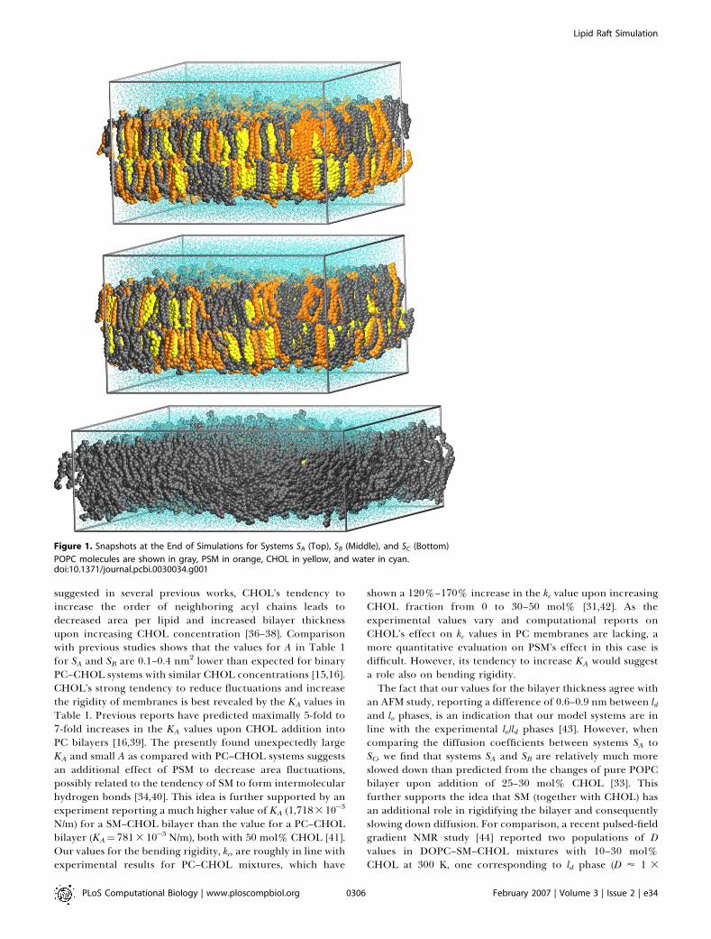
this work, 15–20 nm in lateral dimensions and 100 ns in time,enables a reliable quantitative analysis of the properties ofraft-like membranes not accomplished before. We employlarge-scale atom level simulations for three mixtures ofpalmitoyloleoylphosphatidylcholine (POPC), PSM, andCHOL. The molar fractions are POPC:PSM:CHOL ¼ 1:1:1,2:1:1, and 62:1:1 for systems that we call SA, SB, and SC,respectively (see Figure 1). Based on a recent experimentalphase diagram [27], these mixtures are expected to displaythe coexistent lo and ld phase domains (SA and SB) or a single ldphase (SC). Here, we illustrate the distinct nature of raft-likedomains in three parts. First, we consider the elastic,thermodynamic, and dynamic properties of rafts that turnout to be very different from those of nonraft-like mem-branes. Second, we provide evidence that the presence ofPSM and CHOL in raft-like membranes leads to stronglypacked and rigid bilayers, characterized by significant nano-scale lateral heterogeneity within the raft domains. Thesefindings express the prominent role of favored lipid–lipidinteractions within rafts and highlight the significant role ofCHOL in promoting the formation of rafts. Third, we providecompelling evidence that the lateral pressure profiles can bealtered by a change in lipid content. In particular, we showhow the presence of PSM and CHOL leads to intriguinglateral pressure profiles that are distinctly different fromcorresponding lateral pressure profiles in nonraft-like mem-branes, proposing that lipid membranes may regulate thefunctioning of certain classes of membrane proteins such asmechanosensitive channels through changes in lipid compo-sition, and hence the lateral pressure profile.
Results
Elastic, Thermodynamic, and Dynamic PropertiesSelected properties of the simulated membranes are
summarized in Table 1. For system SC, the average area perlipid, A, and the bilayer thickness, d, are in agreement withprevious findings on pure POPC bilayers [28,29], indicatingnegligible effects of PSM and CHOL on the bilayerdimensions. Also, the area compressibility modulus, KA, andthe bending rigidity, kc, are in line with previous studies ofpure PC bilayers, reporting KA¼ 140–3003 10�3 N/m and kc¼4–9 3 10�20 J [30–32]. The lateral diffusion coefficient, D, forPOPC in system SC is about 50% lower than the value of 1.43
10�7 cm2/s measured for pure POPC bilayer at 313 K [33]. Asimilar trend was found in comparison of our previoussimulations on pure SM and PC bilayers [34] with thisparticular study [33]. This suggests that bilayer SC is close tothe liquid disordered state of a POPC bilayer. This is alsosupported by the finding that small CHOL [33] or SM [35]concentrations have minor effects on D values of PC abovemelting temperatures.The condensing effect of CHOL becomes evident when
comparing the values of A and d between systems SA to SC. As
PLoS Computational Biology | www.ploscompbiol.org February 2007 | Volume 3 | Issue 2 | e340305
Author Summary
Biological membranes are complex 2-D assemblies of various lipidspecies and membrane proteins. For long, it was thought that themain role of lipid membranes is to provide a homogeneous, liquid-like platform for membrane proteins to carry out their functions asthey diffuse freely in the membrane plane. Recently, that view haschanged. It has become evident that several lipid environments withdifferent physical properties may coexist, and that the properties ofthe different lipid domains may play an active role in regulating theconformational state and dynamic sorting of membrane proteins.We have carried out atom-scale computer simulations for three-component lipid bilayers, so-called lipid rafts, rich in cholesterol andsphingolipids. They show that arising from the local interactionsbetween the lipid species, the elastic and dynamic properties of themembranes depend strongly on the lipid composition. The changesin elastic properties are suggested to alter the functional states ofvarious membrane proteins. Changes in lipid composition are alsoshown to alter the distribution of local pressure inside themembrane. This is likely to affect proteins that undergo largeanisotropic conformational changes between the functional states,such as the ion channel MscL, used as an example here. A greatnumber of important physiological phenomena, such as trans-mitting neural impulses or trafficking molecules in and out of thecell, involve activation of membrane proteins, so it is relevant tounderstand all factors affecting them. Our findings support the ideathat general physical properties of the lipid environment are capableof regulating membrane proteins.
Lipid Raft Simulation
suggested in several previous works, CHOL’s tendency toincrease the order of neighboring acyl chains leads todecreased area per lipid and increased bilayer thicknessupon increasing CHOL concentration [36–38]. Comparisonwith previous studies shows that the values for A in Table 1for SA and SB are 0.1–0.4 nm2 lower than expected for binaryPC–CHOL systems with similar CHOL concentrations [15,16].CHOL’s strong tendency to reduce fluctuations and increasethe rigidity of membranes is best revealed by the KA values inTable 1. Previous reports have predicted maximally 5-fold to7-fold increases in the KA values upon CHOL addition intoPC bilayers [16,39]. The presently found unexpectedly largeKA and small A as compared with PC–CHOL systems suggestsan additional effect of PSM to decrease area fluctuations,possibly related to the tendency of SM to form intermolecularhydrogen bonds [34,40]. This idea is further supported by anexperiment reporting a much higher value of KA (1,718310�3
N/m) for a SM–CHOL bilayer than the value for a PC–CHOLbilayer (KA¼ 7813 10�3 N/m), both with 50 mol% CHOL [41].Our values for the bending rigidity, kc, are roughly in line withexperimental results for PC–CHOL mixtures, which have
shown a 120%–170% increase in the kc value upon increasingCHOL fraction from 0 to 30–50 mol% [31,42]. As theexperimental values vary and computational reports onCHOL’s effect on kc values in PC membranes are lacking, amore quantitative evaluation on PSM’s effect in this case isdifficult. However, its tendency to increase KA would suggesta role also on bending rigidity.The fact that our values for the bilayer thickness agree with
an AFM study, reporting a difference of 0.6–0.9 nm between ldand lo phases, is an indication that our model systems are inline with the experimental lo/ld phases [43]. However, whencomparing the diffusion coefficients between systems SA toSC, we find that systems SA and SB are relatively much moreslowed down than predicted from the changes of pure POPCbilayer upon addition of 25–30 mol% CHOL [33]. Thisfurther supports the idea that SM (together with CHOL) hasan additional role in rigidifying the bilayer and consequentlyslowing down diffusion. For comparison, a recent pulsed-fieldgradient NMR study [44] reported two populations of Dvalues in DOPC–SM–CHOL mixtures with 10–30 mol%CHOL at 300 K, one corresponding to ld phase (D ’ 1 3
Figure 1. Snapshots at the End of Simulations for Systems SA (Top), SB (Middle), and SC (Bottom)
POPC molecules are shown in gray, PSM in orange, CHOL in yellow, and water in cyan.doi:10.1371/journal.pcbi.0030034.g001
PLoS Computational Biology | www.ploscompbiol.org February 2007 | Volume 3 | Issue 2 | e340306
Lipid Raft Simulation
10�7 cm2/s) and the other to lo phase (D ’ 1 3 10�8 cm2/s). Asthe exact lipid composition within the proposed domains isunknown, our simulated D values for systems SA and SB are ingood agreement with the proposed lo phase. This is interest-ing, since the lo phase is usually characterized as havingsimilar diffusion rates with the ld phase. Recent evidence onlarge variations in the properties of a single lo phase [45] alsosupports the idea that bilayers SA and SB do display the lophase. Clearly, diffusion within raft domains is stronglysuppressed due to the presence of PSM and CHOL.
The material properties of lipid bilayers have beensuggested to play a major role in regulating the activity andpartitioning of membrane proteins. First, the thicknessdifference of raft and nonraft membranes may be relevantdue to the effects of hydrophobic matching [46,47]. Forexample, the free energy of opening of a bacterial stretch-activated channel has been observed to change from 4 to 20kBT when the acyl chain length of the surrounding PC-lipidschanges from 16 to 20 carbons [48]. Another example is thetransmembrane protein OmpA, whose free energy ofunfolding was reported to change by about 5 kBT per nmwhen the hydrophobic thickness of the surrounding saturatedPC-membrane was varied [49]. Using this value as a simplisticestimate for the effect of hydrophobic thickness, one gets adifference of about 4 kBT in the free energy of unfoldingwhen this particular protein would be transferred fromnonraft to raft membrane. As the higher bending rigidity ofthe raft membrane probably decreases the ability of themembrane to adapt its thickness to match the hydrophobicthickness of the protein, the actual value should be largerthan the above estimate. The role of membrane elasticity inprotein functionality is further emphasized by the fact that,based on recent studies, it costs much more energy to deforma membrane by changing its area per lipid than by bending orchain tilting [50]. It has been suggested that the free energy tocreate a protein-shaped cavity in a bilayer is proportional toKA [51], and evidence exists that the binding free energy ofcertain amphipathic peptides indeed depends linearly on KA
[52]. Our data suggests a 5-fold to 14-fold difference in thevalues of KA between raft and nonraft membranes (see Table1), which practically means a free energy cost of about 4–8kBT when a membrane protein (Mellitin) is transferred from anonraft to a raft environment [52]. Summarizing, theelasticity of raft-like membranes is substantially different
from that of nonraft membranes, and this likely influencesmembrane protein functionality.
Lateral HeterogeneityThe above results highlight the different bulk properties of
raft-like domains with respect to more disordered bilayers.However, as becomes evident below, raft domains are alsocharacterized by strong spatial and temporal variations.Figure 2 reveals lateral heterogeneity in the calculateddeuterium-order parameter values (SCD) when averaged over10 ns. The nature of chain ordering varies in differentsystems. System SA exhibits the highest overall order (averageSCD ¼ �0.41) that is almost uniformly distributed over thebilayer plane and broken only by a few small low-order areasand empty points due to poor sampling. System SB is slightlyless ordered (SCD ¼ �0.36) and contains domains of a fewnanometers in size, differing significantly in their SCD values.The overall ordering in SC is much weaker (SCD¼�0.18) thanin the two other systems, but even SC displays lateralheterogeneity, though the domains appear larger, smoother,and with smaller variations in the SCD values. The average SCDvalues are in line with corresponding experimental orderparameter profiles of fluid POPC [53,54] and DPPC–CHOLmixtures with similar CHOL concentrations [45,55].In Figure 2, the more ordered regions in SCD plots are
clearly correlated with a higher density of CHOL. This is inline with a previous study showing CHOL’s ability to orderthe neighboring acyl chains within a radius of few nano-meters [56]. The rchain plots in Figure 2 reveal highlocalization of the chains in SA, whereas in SB some of theregions are smeared out. The SC plot is much morehomogeneous, indicating higher overall mobility and moreisotropic distribution of the chains. In SC, the smallconcentration of CHOL does not seem sufficient to accountfor the observed large-scale lateral heterogeneity in chain-order parameters. Instead, we find that the SCD value is clearlycorrelated with bilayer thickness. This is particularly sup-ported by the fact that the amplitudes of the large-scaleperistaltic wave modes are significantly larger for system SCthan for the other systems (see Figure S6). Even though theautocorrelation functions for most of the largest undulationsand peristaltic modes decay roughly within a few nano-seconds (unpublished data), some modes display much longerdecay times. In particular for system SC, this may be related tothe heterogeneity induced by the few CHOL and SMmolecules that are embedded in the bilayer.To judge our findings for lateral heterogeneity, it is
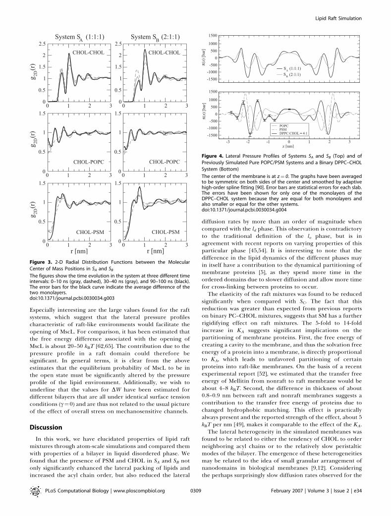
worthwhile to stress the slow dynamics in the bilayer plane:despite the extensive time scale simulated, the lateraldiffusion coefficients indicate that the molecules move inthe plane of the membrane approximately over only theirown size within the simulated time scale. Hence, it is evidentthat the simulation time is not long enough to adequatelyrelax the large-scale structure of the initial configuration andlead to complete mixing of the lipids. The nanoscaleheterogeneity observed in this work could thus be debated.However, there is reason to emphasize that while systems SAand SB were started from different initial configurations, theylead to similar conclusions. Further, the small-scale move-ments of the molecules relative to each other can becharacterized; see the 2-D radial distribution functions inFigure 3. The unfavorable close contacts of CHOL–CHOL
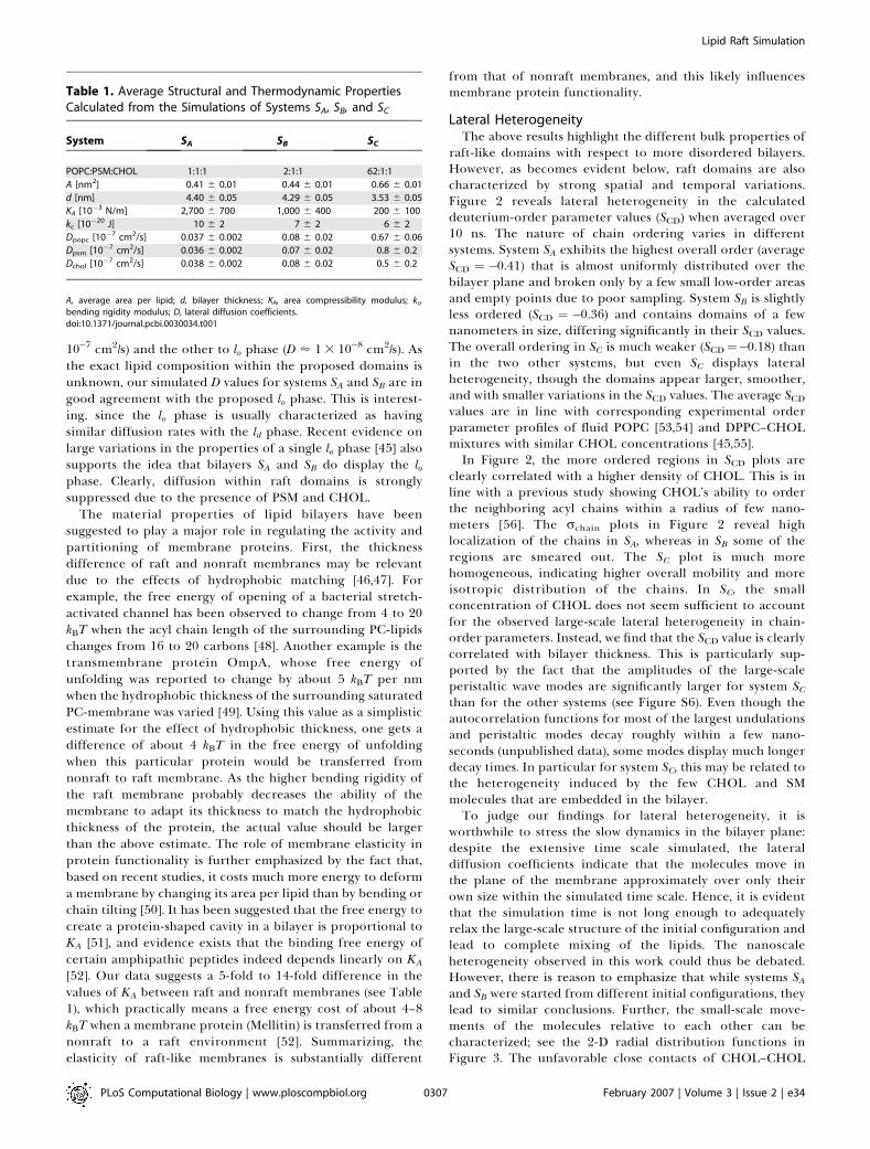
Table 1. Average Structural and Thermodynamic PropertiesCalculated from the Simulations of Systems SA, SB, and SC
System SA SB SC
POPC:PSM:CHOL 1:1:1 2:1:1 62:1:1
A [nm2] 0.41 6 0.01 0.44 6 0.01 0.66 6 0.01
d [nm] 4.40 6 0.05 4.29 6 0.05 3.53 6 0.05
KA [10�3 N/m] 2,700 6 700 1,000 6 400 200 6 100
kc [10�20 J] 10 6 2 7 6 2 6 6 2
Dpopc [10�7 cm2/s] 0.037 6 0.002 0.08 6 0.02 0.67 6 0.06
Dpsm [10�7 cm2/s] 0.036 6 0.002 0.07 6 0.02 0.8 6 0.2
Dchol [10�7 cm2/s] 0.038 6 0.002 0.08 6 0.02 0.5 6 0.2
A, average area per lipid; d, bilayer thickness; KA, area compressibility modulus; kc,bending rigidity modulus; D, lateral diffusion coefficients.doi:10.1371/journal.pcbi.0030034.t001
PLoS Computational Biology | www.ploscompbiol.org February 2007 | Volume 3 | Issue 2 | e340307
Lipid Raft Simulation

pairs are revealed by the lowering of the nearest neighborpeak in time. Simultaneously, the secondary peak at 1.0 nmincreases, indicating small-scale reorganization of CHOLmolecules. Significant changes in time can also be seen in theother plots of Figure 3, revealing the tendency of closercontacts between CHOL–POPC center of mass pairs withrespect to PSM–CHOL pairs. In all, this provides furthersupport for lateral reorganization and heterogeneity. Thedetails of the lipid–lipid interactions are related to the widelyspeculated specific interaction between SM and CHOL, whichis discussed elsewhere [57,58].
Lateral Pressure ProfilesStructure and dynamics of membrane proteins are likely to
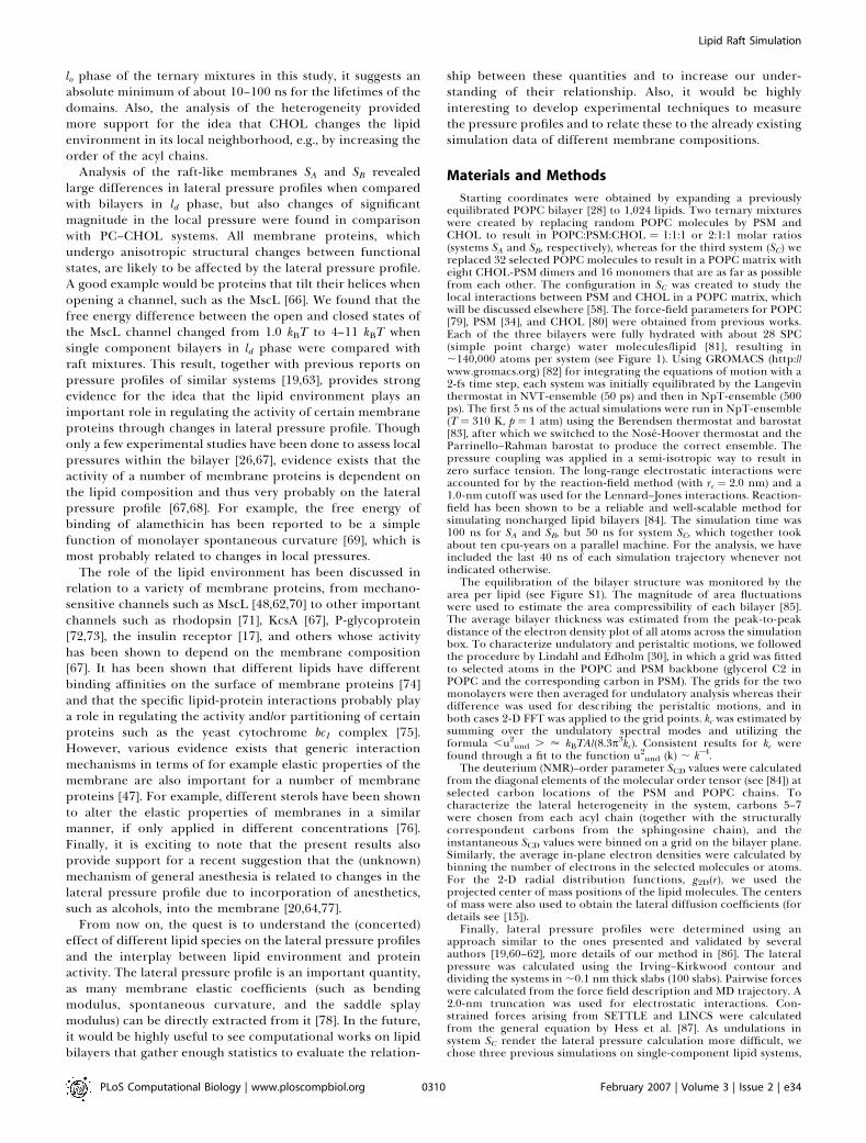
be influenced by the lateral pressure profile, which has beenproposed as a mechanism for, e.g., general anesthesia [20,59].To elucidate this issue, we computed the lateral pressureprofiles of various lipid membrane systems (see Figure 4). Fora discussion of the coupling of the peaks in the lateralpressure profile with the molecular groups and differentinteraction types, see previous related simulation studies[19,60–63]. Here, we focus on a more generic issue, that is, thejoint effect of CHOL and PSM on the pressure profile.
The pressure profiles across the membranes of SA and SB,shown in Figure 4A, indicate a striking difference comparedwith profiles in nonraft membranes (see Figure 4B): raftbilayers display qualitatively different behavior with a greaternumber of peaks as compared with single component POPCand PSM bilayers in ld phase. Rather, raft systems display a
qualitative similarity to the DPPC–CHOL system, shown inFigure 4B. These observations are in line with previoussimulation studies, if reports on other single component ldbilayers [60–62] are compared with binary PC–CHOL systems[19,63]. A remarkable difference found here is the significantincrease of positive (repulsive) pressure at the middle of raftbilayers compared with pure POPC, the effect beingparticularly large in the case of raft-mixture SA.Notably, the peak heights in the lateral pressure profile are
of the order of 1,000 bar. Thus, molecules such as integralmembrane proteins are under the influence of huge localpressures that likely affect their conformational state.Particularly, proteins whose cross-sectional area undergoessignificant anisotropic changes when shifting from active toinactive state are likely to be governed or regulated by thepressure profile [20,64]. To further quantify this idea, weestimated the lateral pressure profile–induced component ofthe energy between open and closed conformations of achannel protein MscL (see Methods). For this quantity, we getDW ¼ (11 6 2) kBT and (4 6 1) kBT for systems SA and SB,respectively. These are significantly higher than the valuesfound for the pure POPC bilayer (1.9 6 0.2) kBT, the purePSM bilayer (1.0 6 0.6) kBT, or the binary DPPC–CHOLbilayer (1.0 6 0.4) kBT. The above result for a POPC bilayer isin agreement with the previous calculation by Gullingsrudand Schulten [62], who found 1.7 kBT for POPC. The positivevalues of DW indicate that the lateral pressure profiles ofthese bilayers lower the open state energy of MscL relative tothe closed state; that is, they are in favor of the open state.
Figure 2. Snapshots Averaged over the Last 10 ns from the End of Each Simulation
The deuterium order parameters, SCD, of selected carbons (C5–C7) of POPC and PSM chains were binned in the xy-plane (column 1, from left). The in-plane electron densities, r, have been plotted separately for CHOL (column 2) and the selected chain carbons (column 3). The average bilayer thickness,d, was obtained from the grid of the undulation analysis (column 4). Systems SA to SC are represented on rows from top to bottom, respectively. Onlythe bottom leaflet has been used for columns 1–3, whereas both leaflets were used for column 4. The equivalent plots for the top leaflet have beenpresented in Figure S2.doi:10.1371/journal.pcbi.0030034.g002
PLoS Computational Biology | www.ploscompbiol.org February 2007 | Volume 3 | Issue 2 | e340308
Lipid Raft Simulation
Especially interesting are the large values found for the raftsystems, which suggest that the lateral pressure profilescharacteristic of raft-like environments would facilitate theopening of MscL. For comparison, it has been estimated thatthe free energy difference associated with the opening ofMscL is about 20–50 kBT [62,65]. The contribution due to thepressure profile in a raft domain could therefore besignificant. In general terms, it is clear from the aboveestimates that the equilibrium probability of MscL to be inthe open state must be significantly altered by the pressureprofile of the lipid environment. Additionally, we wish tounderline that the values for DW have been estimated fordifferent bilayers that are all under identical surface tensionconditions (c¼0) and are thus not related to the usual pictureof the effect of overall stress on mechanosensitive channels.
Discussion
In this work, we have elucidated properties of lipid raftmixtures through atom-scale simulations and compared themwith properties of a bilayer in liquid disordered phase. Wefound that the presence of PSM and CHOL in SA and SB notonly significantly enhanced the lateral packing of lipids andincreased the acyl chain order, but also reduced the lateral
diffusion rates by more than an order of magnitude whencompared with the ld phase. This observation is contradictoryto the traditional definition of the lo phase, but is inagreement with recent reports on varying properties of thisparticular phase [45,54]. It is interesting to note that thedifference in the lipid dynamics of the different phases mayin itself have a contribution to the dynamical partitioning ofmembrane proteins [5], as they spend more time in theordered domains due to slower diffusion and allow more timefor cross-linking between proteins to occur.The elasticity of the raft mixtures was found to be reduced
significantly when compared with SC. The fact that thisreduction was greater than expected from previous reportson binary PC–CHOL mixtures, suggests that SM has a furtherrigidifying effect on raft mixtures. The 5-fold to 14-foldincrease in KA suggests significant implications on thepartitioning of membrane proteins. First, the free energy ofcreating a cavity to the membrane, and thus the solvation freeenergy of a protein into a membrane, is directly proportionalto KA, which leads to unfavored partitioning of certainproteins into raft-like membranes. On the basis of a recentexperimental report [52], we estimated that the transfer freeenergy of Mellitin from nonraft to raft membrane would beabout 4–8 kBT. Second, the difference in thickness of about0.8–0.9 nm between raft and nonraft membranes suggests acontribution to the transfer free energy of proteins due tochanged hydrophobic matching. This effect is practicallyalways present and the reported strength of the effect, about 5kBT per nm [49], makes it comparable to the effect of the KA.The lateral heterogeneity in the simulated membranes was
found to be related to either the tendency of CHOL to orderneighboring acyl chains or to the relatively slow peristalticmodes of the bilayer. The emergence of these heterogeneitiesmay be related to the idea of small granular arrangement ofnanodomains in biological membranes [9,12]. Consideringthe perhaps surprisingly slow diffusion rates observed for the
Figure 3. 2-D Radial Distribution Functions between the Molecular
Center of Mass Positions in SA and SB
The figures show the time evolution in the system at three different timeintervals: 0–10 ns (gray, dashed), 30–40 ns (gray), and 90–100 ns (black).The error bars for the black curve indicate the average difference of thetwo monolayers.doi:10.1371/journal.pcbi.0030034.g003
Figure 4. Lateral Pressure Profiles of Systems SA and SB (Top) and of
Previously Simulated Pure POPC/PSM Systems and a Binary DPPC–CHOL
System (Bottom)
The center of the membrane is at z¼ 0. The graphs have been averagedto be symmetric on both sides of the center and smoothed by adaptivehigh-order spline fitting [90]. Error bars are statistical errors for each slab.The errors have been shown for only one of the monolayers of theDPPC–CHOL system because they are equal for both monolayers andalso smaller or equal for the other systems.doi:10.1371/journal.pcbi.0030034.g004
PLoS Computational Biology | www.ploscompbiol.org February 2007 | Volume 3 | Issue 2 | e340309
Lipid Raft Simulation
lo phase of the ternary mixtures in this study, it suggests anabsolute minimum of about 10–100 ns for the lifetimes of thedomains. Also, the analysis of the heterogeneity providedmore support for the idea that CHOL changes the lipidenvironment in its local neighborhood, e.g., by increasing theorder of the acyl chains.
Analysis of the raft-like membranes SA and SB revealedlarge differences in lateral pressure profiles when comparedwith bilayers in ld phase, but also changes of significantmagnitude in the local pressure were found in comparisonwith PC–CHOL systems. All membrane proteins, whichundergo anisotropic structural changes between functionalstates, are likely to be affected by the lateral pressure profile.A good example would be proteins that tilt their helices whenopening a channel, such as the MscL [66]. We found that thefree energy difference between the open and closed states ofthe MscL channel changed from 1.0 kBT to 4–11 kBT whensingle component bilayers in ld phase were compared withraft mixtures. This result, together with previous reports onpressure profiles of similar systems [19,63], provides strongevidence for the idea that the lipid environment plays animportant role in regulating the activity of certain membraneproteins through changes in lateral pressure profile. Thoughonly a few experimental studies have been done to assess localpressures within the bilayer [26,67], evidence exists that theactivity of a number of membrane proteins is dependent onthe lipid composition and thus very probably on the lateralpressure profile [67,68]. For example, the free energy ofbinding of alamethicin has been reported to be a simplefunction of monolayer spontaneous curvature [69], which ismost probably related to changes in local pressures.
The role of the lipid environment has been discussed inrelation to a variety of membrane proteins, from mechano-sensitive channels such as MscL [48,62,70] to other importantchannels such as rhodopsin [71], KcsA [67], P-glycoprotein[72,73], the insulin receptor [17], and others whose activityhas been shown to depend on the membrane composition[67]. It has been shown that different lipids have differentbinding affinities on the surface of membrane proteins [74]and that the specific lipid-protein interactions probably playa role in regulating the activity and/or partitioning of certainproteins such as the yeast cytochrome bc1 complex [75].However, various evidence exists that generic interactionmechanisms in terms of for example elastic properties of themembrane are also important for a number of membraneproteins [47]. For example, different sterols have been shownto alter the elastic properties of membranes in a similarmanner, if only applied in different concentrations [76].Finally, it is exciting to note that the present results alsoprovide support for a recent suggestion that the (unknown)mechanism of general anesthesia is related to changes in thelateral pressure profile due to incorporation of anesthetics,such as alcohols, into the membrane [20,64,77].
From now on, the quest is to understand the (concerted)effect of different lipid species on the lateral pressure profilesand the interplay between lipid environment and proteinactivity. The lateral pressure profile is an important quantity,as many membrane elastic coefficients (such as bendingmodulus, spontaneous curvature, and the saddle splaymodulus) can be directly extracted from it [78]. In the future,it would be highly useful to see computational works on lipidbilayers that gather enough statistics to evaluate the relation-
ship between these quantities and to increase our under-standing of their relationship. Also, it would be highlyinteresting to develop experimental techniques to measurethe pressure profiles and to relate these to the already existingsimulation data of different membrane compositions.
Materials and Methods
Starting coordinates were obtained by expanding a previouslyequilibrated POPC bilayer [28] to 1,024 lipids. Two ternary mixtureswere created by replacing random POPC molecules by PSM andCHOL to result in POPC:PSM:CHOL ¼ 1:1:1 or 2:1:1 molar ratios(systems SA and SB, respectively), whereas for the third system (SC) wereplaced 32 selected POPC molecules to result in a POPC matrix witheight CHOL-PSM dimers and 16 monomers that are as far as possiblefrom each other. The configuration in SC was created to study thelocal interactions between PSM and CHOL in a POPC matrix, whichwill be discussed elsewhere [58]. The force-field parameters for POPC[79], PSM [34], and CHOL [80] were obtained from previous works.Each of the three bilayers were fully hydrated with about 28 SPC(simple point charge) water molecules/lipid [81], resulting in;140,000 atoms per system (see Figure 1). Using GROMACS (http://www.gromacs.org) [82] for integrating the equations of motion with a2-fs time step, each system was initially equilibrated by the Langevinthermostat in NVT-ensemble (50 ps) and then in NpT-ensemble (500ps). The first 5 ns of the actual simulations were run in NpT-ensemble(T ¼ 310 K, p ¼ 1 atm) using the Berendsen thermostat and barostat[83], after which we switched to the Nose-Hoover thermostat and theParrinello–Rahman barostat to produce the correct ensemble. Thepressure coupling was applied in a semi-isotropic way to result inzero surface tension. The long-range electrostatic interactions wereaccounted for by the reaction-field method (with rc ¼ 2.0 nm) and a1.0-nm cutoff was used for the Lennard–Jones interactions. Reaction-field has been shown to be a reliable and well-scalable method forsimulating noncharged lipid bilayers [84]. The simulation time was100 ns for SA and SB, but 50 ns for system SC, which together tookabout ten cpu-years on a parallel machine. For the analysis, we haveincluded the last 40 ns of each simulation trajectory whenever notindicated otherwise.
The equilibration of the bilayer structure was monitored by thearea per lipid (see Figure S1). The magnitude of area fluctuationswere used to estimate the area compressibility of each bilayer [85].The average bilayer thickness was estimated from the peak-to-peakdistance of the electron density plot of all atoms across the simulationbox. To characterize undulatory and peristaltic motions, we followedthe procedure by Lindahl and Edholm [30], in which a grid was fittedto selected atoms in the POPC and PSM backbone (glycerol C2 inPOPC and the corresponding carbon in PSM). The grids for the twomonolayers were then averaged for undulatory analysis whereas theirdifference was used for describing the peristaltic motions, and inboth cases 2-D FFT was applied to the grid points. kc was estimated bysumming over the undulatory spectral modes and utilizing theformula ,u2und . ’ kBTA/(8.3p
3kc). Consistent results for kc werefound through a fit to the function u2und (k) ; k�4.
The deuterium (NMR)–order parameter SCD values were calculatedfrom the diagonal elements of the molecular order tensor (see [84]) atselected carbon locations of the PSM and POPC chains. Tocharacterize the lateral heterogeneity in the system, carbons 5–7were chosen from each acyl chain (together with the structurallycorrespondent carbons from the sphingosine chain), and theinstantaneous SCD values were binned on a grid on the bilayer plane.Similarly, the average in-plane electron densities were calculated bybinning the number of electrons in the selected molecules or atoms.For the 2-D radial distribution functions, g2D(r), we used theprojected center of mass positions of the lipid molecules. The centersof mass were also used to obtain the lateral diffusion coefficients (fordetails see [15]).
Finally, lateral pressure profiles were determined using anapproach similar to the ones presented and validated by severalauthors [19,60–62], more details of our method in [86]. The lateralpressure was calculated using the Irving–Kirkwood contour anddividing the systems in ;0.1 nm thick slabs (100 slabs). Pairwise forceswere calculated from the force field description and MD trajectory. A2.0-nm truncation was used for electrostatic interactions. Con-strained forces arising from SETTLE and LINCS were calculatedfrom the general equation by Hess et al. [87]. As undulations insystem SC render the lateral pressure calculation more difficult, wechose three previous simulations on single-component lipid systems,
PLoS Computational Biology | www.ploscompbiol.org February 2007 | Volume 3 | Issue 2 | e340310
Lipid Raft Simulation
POPC [86], PSM [34], and a binary 1:4 DPPC–CHOL [17] forreference. For each system, the pressure profile was calculated thesame way. To estimate the effect of pressure profile on membraneproteins, we followed the approach introduced by Cantor [88] andlater applied to molecular simulation data of single-componentbilayers by Gullingsrud et al. [62]. As a model we use themechanosensitive ion channel MscL, whose conformation has beenfound to change anisotropically between cylindrical (open) and cone(closed) shapes [89]. Here we calculate the work, DW, done against thelateral pressure profile to alter the shape of the membrane cavityoccupied by the protein as it changes conformation from the closedto an open state. Then DW can be written as:
DW ¼Z
pðzÞDAðzÞ dz; ð1Þ
where DA(z) is the change in the cross-sectional area of the proteinand p(z) is the pressure profile. Here, we use an approach identical tothat used in [62], and identical values for DA(z) for MscL as used in[62], in which the area is kept unchanged in the middle of themembrane between the two states. Error bars for DW have beencalculated using results for different monolayers. It is, however,important to realize that DW depends on the second moment of thelateral pressure profile [62] and thus is susceptible to small changes oflateral pressure far from the bilayer center. Therefore, extra cautionmust be followed when interpreting these results. Also, in thisapproach the influence of inserting a protein into the membrane onthe lateral pressure profile is not taken into account.
Supporting Information
Figure S1. The Area per Lipid versus Simulation Time
Found at doi:10.1371/journal.pcbi.0030034.sg001 (509 KB EPS).
Figure S2. Averaged Snapshots from the Last 10 ns of EachSimulation
The data is represented as in Figure 2 of the main article, but plottedfor the top monolayer (columns 1–3) instead of the bottommonolayer.
Found at doi:10.1371/journal.pcbi.0030034.sg002 (212 KB EPS).
Figure S3. Snapshots (1-ns Averages) Revealing the In-Plane ElectronDensity of CHOL at 10-ns Time Intervals
Columns A1–C1 are the bottom monolayer and columns A2–C2 thetop monolayer in systems SA to SC, respectively.
Found at doi:10.1371/journal.pcbi.0030034.sg003 (1.2 MB EPS).
Figure S4. Snapshots (1-ns Averages) Revealing the Undulation andPeristaltic Motions at 10-ns Time Intervals
Columns A1–C1 are the average bilayer height (z(x;y), the mean heightof the two monolayers), whereas columns A2–C2 are the bilayerthickness (d(x;y), the difference in height of the two monolayers) insystems SA to SC, respectively. For calculating z(x;y) and d(x;y), we usedthe grid method discussed in the Methods section.
Found at doi:10.1371/journal.pcbi.0030034.sg004 (725 KB EPS).
Figure S5. Undulatory Spectral Intensity per Wave Mode versus WaveVector Magnitude for Systems SA to SCThe legend shows kc values calculated by two different methods, thesumming method utilizing Equation 4 and fitting Equation 3 in [30].
Found at doi:10.1371/journal.pcbi.0030034.sg005 (183 KB EPS).
Figure S6. Peristaltic Spectral Intensity per Wave Mode versus WaveVector Magnitude for Systems SA to SCFound at doi:10.1371/journal.pcbi.0030034.sg006 (97 KB EPS).
Acknowledgments
We thank Dr. Tomasz Rog for providing the simulation trajectory ofthe DPPC–CHOL system and Jussi Aittoniemi for help in running theraft simulations.
Author contributions. PSN, MTH, MK, and IV conceived anddesigned the experiments. PSN performed the experiments andanalyzed the data, except for the lateral pressure profiles computedby SO. PSN wrote the paper together with the other authors.
Funding. This work has, in part, been supported by the Academy ofFinland (PSN, MTH, MK, and IV), the Academy of Finland Center ofExcellence Program (SO, PSN, and IV), the Jenny and Antti WihuriFoundation (MTH), the Finnish Academy of Science and Letters(PSN), the Emil Aaltonen Foundation (MK), and the Natural Sciencesand Engineering Council (NSERC) of Canada (MK). We acknowledgethe Finnish IT Center for Science and the HorseShoe (DCSC)supercluster computing facility at the University of Southern Den-mark for computer resources.
Competing interests. The authors have declared that no competinginterests exist.
References1. Singer SJ, Nicolson GL (1972) The fluid mosaic model of the structure of
cell membranes. Science 175: 720–731.2. Simons K, Ikonen E (1997) Functional rafts in cell membranes. Nature 387:
569–572.3. Edidin M (2003) The state of lipid rafts: From model membranes to cells.
Annu Rev Biophys Biomol Struct 32: 257–283.4. Pike LJ (2004) Lipid rafts: Heterogeneity on the high seas. Biochem J 378:
281–292.5. Hancock JF (2006) Lipid rafts: Continuous only from simplistic stand-
points. Nat Rev Mol Cell Biol 7: 456–462.6. Simons K, Vaz WLC (2004) Model system, lipid rafts, and cell membranes.
Annu Rev Biophys Biomol Struct 33: 269–295.7. Gaus K, Gratton E, Kable EPW, Jones AS, Gelissen I, et al. (2003) Visualizing
lipid structure and raft domains in living cells with two-photon micro-scopy. Proc Natl Acad Sci U S A 100: 15554–15559.
8. Brown RE (1998) Sphingolipid organization in biomembranes: Whatphysical studies of model membranes reveal. J Cell Sci 111: 1–9.
9. London E (2005) How principles of domain formation in modelmembranes may explain ambiguities concerning lipid raft formation incells. Biochim Biophys Acta 1746: 203–220.
10. Almeida PFF, Pokorny A, Hinderliter A (2005) Thermodynamics ofmembrane domains. Biochim Biophys Acta 1720: 1–13.
11. Shaikh SR, Edidin MA (2006) Membranes are not just rafts. Chem PhysLipids 144: 1–3.
12. Hevonoja T, Pentikainen MO, Hyvonen MT, Kovanen PT, Ala-Korpela M(2000) Structure of low density lipoprotein (LDL) particles: Basis forunderstanding molecular changes in modified LDL. Biochim Biophys Acta1488: 189–210.
13. Ramstedt B, Slotte JP (2002) Membrane properties of sphingomyelins. FEBSLett 531: 33–37.
14. Holopainen JM, Metso AJ, Mattila JP, Jutila A, Kinnunen PKJ (2004)Evidence for the lack of a specific interaction between cholesterol andsphingomyelin. Biophys J 86: 1510–1520.
15. Falck E, Patra M, Karttunen M, Hyvonen MT, Vattulainen I (2004) Lessons
of slicing membranes: Interplay of packing, free area, and lateral diffusionin phospholipid/cholesterol bilayers. Biophys J 87: 1076–1091.
16. Hofsass C, Lindahl E, Edholm O (2003) Molecular dynamics simulations ofphospholipid bilayers with cholesterol. Biophys J 84: 2192–2206.
17. Vainio S, Jansen M, Koivusalo M, Rog T, Karttunen M, et al. (2006)Significance of sterol structural specificity. Desmosterol cannot replacecholesterol in lipid rafts. J Biol Chem 281: 348–355.
18. Kupiainen M, Falck E, Ollila S, Niemela P, Gurtovenko AA, et al. (2005) Freevolume properties of sphingomyelin, DMPC, DPPC, and PLPC bilayers. JComput Theor Nanosci 2: 401–413.
19. Patra M (2005) Lateral pressure profiles in cholesterol–dppc bilayers. EurBiophys J 35: 79–88.
20. Cantor RS (1997) The lateral pressure profile in membranes: A physicalmechanism of general anesthesia. Biochemistry 36: 2339–2344.
21. Varma R, Mayor S (1998) GPI-anchored proteins are organized insubmicron domains at the cell surface. Nature 394: 798–801.
22. Plowman SJ, Muncke C, Parton RG, Hancock JF (2005) H-ras, K-ras, andinner plasma membrane raft proteins operate in nanoclusters withdifferential dependence on the actin cytoskeleton. Proc Natl Acad Sci US A 102: 15500–15505.
23. Pandit SA, Jakobsson E, Scott HL (2004) Simulation of the early stages ofnano-domain formation in mixed bilayers of sphingomyelin, cholesterol,and dioleylphosphatidylcholine. Biophys J 87: 3312–3322.
24. Pandit SA, Vasudevan S, Chiu SW, Mashl RJ, Jakobsson E, et al. (2004)Sphingomyelin–cholesterol domains in phospholipid membranes: Atom-istic simulation. Biophys J 87: 1092–1100.
25. Cantor RS (1997) Lateral pressures in cell membranes: A mechanism formodulation of protein function. J Phys Chem B 101: 1723–1725.
26. Templer RH, Castle SJ, Curran AR, Rumbles G, Klug DR (1998) Sensingisothermal changes in the lateral pressure in model membranes using di-pyrenyl phosphatidylcholine. Faraday Discuss 111: 41–53.
27. de Almeida RFM, Fedorov A, Prieto M (2003) Sphingomyelin/phosphati-dylcholine/cholesterol phase diagram: Boundaries and composition of lipidrafts. Biophys J 85: 2406–2416.
28. Patra M, Salonen E, Terama E, Vattulainen I, Faller R, et al. (2006) Under
PLoS Computational Biology | www.ploscompbiol.org February 2007 | Volume 3 | Issue 2 | e340311
Lipid Raft Simulation
the influence of alcohol: The effect of ethanol and methanol on lipidbilayers. Biophys J 90: 1121–1135.
29. Kucerka N, Tristram-Nagle S, Nagle JF (2005) Structure of fully hydratedfluid phase lipid bilayers with monounsaturated chains. J Membrane Biol208: 193–202.
30. Lindahl E, Edholm O (2000) Mesoscopic undulations and thicknessfluctuations in lipid bilayers from molecular dynamics simulations. BiophysJ 79: 426–433.
31. Evans E, Rawicz W (1990) Entropy driven tension and bending elasticity incondensed-fluid membranes. Phys Rev Lett 64: 2094–2097.
32. Rawicz W, Olbrich KC, McIntosh T, Needham D, Evans E (2000) Effect ofchain length and unsaturation on elasticity of lipid bilayers. Biophys J 79:328–339.
33. Filippov A, Oradd G, Lindblom G (2003) The effect of cholesterol on thelateral diffusion of phospholipids in oriented bilayers. Biophys J 84: 3079–3086.
34. Niemela P, Hyvonen MT, Vattulainen I (2004) Structure and dynamics ofsphingomyelin bilayer: Insight gained through systematic comparison tophosphatidylcholine. Biophys J 87: 2976–2989.
35. Filippov A, Oradd G, Lindblom G (2004) Lipid lateral diffusion in orderedand disordered phases in raft mixtures. Biophys J 86: 891–896.
36. McIntosh TJ (1978) The effect of cholesterol on the structure ofphosphatidylcholine bilayers. Biochim Biophys Acta 513: 43–58.
37. Sankaram MB, Thompson TE (1990) Modulation of phospholipid acyl chainorder by cholesterol. A solid-state 2H nuclear magnetic resonance study.Biochemistry 29: 10676–10684.
38. Smaby JM, Momsen M, Kulkarni VS, Brown RE (1996) Cholesterol-inducedinterfacial area condensations of galactosylceramides sphingomyelins withidentical acyl chains. Biochemistry 35: 5696–5704.
39. Needham D, McIntosh TJ, Evans E (1988) Thermomechanical and transitionproperties of dimyristoylphosphatidylcholine/cholesterol bilayers. Bio-chemistry 27: 4668–4673.
40. Niemela P, Hyvonen MT, Vattulainen I (2006) Influence of chain length andunsaturation on sphingomyelin bilayers. Biophys J 90: 851–863.
41. Needham D, Nunn RS (1990) Elastic deformation and failure of lipidbilayer membranes containing cholesterol. Biophys J 58: 997–1009.
42. Henriksen J, Rowat AC, Ipsen JH (2004) Vesicle fluctuation analysis of theeffect of sterols on membrane bending rigidity. Eur Biophys J 33: 732–741.
43. Rinia HA, Snel MME, van der Eerden JPJM, de Kruijff B (2001) Visualizingdetergent resistant domains in model membranes with atomic forcemicroscopy. FEBS Lett 501: 92–96.
44. Filippov A, Oradd G, Lindblom G (2006) Sphingomyelin structureinfluences the lateral diffusion and raft formation in lipid bilayers. BiophysJ 90: 2086–2092.
45. Clarke JA, Heron AJ, Seddon JM, Law RW (2006) The diversity of the liquidordered (lo) phase of phosphatidylcholine/cholesterol membranes: Avariable temperature multinuclear solid-state nmr and X-ray diffractionstudy. Biophys J 90: 2383–2393.
46. Jensen MO, Mouritsen OG (2004) Lipids do influence protein function—The hydrophobic matching hypothesis revisited. Biochim Biophys Acta1666: 205–226.
47. McIntosh TJ, Simon SA (2006) Roles of bilayer material properties infunction and distribution of membrane properties. Annu Rev BiophysBiomol Struct 35: 177–198.
48. Perozo E, Kloda A, Cortes DM, Martinac B (2002) Physical principlesunderlying the transduction of bilayer deformation forces duringmechanosensitive channel gating. Nature Struct Biol 9: 696–703.
49. Hong H, Tamm LK (2004) Elastic coupling of integral membrane proteinstability to lipid bilayer forces. Proc Natl Acad Sci U S A 101: 4065–4070.
50. Kuzmin PI, Akimov SA, Chizmadzhev YA, Zmmerberg J, Cohen FS (2005)Line tension and interaction energies of membrane rafts calculated fromlipid splay and tilt. Biophys J 88: 1120–1133.
51. Zhelev DV (1998) Material property characteristics for lipid bilayerscontaining lysolipid. Biophys J 75: 321–330.
52. Allende D, McIntosh TJ (2003) Mellitin-induced bilayer leakage depends onlipid material properties: Evidence for toroidal pores. Biophys J 88: 1828–1837.
53. Seelig J, Waespe-Sarcevic N (1978) Molecular order in cis and transunsaturated phospholipid bilayers. Biochemistry 17: 3310–3315.
54. Mehnert T, Jacob K, Bittman R, Beyer K (2006) Structure and lipidinteraction of N-palmitoylsphingomyelin in bilayer membranes as revealedby 2H-NMR spectroscopy. Biophys J 90: 939–946.
55. Guo W, Kurze V, Huber T, Afdhal NH, Beyer K, et al. (2002) A solid-stateNMR study of phospholipid–cholesterol interactions: Sphingomyelin–cholesterol binary systems. Biophys J 83: 1465–1478.
56. Pitman MC, Suits F, MacKerell AD Jr, Feller SE (2004) Molecular-levelorganization of saturated and polyunsaturated fatty acids in a phospha-tidylcholine bilayer containing cholesterol. Biochemistry 43: 15318–15328.
57. Rog T, Pasenkiewicz-Gierula M (2006) Cholesterol–sphingomyelin inter-actions: A molecular dynamics simulation study. Biophys J 91: 3756–3767.
58. Aittoniemi J, Niemela PS, Hyvonen MT, Karttunen M, Vattulainen I (2007)Insight into the putative specific interactions between cholesterol,sphingomyelin and palmitoyl–oleoyl phosphatidylcholine. Biophys J 92:1125–1127.
59. Eckenhoff RG (2001) Promiscuous ligands and attractive cavities. How dothe inhaled anesthetics work? Mol Interv 1: 258–268.
60. Lindahl E, Edholm O (2000) Spatial and energetic–entropic decompositionof surface tension in lipid bilayers from molecular dynamics simulations. JChem Phys 113: 3882–3893.
61. Sonne J, Hansen FY, Peters GH (2005) Methodological problems inpressure profile calculations for lipid bilayers. J Chem Phys 122: 124903.
62. Gullingsrud J, Schulten K (2004) Lipid bilayer pressure profiles andmechanosensitive channel gating. Biophys J 86: 3496–3509.
63. Carrillo-Tripp M, Feller SE (2005) Evidence for a mechanism by which x-3polyunsaturated lipids may affect membrane protein function. Biochem-istry 44: 10164–10169.
64. Cantor RS (1998) The lateral pressure profile in membranes: A physicalmechanism of general anesthesia. Toxicol Lett 100–101: 451–458.
65. Sukhraev SI, Sigurdson WJ, Kung C, Sachs F (1999) Energetic and spatialparameters for gating of the bacterial large conductance mechanosensitivechannel, MscL. J Gen Physiol 113: 525–593.
66. Doyle DA (2004) Structural changes during ion channel gating. TrendsNeurosci 27: 298–302.
67. van den Brink–van der Laan E, Killian JA, de Kruijff B (2004) Nonbilayerlipids affect peripheral and integral membrane proteins via changes in thelateral pressure profile. Biochim Biophys Acta 1666: 275–288.
68. Bezrukov SM (2000) Functional consequences of lipid packing stress. CurrOpin Coll Int Sci 5: 237–243.
69. Lewis JR, Cafiso DS (1999) Correlation of the free energy of a channel-forming voltage-gated peptide and the spontaneous curvature of bilayerlipids. Biochemistry 38: 5932–5938.
70. Meyer GR, Gullingsrud J, Schulten K, Martinac B (2006) Moleculardynamics study of MscL interactions with a curved lipid bilayer. BiophysJ 91: 1630–1637.
71. Botelho AV, Gibson NJ, Thurmond RL, Wang Y, Brown MF (2002)Conformational energetics of rhodopsin modulated by nonlamellar-forming lipids. Biochemistry 41: 6354–6368.
72. Troost J, Lindenmaier H, Haefeli WE, Weiss J (2004) Modulation of cellularcholesterol alters p-glycoprotein activity in multidrug-resistant cells. MolPharmacol 66: 1332–1339.
73. Kamau SW, Kramer SD, Gunthert M, Wunderli-Allenspach H (2005) Effectof the modulation of the membrane lipid composition on the localizationand function of p-glycoprotein in MDR1-MDCK cells. In Vitro Cell DevBiol Anim 41: 207–216.
74. Powl AM, Carney J, Maurius P, East JM, Lee AG (2005) Lipid interactions withbacterial channels: Fluorescence studies. Biochem Soc Trans 33: 905–909.
75. Palsdottir H, Hunte C (2004) Lipids in membrane protein structures.Biochim Biophys Acta 1666: 2–18.
76. Henriksen J, Rowat AC, Brief E, Hsueh YW, Thewalt JL, et al. (2006)Universal behavior of membranes with sterols. Biophys J 90: 1639–1649.
77. van den Brink–van der Laan E, Chupin V, Killian JA, de Kruijff B (2004)Small alcohols destabilize the KcsA tetramer via their effect on themembrane lateral pressure. Biochemistry 43: 5937–5942.
78. Safran S (1994) Frontiers in physics. Volume 90. Statistical thermodynamicsof surfaces, interfaces, and membranes. New York: Addison-Wesley. 498 p.
79. Tieleman DP, Berendsen HJC (1998) A molecular dynamics study of thepores formed by Escherichia coli OmpF porin in a fully hydratedpalmitoyloleoylphosphatidylcholine bilayer. Biophys J 74: 2786–2801.
80. Holtje M, Forster T, Brandt B, Engels T, von Rybinski W, et al. (2001)Molecular dynamics simulations of stratum corneum lipid models: Fattyacids and cholesterol. Biochim Biophys Acta 1511: 156–167.
81. Berendsen HJC, Postma JPM, van Gunsteren WF, Hermans J (1981)Interaction models for water in relation to protein hydration. In: PullmanB, editor. Intermolecular forces. Dordrecht: Reidel. pp. 331–342.
82. Lindahl E, Hess B, van der Spoel D (2001) Gromacs 3.0: A package formolecular simulation and trajectory analysis. J Mol Mod 7: 306–317.
83. Berendsen HJC, Postma JPM, van Gunsteren WF, DiNola A, Haak JR (1984)Molecular dynamics with coupling to an external bath. J Chem Phys 81:3684–3690.
84. Patra M, Karttunen M, Hyvonen MT, Falck E, Vattulainen I (2004) Lipidbilayers driven to a wrong lane in molecular dynamics simulations by subtlechanges in long-range electrostatic interactions. J Phys Chem B 108: 4485–4494.
85. Feller SE, Pastor RW (1999) Constant surface tension simulations of lipidbilayers: The sensitivity of surface areas and compressibilities. J Chem Phys111: 1281–1287.
86. Ollila S (2006) Lateral pressure profile calculations of lipid membranesfrom atomic scale molecular dynamics simulations. [Master’s thesis].Helsinki: Helsinki University of Technology.
87. Hess B, Bekker H, Berendsen HJC, Fraaije JGEM (1997) LINCS: A linearconstraint solver for molecular simulations. J Comput Chem 18: 1463–1472.
88. Cantor RS (1999) The influence of membrane lateral pressure on simplegeometric models of protein conformational equilibra. Chem Phys Lipids101: 45–56.
89. Sukharev S, Durell SR, Guy HR (2001) Structural models of the MscL gatingmechanism. Biophys J 81: 917–936.
90. Thijsse BJ, Hollanders MA, Hendrikse J (1998) A practical algorithm forleast-squares spline approximation of data containing noise. Comp Phys12: 393–399.
PLoS Computational Biology | www.ploscompbiol.org February 2007 | Volume 3 | Issue 2 | e340312
Lipid Raft Simulation