Embed Size (px)
Citation preview
Assessment, diagnosis and management of leg ulcers
Sarah Gardner, Clinical lead, Tissue viability service
What are the challenges of leg ulcer management?
How do you feel when a patient is referred with a leg ulcer?
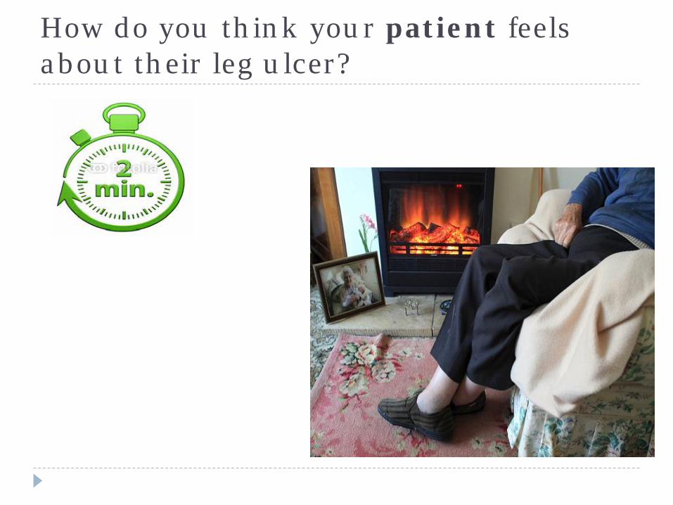
How do you think your patient feels about their leg ulcer?
Aim of the session
To develop a better understanding of the factors that contribute to the development of leg ulceration and how the application of proven treatments can improve clinical outcomes
Objectives -} To gain a better understanding of the anatomy &
Physiology of the vascular system} To understand the function of the veins and how venous
incompetence can lead to skin breakdown} Be able to carry out a full holistic leg ulcer assessment in
order to correctly diagnose the aetiology.} On diagnosis, be able to put an appropriate plan of care
in place in order to support timely wound healing} Be able to differentiate between ‘normal’ and ‘abnormal’
ulceration and recognise when referral is necessary.
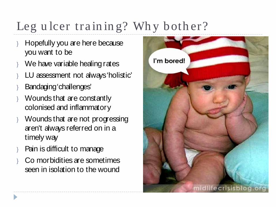
Leg ulcer training? Why bother?} Hopefully you are here because
you want to be} We have variable healing rates } LU assessment not always ‘holistic’} Bandaging ‘challenges’} Wounds that are constantly
colonised and inflammatory} Wounds that are not progressing
aren't always referred on in a timely way
} Pain is difficult to manage} Co morbidities are sometimes
seen in isolation to the wound
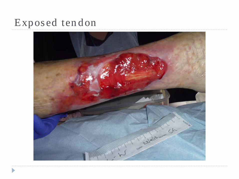
Exposed tendon
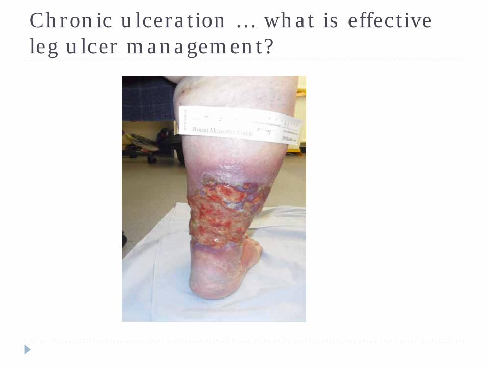
Chronic ulceration … what is effective leg ulcer management?
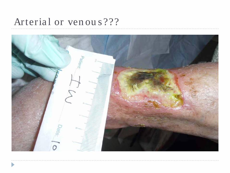
Arterial or venous???
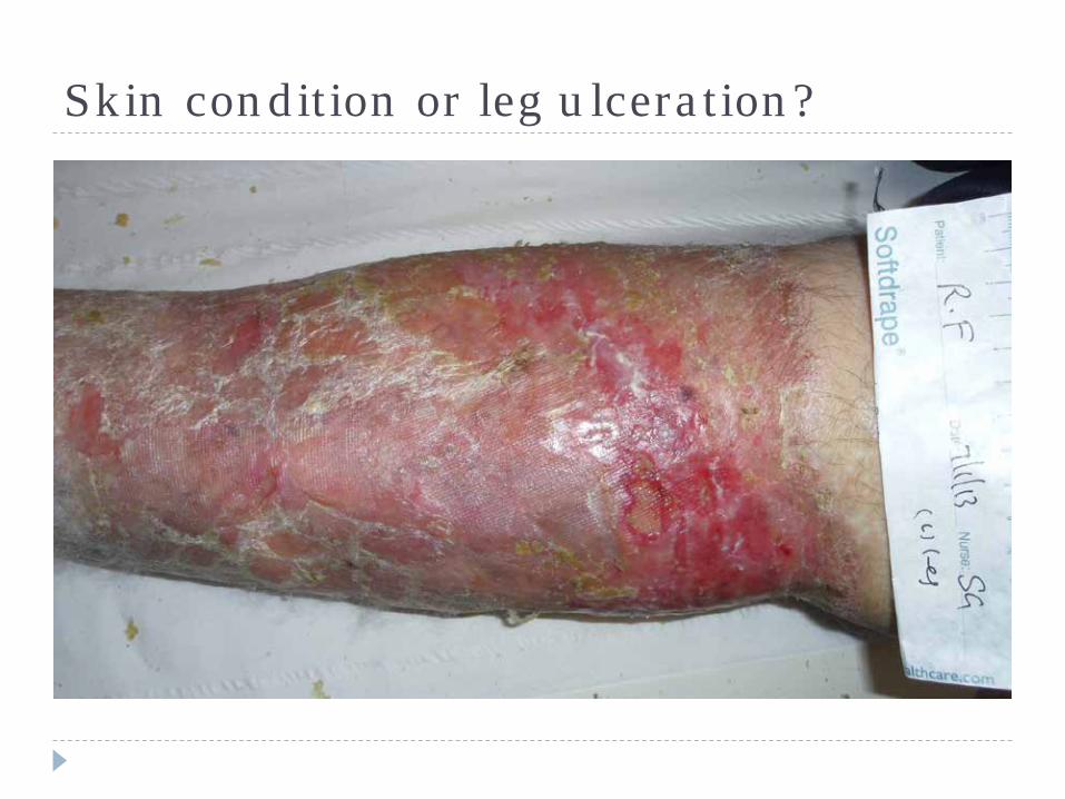
Skin condition or leg ulceration?
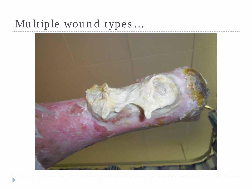
Multiple wound types…
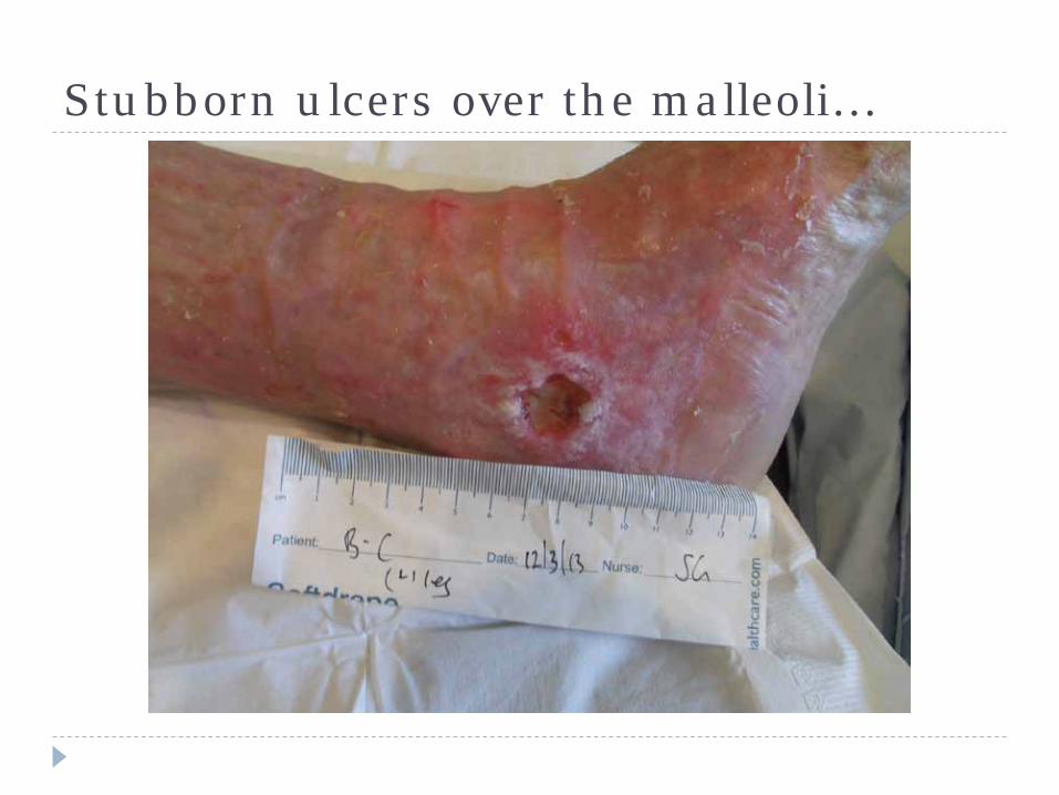
Stubborn ulcers over the malleoli…
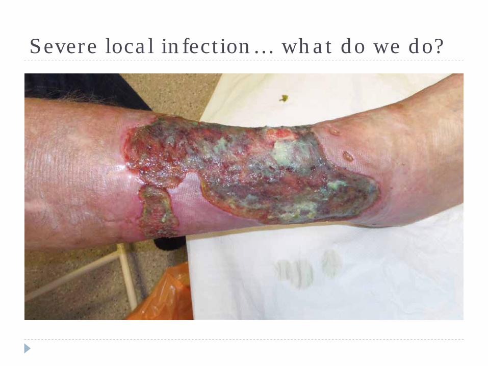
Severe local infection… what do we do?
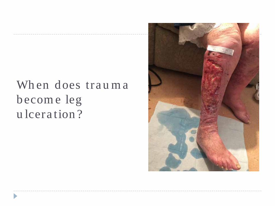
When does trauma become leg ulceration?
The tipping point….
Today you will leave this training session and you will do things differently!
Patient experience film

What is a leg ulcer?
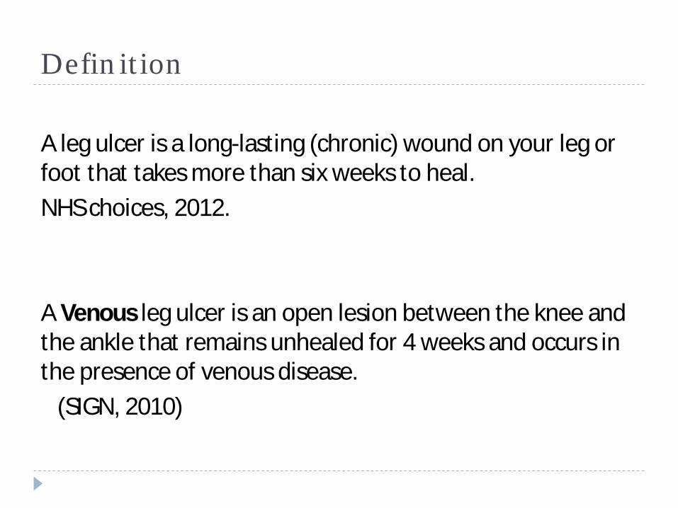
Definition
A leg ulcer is a long-lasting (chronic) wound on your leg or foot that takes more than six weeks to heal.NHS choices, 2012.
A Venous leg ulcer is an open lesion between the knee and the ankle that remains unhealed for 4 weeks and occurs in the presence of venous disease.
(SIGN, 2010)
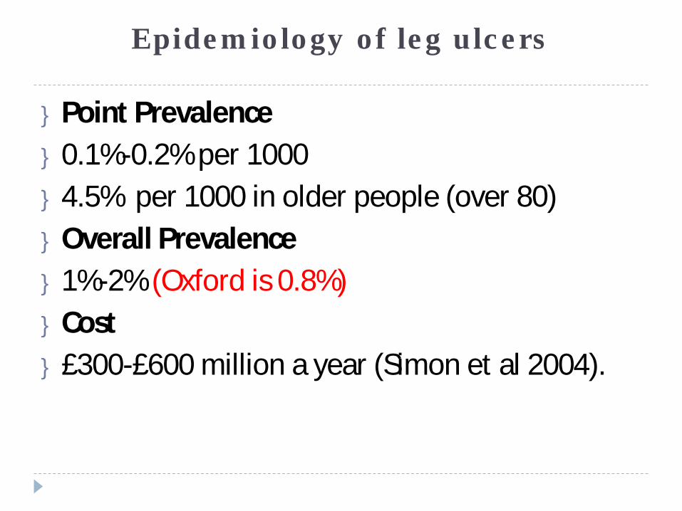
Epidemiology of leg ulcers
} Point Prevalence} 0.1%-0.2% per 1000 } 4.5% per 1000 in older people (over 80)} Overall Prevalence} 1%-2% (Oxford is 0.8%)} Cost} £300-£600 million a year (Simon et al 2004).
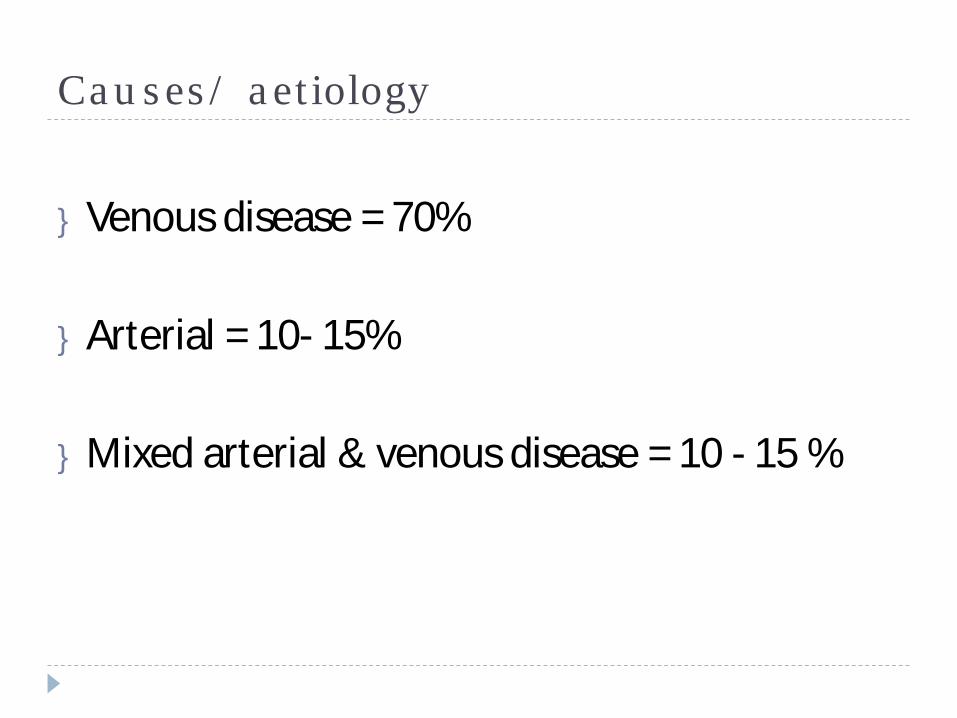
Causes/ aetiology
} Venous disease = 70%
} Arterial = 10- 15%
} Mixed arterial & venous disease = 10 - 15 %
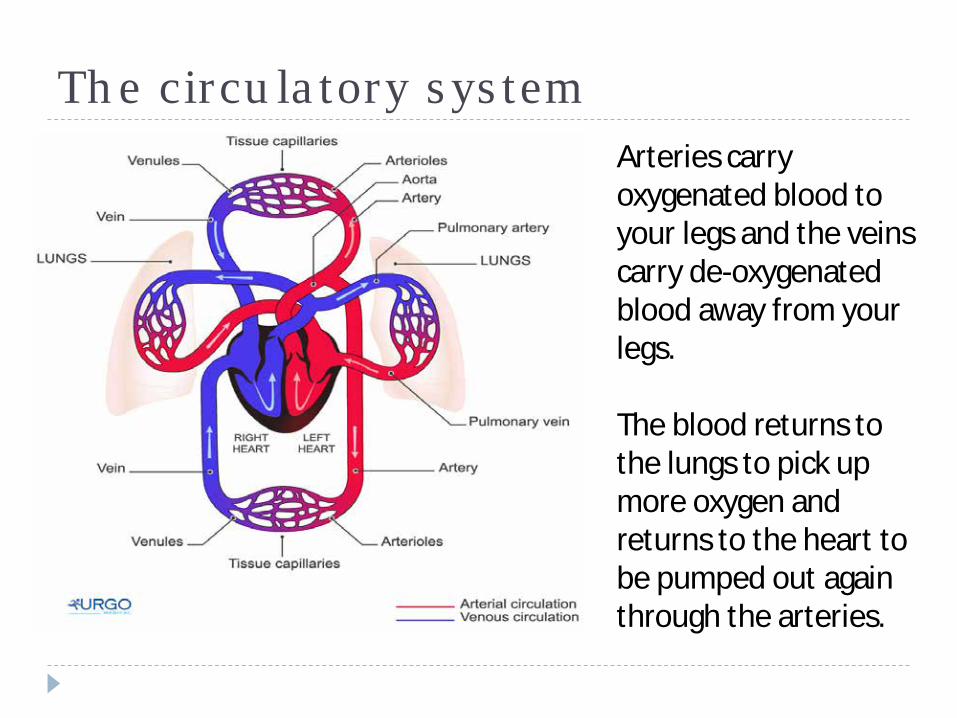
The circulatory system Arteries carry oxygenated blood to your legs and the veins carry de-oxygenated blood away from your legs.
The blood returns to the lungs to pick up more oxygen and returns to the heart to be pumped out again through the arteries.
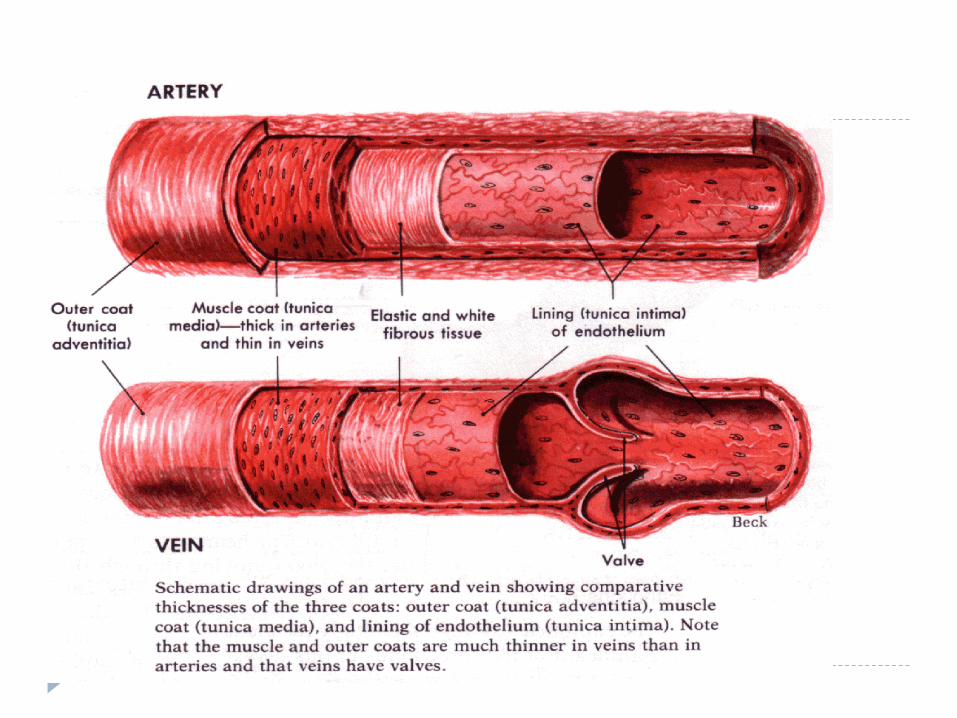
Blood vesselsArteriesArteries take blood away from the heart and are the strongest blood vessels, as they have to withstand the pumping action of the heart. Arterial blood is charged with oxygen and nutrients for healthy tissues. Artery walls consist of three layers, the walls are more muscular and the lumen smaller enabling blood to travel at high pressure.
Arteries branch into smaller vessels known as arterioles, which merge into capillaries.
CapillariesMinute vessels, no wider than a hair, which connect arteries to veins. The exchange of gases (oxygen for carbon dioxide) occurs within the capillaries. A capillary wall is a semi permeable membrane and interstitial fluid moves continually between the blood capillary and the tissue. The rate of fluid leaking depends on several factors, including blood pressure in the blood capillaries and the concentration of plasma proteins in the tissue spaces.
}
Venules - When blood has left the tiny capillaries on its journey back to the heart, the vessels widen into venules and then into the larger vessels known as veins.
Veins – These take blood back to the heart. Their walls are weaker than arteries and take impure blood back to the heart as oxygen has now been exchanged for carbon dioxide in the capillaries. Veins can be classed as superficial or deep. 10% of blood travels through the superficial veins, which are situated just beneath the skin. The remaining 90% of blood travels through deep veins, which are found closer to the bones in the leg and are surrounded and protected by various muscles. These are the veins squeezed by the calf pump on contraction. Veins are also equipped with valvesto assist the flow of blood back to the heart. The valves in the veins open in one direction, therefore helping to prevent backflow of blood. When valves are working as they should, healthy veins are divided into compartments and this helps to relieve strain on the vessel walls.
Perforator -So called because they perforate the deep fascia of muscles, to connect the superficial veins to the deep veins where they drain. Their role is to maintain correct blood drainage. They have valves which prevent blood flowing back (reflux), from deep to superficial veins in muscular systole.
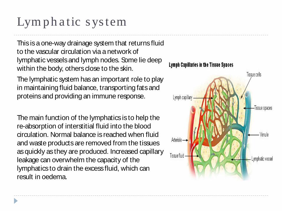
Lymphatic systemThis is a one-way drainage system that returns fluid to the vascular circulation via a network of lymphatic vessels and lymph nodes. Some lie deep within the body, others close to the skin.The lymphatic system has an important role to play in maintaining fluid balance, transporting fats and proteins and providing an immune response.
The main function of the lymphatics is to help the re-absorption of interstitial fluid into the blood circulation. Normal balance is reached when fluid and waste products are removed from the tissues as quickly as they are produced. Increased capillary leakage can overwhelm the capacity of the lymphatics to drain the excess fluid, which can result in oedema.
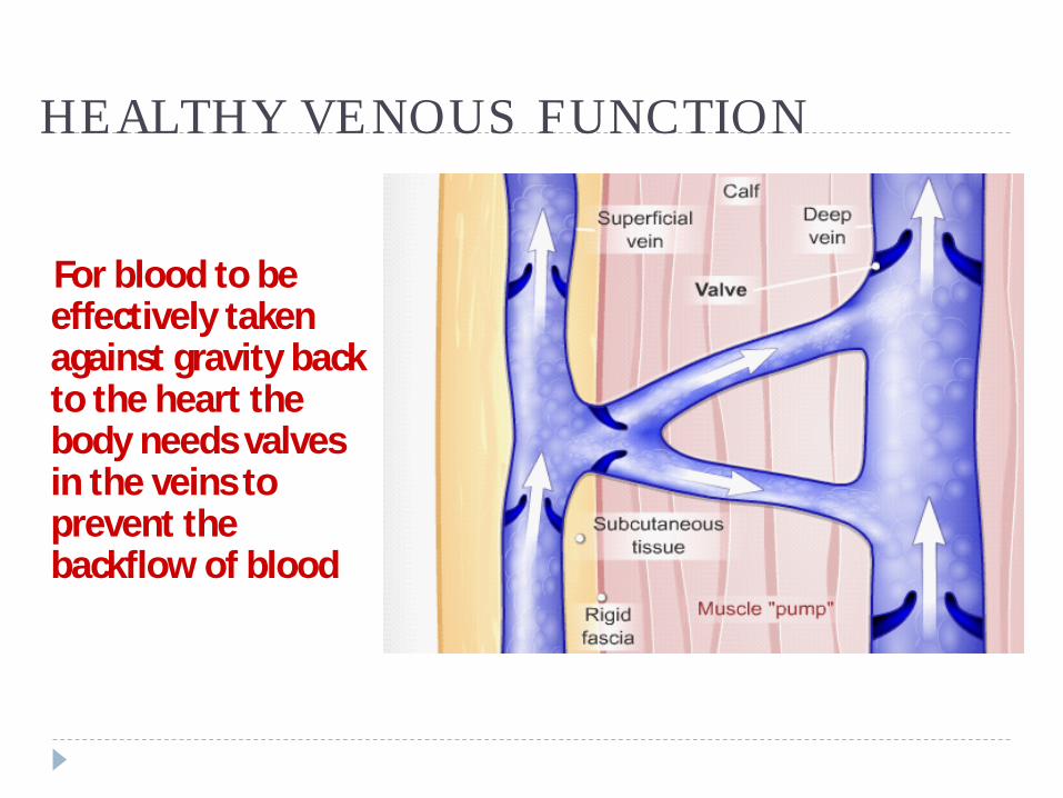
HEALTHY VENOUS FUNCTION
For blood to be effectively taken against gravity back to the heart the body needs valves in the veins to prevent the backflow of blood
Leg Ulcers
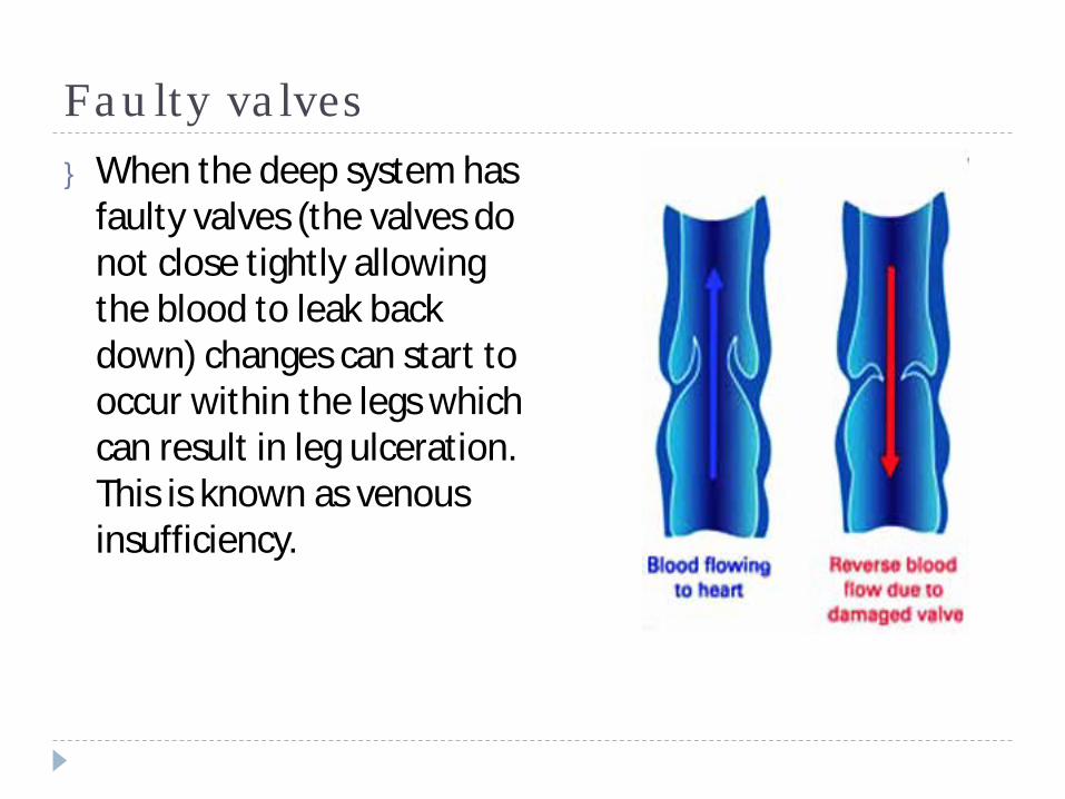
Faulty valves} When the deep system has
faulty valves (the valves do not close tightly allowing the blood to leak back down) changes can start to occur within the legs which can result in leg ulceration. This is known as venous insufficiency.
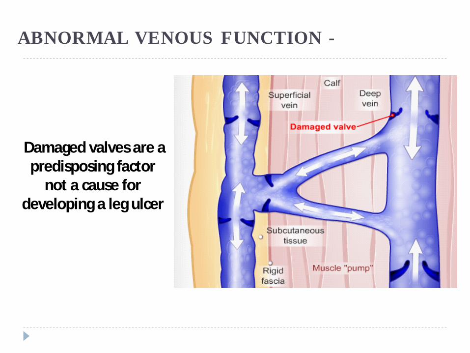
ABNORMAL VENOUS FUNCTION -
Damaged valves are a predisposing factor
not a cause for developing a leg ulcer
Leg Ulcers
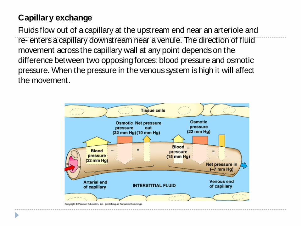
Capillary exchangeFluids flow out of a capillary at the upstream end near an arteriole and re- enters a capillary downstream near a venule. The direction of fluid movement across the capillary wall at any point depends on the difference between two opposing forces: blood pressure and osmotic pressure. When the pressure in the venous system is high it will affect the movement.
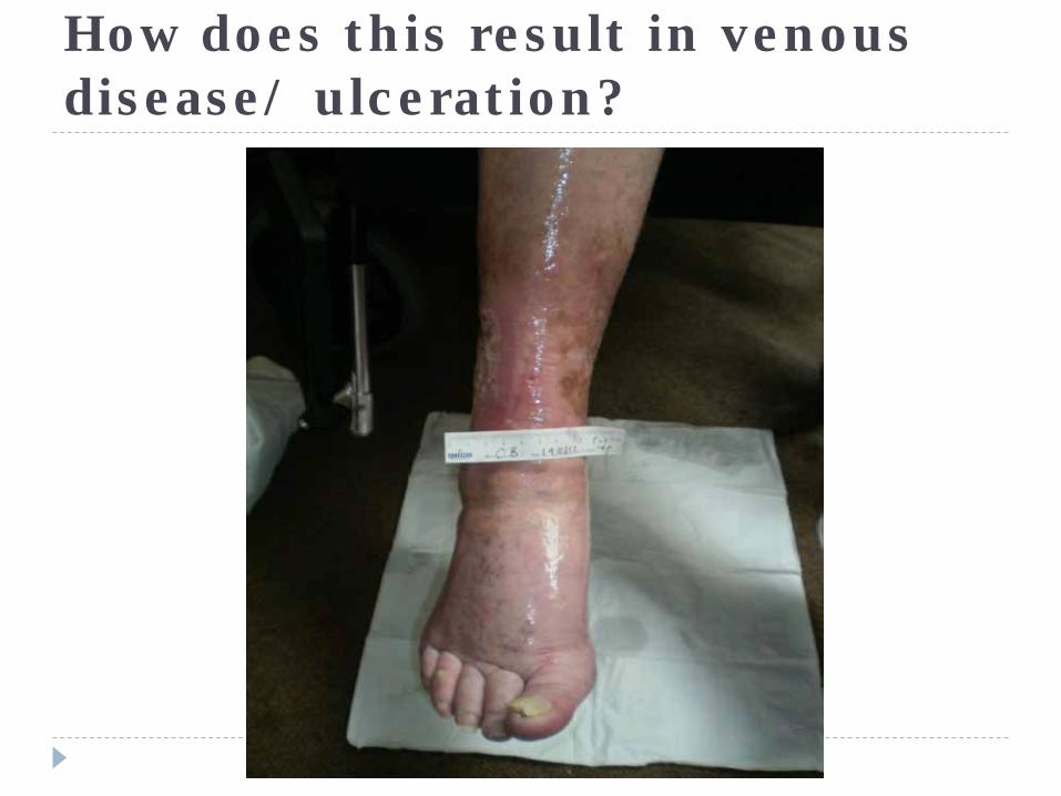
How does this result in venous disease/ ulceration?
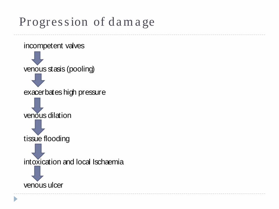
Progression of damage
incompetent valves
venous stasis (pooling)
exacerbates high pressure
venous dilation
tissue flooding
intoxication and local Ischaemia
venous ulcer
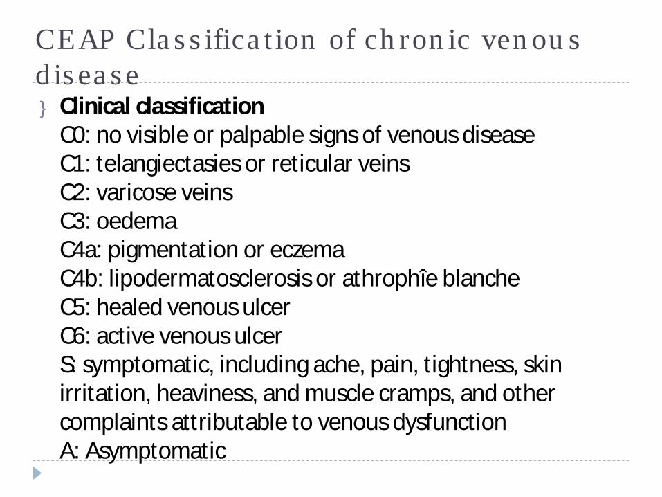
CEAP Classification of chronic venous disease} Clinical classification
C0: no visible or palpable signs of venous diseaseC1: telangiectasies or reticular veinsC2: varicose veinsC3: oedemaC4a: pigmentation or eczemaC4b: lipodermatosclerosis or athrophîe blancheC5: healed venous ulcerC6: active venous ulcerS: symptomatic, including ache, pain, tightness, skin irritation, heaviness, and muscle cramps, and other complaints attributable to venous dysfunctionA: Asymptomatic
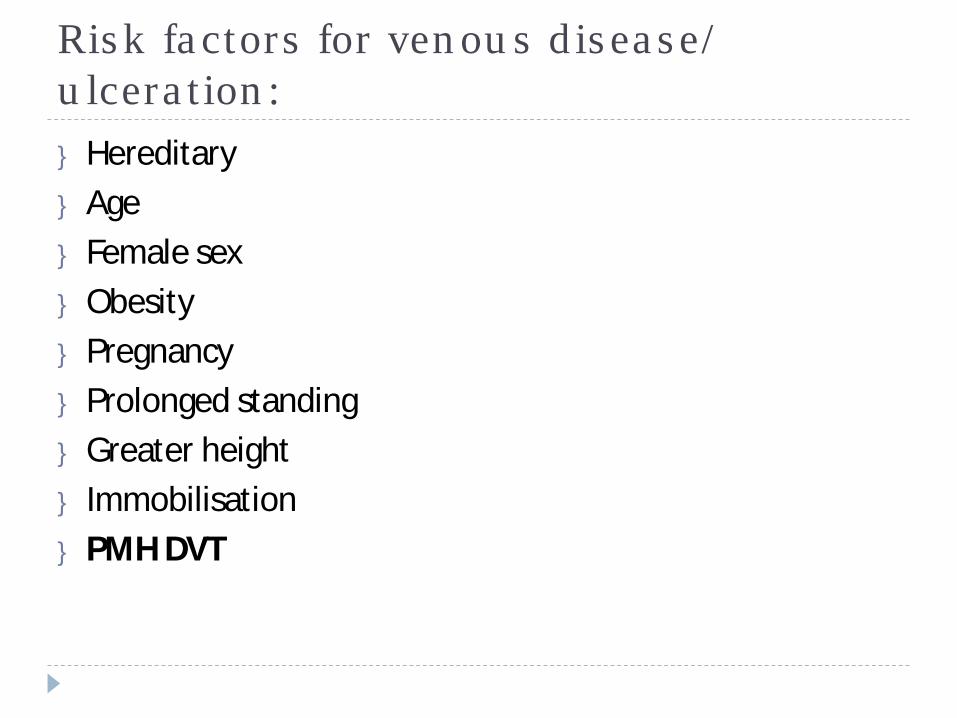
Risk factors for venous disease/ ulceration:} Hereditary} Age} Female sex} Obesity} Pregnancy} Prolonged standing} Greater height} Immobilisation} PMH DVT
Arterial ulcers
} Arterial insufficiency refers to poor blood circulation to the lower leg and foot and is most often due to atherosclerosis. In atherosclerosis the arteries become narrowed from deposits of fatty substances in the arterial vessel walls, often due to high levels of circulating cholesterol and aggravated by smoking and high blood pressure (hypertension). The arteries fail to deliver oxygen and nutrients to the leg and foot resulting in tissue breakdown.
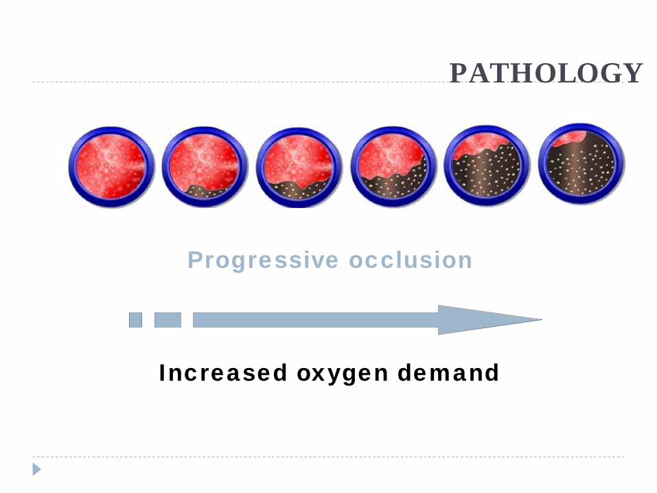
PATHOLOGY
Increased oxygen demand
Progressive occlusion
Leg Ulcers
Risk factors for arterial disease
} Smoking} Diabetes} Obesity} High BP} High cholesterol} Increasing age} Family history
Assessment
} For every 1 mistake for not knowing, 10 are made for not looking!!
Assessment} Obtaining a diagnosis can only
be achieved with a robust leg ulcer assessment
} A leg ulcer assessment, including a doppler and/ or lower limb assessment should be carried out within 1 - 2 weeks of the patient presenting
} Doppler is only an ‘aid’ to diagnosis not the ‘be all and end all’…. LOOK AT THE LIMB –WHAT DOES IT TELL YOU?
Assessing patients with leg ulceration
} 1 – Patient assessment (Extrinsic factors)} 2 – Patient assessment (Intrinsic factors)} 3 – Lower limb assessment} 4 – Wound assessment
Patient assessment
} What information do you need to obtain when you carry out a patient assessment?
} Why is this information important/ relevant?
} From the case study you have in front of you, what information is relevant and why?
} Is this information assisting your diagnosis?
} Do you require more information at this point?
Feedback
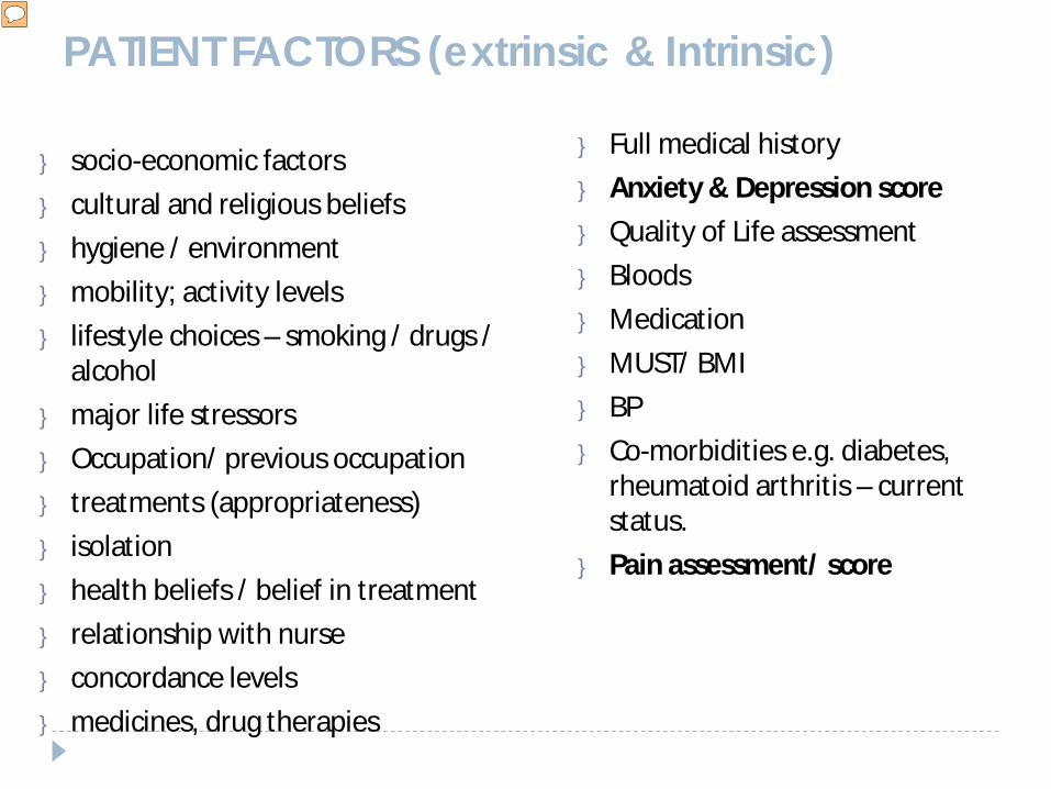
PATIENT FACTORS (extrinsic & Intrinsic)
} socio-economic factors} cultural and religious beliefs} hygiene / environment} mobility; activity levels} lifestyle choices – smoking / drugs /
alcohol} major life stressors} Occupation/ previous occupation} treatments (appropriateness)} isolation } health beliefs / belief in treatment} relationship with nurse} concordance levels} medicines, drug therapies
} Full medical history } Anxiety & Depression score } Quality of Life assessment} Bloods} Medication } MUST/ BMI} BP} Co-morbidities e.g. diabetes,
rheumatoid arthritis – current status.
} Pain assessment/ score
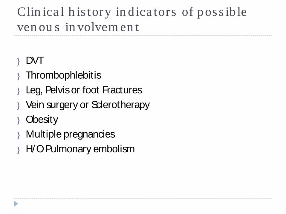
Clinical history indicators of possible venous involvement
} DVT } Thrombophlebitis} Leg, Pelvis or foot Fractures} Vein surgery or Sclerotherapy} Obesity} Multiple pregnancies} H/O Pulmonary embolism
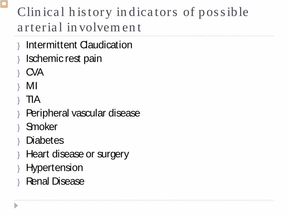
Clinical history indicators of possible arterial involvement} Intermittent Claudication} Ischemic rest pain} CVA} MI} TIA} Peripheral vascular disease} Smoker} Diabetes} Heart disease or surgery} Hypertension} Renal Disease
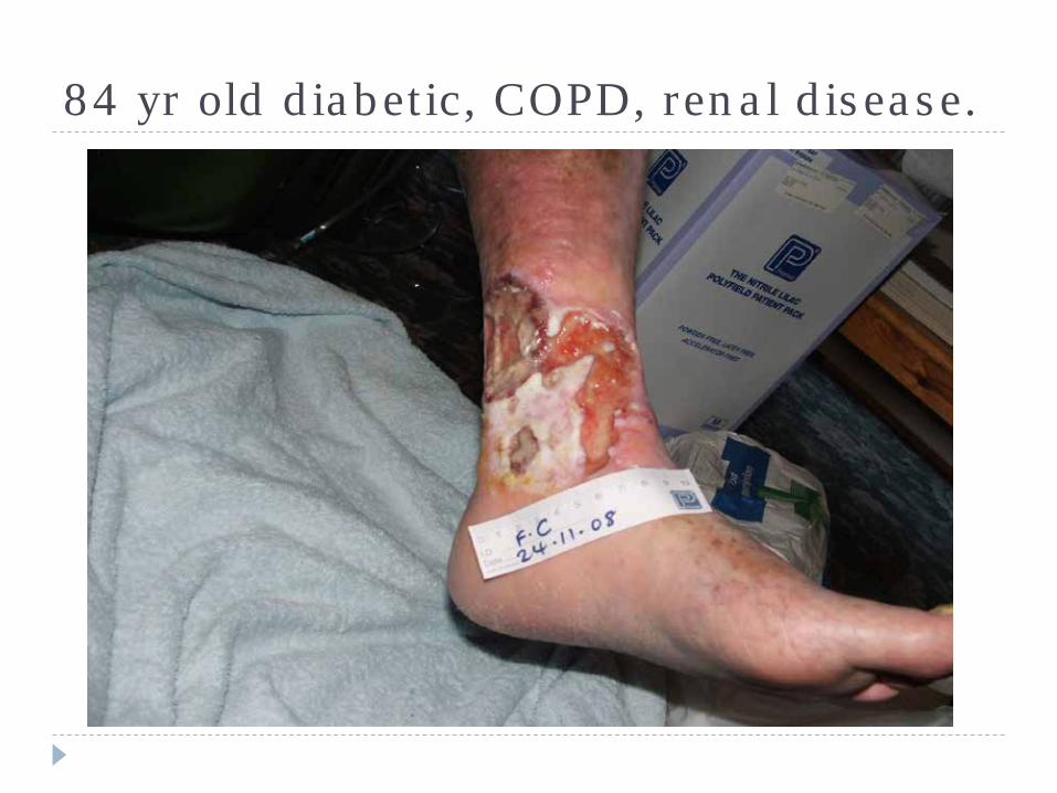
84 yr old diabetic, COPD, renal disease.
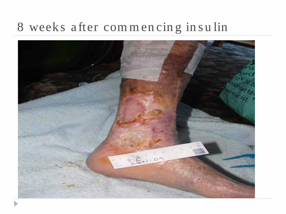
8 weeks after commencing insulin
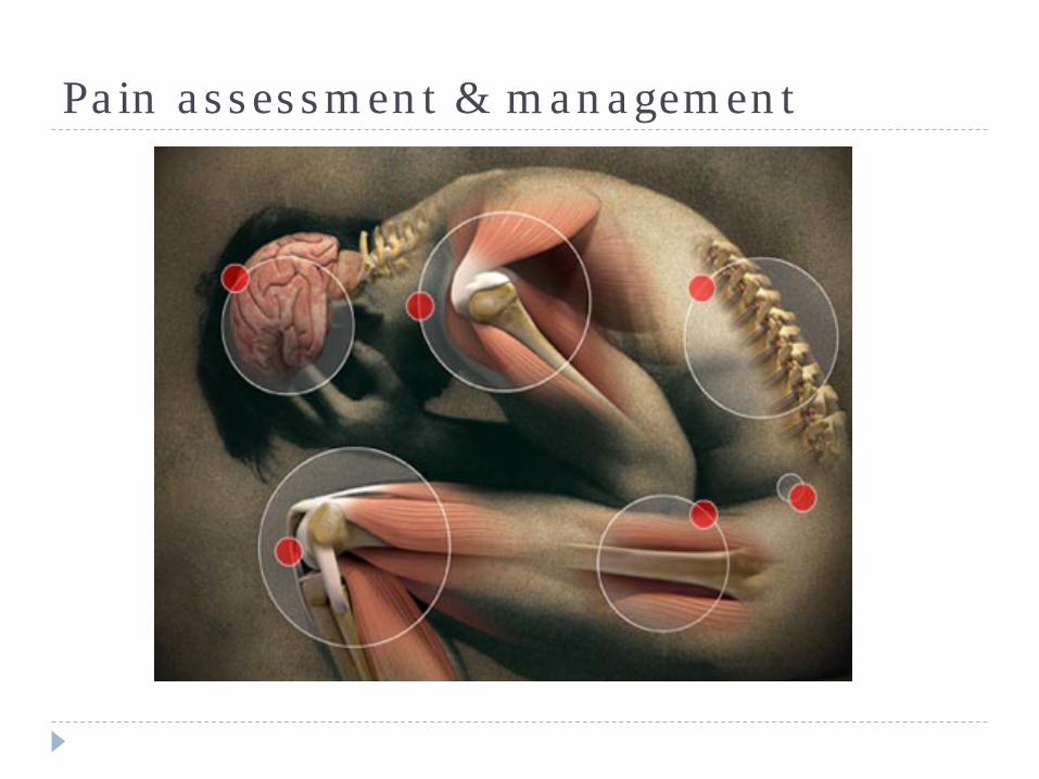
Pain assessment & management
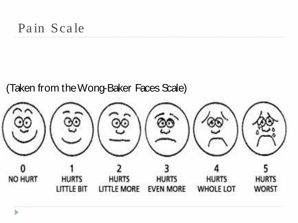
Pain Scale
} (Taken from the Wong-Baker Faces Scale)
Abbey Pain scale} For measurement of pain in people with dementia who
cannot verbalise.} Focusses on: vocalisation (whimpering, groaning, crying)} Facial expression} Changes in body language} Behavioural change} Physiological change (Temp, pulse or BP)} Physical changes (Skin tears, pressure areas, contractures)
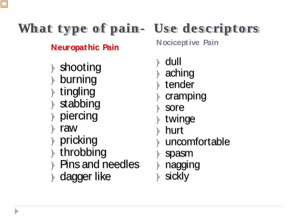
What type of pain- Use descriptorsNeuropathic Pain
} shooting } burning} tingling } stabbing } piercing } raw } pricking} throbbing} Pins and needles } dagger like
Nociceptive Pain
} dull } aching } tender } cramping} sore } twinge} hurt} uncomfortable } spasm} nagging } sickly
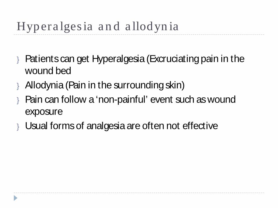
Hyperalgesia and allodynia
} Patients can get Hyperalgesia (Excruciating pain in the wound bed
} Allodynia (Pain in the surrounding skin)} Pain can follow a ‘non-painful’ event such as wound
exposure} Usual forms of analgesia are often not effective
Lower limb assessment
} Signs and symptoms of leg ulceration
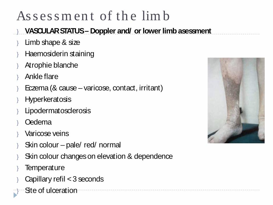
Assessment of the limb} VASCULAR STATUS – Doppler and/ or lower limb asessment} Limb shape & size} Haemosiderin staining} Atrophie blanche} Ankle flare} Eczema (& cause – varicose, contact, irritant)} Hyperkeratosis} Lipodermatosclerosis} Oedema} Varicose veins} Skin colour – pale/ red/ normal} Skin colour changes on elevation & dependence} Temperature} Capillary refil < 3 seconds} Site of ulceration
Acute and chronic wound, Ruth A. Bryant lower extremity ulcers, chapter 12, 2000
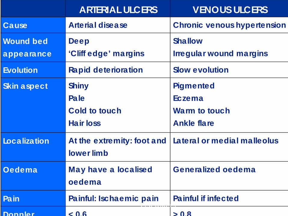
ARTERIAL ULCERS VENOUS ULCERSCause Arterial disease Chronic venous hypertension
Wound bedappearance
Deep‘Cliff edge’ margins
ShallowIrregular wound margins
Evolution Rapid deterioration Slow evolution
Skin aspect ShinyPaleCold to touchHair loss
PigmentedEczemaWarm to touch Ankle flare
Localization At the extremity: foot and lower limb
Lateral or medial malleolus
Oedema May have a localised oedema
Generalized oedema
Pain Painful: Ischaemic pain Painful if infected
Doppler < 0.6 > 0.8 Leg Ulcers
Lymphovenous disease} Venous and lymphovenous disease is progressive} Symptoms manifest as skin changes} These deteriorate as the disease progresses} Following the mid term disease stage (atrophie blanche,
haemosiderin staining etc) legs progress to the advanced stage.
} This includes enhanced skin folds, papillomatosis (superficial lymphatic vessels protruding through the skin), lymphorrhoea (wet legs), cellulitis.
} On reaching this stage your management plan would differ to that of a standard venous leg ulcer patient.
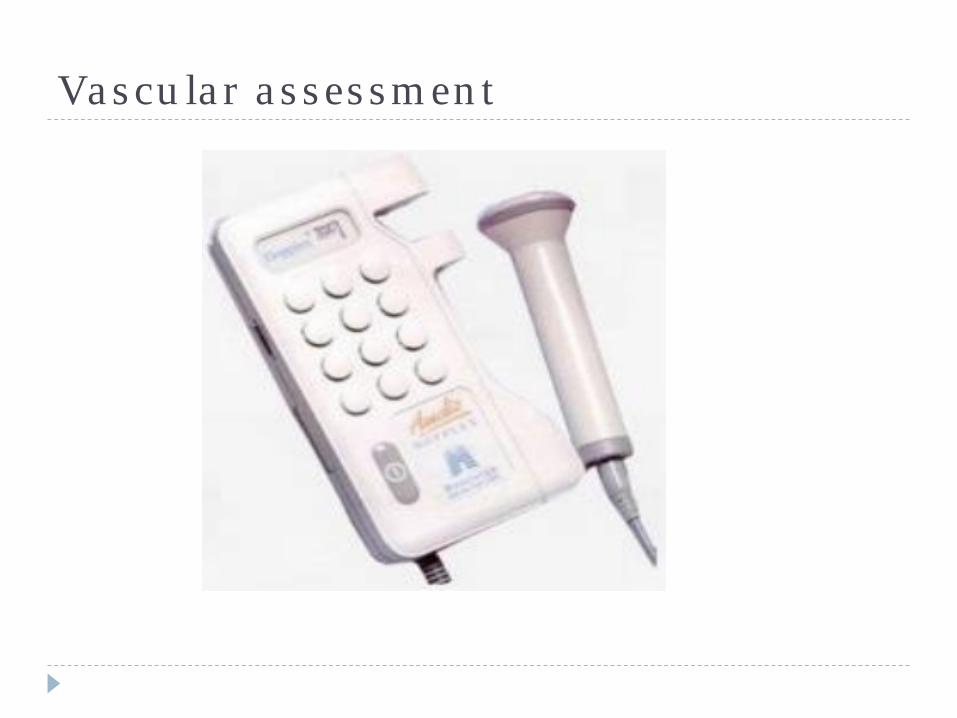
Vascular assessment
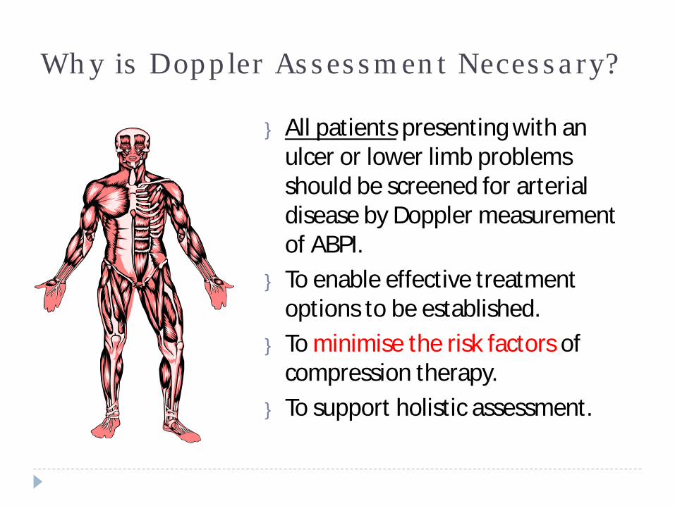
Why is Doppler Assessment Necessary?
} All patients presenting with an ulcer or lower limb problems should be screened for arterial disease by Doppler measurement of ABPI.
} To enable effective treatment options to be established.
} To minimise the risk factors of compression therapy.
} To support holistic assessment.
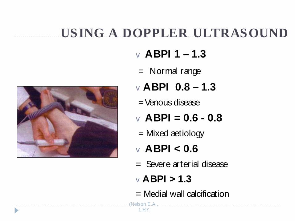
USING A DOPPLER ULTRASOUNDvABPI 1 – 1.3= Normal range
vABPI 0.8 – 1.3= Venous disease
vABPI = 0.6 - 0.8= Mixed aetiology
vABPI < 0.6= Severe arterial disease
vABPI > 1.3= Medial wall calcification
(Nelson E.A., 1996)Graduated Compression Therapy
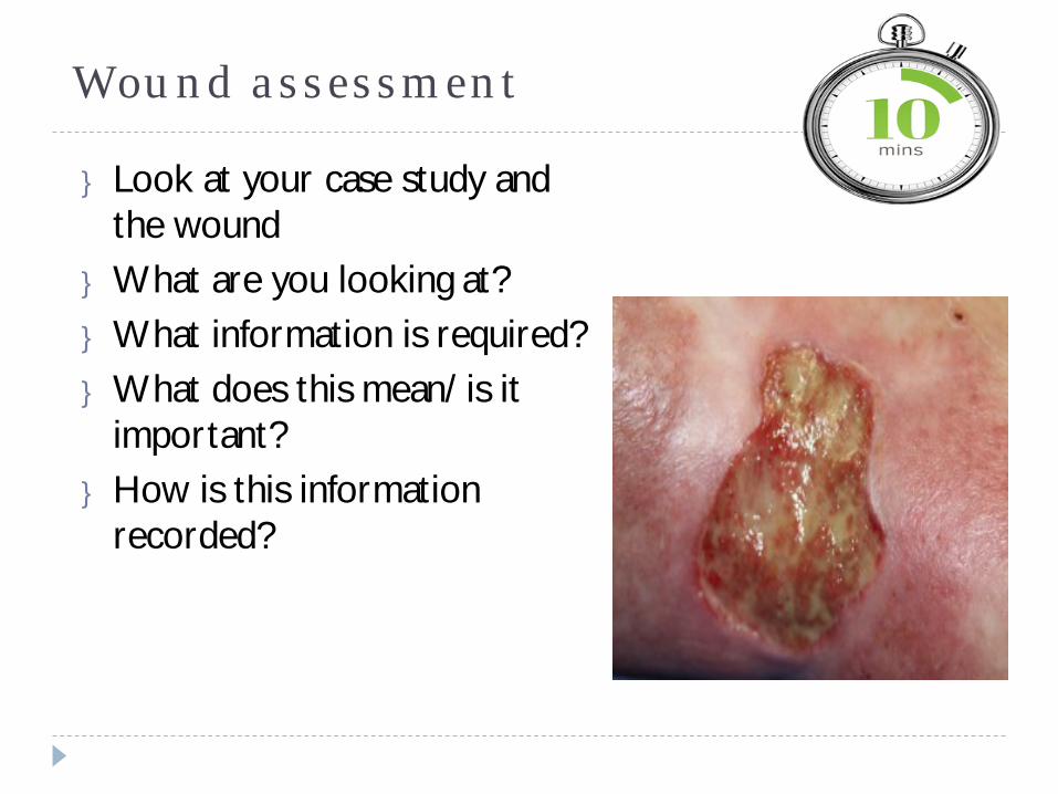
Wound assessment
} Look at your case study and the wound
} What are you looking at?} What information is required?} What does this mean/ is it
important?} How is this information
recorded?

Feedback
Wound assessment} Cause } Is it a reoccurrence?} Duration} Wound area in cm² as a baseline (Is it bigger/ smaller and in what
timescale)} Tissue type (including hypergranulation)} Infection/ biofilm prescence } Wound edges} Odour} Type and level of exudate} Peri wound skin status} Photograph} Previous management regimes} History of healing rates
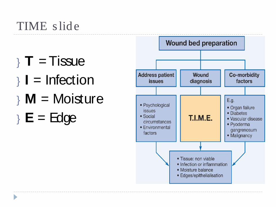
TIME slide
}T = Tissue} I = Infection}M = Moisture} E = Edge
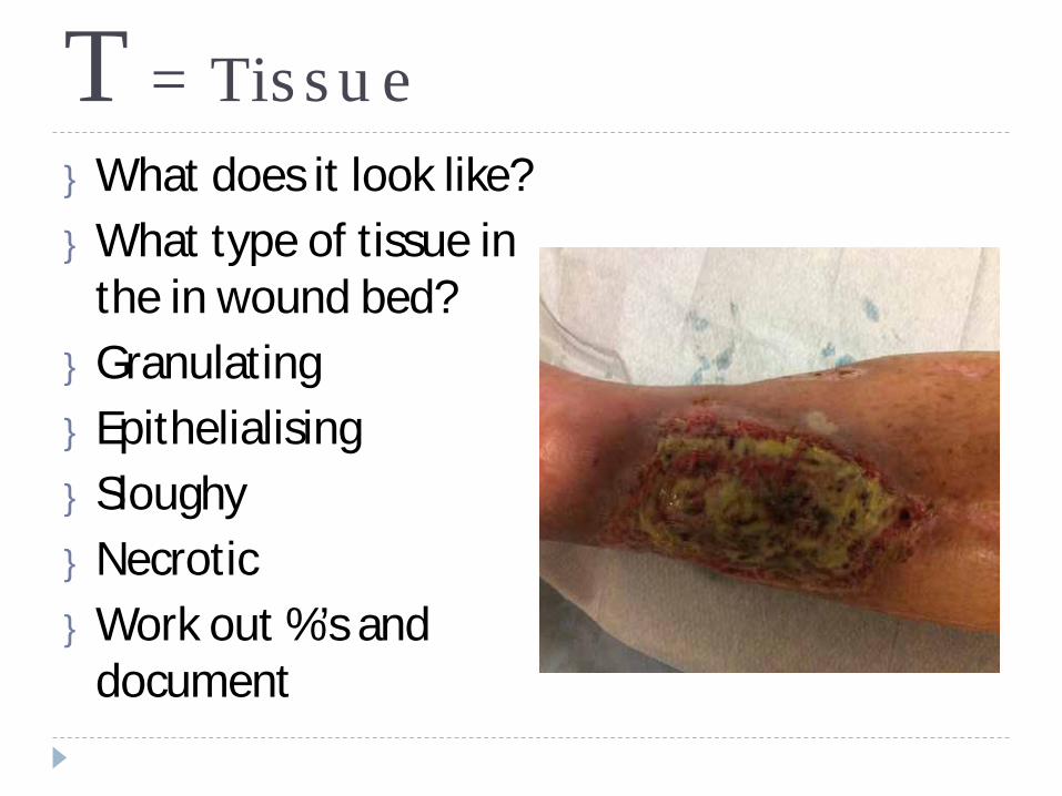
T = Tissue} What does it look like?} What type of tissue in
the in wound bed?} Granulating} Epithelialising} Sloughy} Necrotic} Work out %’s and
document
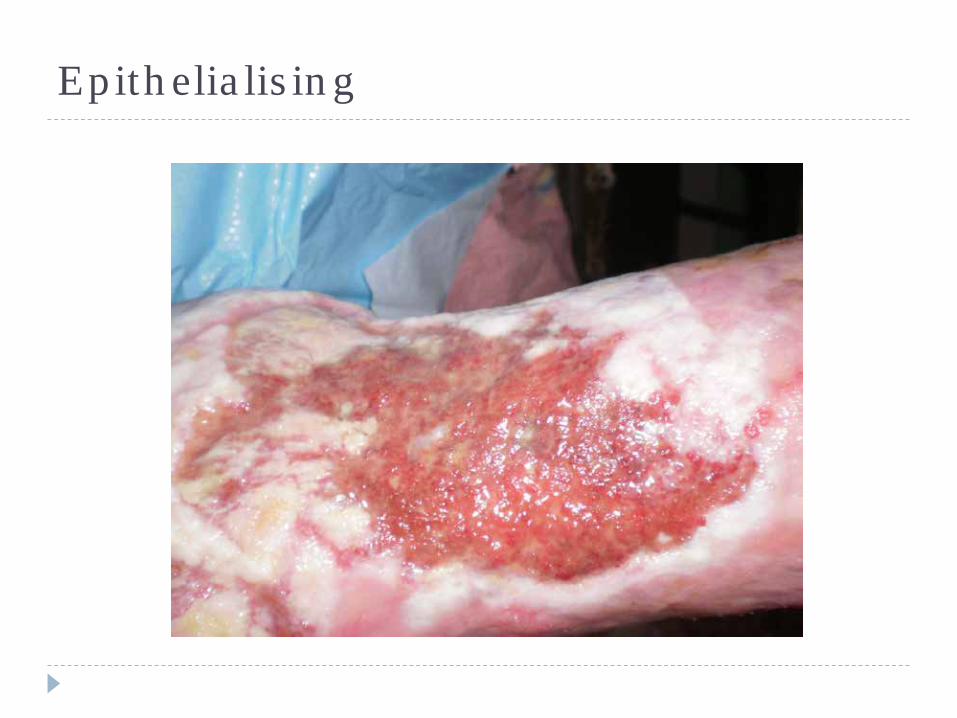
Epithelialising
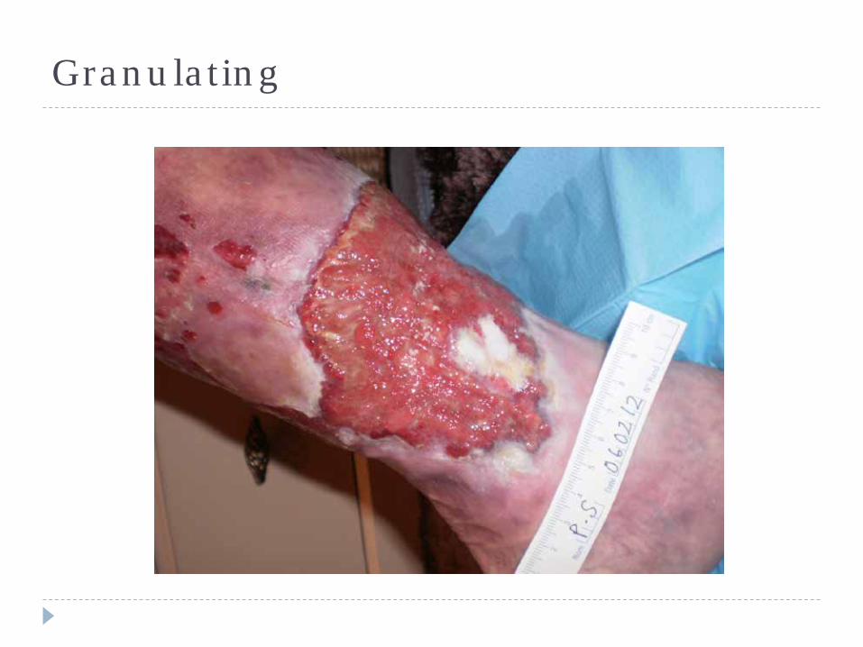
Granulating
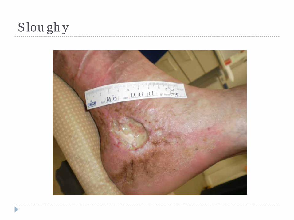
Sloughy
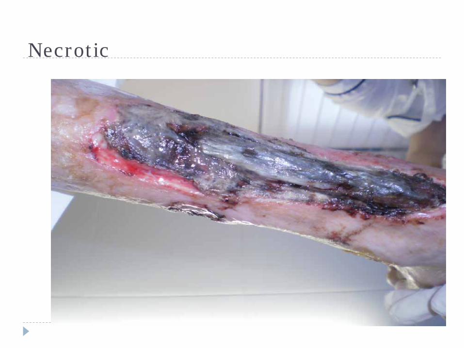
Necrotic
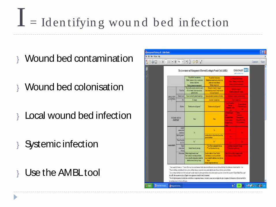
I = Identifying wound bed infection
} Wound bed contamination
} Wound bed colonisation
} Local wound bed infection
} Systemic infection
} Use the AMBL tool
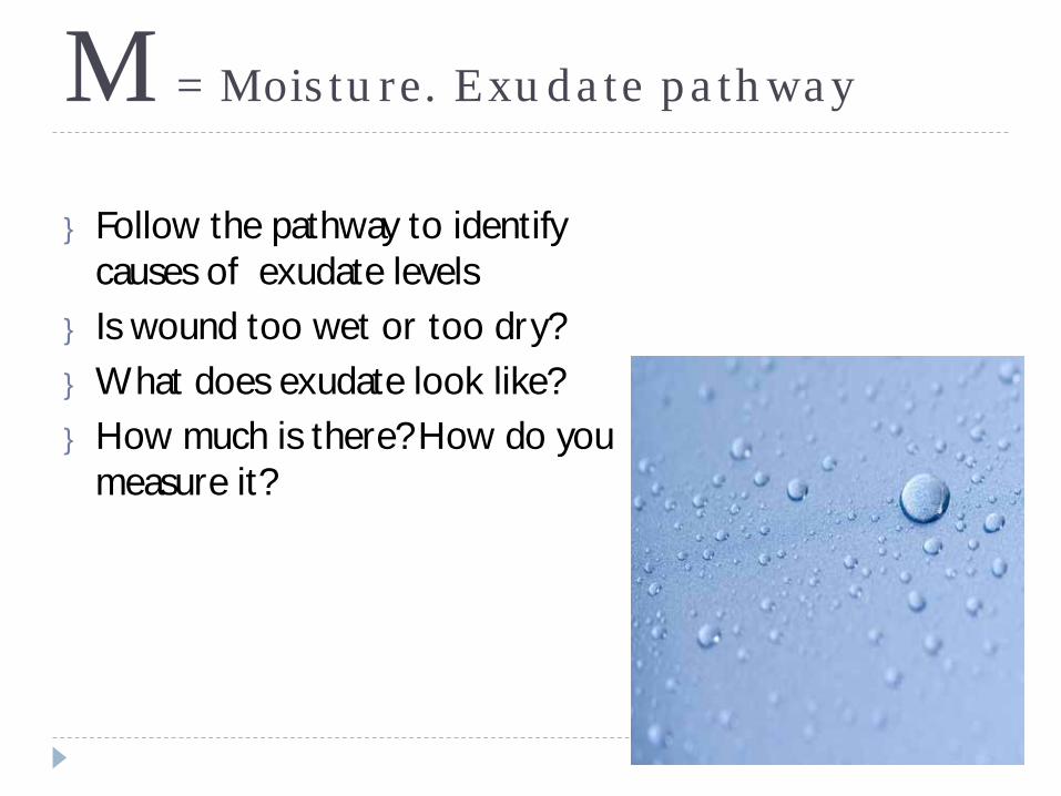
M = Moisture. Exudate pathway
} Follow the pathway to identify causes of exudate levels
} Is wound too wet or too dry?} What does exudate look like?} How much is there? How do you
measure it?
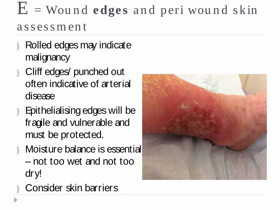
E = Wound edges and peri wound skin assessment} Rolled edges may indicate
malignancy} Cliff edges/ punched out
often indicative of arterial disease
} Epithelialising edges will be fragile and vulnerable and must be protected.
} Moisture balance is essential – not too wet and not too dry!
} Consider skin barriers
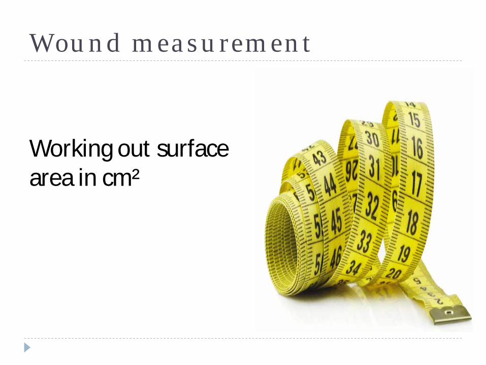
Wound measurement
Working out surface area in cm²
Identifying abnormal or difficult to treat leg ulcers
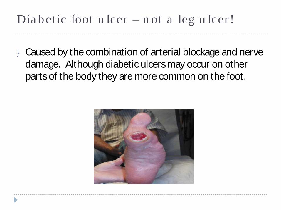
Diabetic foot ulcer – not a leg ulcer!
} Caused by the combination of arterial blockage and nerve damage. Although diabetic ulcers may occur on other parts of the body they are more common on the foot.
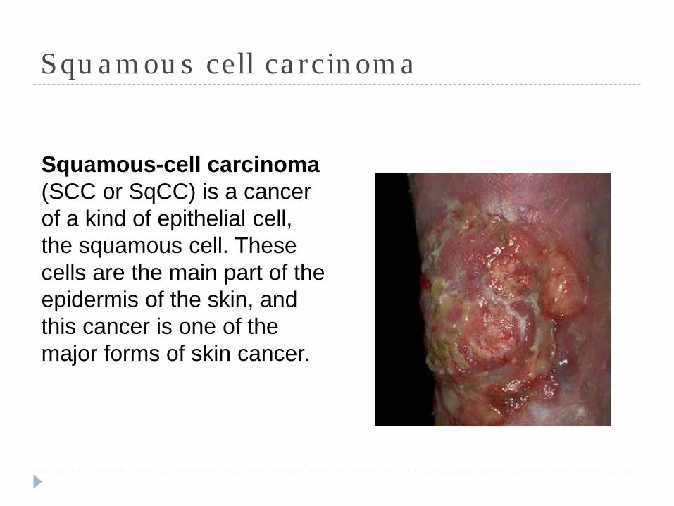
Squamous cell carcinoma
Squamous-cell carcinoma(SCC or SqCC) is a cancer of a kind of epithelial cell, the squamous cell. These cells are the main part of the epidermis of the skin, and this cancer is one of the major forms of skin cancer.
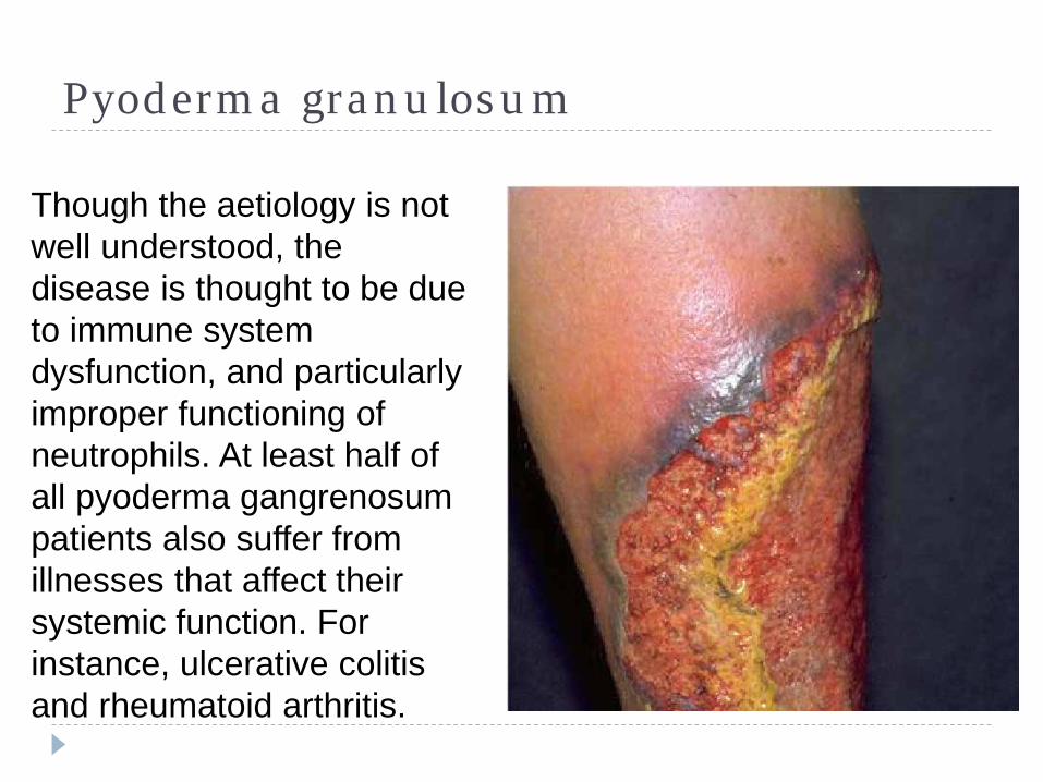
Pyoderma granulosum
Though the aetiology is not well understood, the disease is thought to be due to immune system dysfunction, and particularly improper functioning of neutrophils. At least half of all pyoderma gangrenosum patients also suffer from illnesses that affect their systemic function. For instance, ulcerative colitis and rheumatoid arthritis.
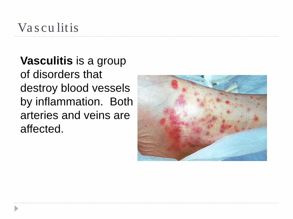
Vasculitis
Vasculitis is a group of disorders that destroy blood vessels by inflammation. Both arteries and veins are affected.
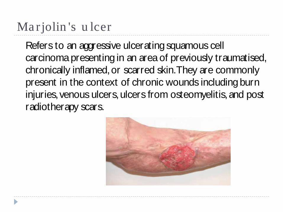
Marjolin's ulcerRefers to an aggressive ulcerating squamous cell carcinoma presenting in an area of previously traumatised, chronically inflamed, or scarred skin. They are commonly present in the context of chronic wounds including burn injuries, venous ulcers, ulcers from osteomyelitis, and post radiotherapy scars.
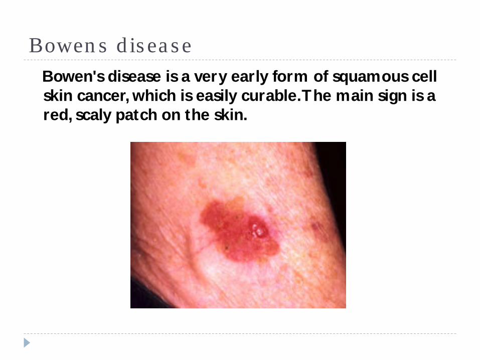
Bowens diseaseBowen's disease is a very early form of squamous cell skin cancer, which is easily curable. The main sign is a red, scaly patch on the skin.
Leg ulcer management
Venous leg ulcer management – using the VLU pathway.

Set out your milestones
Milestones} Assessment} Diagnosis} Consider – Do I need to refer at this stage?} If no…. } Management } Leg ulcer pathway allocation (If venous)} Re-assessment (every 6 weeks)} Progressing? If no then refer to tissue viability.} Healed?} If yes…. Prevention of recurrence care plan
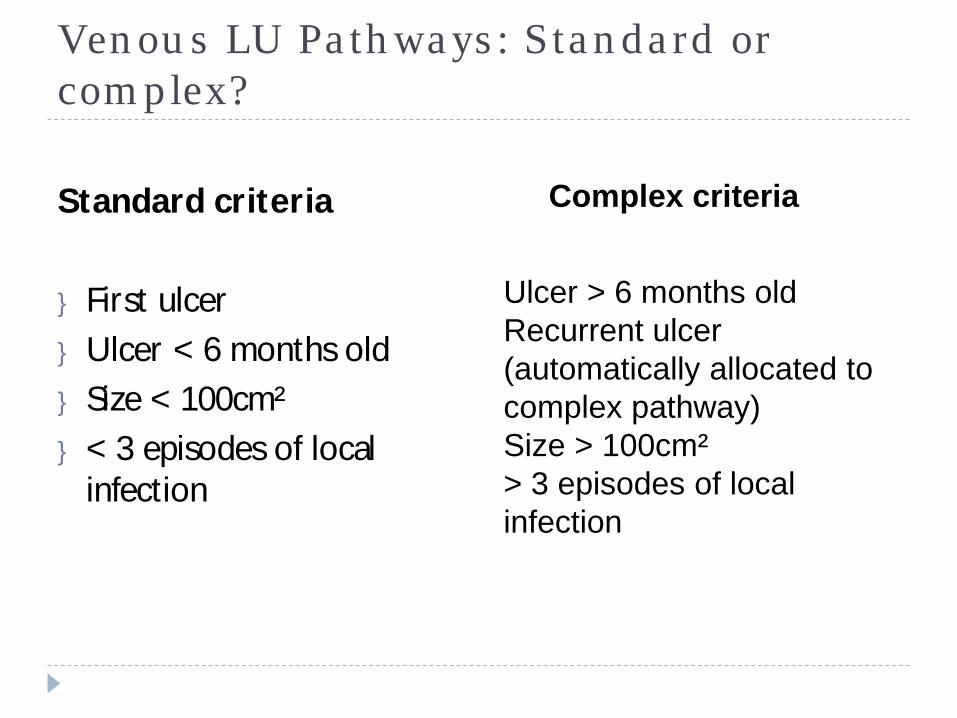
Venous LU Pathways: Standard or complex?
Standard criteria
} First ulcer} Ulcer < 6 months old} Size < 100cm²} < 3 episodes of local
infection
Complex criteria
Ulcer > 6 months oldRecurrent ulcer (automatically allocated to complex pathway)Size > 100cm²> 3 episodes of local infection
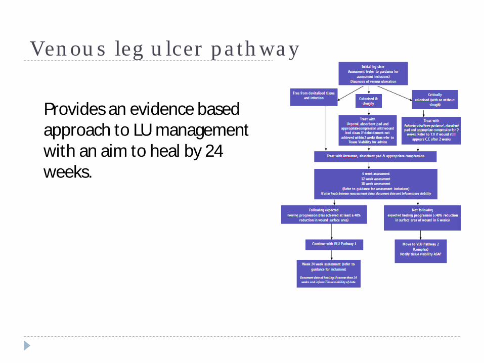
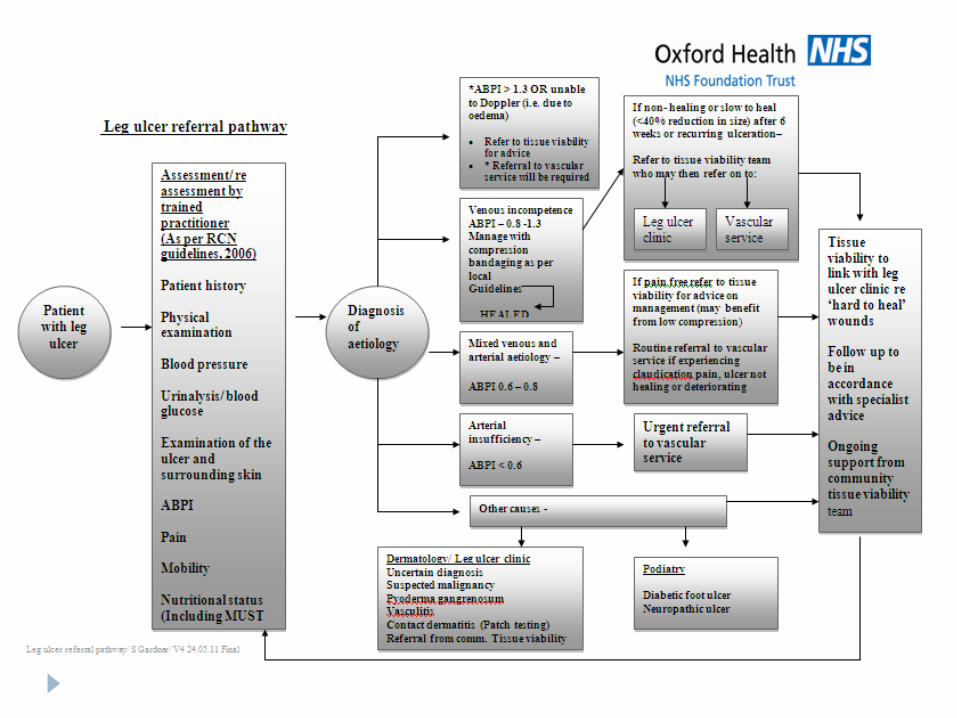
Venous leg ulcer pathway
Provides an evidence based approach to LU management with an aim to heal by 24 weeks.
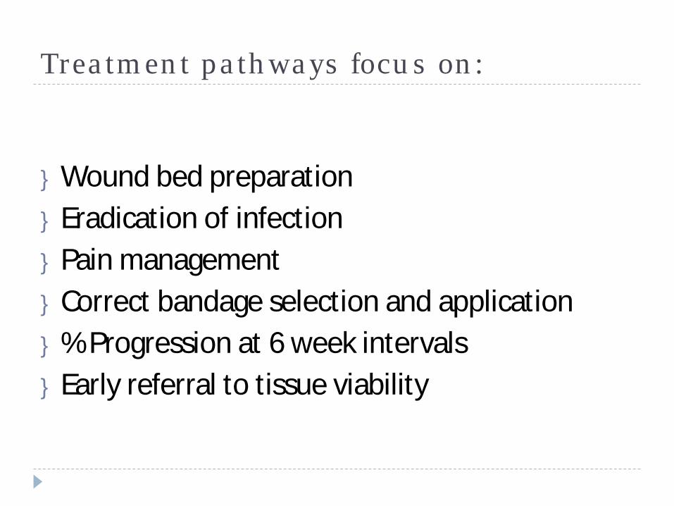
Treatment pathways focus on:
} Wound bed preparation} Eradication of infection} Pain management} Correct bandage selection and application} % Progression at 6 week intervals} Early referral to tissue viability
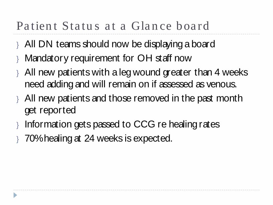
Patient Status at a Glance board} All DN teams should now be displaying a board} Mandatory requirement for OH staff now} All new patients with a leg wound greater than 4 weeks
need adding and will remain on if assessed as venous.} All new patients and those removed in the past month
get reported} Information gets passed to CCG re healing rates} 70% healing at 24 weeks is expected.
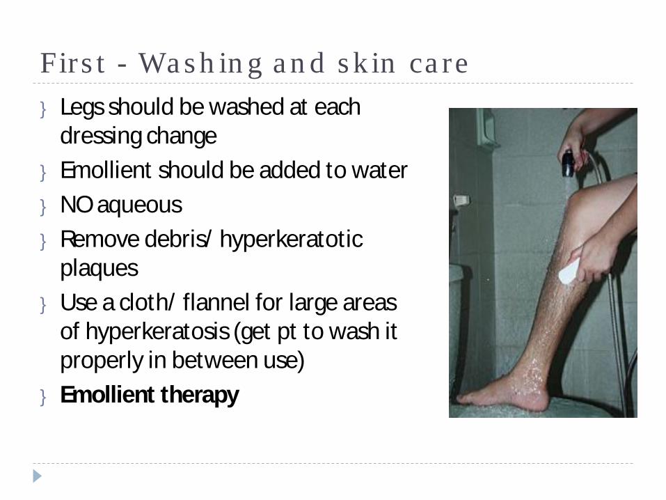
First - Washing and skin care} Legs should be washed at each
dressing change} Emollient should be added to water} NO aqueous} Remove debris/ hyperkeratotic
plaques} Use a cloth/ flannel for large areas
of hyperkeratosis (get pt to wash it properly in between use)
} Emollient therapy
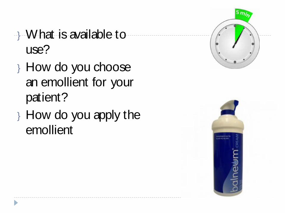
} What is available to use?} How do you choose
an emollient for your patient?} How do you apply the
emollient

Feedback
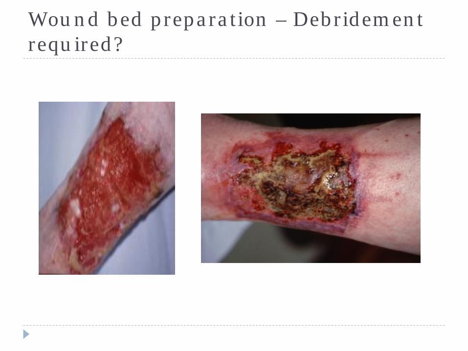
Wound bed preparation – Debridement required?
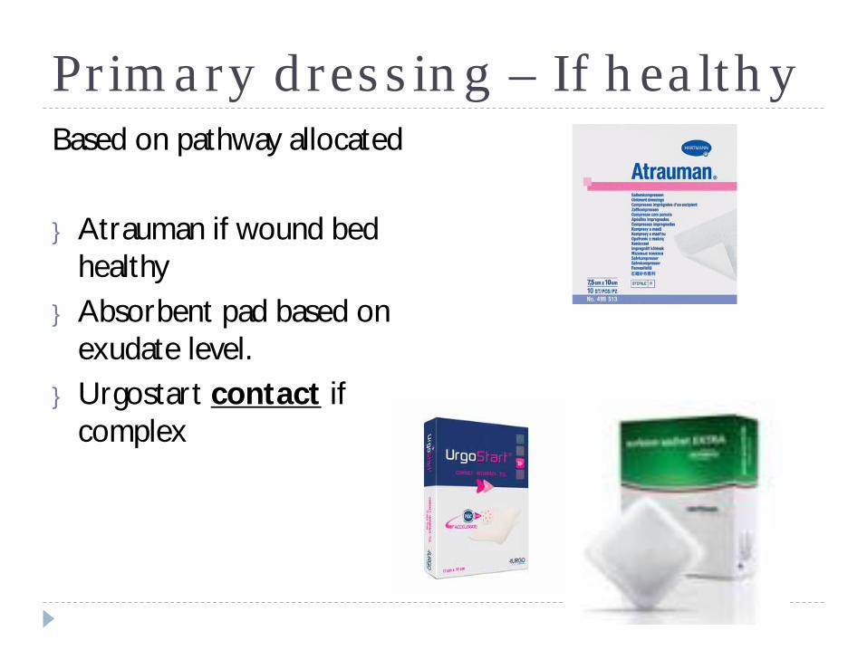
Primary dressing – If healthyBased on pathway allocated
} Atrauman if wound bed healthy
} Absorbent pad based on exudate level.
} Urgostart contact if complex
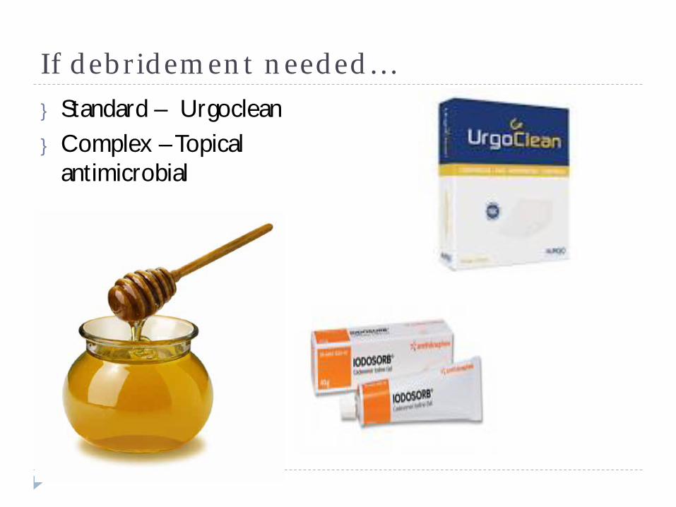
If debridement needed…} Standard – Urgoclean} Complex – Topical
antimicrobial
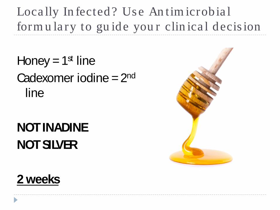
Locally Infected? Use Antimicrobial formulary to guide your clinical decision
Honey = 1st lineCadexomer iodine = 2nd
line
NOT INADINENOT SILVER
2 weeks
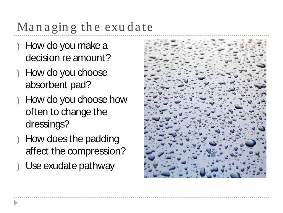
Managing the exudate} How do you make a
decision re amount?} How do you choose
absorbent pad?} How do you choose how
often to change the dressings?
} How does the padding affect the compression?
} Use exudate pathway
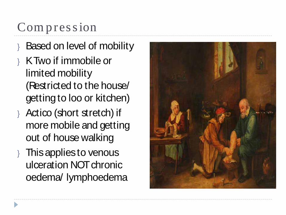
Compression} Based on level of mobility} K Two if immobile or
limited mobility (Restricted to the house/ getting to loo or kitchen)
} Actico (short stretch) if more mobile and getting out of house walking
} This applies to venous ulceration NOT chronic oedema/ lymphoedema
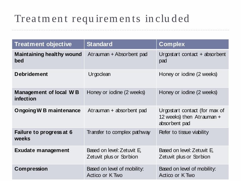
Treatment requirements included
Treatment objective Standard Complex
Maintaining healthy wound bed
Atrauman + Absorbent pad Urgostart contact + absorbent pad
Debridement Urgoclean Honey or iodine (2 weeks)
Management of local WB infection
Honey or iodine (2 weeks) Honey or iodine (2 weeks)
Ongoing WB maintenance Atrauman + absorbent pad Urgostart contact (for max of 12 weeks) then Atrauman + absorbent pad
Failure to progress at 6 weeks
Transfer to complex pathway Refer to tissue viability
Exudate management Based on level: Zetuvit E, Zetuvit plus or Sorbion
Based on level: Zetuvit E, Zetuvit plus or Sorbion
Compression Based on level of mobility: Actico or K Two
Based on level of mobility: Actico or K Two
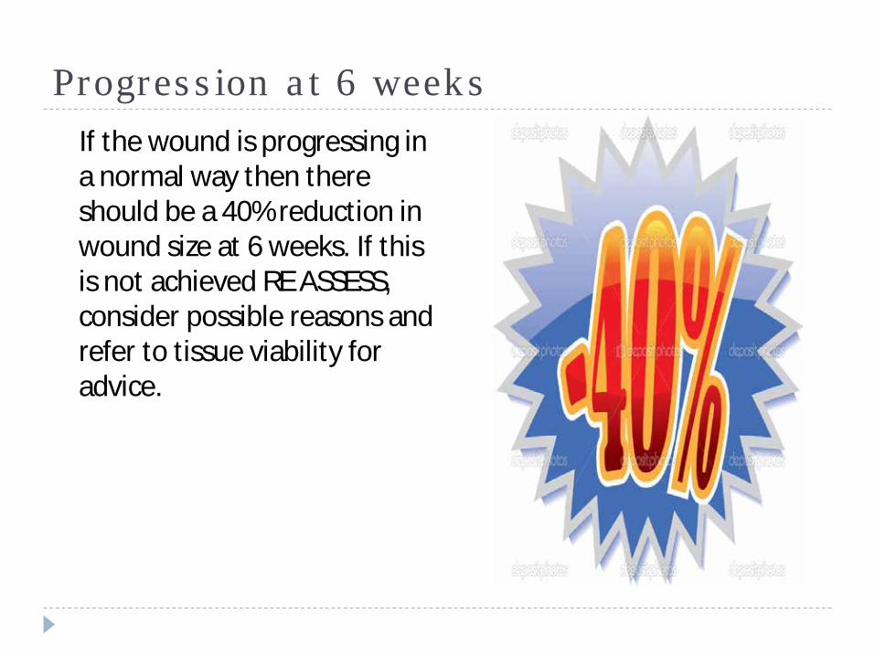
Progression at 6 weeksIf the wound is progressing in a normal way then there should be a 40% reduction in wound size at 6 weeks. If this is not achieved RE ASSESS, consider possible reasons and refer to tissue viability for advice.

Wound size progression
Ø 30 – 40 % = Continue on pathwayØ 20 – 30% = Consider:• Is the wound sloughy or infected?• Is the wound inflamed?• Is the compression on properly?• Has there been a change in Pts health?
Ø 0 – 20% = Refer to Tissue Viability for advice
Management plan should also include:
} Care plan for pain management}Mobility/ exercises} Lifestyle/ QoL
Exercise/ leg elevation
} “ In the case of an ulcer, it is not expedient to stand, especially if the ulcer be situated on the leg”
(Hippocrates, 400 yrs BC)
Topical steroids in wound care…The evidence???} Is known to work but why?} Not licensed to be used directly on wound beds so where do
you stand re accountability?} How much should be used and for how long?} Full of allergens – particularly the ones with antibiotics.} Possibly mask underlying problems} Can have similar long term effects as systemic steroids.} Should not routinely be used unless discussed with tissue
viability first. NEW PROTOCOL AVAILABLE} Trimovate now YELLOW listed (For continuation following
specialist recommendation only)and should be time specific.
Gallop through compression ….
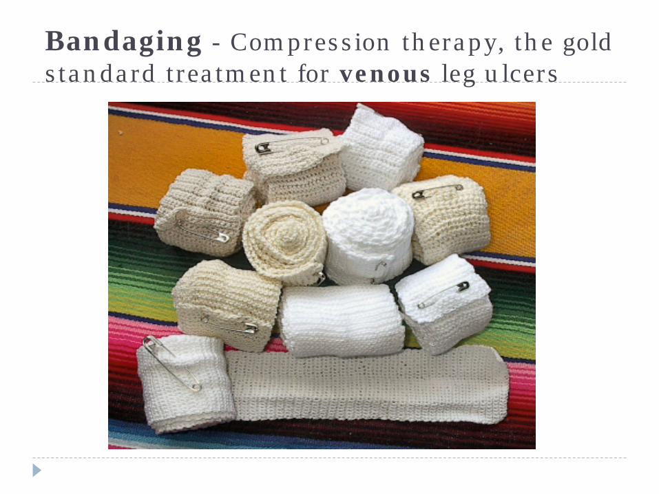
Bandaging - Compression therapy, the gold standard treatment for venous leg ulcers
Factors to be considered before applying compression
} Skin condition – delicate friable skin can be damaged by high levels of pressure
} Shape of the limb – the sub-bandage pressure and the pressure gradient will be altered by the limb shape in accordance with Laplace’s Law. Skin overlying exposed bony prominences may be subject to pressure damage
} Presence of neuropathy – the absence of a protective response increases the risk of sub-bandage pressure damage
} Presence of cardiac failure – rapid fluid shifts can be dangerous as it increases the preload of the heart
How does compression work?

Feedback
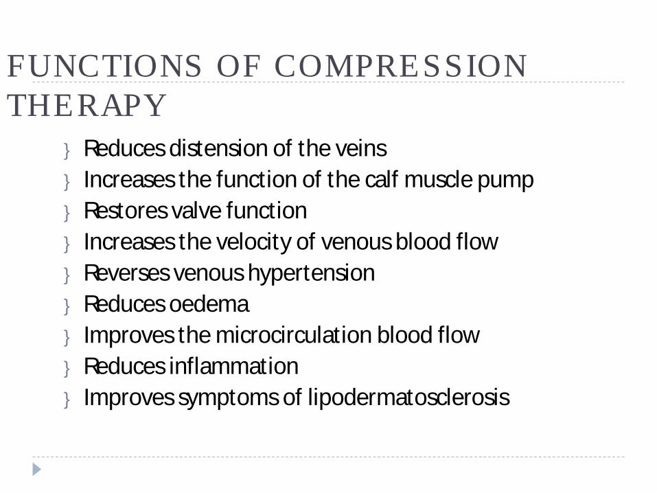
FUNCTIONS OF COMPRESSION THERAPY
} Reduces distension of the veins} Increases the function of the calf muscle pump} Restores valve function} Increases the velocity of venous blood flow} Reverses venous hypertension} Reduces oedema} Improves the microcirculation blood flow} Reduces inflammation} Improves symptoms of lipodermatosclerosis
Graduated Compression Therapy
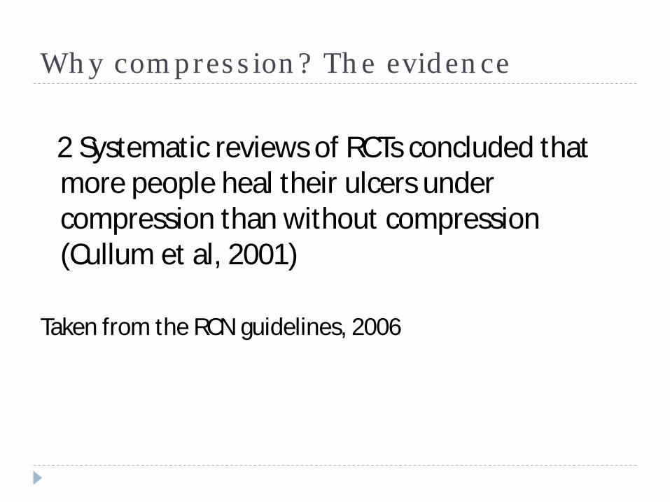
Why compression? The evidence
2 Systematic reviews of RCTs concluded that more people heal their ulcers under compression than without compression (Cullum et al, 2001)
Taken from the RCN guidelines, 2006
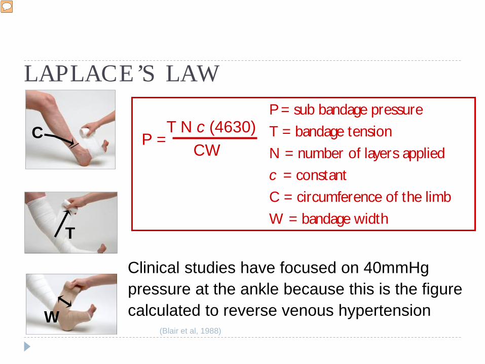
LAPLACE’S LAWP = sub bandage pressureT = bandage tensionN = number of layers appliedc = constant C = circumference of the limbW = bandage width
T N c (4630)CW
P =
Clinical studies have focused on 40mmHg pressure at the ankle because this is the figure calculated to reverse venous hypertension
(Blair et al, 1988)
Graduated Compression Therapy
C
T
W
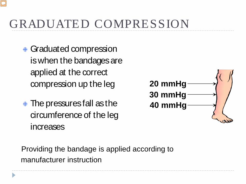
GRADUATED COMPRESSION
Graduated compression is when the bandages are applied at the correct compression up the leg
The pressures fall as the circumference of the leg increases
Providing the bandage is applied according to manufacturer instruction
Graduated Compression Therapy
20 mmHg30 mmHg40 mmHg
Bandages
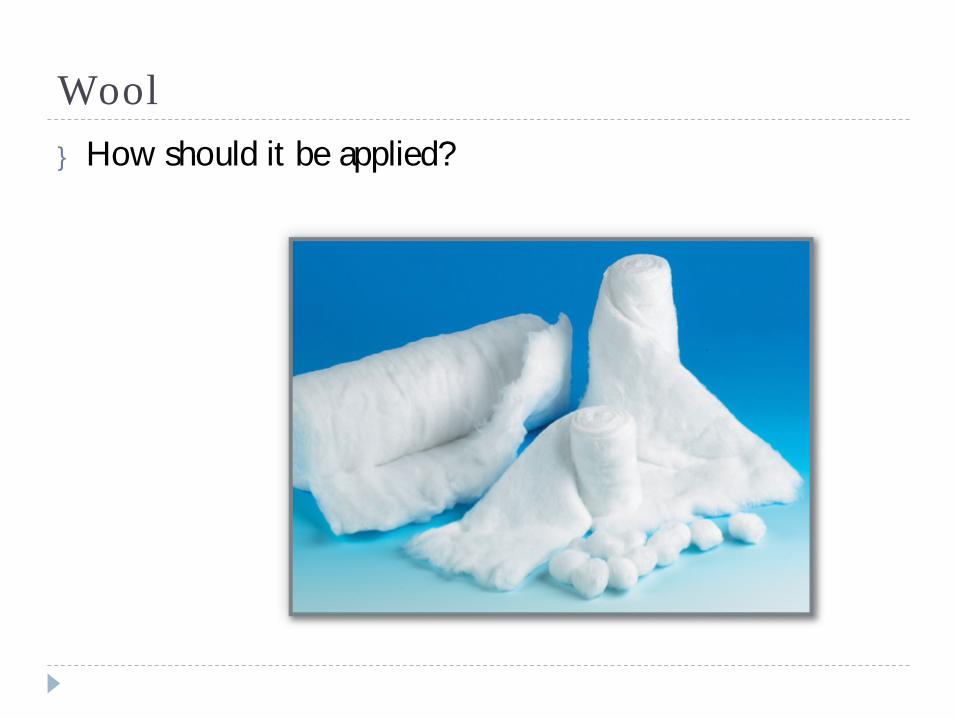
Wool} How should it be applied?
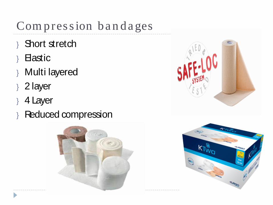
Compression bandages} Short stretch} Elastic} Multi layered} 2 layer} 4 Layer} Reduced compression
What is the difference between long (elastic) and short stretch (inelastic) bandages?
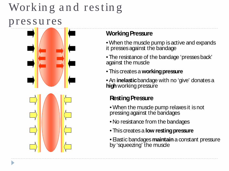
Working and resting pressures
Working Pressure•When the muscle pump is active and expands it presses against the bandage•The resistance of the bandage ‘presses back’ against the muscle•This creates a working pressure•An inelastic bandage with no ‘give’ donates a high working pressure
Resting Pressure•When the muscle pump relaxes it is not pressing against the bandages•No resistance from the bandages•This creates a low resting pressure•Elastic bandages maintain a constant pressure by ‘squeezing’ the muscle
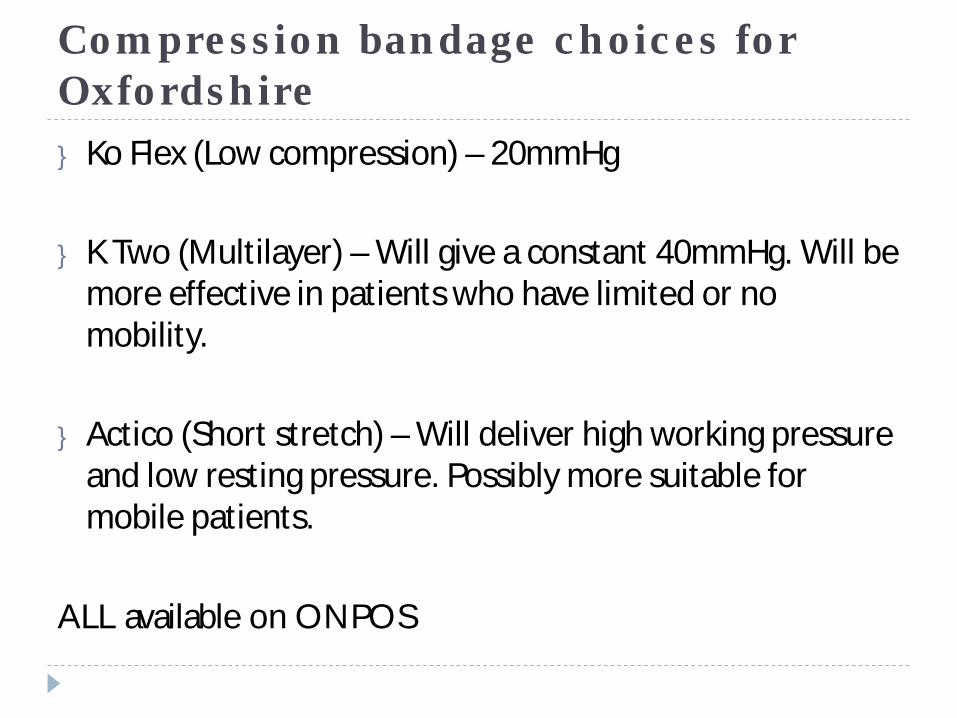
Compression bandage choices for Oxfordshire} Ko Flex (Low compression) – 20mmHg
} K Two (Multilayer) – Will give a constant 40mmHg. Will be more effective in patients who have limited or no mobility.
} Actico (Short stretch) – Will deliver high working pressure and low resting pressure. Possibly more suitable for mobile patients.
ALL available on ONPOS
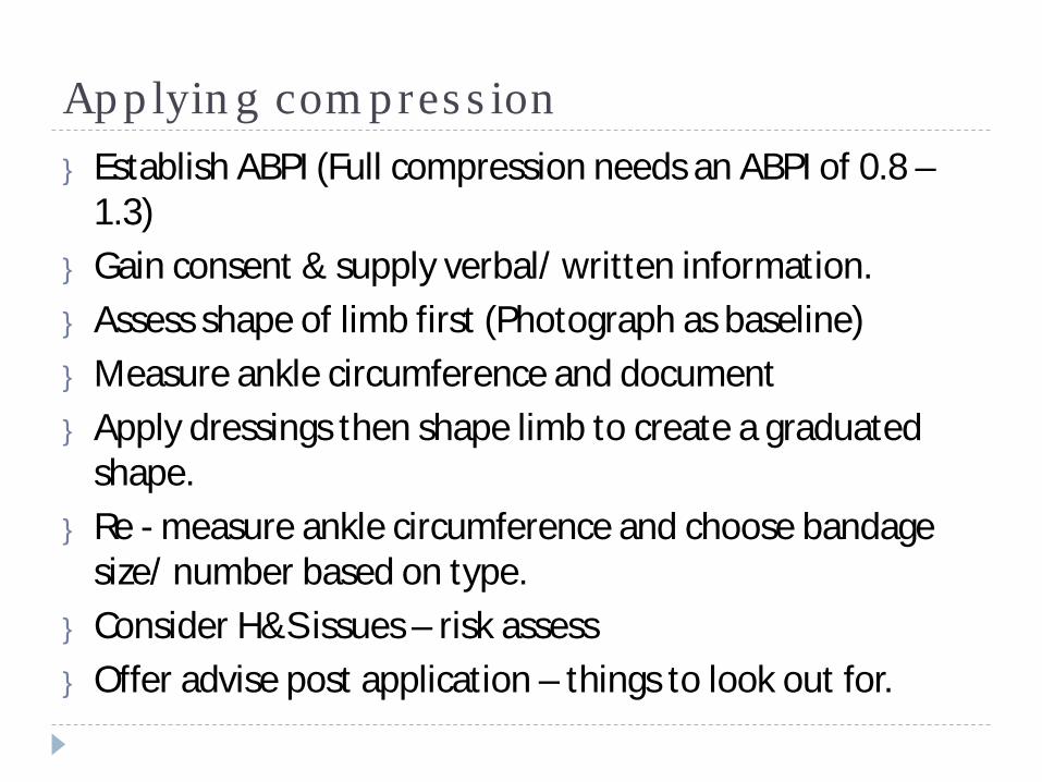
Applying compression} Establish ABPI (Full compression needs an ABPI of 0.8 –
1.3)} Gain consent & supply verbal/ written information.} Assess shape of limb first (Photograph as baseline)} Measure ankle circumference and document} Apply dressings then shape limb to create a graduated
shape.} Re - measure ankle circumference and choose bandage
size/ number based on type.} Consider H&S issues – risk assess} Offer advise post application – things to look out for.
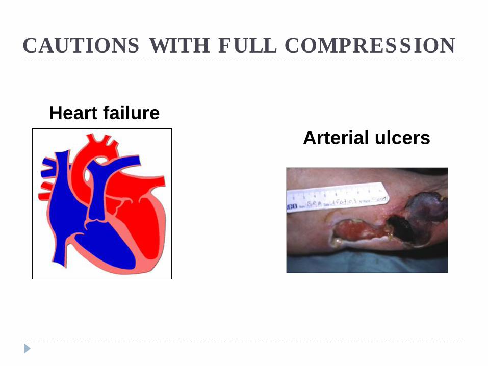
CAUTIONS WITH FULL COMPRESSION
Heart failureArterial ulcers
Graduated Compression Therapy
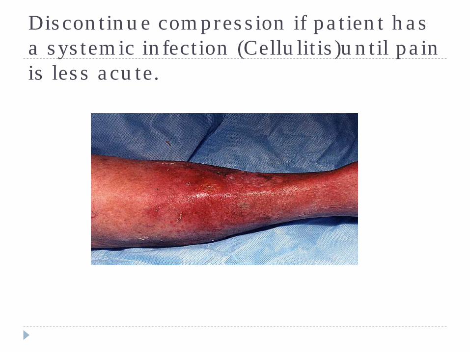
Discontinue compression if patient has a systemic infection (Cellulitis)until pain is less acute.
Concordance
} Assess why patient is not concording} Is pain managed effectively} Is patient anxious or depressed? (HADS score)} Do they need to be referred?} Consider your skills/ your approach to the care} Have you taken time to explain why they have leg
ulceration and how compression works?} Have you issued a patient information leaflet?
Managing non healing, complex ulcers/ wounds
When is a wound ‘palliative’?
Managing complex ulcers} Failure to progress} Exudate management} Pain} Odour} Infection} Dealing with pts anxiety re the problem} Feeling helpless – never ending!} When to refer} When do we ‘give up’?} Palliative wounds
Mixed aetiology} Correct diagnosis} Establish degree of arterial
disease} Consider reduced compression
(Ko flex)} Debride and treat local infection} Skin care} Monitor progress every 6 weeks
(Unlikely to achieve 40%)} Reassess ABPI} Refer to TV if uncertain of plan
Arterial disease- may become palliative} Refer to vascular for an
opinion} Reassess following
intervention re healing potential
} Careplan should be focussed on symptom management (Pain, infection, exudate, skin care etc)
} Evaluate effectiveness of actions
} Refer to TV for advice/ support
Dressings for mixed and arterial ulcers – treat the symptom/ problem} Layering dressings will not
necessarily improve the outcome
} Most advanced dressings are effective on their own
} Use the step up step down approach
} Consider your rationale for choice
} Measure whether its working} Give the dressing time to
work} Is it a formulary dressing?
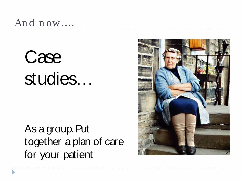
And now….
Case studies…
As a group. Put together a plan of care for your patient

Thank you…
}You have been….