Embed Size (px)
Citation preview
AJVR, Vol 74, No. 1, January 2013 155
An upper respiratory tract disease known as BAOS has been identified in brachycephalic dogs since
the beginning of the 20th century. The syndrome is comprised of morphological and functional alterations involving the upper airways, lower airways, and diges-tive system.1–4 Breeds commonly affected include Eng-lish Bulldog, French Bulldog, Pug, and Boston Terrier, although BAOS has also been detected in Pekingese, Cavalier King Charles Spaniel, Shih Tzu, Lhasa Apso, Boxer, and Shar-Pei. Both sexes are affected. Treatment
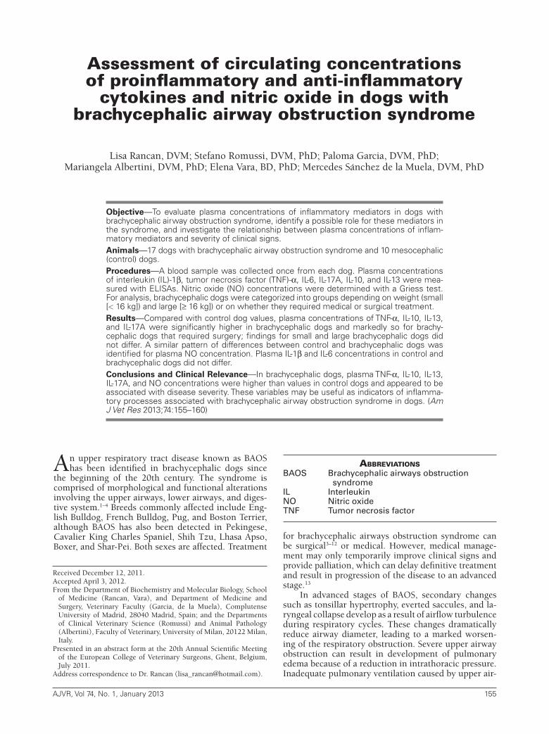
Assessment of circulating concentrations of proinflammatory and anti-inflammatory
cytokines and nitric oxide in dogs with brachycephalic airway obstruction syndrome
Lisa Rancan, DVM; Stefano Romussi, DVM, PhD; Paloma Garcia, DVM, PhD; Mariangela Albertini, DVM, PhD; Elena Vara, BD, PhD; Mercedes Sánchez de la Muela, DVM, PhD
Objective—To evaluate plasma concentrations of inflammatory mediators in dogs with brachycephalic airway obstruction syndrome, identify a possible role for these mediators in the syndrome, and investigate the relationship between plasma concentrations of inflam-matory mediators and severity of clinical signs.Animals—17 dogs with brachycephalic airway obstruction syndrome and 10 mesocephalic (control) dogs. Procedures—A blood sample was collected once from each dog. Plasma concentrations of interleukin (IL)-1β, tumor necrosis factor (TNF)-α, IL-6, IL-17A, IL-10, and IL-13 were mea-sured with ELISAs. Nitric oxide (NO) concentrations were determined with a Griess test. For analysis, brachycephalic dogs were categorized into groups depending on weight (small [< 16 kg]) and large [≥ 16 kg]) or on whether they required medical or surgical treatment.Results—Compared with control dog values, plasma concentrations of TNF-α, IL-10, IL-13, and IL-17A were significantly higher in brachycephalic dogs and markedly so for brachy-cephalic dogs that required surgery; findings for small and large brachycephalic dogs did not differ. A similar pattern of differences between control and brachycephalic dogs was identified for plasma NO concentration. Plasma IL-1β and IL-6 concentrations in control and brachycephalic dogs did not differ. Conclusions and Clinical Relevance—In brachycephalic dogs, plasma TNF-α, IL-10, IL-13, IL-17A, and NO concentrations were higher than values in control dogs and appeared to be associated with disease severity. These variables may be useful as indicators of inflamma-tory processes associated with brachycephalic airway obstruction syndrome in dogs. (Am J Vet Res 2013;74:155–160)
for brachycephalic airways obstruction syndrome can be surgical3–12 or medical. However, medical manage-ment may only temporarily improve clinical signs and provide palliation, which can delay definitive treatment and result in progression of the disease to an advanced stage.13
In advanced stages of BAOS, secondary changes such as tonsillar hypertrophy, everted saccules, and la-ryngeal collapse develop as a result of airflow turbulence during respiratory cycles. These changes dramatically reduce airway diameter, leading to a marked worsen-ing of the respiratory obstruction. Severe upper airway obstruction can result in development of pulmonary edema because of a reduction in intrathoracic pressure. Inadequate pulmonary ventilation caused by upper air-
Received December 12, 2011.Accepted April 3, 2012.From the Department of Biochemistry and Molecular Biology, School
of Medicine (Rancan, Vara), and Department of Medicine and Surgery, Veterinary Faculty (Garcia, de la Muela), Complutense University of Madrid, 28040 Madrid, Spain; and the Departments of Clinical Veterinary Science (Romussi) and Animal Pathology (Albertini), Faculty of Veterinary, University of Milan, 20122 Milan, Italy.
Presented in an abstract form at the 20th Annual Scientific Meeting of the European College of Veterinary Surgeons, Ghent, Belgium, July 2011.
Address correspondence to Dr. Rancan ([email protected]).
ABBREVIATIONSBAOS Brachycephalic airways obstruction syndromeIL InterleukinNO Nitric oxideTNF Tumor necrosis factor
11-12-0409r.indd 155 12/19/2012 2:25:19 PM
156 AJVR, Vol 74, No. 1, January 2013
way obstruction can lead to a reduction of blood oxy-gen content and subsequent hypoxia. Hypoxia may also increase fatigability of upper airway dilator muscle and soft tissue in a manner that has the potential to worsen the disease, as indicated by results of a study14 in adult rats. Furthermore, respiratory obstruction is a risk fac-tor for suffocation during anesthesia and the postsur-gical period.15 Inflammation as well as oxidative and nitrosative stress associated with repeated hypoxia and reoxygenation could be implicated in the pathogenesis of BAOS; however, despite the fact that anatomic and functional alterations are well described,16–23 the mo-lecular mechanisms involved in this syndrome remain unknown. To our knowledge, there are no reports of the molecular mediators implicated in BAOS. Thus, the objectives of the study reported here were to evaluate plasma concentrations of inflammatory mediators in dogs with BAOS, identify a possible role for these medi-ators in the syndrome, and investigate the relationship between plasma concentrations of inflammatory media-tors and severity of clinical signs. We hypothesized that a proinflammatory condition could be related to the pathogenesis and the progression of BAOS.
Materials and Methods
Animals—Other than the control dogs, only pure-bred brachycephalic dogs with BAOS were included in the study. At admission, each dog’s case history was obtained from the owner, focusing on questions about the severity of stertor, stridor, or snoring; degree of exercise intolerance; presence of syncope or cyanosis; and gastrointestinal signs, such as vomiting, regurgita-tion, and signs of nausea. A clinical examination was then performed, with special emphasis on patency and symmetry of the nares, careful palpation of larynx and tracheal rings, and auscultation of the heart and lung fields. By combining the information obtained from the owners and that obtained during the clinical examina-tion, a score was assigned to each brachycephalic dog on the basis of the scoring system proposed by Poncet et al.23 Briefly, the frequency of 4 upper respiratory tract signs (ie, snoring, inspiratory effort, exercise or stress intolerance, and syncope) was each assessed; on the basis of the frequency of each respiratory sign in com-bination, a global grade classification was obtained for each dog: not affected (grade 1), moderately affected (grade 2), or severely affected (grade 3). A complete he-matologic and biochemical analysis was performed for each brachycephalic dog to rule out systemic diseases. Dogs with potential systemic disease (eg, heart, liver, or kidney failure) were excluded from the study. Similarly, dogs with aggressive or particularly excitable behaviors were excluded from the study.
For purposes of data analysis, brachycephalic dogs were categorized into 2 groups on the basis of weight: small (< 16 kg) and large (≥ 16 kg). The brachycephalic dogs were also categorized into 2 groups on the basis of whether they required immediate surgical treatment or received medical treatment. Dogs that underwent medical treatment were further categorized (depend-ing on results of clinical examination) into 3 groups: those with no clinical signs, moderate clinical signs, or severe clinical signs. Ten clinically normal 2- to 3-year-
old Beagles (5 males and 5 females) were used as the control group.
The study was approved by the Complutense Uni-versity Ethical Committee and adhered to the guide-lines of Commission Directive 86/609/EEC (The Coun-cil Directive of the European Community) concerning the protection of animals used for experimental and other scientific purposes. The national legislation, in agreement with this directive, is defined in Royal De-cree No. 1201/2005.
Preparation of samples—A blood sample (10 mL) was collected from a cephalic vein of each dog. The samples were centrifuged at 3,000 X g for 10 minutes at 4°C, and the plasma was transferred to polypropylene tubes and frozen at –80°C until assayed.
Determinations of plasma cytokine concentra-tions—Concentrations of proinflammatory (IL-1β, TNF-α, IL-6, and IL-17A) and anti-inflammatory (IL-10 and IL-13) cytokines were measured in plasma samples with specific ELISA commercial kitsa according to the manufacturer’s protocols. Briefly, plasma samples and standard dilutions of the tested cytokine were in-cubated onto special microwells coated with specific monoclonal antibodies. The sample incubation was performed at 18° to 25°C for 1 to 3 hours, depending on the specific cytokine and the manufacturer’s instruc-tions. A second antibody and a horseradish peroxidase conjugate were added afterward, followed by incuba-tion for 30 minutes at 18° to 25°C.
Plates were developed with 3,3′,5,5′-tetramethyl-benzidine as a chromogen substrate; 12 to 15 minutes later, the reaction was stopped with a sulfuric acid so-lution and plates were evaluated at 450 nm. Each cy-tokine concentration was quantified against a standard curve.
Determination of plasma NO concentration—Plasma NO concentration was measured by the Griess reaction (ie, NO2 concentration was determined after the reduction of NO
3 to NO
2). Plasma samples were
deproteinized by the addition of sulfosalicylic acid, in-cubated for 30 minutes at 4°C, and subsequently cen-trifuged (12,000 X g for 20 minutes). After incubating the supernatants with Escherichia coli NO
3 reductase
(at 37°C for 30 minutes), 1 mL of Griess reagent (0.5% naphthylenediamine dihydrochloride, 5% sulfonil-amide, and 25% H
3PO4) was added. The reaction was
allowed to proceed for 20 minutes at 22°C, and the ab-sorbance at 546 nm was measured; an NaNO
2 solution
was used as a standard. The measured signal was linear from 1 to 150µM (r = 0.994; P < 0.001; n = 5), and the detection threshold was approximately 2µM.
Reproducibility within each assay was evaluated in 3 independent experiments, and each assay was per-formed with 3 replicates. The overall intra-assay coef-ficient of variation was calculated to be < 5%. Interassay reproducibility was evaluated in 3 independent experi-ments. The overall interassay coefficient of variation was calculated to be < 6%.
Statistical analysis—Results are expressed as the mean ± SEM. Significant differences in plasma cyto-kine and NO concentrations between the control and
11-12-0409r.indd 156 12/19/2012 2:25:19 PM
AJVR, Vol 74, No. 1, January 2013 157
brachycephalic dogs, between the control and small or large brachycephalic dogs, and among all groups (con-trol dogs, brachycephalic dogs with no clinical signs, moderate clinical signs, or severe clinical signs and brachycephalic dogs that underwent surgery) were de-termined with the nonparametric Kruskal-Wallis test and subsequently with the Mann-Whitney U test. A confidence level of 95% and values of P < 0.05 were considered significant.
ResultsDogs—Twenty-one brachycephalic dogs were con-
sidered for the present study. Three of the dogs were excluded from the study because they had a systemic illness, and 1 was excluded because it had an excitable character. Seventeen brachycephalic dogs were includ-ed in the study, of which 9 were sexually intact males, 6 were sexually intact females, and 2 were spayed fe-males. The median age of the brachycephalic dogs was 3.3 years (range, 11 months to 7 years). All were pure-bred dogs. English (n = 8) and French (4) Bulldogs were the most common breeds. For purposes of data analysis, the brachycephalic dogs were categorized into 2 groups on the basis of weight: 7 dogs were considered small (< 16 kg), and 10 dogs were considered large (≥ 16 kg). The brachycephalic dogs were also categorized into 2 groups on the basis of whether they required im-mediate surgical treatment (n = 4) or received medical treatment (13). Dogs that underwent medical treatment were further categorized into 3 groups: those with no clinical signs (n = 6), moderate clinical signs (5), and severe clinical signs (2). The control group included 10 clinically normal 2- to 3-year-old university-owned Beagles (5 males and 5 females; all were sexually intact).
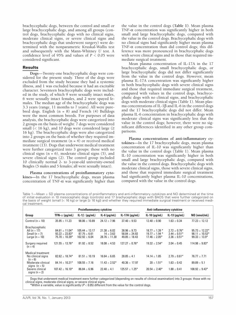
Plasma concentrations of proinflammatory cyto-kines—In the 17 brachycephalic dogs, mean plasma concentration of TNF-α was significantly higher than
the value in the control dogs (Table 1). Mean plasma TNF-α concentration was significantly higher in both small and large brachycephalic dogs, compared with the value in the control dogs. Brachycephalic dogs with no clinical signs had significantly higher mean plasma TNF-α concentration than did control dogs; this dif-ference was more pronounced in brachycephalic dogs with severe clinical signs and in those that required im-mediate surgical treatment.
Mean plasma concentration of IL-17A in the 17 brachycephalic dogs, small brachycephalic dogs, or large brachycephalic dogs did not differ significantly from the value in the control dogs. However, mean plasma IL-17A concentration was significantly higher in both brachycephalic dogs with severe clinical signs and those that required immediate surgical treatment, compared with values in the control dogs, brachyce-phalic dogs with no clinical signs, and brachycephalic dogs with moderate clinical signs (Table 1). Mean plas-ma concentrations of IL-1β and IL-6 in the control dogs and the 17 brachycephalic dogs did not differ. Mean plasma IL-6 concentration in brachycephalic dogs with moderate clinical signs was significantly less that the value in the control dogs; however, there were no sig-nificant differences identified in any other group com-parisons.
Plasma concentrations of anti-inflammatory cy-tokines—In the 17 brachycephalic dogs, mean plasma concentration of IL-10 was significantly higher than the value in the control dogs (Table 1). Mean plasma IL-10 concentration was significantly higher in both small and large brachycephalic dogs, compared with the value in the control dogs. Brachycephalic dogs with moderate clinical signs, those with severe clinical signs, and those that required immediate surgical treatment had significantly higher plasma IL-10 concentrations, compared with the value in the control dogs.
Proinflammatory cytokine Anti-inflammatory cytokine
Group TFN-α (pg/mL) IL-1β (pg/mL) IL-6 (pg/mL) IL-17A (pg/mL) IL-10 (pg/mL) IL-13 (pg/mL) NO (nmol/mL)
Control (n = 10) 35.95 ± 11.23 98.06 ± 10.89 24.12 ± 7.96 37.40 ± 9.53 12.40 ± 0.90 1.63 ± 0.34 77.23 ± 12.12
Brachycephalic All (n = 17) 84.61 ± 11.04* 105.44 ± 13.17 21.38 ± 6.02 38.96 ± 9.73 18.77 ± 1.39 * 2.72 ± 0.76* 95.73 ± 12.22* Small (n = 7) 93.22 ± 23.83* 87.75 ± 6.61 14 ± 3.63 58.84 ± 24.93 19.77 ± 1.94 * 2.44 ± 0.51* 98.11 ± 10.53* Large (n = 10) 75.70 ± 10.39* 102.92 ± 6.04 28.76 ± 11.30 49.85 ± 18.43 17.46 ± 2.05* 2.36 ± 0.51* 99.33 ± 13.9* Surgery required 121.55 ± 13.76* 81.92 ± 8.52 18.08 ± 4.53 127.27 ± 0.76* 19.32 ± 2.54* 2.04 ± 0.45 114.98 ± 9.83* (n = 4)
Medical treatment No clinical signs 82.62 ± 18.74* 81.51 ± 10.19 16.64 ± 6.65 28.85 ± 4.1 14.14 ± 1.85 2.79 ± 0.61* 76.77 ± 7.71 (n = 6) Moderate clinical 84.14 ± 10.21* 108.55 ± 7.16 11.43 ± 2.52* 40.36 ± 17.97 20 ± 1.51* 1.63 ± 0.42 89.89 ± 5.1 signs (n = 5) Severe clinical 107.42 ± 16.10* 86.84 ± 6.96 22.40 ± 4.1 125.57 ± 1.25* 20.54 ± 2.40* 1.88 ± 0.41 108.92 ± 9.43* signs (n = 2)
Dogs that underwent medical treatment were further categorized (depending on results of clinical examination) into 3 groups: those with no clinical signs, moderate clinical signs, or severe clinical signs.
*Within a variable, value is significantly (P < 0.05) different from the value for the control dogs.
Table 1—Mean ± SD plasma concentrations of proinflammatory and anti-inflammatory cytokines and NO determined at the time of initial evaluation in 10 mesocephalic dogs (controls) and 17 brachycephalic dogs with BAOS that were further categorized on the basis of weight (small [< 16 kg] or large [≥ 16 kg]) and whether they required immediate surgical treatment or received medi-cal treatment.
11-12-0409r.indd 157 12/19/2012 2:25:19 PM
158 AJVR, Vol 74, No. 1, January 2013
Mean plasma concentration of IL-13 in the 17 brachycephalic dogs, small brachycephalic dogs, or large brachycephalic dogs was significantly higher than the value in the control dogs (Table 1). Mean plasma IL-6 concentration in brachycephalic dogs with no clinical signs was significantly higher than the value in the control dogs; however, there were no significant differences identified in any other group comparisons.
Plasma concentration of NO—In the 17 brachy-cephalic dogs, mean plasma concentration of NO was significantly higher than the value in the control dogs (Table 1). Mean plasma NO concentration was signifi-cantly higher in both small and large brachycephalic dogs, compared with the value in the control dogs. Al-though there was a progressive increase in mean plasma NO concentration in brachycephalic dogs with increas-ing severity of clinical signs, compared with the value in the control dogs, the difference was significant only for brachycephalic dogs with severe signs. Brachyce-phalic dogs that required immediate surgical treatment had significantly higher plasma NO concentration, compared with the value in the control dogs.
Discussion
Brachycephalic airway obstruction syndrome is a serious and potentially deadly combination of upper airway disorders in predisposed breeds. In the present study, 17 brachycephalic dogs were evaluated, 12 of which were Bulldogs. This study group was not rep-resentative of all brachycephalic breeds or the breeds that are commonly affected with BAOS, which was a limitation of this investigation. In the present study, we hypothesized that a proinflammatory condition could be related to the pathogenesis and the progression of BAOS.
Inflammation is a complex host’s normal defense reaction to insult and stress. Inflammation responses, whether acute or chronic, are activated by well-coor-dinated, sequential events that are controlled by hu-moral and cellular reactions. Cytokines are the major communication channels that provide links within and between the immune system and other organs. An inflammatory response is associated with a spon-taneous increase in concentrations of proinflammatory cytokines; those that have obtained most attention are IL-1β, IL-6, and TNF-α. In the present study, plasma TNF-α concentration in brachycephalic dogs was sig-nificantly higher than the value in clinically normal nonbrachycephalic control dogs; this significant dif-ference in plasma TNF-α concentration was apparent between control dogs and both small (weight < 16 kg) and large (weight ≥ 16 kg) brachycephalic dogs. These findings agree with results of studies24–27 performed in other species, including humans, which identified an association between the concentration of TNF-α in plasma and advanced stages of respiratory obstructive disease. The possible relationship between plasma con-centrations of TNF-α and the degree of BAOS disease in dogs has not been described.
Tumor necrosis factor-α is a pivotal proinflamma-tory cytokine, which is centrally involved in local and
systemic responses in the immune system that lead to typical effects of inflammation. Unregulated, prolonged synthesis and release of TNF-α in chronic inflamma-tory conditions may contribute to the development of many diseases. Tumor necrosis factor-α can directly in-duce tissue injury by inducing accelerated cell apopto-sis but may also initiate and perpetuate an inflamma-tory response through upregulation of the expression of other inflammatory mediators. It is known that the TNF-α triggers a cascade of cytokines and angiogen-ic factors that contribute to endothelial changes first within the lung parenchyma and then systemically.27,28 However, these effects have never been investigated in relation to BAOS, to our knowledge. In the study re-ported here of brachycephalic dogs with BAOS, plas-ma TNF-α concentration was significantly higher in dogs with no clinical signs, compared with unaffected control dogs; this difference was more pronounced in brachycephalic dogs with severe clinical signs and in those that required immediate surgical treatment, sug-gesting that TNF-α could play an important role in the pathogenesis of BAOS.
Because of its ability to stimulate the production of other proinflammatory cytokines and activate nuclear factor-κB, TNF-α could contribute to the maintenance and exacerbation of ongoing inflammatory processes. This increase in chronic inflammatory process may cause changes of the lung parenchyma that lead to pul-monary hypertension, respiratory failure, or cardiac dysfunction. These pulmonary or cardiac alterations are often present in advanced stages of BAOS, suggest-ing an important role of inflammation in their develop-ment. In the present study, grade 1 and grade 2 brachy-cephalic dogs had significantly higher plasma TNF-α concentrations, compared with control dogs; therefore, it is possible that even in the early stages of BAOS, there could be alterations in the lung parenchyma (allowing passage of mediators to the bloodstream), implying the presence of systemic disorders. Increases in circulating concentrations of TNF-α are often accompanied by in-creases in concentrations of IL-1β and IL-6; however, in the present study, plasma IL-1β and Il-6 concentra-tions were not altered in dogs with BAOS, compared with values in the control dogs.
Interleukin-17 has been characterized as a proin-flammatory cytokine that acts on a variety of tissues. In chronic inflammatory processes associated with the respiratory tract in humans, such as allergic asthma, IL-17A, IL-17F, and IL-17E have been demonstrated to have an important role.29,30 Findings of those studies29,30 suggest that IL-17A and IL-17F are involved in subepi-thelial fibrosis that leads to airway remodeling, a pro-cess related to the severity of the disease. Results of the present study seem to be in agreement, at least partially, with those of the aforementioned studies.29,30 In fact, plasma IL-17A concentration was significantly higher in both brachycephalic dogs with severe clinical signs and those that required immediate surgical treatment, compared with the value in control dogs. This could be explained by hypothesizing that IL-17A acts in the later stages of the disease and promotes vascular remodeling, which in turn would then cause hypertension typical of advanced stages of obstructive respiratory syndromes.
11-12-0409r.indd 158 12/19/2012 2:25:19 PM
AJVR, Vol 74, No. 1, January 2013 159
The mechanism of signaling and activation of IL-17A has not been fully defined.31 Therefore, it is difficult to understand the role of IL-17A in obstructive respiratory syndromes, especially in veterinary medicine, wherein the only study32 of this proinflammatory cytokine, to our knowledge, investigated its role in osteoarthritis in canine hip joints.
Interleukin-10 is regarded as an excellent immu-nosuppressive cytokine. It acts on macrophages by sup-pressing the secretion of proinflammatory cytokines and promoting the production of cytokine inhibitors. Its contribution to the resolution of airway inflam-matory processes33 and its anti-inflammatory activity in chronic obstructive pulmonary disease34 have been demonstrated. This cytokine is so important for the control and resolution of obstructive respiratory syn-dromes, it has been proposed in human medicine as a marker for monitoring chronic obstructive pulmo-nary disease and for use in the treatment of the disease (as well as other nonsteroidal drugs, such as inhibi-tors of phosphodiesterase-4 and inhibitors of nuclear factor-κB35).
Interleukin-13 has immunosuppressive activity to-gether with IL-4 and IL-10 and inhibits production of inflammatory cytokines by monocytes. It also increases the proliferation and differentiation of monocytes and B cells. Data from several studies have highlighted the role of IL-13 in the pathogenesis of respiratory tract dis-eases (eg, asthma and chronic obstructive pulmonary disease)36 and established a direct association between plasma concentrations of this cytokine and degree of disease.37 In the study reported here, both plasma IL-10 and IL-13 concentrations were significantly higher in brachycephalic dogs, compared with values in control dogs, which suggested that anti-inflammatory cyto-kines could play an important role in the pathogenesis of BAOS. On the other hand, reactive nitrogen species are heavily implicated in the inflammatory process. Excessive NO production by inducible NO synthase may have an important role in tissue injury second-ary to an inflammatory response. In the present study, plasma concentrations of nitrate and nitrite (the stable end products of NO) were assessed to estimate the dif-ference in NO production between brachycephalic and control dogs. The plasma concentrations of NO were significantly increased in brachycephalic dogs, com-pared with the value in control dogs. Plasma concentra-tions of NO increased progressively in brachycephalic dogs with no, moderate, and severe clinical signs, com-pared with control dog findings; plasma NO concentra-tions in brachycephalic dogs with severe clinical signs and those that required immediate surgical treatment were significantly greater than the value in control dogs. A similar pattern of increasing increase plasma TNF-α concentration with worsening clinical signs of BAOS was identified, suggesting the existence of a pos-sible association between the production of NO and the expression of some cytokines. This association may un-derlie the regulation of the disease process, increasing the inflammatory response and thus contributing to the development of secondary changes.
Results of the present study have suggested that BAOS is not, as hitherto considered, a local process but
rather a set of changes at the local level that result in systemic pathological changes through the production of inflammatory mediators. An improved understand-ing of the mechanisms involved in worsening BAOS may aid in the identification of dogs at risk for disease progression and lead to alternative treatments that could prevent development of secondary changes.
a. Bio-NOVA Cientifica Ltd, Madrid, Spain.
References1. Hendricks J. Brachycephalic airway syndrome. In: King LG, ed.
Saunders textbook of respiratory disease in dogs and cats. Philadel-phia: Saunders, 2004;310–318.
2. Koch DA, Arnold S, Hubler M, et al. Brachycephalic syndrome in dogs. Compend Contin Educ Pract Vet 2003;25:48–55.
3. Hedlund CS. Surgery of the upper respiratory system. In: Fossum TW, ed. Small animal surgery. 3rd ed. St Louis: Elsevier, 2007;817–866.
4. Hobson H. Brachycephalic syndrome. Semin Vet Med Surg (Small Anim) 1995;10:109–114.
5. Monnet E. Brachycephalic airway syndrome. In Slatter D, ed. Saunders textbook of small animal surgery. 3rd ed. Philadelphia: WB Saunders Co, 2000;808–813.
6. Huck JL, Stanley BJ, Hauptman JG. Technique and outcome of nares amputation (Trader’s technique) in immature Shih Tzus. J Am Anim Hosp Assoc 2008;44:82–85.
7. Ellison GW. Alapexy: an alternative technique for repair of ste-notic nares in dogs. J Am Anim Hosp Assoc 2004;40:484–489.
8. Davidson EB, Davis MS, Campbell GA, et al. Evaluation of car-bon dioxide laser and conventional incisional techniques for resection of soft palates in brachycephalic dogs. J Am Vet Med Assoc 2001;219:776–781.
9. Dupré G, Findji L, Poncet C. The folded flap palatoplasty: a new technique for treatment of elongated soft palate in dogs, in Pro-ceedings. 14th Annu Sci Meet Eur Coll Vet Surg 2005;265–267.
10. Findji L, Duprè G. Folded flap palatoplasty for treatment of elongated soft palates in 55 dogs. Wien Tierarztl Monatsschr 2008;95:56–63.
11. Riecks TW, Birchard SJ, Stephens JA. Surgical correction of brachycephalic syndrome in dogs: 62 cases (1991–2004). J Am Vet Med Assoc 2007;230:1324–1328.
12. Pink J, Doyle R, Hughes J, et al. Laryngeal collapse in seven brachycephalic puppies. J Small Anim Pract 2006;47:131–135.
13. Torrez CV, Hunt GB. Results of surgical correction of abnor-malities associated with brachycephalic airway obstruction syndrome in dogs in Australia. J Small Anim Pract 2006;47:150–154.
14. McGuire M, MacDermott M, Bradford A. Effects of chronic epi-sodic hypoxia on rat upper airway muscle contractile properties and fiber-type distribution. Chest 2002;22:1012–1017.
15. de Carvalho AD, de Araujo ACP, Gaiga LH, et al. Brachycephalic syndrome—nostril stenosis in dog. Acta Sci Vet 2008;38:69–72.
16. Monnet E. Brachycephalic airway syndrome. In: Slatter D, ed. Saunders textbook of small animal surgery. 3rd ed. Philadelphia: WB Saunders Co, 2000;808–813.
17. Fasanella FJ, Shivley JM, Wardlaw JL, et al. Brachycephalic air-way obstructive syndrome in dogs: 90 cases (1991–2008). J Am Vet Med Assoc 2010;237:1048–1051.
18. Parnell N. Diseases of the throat. In: Ettinger SJ, Feldman EC, eds. Textbook of veterinary internal medicine. 6th ed. St Louis: Elsevier, 2005;1197–1204.
19. Ginn JA, Kumar MSA, McKiernan BC, et al. Nasopharyngeal turbinates in brachycephalic dogs and cats. J Am Anim Hosp As-soc 2008;44:243–249.
20. White RAS. La laringe. In: Hedlund CS, Taboada J, Merchant S, et al, eds. Malattie dell’orecchio, naso e gola del cane e del gatto. Torino, Italy: Unione Tipografico-Editrice Torinese, 2002;113–131.
21. Lecoindre P, Richard S. Digestive disorders associated with the chronic obstructive respiratory syndrome of brachycephalic
11-12-0409r.indd 159 12/19/2012 2:25:19 PM
160 AJVR, Vol 74, No. 1, January 2013
dogs: 30 cases (1999–2001). Rev Méd Vét 204;155:141–146.
22. Poncet CM, Dupré GP, Freiche VG, et al. Prevalence of gastro-intestinal tract lesions in 73 brachycephalic dogs with upper respiratory syndrome. J Small Anim Pract 2005;46:273–279.
23. Poncet CM, Dupré GP, Freiche VG, et al. Long-term results of upper respiratory syndrome surgery and gastrointestinal tract medical treatment in 51 brachycephalic dogs. J Small Anim Pract 2006;47:137–142.
24. Deloron P, Roux Lombard P, Ringwald P, et al. Plasma levels of TNF-alpha soluble receptors correlate with outcome in human falciparum malaria. Eur Cytokine Netw 1994;5:331–336.
25. de Godoy I, Donahoe M, Calhoun WJ, et al. Elevated TNF- alpha production by peripheral blood monocytes of weight-los-ing COPD patients. Am J Respir Crit Care Med 196;153:633–637.
26. Fujita M, Mason RJ, Cool C, et al. Pulmonary hypertension in TNF-α-overexpressing mice is associated with decreased VEGF gene expression. J Appl Physiol 2002;93:2162–2170.
27. Brindicci C, Kharitonov SA, Ito M, et al. Nitric oxide synthase isoenzyme expression and activity in peripheral lung tissue of patients with chronic obstructive pulmonary disease. Am J Respir Crit Care Med 2010;181:21–30.
28. Jasielska M, Semkova I, Shi X, et al. Differential role of tumor necrosis factor (TNF)-α receptors in the development of choroi-dal neovascularization. Invest Ophthalmol Vis Sci 2010;51:3874–3883.
29. Chakir J, Shannon J, Molet S, et al. Airway remodeling-associat-ed mediators in moderate to severe asthma: effect of steroids on TGF-β, IL-11, IL-17A, and type I and type III collagen expres-sion. J Allergy Clin Immunol 2003;111:1293–1298.
30. Linden A. Interleukin-17 and airway remodelling. Pulm Phar-macol Ther 2006;19:47–50.
31. McKenzie BS, Kastelein RA, Cua DJ. Understanding the IL-23-IL-17A immune pathway. Trends Immunol 2006;27:17–23.
32. Maccoux LJ, Salway F, Day PJ, et al. Expression profiling of se-lect cytokines in canine osteoarthritis tissues. Vet Immunol Im-munopathol 2007;118:59–67.
33. Ogawa Y, Duru EA, Ameredes BT. Role of IL-10 in the resolution of airway inflammation. Curr Mol Med 2008;8:437–445.
34. Lane N, Robins RA, Corne J, et al. Regulation in chronic ob-structive pulmonary disease: the role of regulatory T-cells and Th17 cells. Clin Sci (Lond) 2010;119:75–86.
35. Barnes PJ. Novel approaches and targets for treatment of chron-ic obstructive pulmonary disease. Am J Respir Crit Care Med 1999;160:S72–S79.
36. Hoshino T, Kato S, Oka N, et al. Pulmonary inflammation and emphysema: role of the cytokines IL-18 and IL-13. Am J Respir Crit Care Med 2007;176:49–62.
37. Lee JS, Rosengart MR, Kondragunta V, et al. Inverse association of plasma IL-13 and inflammatory chemokines with lung func-tion impairment in stable COPD: a cross-sectional cohort study. Respir Res 2007;8:64.
11-12-0409r.indd 160 12/19/2012 2:25:20 PM