Embed Size (px)
Citation preview
Donald J. Widder 1, 2
Kenneth R. Davis Juan M. Taveras
This article appears in the May/June 1986 issue of AJNR and in the August 1985 issue of AJR.
Received May 23 , 1985; accepted after revision September 12, 1985.
Presented in part at the American Society of Neuroradiology, Scientific Session, New Orleans, LA, February 1985.
1 All authors: Department of Radiology, Massachusetts General Hospital, Boston, MA 02114. Address reprint requests to D. J. Widder.
2 Present address: Department of Radiology, Santa Barbara Cottage Hospital , Pueblo at Beth St. , Santa Barbara, CA 93102.
AJNR 7:439-442, May/June 1986 0195-6108/86/0703-439 © American Society of Neuroradiology
439
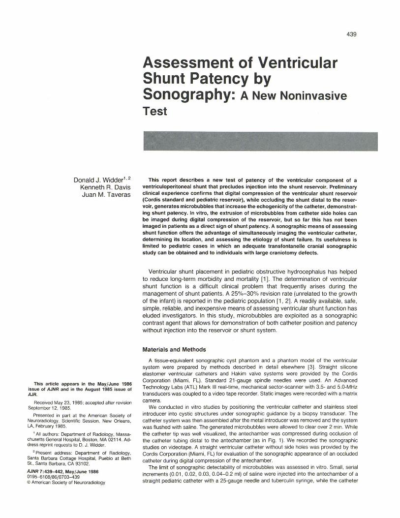
Assessment of Ventricular Shunt Patency by Sonography: A New Noninvasive Test
This report describes a new test of patency of the ventricular component of a ventriculoperitoneal shunt that precludes injection into the shunt reservoir. Preliminary clinical experience confirms that digital compression of the ventricular shunt reservoir (Cordis standard and pediatric reservoir), while occluding the shunt distal to the reservoir, generates microbubbles that increase the echogenicity of the catheter, demonstrating shunt patency. In vitro, the extrusion of microbubbles from catheter side holes can be imaged during digital compression of the reservoir, but so far this has not been imaged in patients as a direct sign of shunt patency. A sonographic means of assessing shunt function offers the advantage of simultaneously imaging the ventricular catheter, determining its location, and assessing the etiology of shunt failure. Its usefulness is limited to pediatric cases in which an adequate transfontanelle cranial sonographic study can be obtained and to individuals with large craniotomy defects.
Ventricular shunt placement in pediatric obstructive hydrocephalus has helped to reduce long-term morbidity and mortality [1]. The determination of ventricular shunt function is a difficult clinical problem that frequently arises during the management of shunt patients. A 25%-30% revision rate (unrelated to the growth of the infant) is reported in the pediatric population [1 , 2]. A readily available, safe, simple, reliable, and inexpensive means of assessing ventricular shunt function has eluded investigators. In this study, microbubbles are exploited as a sonographic contrast agent that allows for demonstration of both catheter position and patency without injection into the reservoir or shunt system.
Materials and Methods
A tissue-equivalent sonographic cyst phantom and a phantom model of the ventricular system were prepared by methods described in detail elsewhere [3] . Straight silicone elastomer ventricular catheters and Hakim valve systems were provided by the Cordis Corporation (Miami, FL). Standard 21-gauge spindle needles were used. An Advanced Technology Labs (ATL) Mark III real-time, mechanical sector-scanner with 3.5- and 5.0-MHz transducers was coupled to a video tape recorder. Static images were recorded with a matrix camera.
We conducted in vitro studies by positioning the ventricular catheter and stainless steel introducer into cystic structures under sonographic guidance by a biopsy transducer. The catheter system was then assembled after the metal introducer was removed and the system was flushed with saline. The generated microbubbles were allowed to clear over 2 min. While the catheter tip was well visualized , the antechamber was compressed during occlusion of the catheter tubing distal to the antechamber (as in Fig . 1). We recorded the sonographic studies on videotape. A straight ventricular catheter without side holes was provided by the Cordis Corporation (Miami , FL) for evaluation of the sonographic appearance of an occluded catheter during digital compression of the antechamber.
The limit of sonographic detectability of microbubbles was assessed in vitro. Small , serial increments (0 .01 , 0.02, 0.03, 0.04-0.2 ml) of saline were injected into the antechamber of a straight pediatric catheter with a 25-gauge needle and tuberculin syringe, while the catheter
440 WIDDER ET AL. AJNR :7, May/June 1986
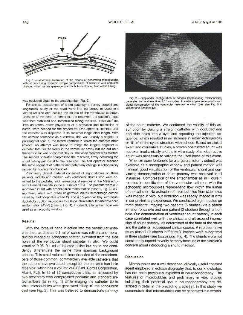
Fig. 1.- Schematic illustration of the means of generating microbubbles without puncturing reservoir. Simple compression of reservoir with occlusion of shunt tubing distally generates microbubbles in flowing fluid within tubing.
was occluded distal to the antechamber (Fig. 2). For clinical assessment of shunt patency, a survey coronal and
longitudinal study of the head were first performed to document ventricular size and localize the course of the ventricular catheter. Because of the need to compress the reservoir, the patient' s head was then stabilized and immobilized facing the side, "reservoir" up. Two operators, either physicians or a physician and technician or nurse, were needed for the procedure. One operator scanned until the catheter was displayed in its maximal longitudinal length. With the anterior fontanelle as a window, this was usually a sagittal or parasagittal scan of the lateral ventricle in which the catheter often resides. An attempt was made to image the longest segment of catheter that floated freely in the ventricular cavity but did not abut the ventricular wall or choroid plexus. The video recorder was started. The second operator compressed the reservoir, firmly occluding the shunt tubing just distal to the reservoir. The first operator scanned the same segment of catheter and noted any change in echogenicity caused by flowing microbubbles.
Preliminary clinical material consisted of eight studies on three patients, infants and children with ventricular shunts who were admitted to the pediatric and neurosurgical services at the Massachusetts General Hospital in the autumn of 1984. The patients were a 2-month-old infant with Arnold-Chiari malformation (case 1, Fig. 3), a 7-month-old infant with grade IV germinal matrix hemorrhage complicated by hydrocephalus (case 2), and a 15-year-old boy with aqueductal obstruction secondary to a large intraventricular arteriovenous malformation (AVM) (case 3, Fig. 4). In case 3, a large burr hole was used as an acoustic window.
Results
With the force of hand injection into the ventricular antechamber, as little as 0.1 ml of saline was reliably and reproducibly imaged as echogenic scatter, extruded from the side holes of the ventricular shunt catheter in vitro. We could visualize 0.05- 0.1 ml of injected saline but could not confidently differentiate the saline from spurious background echoes. This small volume is less than that of the antechambers of those common, commercially available catheters that the authors have evaluated (except for the pediatric burr hole reservoir, which has a volume of 0.08 ml [Cordis Corporation , Miami, FLJ). In 15 of 15 consecutive trials, as assessed by two observers who compressed pediatric and standard antechambers (as in Fig . 1) while imaging the catheter tip in vitro, microbubbles were generated "filling in" the sonolucent cyst (see Fig . 3). This was believed to demonstrate patency
Fig. 2.-Stepladder configuration of echoes (representing microbubbles) generated by hand injection of O.1-ml saline. A similar appearance results from digital compression of the ventricular reservoir in vitro. (See also Fig. 5 in Widder and Simeone [3]).
of the shunt catheter. We confirmed the validity of this assumption by placing a straight catheter with occluded end and side holes into a cyst and repeating the injection sequence, which resulted in no increase in either echogenicity or "fill-in " of the cystic structure with echoes. Based on clinical exam and correlative studies, a proven obstructed shunt was not examined clinically and the in vitro study of an obstructive shunt was necessary to validate the usefulness of this exam.
When an open fontanelle (or a large craniotomy defect) was available as a sonographic window and patient motion was minimal, good visualization of the ventricular shunt and convincing demonstration of shunt patency was achieved in all instances. Compression of the antechamber as in Figure 1 resulted in opacification of the ventricular catheter, with the echogenic microbubbles representing flow within the lumen of the catheter. No extrusion of microbubbles from side holes was imaged in vivo, but extrusion was readily imaged in vitro in our preliminary experience. We conducted eight studies on three patients, imaging two patients (6 studies) via a patent anterior fontanelle and one patient (2 studies) through a burr hole. Our demonstration of ventricular shunt patency in each case correlated well with the clinical and ultrasound impression of shunt patency, as determined at the time of the study and the patients ' subsequent clinical course. A representative study (case 1) is shown in Figure 3. Images were suboptimal in three studies (see Discussion ; Fig. 4). The shunts were not consistently tapped to verify patency because of the clinician 's concern about introducing a shunt infection.
Discussion
Microbubbles are a well described, clinically useful contrast agent employed in echocardiography that , to our knowledge, has not been previously exploited in neurosonography. The features of microbubbles and preliminary in vitro studies indicating their potential use in neurosonography are described in detail in the preceding article [3] . In this study we demonstrate that microbubbles can be generated in a ventric-
AJNR :7, May/June 1986 VENTRICULAR SHUNT PLACEMENT BY SONOGRAPHY 441
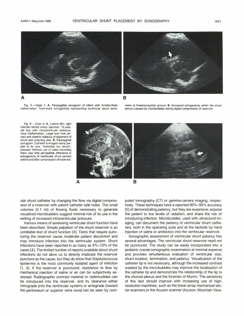
A B Fig. 3.-Case 1. A, Para sagittal sonogram of infant with Arnold-Chiari nates at thalamocaudate groove. B, Increased echogenicity within the shunt
malformation. Tram-track echogenicity representing ventricular shunt termi- (arrow) caused by microbubbles during digital compression of reservoir.
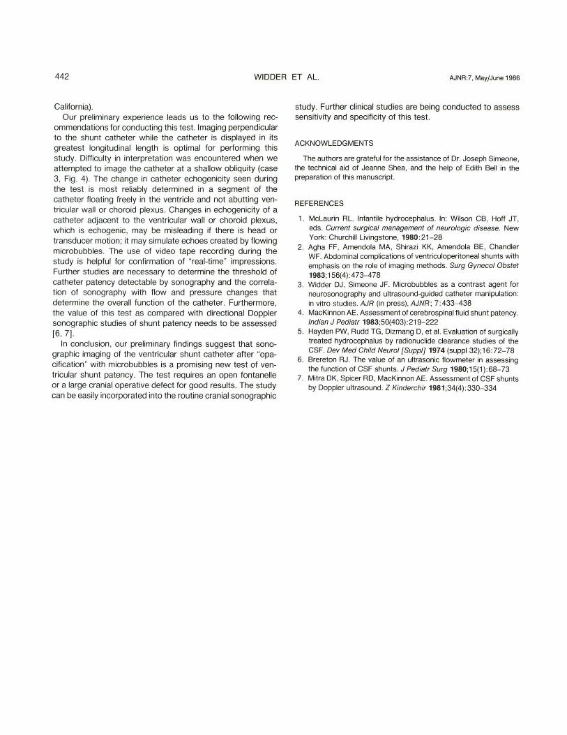
Fig. 4.-Case 3. A, Lateral film ; right internal carotid artery injection; 1S-yearold boy with intraventricular arteriovenous malformation. Large burr hole (arrow ) and shallow obliquity of alignment of shunt and scanning axis. B, Parasagittal sonogram. Catheter is imaged nearly parallel to its axis. Ventricles are decompressed. Without use of video recording there was little perceptible difference in echogenicity of ventricular shunt (arrow) before and after compression of reservoir.
A
ular shunt catheter by changing the flow via digital compression of a reservoir with patent catheter side holes. The small volumes (0.1 ml) of flowing fluids necessary to generate visualized microbubbles suggest minimal risk of its use in the setting of increased intraventricular pressure.
Various means of assessing ventricular shunt function have been described. Simple palpation of the shunt reservoir is an unreliable test of shunt function [4] . Tests that require puncturing the reservoir cause moderate patient discomfort and may introduce infection into the ventricular system. Shunt infections have been reported in as many as 5%-10% of the cases [4]. The limited number of reports available about shunt infections do not allow us to directly implicate the reservoir puncture as the cause, but they do show that Staphylococcus epidermis is the most commonly isolated agent of infection [1 , 4] . If the reservoir is punctured, resistance to flow by mechanical injection of saline or air can be subjectively assessed. Radiographic contrast material or radionuclides can be introduced into the reservoir, and its clearance either retrograde (into the ventricular system) or antegrade (toward the peritoneum or superior vena cava) can be seen by com-
B
puted tomography (CT) or gamma-camera imaging, respectively. These techniques have a reported 90%-95% accuracy [5] of demonstrating patency, but they are expensive, expose the patient to low levels of radiation, and share the risk of introducing infection. Microbubbles , used with ultrasound imaging , can document the patency of ventricular shunt catheters, both in the operating suite and at the bedside by hand injection of saline or antibiotics into the ventricular reservoir.
Sonographic assessment of ventricular shunt patency has several advantages. The ventricular shunt reservoir need not be punctured. The study can be easily incorporated into a pediatric cranial sonographic examination at minimal expense and provides simultaneous evaluation of ventricular size, shunt location, termination , and patency. Visualization of the catheter tip is not necessary, although the increased contrast created by the micro bubbles may improve the localization of the catheter tip and demonstrate the relationship of the tip to the choroid plexus and the foramen of Munro. The sensitivity of this test should improve with increasing use of highresolution machines, such as the linear-array mechanical sector-scanners or the Acuson scanner (Acuson , Mountain View,
442 WIDDER ET AL. AJNR:7, May/June 1986
California). Our preliminary experience leads us to the following rec
ommendations for conducting this test. Imaging perpendicular to the shunt catheter while the catheter is displayed in its greatest longitudinal length is optimal for performing this study. Difficulty in interpretation was encountered when we attempted to image the catheter at a shallow obliquity (case 3, Fig. 4). The change in catheter echogenicity seen during the test is most reliably determined in a segment of the catheter floating freely in the ventricle and not abutting ventricular wall or choroid plexus. Changes in echogenicity of a catheter adjacent to the ventricular wall or choroid plexus, which is echogenic, may be misleading if there is head or transducer motion; it may simulate echoes created by flowing microbubbles. The use of video tape recording during the study is helpful for confirmation of "real-time" impressions. Further studies are necessary to determine the threshold of catheter patency detectable by sonography and the correlation of sonography with flow and pressure changes that determine the overall function of the catheter. Furthermore, the value of this test as compared with directional Doppler sonographic studies of shunt patency needs to be assessed [6, 7] .
In conclusion, our preliminary findings suggest that sonographic imaging of the ventricular shunt catheter after "opacification" with microbubbles is a promising new test of ventricular shunt patency. The test requires an open fontanelle or a large cranial operative defect for good results . The study can be easily incorporated into the routine cranial sonographic
study. Further clinical studies are being conducted to assess sensitivity and specificity of this test.
ACKNOWLEDGMENTS
The authors are grateful for the assistance of Dr. Joseph Simeone, the technical aid of Jeanne Shea, and the help of Edith Bell in the preparation of this manuscript.
REFERENCES
1. McLaurin RL. Infantile hydrocephalus. In: Wilson CB, Hoff JT, eds. Current surgical management of neurologic disease. New York: Churchill Livingstone, 1980:21-28
2. Agha FF, Amendola MA, Shirazi KK, Amendola BE, Chandler WF. Abdominal complications of ventriculoperitoneal shunts with emphasis on the role of imaging methods. Surg Gynecol Obstet 1983;156(4):473-478
3. Widder OJ , Simeone JF. Microbubbles as a contrast agent for neurosonography and ultrasound-guided catheter manipulation: in vitro studies . AJR (in press), AJNR ; 7:433-438
4. MacKinnon AE. Assessment of cerebrospinal fluid shunt patency. Indian J Pediatr 1983;50(403):219-222
5. Hayden PW, Rudd TG, Dizmang 0, et al. Evaluation of surgically treated hydrocephalus by radionuclide clearance studies of the CSF. Dev Med Child Neurol [Suppl] 1974 (suppI 32);16:72-78
6. Brereton RJ . The value of an ultrasonic flowmeter in assessing the function of CSF shunts. J Pediatr Surg 1980; 15(1): 68-73
7. Mitra OK, Spicer RD, MacKinnon AE. Assessment of CSF shunts by Doppler ultrasound. Z Kinderchir 1981 ;34(4) :330-334