Embed Size (px)
Citation preview
iPSC lines of a Klinefelter’s Syndrome patient
1
Aberrant gene expression profiles in pluripotent stem cells induced from fibroblasts of a Klinefelter’s
Syndrome patient*
Yu Ma ‡,§, Chunliang Li‡,§,1, Junjie Gu ‡,§, Fan Tang ‡,§, Chun Li ‡,§, Peng Li¶, Ping Ping¶, Shi
Yang¶, Zheng Li¶,2 and Ying Jin‡,§,3
From the ‡Key Laboratory of Stem Cell Biology, Institute of Health Sciences, Shanghai Jiao Tong University School of Medicine & Shanghai Institutes of Biological Sciences, Chinese Academy of
Sciences, 225 South Chongqing Road, Shanghai, 200025, the §Shanghai Stem Cell Institute, Shanghai Jiao Tong University School of Medicine, 225 South Chongqing Road, Shanghai, 200025 and the
¶Department of Urology, Sperm Development and Genetics Laboratory, Shanghai Human Sperm Bank, Shanghai Institute of Andrology, Renji Hospital, Shanghai Jiao Tong University School of Medicine, 145
Shandong Zhong Road, Shanghai, 200001, China. *Running title: iPSC lines of a Klinefelter’s Syndrome patient 2To whom correspondence may be addressed: Zheng Li, M.D., Ph.D.,145 Shandong Zhong Road, Shanghai, 200001, China, Tel: +86-21-6373-2926; Email: [email protected], [email protected] 3To whom correspondence may be addressed: Ying Jin, M.D., Ph.D., Building 1, Room 607, 225 South Chongqing Road, Shanghai, 200025, China, Tel: +86-21-6385-2591; Fax: +86-21-6385-2591; Email: [email protected] Keywords: induced pluripotent stem cells; Klinefelter’s Syndrome; transcriptome analysis Background: Pathophysiology for Klinefelter’s Syndrome (KS) is poorly explained due to the lack of adequate models. Results: KS-iPSCs exhibit aberrantly expressed genes associated with KS clinic features. Conclusion: KS-iPSCs can potentially serve as a cellular model for KS research. Significance: Our study will significantly accelerate understanding, diagnosis and treatment of Klinfelter’s Syndrome. SUMMARY
Klinefelter’s Syndrome (KS) is the most
common male chromosome aneuploidy. Its pathophysiology is largely unexplained due to the lack of adequate models. Here, we report derivation of iPSC lines from a KS patient with a karyotype of 47, XXY. Derived KS-iPSCs meet all criteria of normal iPSCs with potential for germ cell differentiation. Although X chromosome inactivation occurs in all KS-iPSCs, genome-wide transcriptome analysis identifies aberrantly expressed genes associated with KS clinic features. Our KS-iPSCs can serve as a cellular model for KS research. Identified genes may become
http://www.jbc.org/cgi/doi/10.1074/jbc.M112.380204The latest version is at JBC Papers in Press. Published on September 27, 2012 as Manuscript M112.380204
Copyright 2012 by The American Society for Biochemistry and Molecular Biology, Inc.
by guest on August 26, 2018
http://ww
w.jbc.org/
Dow
nloaded from
iPSC lines of a Klinefelter’s Syndrome patient
2
biomarkers for early diagnosis or potential therapeutic targets for KS and significantly accelerate understanding, diagnosis and treatment of Klinfelter’s Syndrome.
Klinefelter’s Syndrome (KS) is the most common genetic form of male hypogonadism due to a supernumerary X chromosome, with an estimated frequency between 1/1000 and 1/500 (1). About 90% of KS patients bear the 47, XXY karyotype, whereas the 46, XX/47, XXY mosaic karyotype and, less frequently, additional X chromosomes (48,XXXY, 49, XXXXY) are found in the remaining KS patients (2). Clinically, KS is generally characterized by a reduced testicular volume, azoospermia and some other features such as tall stature, gynecomastia, increased serum FSH excretion and androgen deficiency, although its clinical picture exhibits a broader spectrum of phenotypes (2). Moreover, compared with normal males, KS patients have a higher risk to suffer from many other disorders, such as osteoporosis, metabolic syndrome, diabetes, breast cancer and subtle cognitive deficits (3-6). There is currently no effective treatment for KS. Intracytoplasmic sperm injection (ISCI) with spermatozoa obtained by testicular sperm extraction (TESE) techniques would give some KS males a chance to father a child, while for most of the patients, infertility is still unresolved due to the failure of sperm retrievals or spermatogenesis (1-2). Therefore, further understanding the disease and developing new strategies are required for effective treatment of KS.
Despite extensive studies in the past decades, the link of supernumerary X chromosome to KS phenotypes and molecular mechanisms underlying the testicular degeneration remain a mystery due to the lack of adequate and convenient experimental models. The advent of induced pluripotent stem cells (iPSCs) by defined
transcriptional factors, particularly those derived from patients, provides a new and attractive alternative for the in vitro disease model (7-8). Similar to embryonic stem cells (ESCs), iPSCs have characteristics of unlimited self-renewal and pluripotent developmental potential. Disease-specific iPSCs would not only provide a renewable cell source but also recapitulate the disease in a petri dish to model the development of the disease in vitro (9). Till now, iPSCs derived from somatic cells of various diseases have been applied in disease models and presented the unique opportunity to develop novel disease treatment strategies which have thus far not been possible (10).
In this study, we generated four lines of iPSCs from foreskin fibroblast cells of a KS patient with the 47, XXY karyotype and explored their potential usage for modeling the development of KS disease in vitro. The results obtained here will facilitate our understanding of this particular disease and identify potential new targets for its treatment.
EXPERIMENTAL PROCEDURES
Derivation and culture of primary fibroblast cells from the foreskin-- Discarded foreskins of a normal subject and a Klinefelter’s Syndrome patient were obtained from the Renji Hospital at the approval of the Reproductive Ethical Committee of the hospital after getting the written informed consents from the donors. The procedures of the derivation and culture of the fibroblast cells were same as previously described (11).
iPSC derivation and culture-- For iPSC derivation, virus was prepared as described previously (12-13). About 105 fibroblast cells were cultured in the medium with 4 viruses-containing supernatant and polybrene overnight. About 24 hours later, the fibroblast
by guest on August 26, 2018
http://ww
w.jbc.org/
Dow
nloaded from
iPSC lines of a Klinefelter’s Syndrome patient
3
cells were digested into single cells and replated onto gelatin-coated dishes. Culture medium was replaced by the iPSC culture medium in the next day. The iPSC culture medium was the KO-DMEM medium with 20% Knockout Serum Replacement (GIBCO), 100 U/ml penicillin, 100 µg/ml streptomycin, 0.1 mM β-mercaptoethanol (Sigma), 2 mM L-glutamine, 1% non essential amino acid (GIBCO) and 4 ng/ml bFGF (R&D). iPSC colonies were passaged by collagenase IV (5 mg/ml, Invitrogen) or the mechanical method every five days.
Alkaline phosphatase (AKP) staining-- iPSC colonies were fixed with 4% PFA in PBS and permeabilized by 0.1% TritonX-100 in PBS. The colonies were treated with AKP staining solution as instructed by the manual of ALKALINE PHOSPHATASE SUBSTRATE KIT III (Vector Laboratories, Inc) for 30-45 minutes in 37 oC.
G banding analysis-- Cells were cultured with 100 ng/ml colchicine (Sigma) for 12-16 hours. Then the cells were digested by 0.25% trypsin-EDTA (GIBCO) into single cells, treated by hypotonic solution including 0.16 g potassium chloride and 0.125 g sodium citrate in 50 ml deionized water for 30 minutes and fixed by glacial acetic acid and methyl alcohol in 1:3 ratio for 30 minutes. Later the samples were analyzed in Da An Company (Shanghai, China).
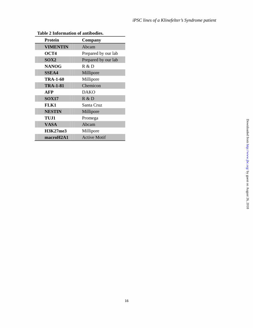
Immunofluorescence staining-- Cells were plated on glass slides and fixed with 4% PFA in PBS. Immunofluorescence staining was performed as reported previously (12). The information of primary antibodies is shown in table 2.
RT-PCR and quantitative RT-PCR-- Total RNA of cells was extracted by Trizol (Invitrogen) and reverse transcribed into cDNA using ReverTra Ace reverse transcriptase and oligo d(T)15. PCR reactions were carried out in the systems reported previously (14). For quantitative
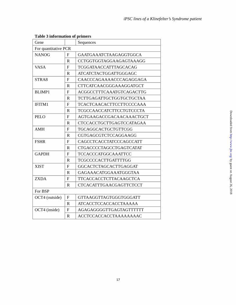
PCR, manufacturer’s instructions (ABI PRISM 7900) were followed. For the detection of the expression levels of germ cell lineage markers, GAPDH and RPLPO were used as internal controls and the average of the △Ct values were calculated for analysis. For other detections, only GAPDH was used as an internal control. The sequences of the primers for RT-PCR, pMXs-OCT4, pMXs-SOX2, pMXs-KLF4, pMXs-C-MYC, SOX9, and RPLPO have been reported previously (12, 15-18). The sequences of primers used to amplify MAGEA2B, MAGEH1, NRK and TMEM47 were from PrimerBank (http://pga.mgh.harvard.edu/primerbank/). The sequences of other primers are shown in table 3.
Bisulfite sequencing PCR-- Two µg of genomic DNA was treated according to the procedures of manufacture Active Motif. Nested PCR was carried out with the primers provided in table 3. PCR products were purified and ligated to the pGEM-T easy vector (Promega) for sequencing.
DNA FISH assay-- Cells were digested into single cells with 0.25% trypsin-EDTA and suspended in PBS. Then the cells were treated with hypotonic solution with 0.16 g of potassium chloride and 0.125 g of sodium citrate in 50 ml deionized water, and fixed by solution with glacial acetic acid and methanol in 1:3 volume ratio. Cell nucleuses were collected and DNA FISH assays were performed by Da An Company (Shanghai, China).
EB formation-- iPSCs were cultured on low-attachment dishes with the human EB medium containing KO-DMEM, 20% fetal bovine serum (Hyclone), 100 U/ml penicillin, 100 µg/ml streptomycin, 0.1 mM β-mercaptoethanol, 2 mM L-glutamine, 1% non essential amino acid for 9 days. Then EBs were collected and replated onto matrigel-coated glass covers for additional two days.
by guest on August 26, 2018
http://ww
w.jbc.org/
Dow
nloaded from
iPSC lines of a Klinefelter’s Syndrome patient
4
Teratoma formation-- About 5x10⁶ iPSCs were cultured in the presence of 10 µM Y27632 (Calbiochem) overnight. Then the cells were collected and injected intramuscularly into SCID mice. About two months later, teratomas were collected for H&E staining.
Microarray analysis-- For each kind of cells, three biological repeats of samples were prepared. All experiments were performed with Affymetrix U133 plus 2.0 gene chips in Shanghai Biotechnology Corporation. Raw data were normalized by MAS 5.0 algorithm and DEGs were analyzed by Gene Spring Software 11.0 (Agilent technologies, Santa Clara, CA, US). Scatter plots comparing the global gene expression profiles were constructed by R software. Differentially expressed genes with a fold change of 1.5 were analyzed in the context of Gene Ontology and KEGG pathway using DAVID 6.7 (http://david.abcc.ncifcrf.gov/). The microarray data from this publication have been submitted to GEO database (http://www.ncbi.nlm.nih.gov/geo/) with the accession number GSE37258.
Germ cell lineage differentiation-- For spontaneous differentiation, iPSCs in the number of 5 x 10⁴ were plated to 6-well plates and cultured in iPSC medium without bFGF. For BMP induction, iPSCs in the number of 5 x 10⁴ were plated to 6-well plates and cultured in the human EB medium supplemented with 10 µM of BMP4, BMP7 and BMP8a (R&D). For both methods, the medium was replaced every 7 days. Samples in the 7th, 14th or 21th day were collected for gene expression analysis.
VASA immunofluorescence staining-- Cells were digested by 0.25% Trypsin-EDTA into single cells and plated on glass slides by cytospin with 200 g for 3 minutes. The prepared samples were fixed with 4% PFA in PBS for immunofluorescence staining.
Statistical analysis-- All values except for otherwise indicated were analyzed by Student’s t test to determine the significance of the differences. P value < 0.05 was considered statistically significant.
RESULTS
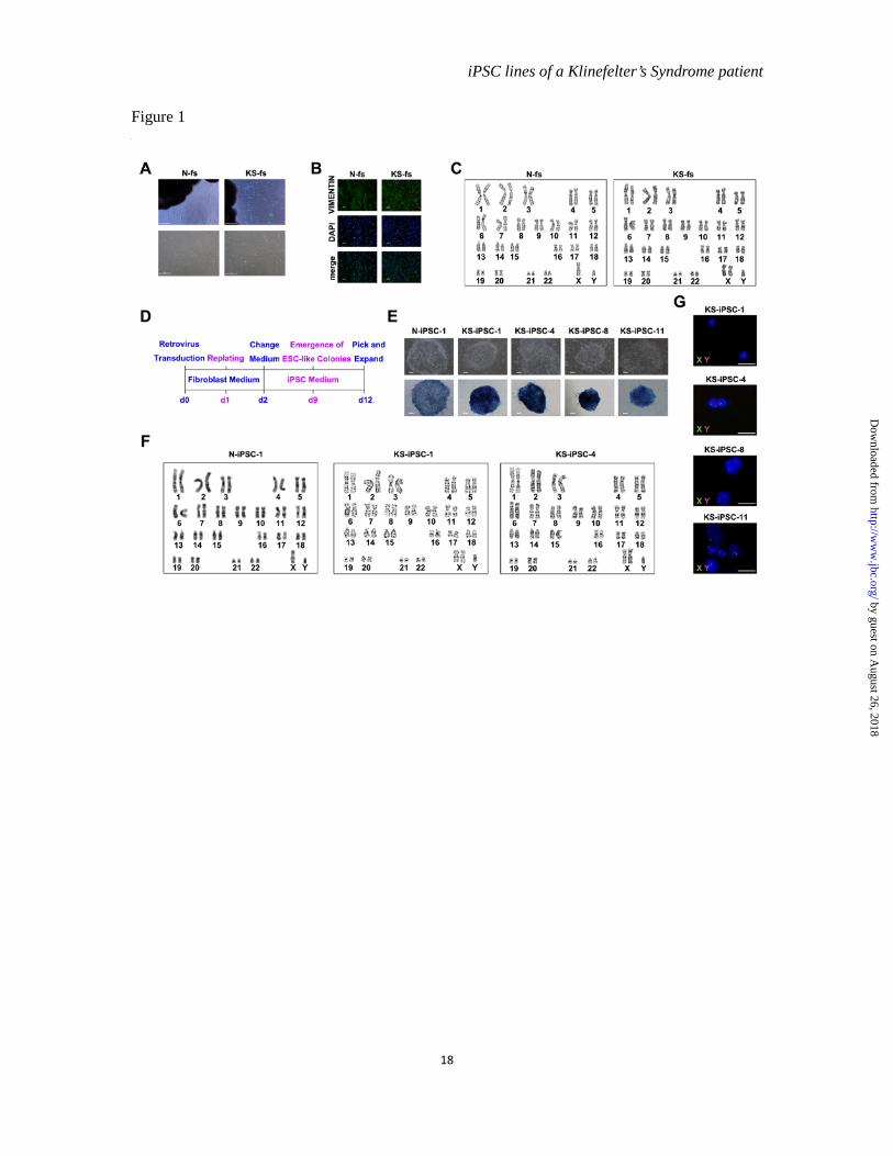
Derivation of iPSC lines from foreskin fibroblast cells of a KS patient and a normal subject-- We established fibroblast cell lines from the foreskin tissue of a normal male and a KS patient, designating them as normal fibroblasts (N-fs) and KS fibroblasts (KS-fs), respectively (Figure 1A and B). The KS patient is 27 years old with typical KS symptoms such as hypogonadism and small size of testes in the clinical diagnosis. G banding examination indicated the karyotype of 46, XY for N-fs and 47, XXY for KS-fs (Figure 1C). All of the cells of KS-fs carried the 47, XXY karyotype, implying that the KS foreskin donor might belong to the group of the most common KS cases with a homogenous 47, XXY karyotype.
Reprogramming of both types of the fibroblasts was induced by transduction of retroviral OCT4, SOX2, KLF4 and C-MYC as shown in Figure 1D. The human embryonic stem cell (hESC)-like colonies were picked on day 12 of infection and expanded to establish stable iPSC lines. Totally, we established 1 normal iPSC line (N-iPSC-1) and 4 KS iPSC lines (KS-iPSC-1, 4, 8, 11). There was no discernible difference in terms of reprogramming speed and efficiency between N-fs and KS-fs. iPSCs from both N-fs and KS-fs exhibited a typical morphology of hESCs and had the same karyotypes as their cognate fibroblasts, suggesting the maintenance of initial karyotypes throughout the reprogramming process (Figure 1E and F). The XXY karyotype of KS-iPSCs was further verified by fluorescence in situ hybridization assays (Figure 1G). In addition, the pattern of seven short tandom repeat (STR) sites
by guest on August 26, 2018
http://ww
w.jbc.org/
Dow
nloaded from
iPSC lines of a Klinefelter’s Syndrome patient
5
further verified the origin of N-iPSCs and KS-iPSCs (Table 1).
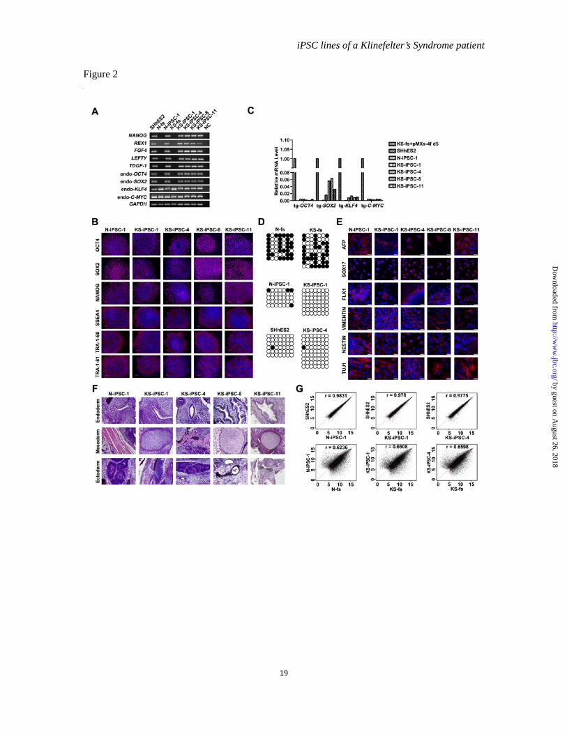
Characterization of iPSC lines-- To characterize iPSC lines described above, we first determined the expression of pluripotency-associated markers in comparison with hESCs of SHhES2 line, which carried a normal 46, XY karyotype and was fully characterized previously (15). Cells of all established iPSC lines were alkaline phosphatase positive (Figure 1E) and endogenous expression of NANOG, REX1, FGF4, LEFTY and TDGF-1 as well as OCT4, SOX2, KLF4 and C-MYC was activated (Figure 2A). Immunofluorescence staining assays also demonstrated the expression of OCT4, SOX2, NANOG, SSEA4, TRA-1-60 and TRA-1-81 in iPSCs (Figure 2B). Simultaneously, the expression of transgenic genes in iPSCs was silenced significantly (Figure 2C). Furthermore, similar to hESCs, the OCT4 promoter was hypomethylated in all iPSCs, whereas it was hypermethylated in the starting fibroblasts, further verifying the reprogramming of fibroblasts (Figure 2D).
Next, we assessed the developmental potential of our iPSCs through both in vitro and in vivo assays. Embryoid body (EB) formed when iPSCs were cultured in suspension. Various types of cells grew out of EBs after attachment. Immunofluorescence staining revealed the presence of cells expressing endoderm (SOX17 and AFP), mesoderm (VIMENTIN and FLK1) and ectoderm (NESTIN and TUJ1) markers (Figure 2E). In addition, teratomas, which contained respiratory epithelium and goblet cells (endoderm), muscles and cartilages (mesoderm), neural rosette and pigmented cells (ectoderm), were detected 4-8 weeks after the iPSCs were injected into immune-deficient mice (Figure 2F).
To further define the iPSCs at a global transcriptional level, we compared the
transcriptomic feature by scatter plots. As a result, all iPSCs tested approximated to hESCs (correlation coefficient > 0.97), but not to their cognate fibroblasts (correlation coefficient < 0.65) (Figure 2G). Therefore, N-fs and KS-fs were successfully reprogrammed into the pluripotent state at a genome-wide transcriptional level.
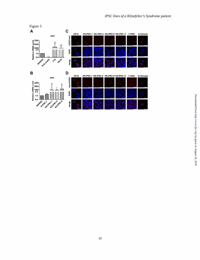
X chromosome inactivation in KS-iPSCs-- In female cells with a 46, XX karyotype, one of two X chromosomes is inactivated to balance the expression dosage of X chromosome linked genes with 46, XY male cells. X chromosome inactivation (XCI) is one of the epigenetic regulations in mammalian cells and is initiated by coating of non-coding XIST RNA on X chromosomes followed by exclusion of active chromatin markers and subsequent accumulation of repressive markers, such as H3K27me3 and macroH2A1 (19). It has been shown that the mosaic pattern of XCI in normal female fibroblasts was converted to be clonal in human iPSCs after reprogramming (20). The question whether XCI occurs in KS-iPSCs has not been addressed. We began with examining XIST expression levels in different types of fibroblasts and found that the level of XIST in KS-fs was similar to that in normal female fibroblasts (F-fs), but significantly higher than that in male normal fibroblasts (N-fs) and hESCs (SHhES2) (Figure 3A). Moreover, we found an extraordinarily high mRNA level of XIST in KS-iPSCs (Figure 3B). These observations suggested the undergoing of XCI in both normal female and KS cells. Furthermore, inactive X chromosome (Xi)-like accumulation of H3K27me3 and macroH2A1 were detected in almost all of the KS-iPSCs, whereas only part of KS-fs displayed this enrichment pattern (Figure 3C and 3D). This phenomenon of differential XCI patterns detected between KS-fs and KS-iPSCs was similar to that observed in normal female fibroblast (data not
by guest on August 26, 2018
http://ww
w.jbc.org/
Dow
nloaded from
iPSC lines of a Klinefelter’s Syndrome patient
6
shown) and normal female iPSCs (F-iPSC), a previously reported iPSC line derived from human amniotic fluid-derived cells (hAFDC-iPS-4) (12). As a negative control, the staining for H3K27me3 and macroH2A1 was not found in normal male fibroblasts (N-fs) (Figure 3C and 3D). Thus, one of X chromosomes in our KS-iPSCs was inactive as in normal female cells.
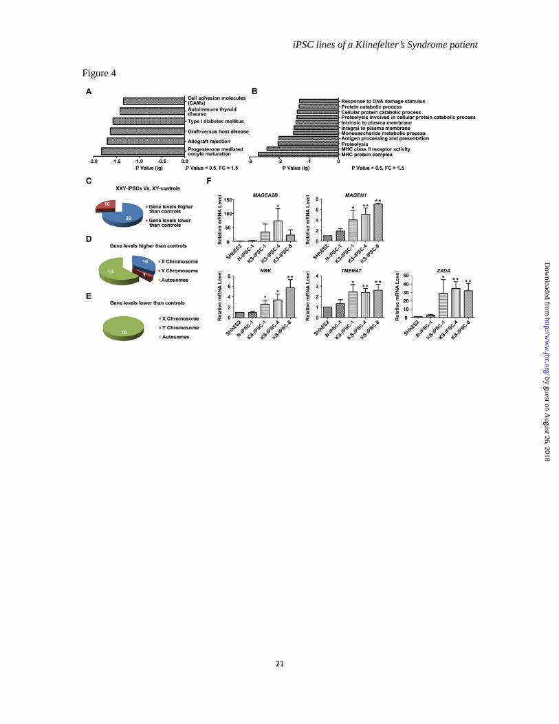
The aberrant transcriptome of KS-iPSCs-- Although we detected the Xi-like accumulation of repressive markers in all KS-iPSCs, which differentiated into various cell types of the three germ layers in a manner similar to N-iPSCs, there might be aberrant gene expression in KS-iPSCs. First, XCI escaping has been demonstrated in females as well as in KS patients, implying that cells from KS patients may have two active copies of strictly X-linked genes (21). Second, KS-iPSCs could have three active copies of X-Y homologous genes from pseudoautosomal regions (PARs) (22). To address this issue, global gene expression profiles of KS-iPSCs (KS-iPSC-1, 4) were compared to those of normal control cells (N-iPSC-1 and SHhES2). A total of 105 genes were identified as differentially expressed genes (DEGs) with more than a 1.5-fold difference (p<0.05). Among them, 76 genes had higher expression levels and 29 genes had lower expression levels in KS-iPSCs than in control cells. The pathway analysis in DAVID system indicated the enrichment of the DEGs in some diseases, especially in autoimmune diseases (Figure 4A), which might reflect an increased risk for KS patients to suffer from autoimmune diseases such as systemic lupus erythematosis (23). Additionally, progesterone mediated oocyte maturation was one of the enriched pathways (Figure 4A), which could be linked to the female-like features of KS patients. Moreover, genes associated with the protein catabolic process were also differentially expressed (Figure
4B). In fact, the metabolic syndrome is one of important clinic features of KS (1). Gene Ontology analysis also revealed distinct properties of the KS-iPSCs. For instance, DEGs encoded molecules with properties of being intrinsic to plasma membrane parts of the cells. The enriched biological processes included the immune regulation, protein catabolic process, and response to DNA damage stimulus (Figure 4B). The aberrant expression of genes associated with these biological processes might correspond to the abnormalities of early embryo development in KS patients and cause complicated disease phenotypes during later development of embryos or post puberty.
We also analyzed the DEGs between KS-iPSCs and normal controls based on a 2-fold difference criterion, and focused on the X-linked genes. A total of 39 DEGs were identified. Twenty-nine of them had higher levels and the rest ten genes had lower levels in KS-iPSCs as compared to the control group (Figure 4C). Intriguingly, 10 out of 29 genes having higher levels but none of genes with lower levels in KS-iPSCs were located on X chromosomes (Figure 4D and E), in line with the overdosage of X chromosomes in KS cells. The RT-qPCR analysis further verified higher expression levels of X-linked genes, such as XIST, MAGEA2B, MAGEH1, NRK, TMEM47 and ZXDA in KS-iPSCs (Figure 3B and 4F). These genes have been known to participate in the pathways or biological processes which were enriched by the DEGs between KS-iPSCs and normal controls. For instance, MAGEH1 belongs to the type II MAGE gene family and plays important roles in cell survival, cell cycle progression and apoptosis (24), being related to the enriched biological process of response to DNA damage stimulus. Also, ZXDA is associated with ZXDC to positively regulate the transcription of MHC II
by guest on August 26, 2018
http://ww
w.jbc.org/
Dow
nloaded from
iPSC lines of a Klinefelter’s Syndrome patient
7
gene (25), which corresponds to the autoimmune disease pathway and MHC associated biological processes. Hence, the abnormal expression of these genes may provide molecular basis for the pathogenesis of KS.
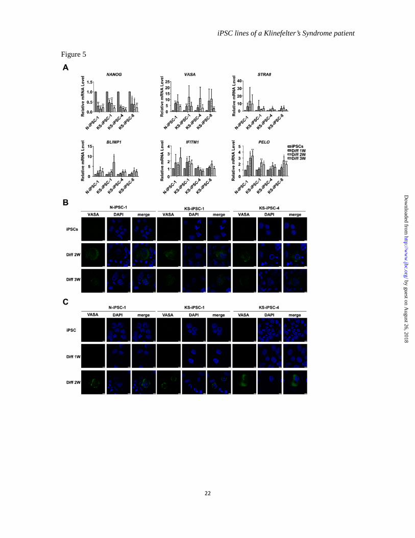
Potentials of iPSCs to differentiate into germ cell lineages-- We next explored the possibility of utilization of KS-iPSCs to either model KS disease development or generate normal germ cells in vitro as potential treatment for the patients. To this end, we differentiated our iPSCs into germ cell lineages. Previous studies have shown the generation of primordial germ cells (PGCs) and haploid gamete-like cells from hESCs and iPSCs in the early stage of their spontaneous differentiation (26). Consistently, differentiation of N- and KS-iPSCs took place spontaneously after withdrawal of bFGF, as evidenced by the down regulation of pluripotency marker Nanog. Simultaneously, expression of germ cell lineage markers was obviously upregulated without significant difference between N- and KS-iPSCs (Figure 5A). Furthermore, we examined the expression of VASA by immunofluorescence staining, which is specially expressed in the germ cell lineage of human beings (27). The differentiated cells expressing VASA were detected at two and three weeks after induction of differentiation for both N- and KS-iPSCs in a tiny proportion of cells (Figure 5B). In addition, we also treated iPSCs with BMPs to induce germ cell differentiation, since they were reported to promote hESCs and iPSCs to differentiate into PGCs (28-29). A small number of VASA positive PGC-like cells was found in BMP-treated cells of both N- and KS-iPSCs (Figure 5C). Thus, our results suggested the potential of KS-iPSCs to differentiate into germ cell lineages.
DISCUSSION
In the present study, we generated 4 lines of
KS-patient specific iPSCs and fully characterized them in terms of pluripotency both in vitro and in vivo, homogeneous XCI after reprogramming, global gene expression profiles and the potential to differentiate into germ cell lineages. To the best of our knowledge, this is the first study of the XCI, gene expression patterns and germ cell differentiation potential of iPSCs from the KS patient. The findings obtained here pave ways to our further elucidating the molecular mechanisms of KS and developing novel therapies for KS patients.
Female cells carry an X chromosome from each parent, but male cells inherit a single maternal X chromosome. For normal females, the XCI process randomly silences gene expression on one of two X chromosomes early in the development to equalize the dosage of X-linked genes to that of males (22). With respect to KS, a few studies have addressed the issue of XCI in somatic cells of KS patients, favoring the notion that XCI in KS follows the same pattern as in females (30-31). Here, we report the enrichment of XCI markers (H3K27me3 and macroH2A1) on an inactivated X chromosome (Xi) in all KS iPSCs. By contrast, the proportion of KS fibroblasts with Xi-accumulation of H3K27me3 and macroH2A1 was substantially lower than in KS-iPSCs, being 22% versus 100%. Similarly, we also noticed significantly lower Xi-accumulation of H3K27me3 and macroH2A1 in normal female fibroblasts than in normal female iPSCs. Currently, we do not know whether the reprogramming process altered the percentage of cells carrying these XCI markers or whether our iPSCs originated from single fibroblasts carrying them. Additionally, it remains elusive whether the fibroblasts without the Xi-enrichment of H3K27me3 and macroH2A1 retained an inactive X chromosome or not, as human cells have been shown to display a highly dynamic and variable
by guest on August 26, 2018
http://ww
w.jbc.org/
Dow
nloaded from
iPSC lines of a Klinefelter’s Syndrome patient
8
epigenetic state of the X chromosome (32-33). Recently, Tchieu et al. showed that reprogramming of human somatic cells could return the inactive X chromosome from the maintenance phase to a state resembling the initiation of XCI (20). The detection of the Xi-enrichment of the repressive markers in all KS-iPSCs may also explain their comparable differentiation potential to that of normal iPSCs. Further studies are warranted to define the XCI process in more details during somatic cell reprogramming in cells with supernumerary X chromosomes.
Nevertheless, the genome wide comparison of transcriptional profiles of KS-iPSCs with normal 46 XY controls identified significant DEGs, which are overrepresented on the X chromosome and known to participate in the biological processes or pathways related to certain clinical features of KS. The overexpression of X-linked genes could be explained, at least partially, by XCI escaping in KS. However, it remains unclear how remaining autosomal genes were differentially regulated. One possibility is that the aberrant expression of the autosomal genes was caused by the disturbed expression of X-linked genes. Further exploration of correlations among identified DEGs will answer this question. Previously, Vawter et al. found the differential expression of 129 genes by comparing whole genome expression profiles of lymphoblastic cells in KS patients with those in control XY males. They observed dysregulation of X-linked genes and the correlation of 12 genes with measures of verbal cognition in KS patients (34). There were no overlaps of X-linked DEGs between our and their data, which may be ascribed to the different
cell types tested in these two studies. The DEGs in KS samples identified in ours and other studies will contribute to the elucidation of molecular mechanisms of KS and may serve as biomarkers of KS for early diagnosis or potential therapeutic targets for KS treatment.
It appears that aberrant gene expression in KS-iPSC did not affect their early developmental potential and germ cell lineage commitment as well. The observation is in agreement with the fact that KS patients generally do not display significant symptoms until puberty and hints at the possibility to generate germ cells from KS-iPSCs for the treatment of KS patients suffering from azoospermia. As a matter of fact, mechanisms behind the degeneration of germ cells in KS currently remain unsolved. The supernumerary X itself could prevent the completion of meiosis; or, on the other hand, an abnormal testicular environment involving somatic Sertoli and Leydig cells could also lead to the failure of generating germ cells. In principal, KS-iPSCs may be induced to differentiate into spermatogonia in vitro, and then proceed through meiosis. This assumption is supported by studies showing that 47, XXY human cells could lose the supernumerary X chromosome to become 46, XY cells (35). Obviously, more extensive investigations, in particular, cell type-specific differentiation from KS-iPSCs, are needed to find out which cell type (s) is affected by altered gene expression in KS cells. The availability of KS-iPSCs and their utilization in disease modeling will greatly accelerate our understanding, diagnosis and treatment of Klinfelter’s Syndrome.
by guest on August 26, 2018
http://ww
w.jbc.org/
Dow
nloaded from
iPSC lines of a Klinefelter’s Syndrome patient
9
REFERENCES 1. Giltay, J. C., and Maiburg, M. C. (2010) Klinefelter syndrome: clinical and molecular
aspects, Expert review of molecular diagnostics 10, 765-776. 2. Forti, G., Corona, G., Vignozzi, L., Krausz, C., and Maggi, M. (2010) Klinefelter's
syndrome: a clinical and therapeutical update, Sex Dev 4, 249-258. 3. Bojesen, A., Birkebaek, N., Kristensen, K., Heickendorff, L., Mosekilde, L., Christiansen,
J. S., and Gravholt, C. H. (2011) Bone mineral density in Klinefelter syndrome is reduced and primarily determined by muscle strength and resorptive markers, but not directly by testosterone, Osteoporos Int 22, 1441-1450.
4. Gravholt, C. H., Jensen, A. S., Host, C., and Bojesen, A. (2011) Body composition, metabolic syndrome and type 2 diabetes in Klinefelter syndrome, Acta Paediatr 100, 871-877.
5. Brinton, L. A. (2011) Breast cancer risk among patients with Klinefelter syndrome, Acta Paediatr 100, 814-818.
6. Ross, J. L., Roeltgen, D. P., Stefanatos, G., Benecke, R., Zeger, M. P., Kushner, H., Ramos, P., Elder, F. F., and Zinn, A. R. (2008) Cognitive and motor development during childhood in boys with Klinefelter syndrome, Am J Med Genet A 146A, 708-719.
7. Takahashi, K., and Yamanaka, S. (2006) Induction of pluripotent stem cells from mouse embryonic and adult fibroblast cultures by defined factors, Cell 126, 663-676.
8. Takahashi, K., Tanabe, K., Ohnuki, M., Narita, M., Ichisaka, T., Tomoda, K., and Yamanaka, S. (2007) Induction of pluripotent stem cells from adult human fibroblasts by defined factors, Cell 131, 861-872.
9. Stadtfeld, M., and Hochedlinger, K. (2010) Induced pluripotency: history, mechanisms, and applications, Genes Dev 24, 2239-2263.
10. Grskovic, M., Javaherian, A., Strulovici, B., and Daley, G. Q. (2011) Induced pluripotent stem cells--opportunities for disease modelling and drug discovery, Nat Rev Drug Discov 10, 915-929.
11. Li, C., Yu, H., Ma, Y., Shi, G., Jiang, J., Gu, J., Yang, Y., Jin, S., Wei, Z., Jiang, H., Li, J., and Jin, Y. (2009) Germline-competent mouse-induced pluripotent stem cell lines generated on human fibroblasts without exogenous leukemia inhibitory factor, PLoS One 4, e6724.
12. Li, C., Zhou, J., Shi, G., Ma, Y., Yang, Y., Gu, J., Yu, H., Jin, S., Wei, Z., Chen, F., and Jin, Y. (2009) Pluripotency can be rapidly and efficiently induced in human amniotic fluid-derived cells, Hum Mol Genet 18, 4340-4349.
13. Kitamura, T., Koshino, Y., Shibata, F., Oki, T., Nakajima, H., Nosaka, T., and Kumagai, H. (2003) Retrovirus-mediated gene transfer and expression cloning: powerful tools in functional genomics, Exp Hematol 31, 1007-1014.
14. Li, L., Sun, L., Gao, F., Jiang, J., Yang, Y., Li, C., Gu, J., Wei, Z., Yang, A., Lu, R., Ma, Y., Tang, F., Kwon, S. W., Zhao, Y., Li, J., and Jin, Y. (2010) Stk40 links the pluripotency factor Oct4 to the Erk/MAPK pathway and controls extraembryonic endoderm differentiation, Proc Natl Acad Sci U S A 107, 1402-1407.
by guest on August 26, 2018
http://ww
w.jbc.org/
Dow
nloaded from
iPSC lines of a Klinefelter’s Syndrome patient
10
15. Li, C., Yang, Y., Lu, X., Sun, Y., Gu, J., Feng, Y., and Jin, Y. (2010) Efficient derivation of Chinese human embryonic stem cell lines from frozen embryos, In Vitro Cell Dev Biol Anim 46, 186-191.
16. Cheung, A. Y., Horvath, L. M., Grafodatskaya, D., Pasceri, P., Weksberg, R., Hotta, A., Carrel, L., and Ellis, J. (2011) Isolation of MECP2-null Rett Syndrome patient hiPS cells and isogenic controls through X-chromosome inactivation, Hum Mol Genet 20, 2103-2115.
17. Bucay, N., Yebra, M., Cirulli, V., Afrikanova, I., Kaido, T., Hayek, A., and Montgomery, A. M. (2009) A novel approach for the derivation of putative primordial germ cells and sertoli cells from human embryonic stem cells, Stem Cells 27, 68-77.
18. Lossos, I. S., Czerwinski, D. K., Wechser, M. A., and Levy, R. (2003) Optimization of quantitative real-time RT-PCR parameters for the study of lymphoid malignancies, Leukemia 17, 789-795.
19. Heard, E., and Disteche, C. M. (2006) Dosage compensation in mammals: fine-tuning the expression of the X chromosome, Genes Dev 20, 1848-1867.
20. Tchieu, J., Kuoy, E., Chin, M. H., Trinh, H., Patterson, M., Sherman, S. P., Aimiuwu, O., Lindgren, A., Hakimian, S., Zack, J. A., Clark, A. T., Pyle, A. D., Lowry, W. E., and Plath, K. (2010) Female human iPSCs retain an inactive X chromosome, Cell Stem Cell 7, 329-342.
21. Tuttelmann, F., and Gromoll, J. (2010) Novel genetic aspects of Klinefelter's syndrome, Molecular human reproduction 16, 386-395.
22. Ross, M. T., Grafham, D. V., Coffey, A. J., Scherer, S., McLay, K., Muzny, D., Platzer, M., Howell, G. R., Burrows, C., Bird, C. P., Frankish, A., Lovell, F. L., Howe, K. L., Ashurst, J. L., Fulton, R. S., Sudbrak, R., Wen, G., Jones, M. C., Hurles, M. E., Andrews, T. D., Scott, C. E., Searle, S., Ramser, J., Whittaker, A., Deadman, R., Carter, N. P., Hunt, S. E., Chen, R., Cree, A., Gunaratne, P., Havlak, P., Hodgson, A., Metzker, M. L., Richards, S., Scott, G., Steffen, D., Sodergren, E., Wheeler, D. A., Worley, K. C., Ainscough, R., Ambrose, K. D., Ansari-Lari, M. A., Aradhya, S., Ashwell, R. I., Babbage, A. K., Bagguley, C. L., Ballabio, A., Banerjee, R., Barker, G. E., Barlow, K. F., Barrett, I. P., Bates, K. N., Beare, D. M., Beasley, H., Beasley, O., Beck, A., Bethel, G., Blechschmidt, K., Brady, N., Bray-Allen, S., Bridgeman, A. M., Brown, A. J., Brown, M. J., Bonnin, D., Bruford, E. A., Buhay, C., Burch, P., Burford, D., Burgess, J., Burrill, W., Burton, J., Bye, J. M., Carder, C., Carrel, L., Chako, J., Chapman, J. C., Chavez, D., Chen, E., Chen, G., Chen, Y., Chen, Z., Chinault, C., Ciccodicola, A., Clark, S. Y., Clarke, G., Clee, C. M., Clegg, S., Clerc-Blankenburg, K., Clifford, K., Cobley, V., Cole, C. G., Conquer, J. S., Corby, N., Connor, R. E., David, R., Davies, J., Davis, C., Davis, J., Delgado, O., Deshazo, D., Dhami, P., Ding, Y., Dinh, H., Dodsworth, S., Draper, H., Dugan-Rocha, S., Dunham, A., Dunn, M., Durbin, K. J., Dutta, I., Eades, T., Ellwood, M., Emery-Cohen, A., Errington, H., Evans, K. L., Faulkner, L., Francis, F., Frankland, J., Fraser, A. E., Galgoczy, P., Gilbert, J., Gill, R., Glockner, G., Gregory, S. G., Gribble, S., Griffiths, C., Grocock, R., Gu, Y., Gwilliam, R., Hamilton, C., Hart, E. A., Hawes, A., Heath, P. D.,
by guest on August 26, 2018
http://ww
w.jbc.org/
Dow
nloaded from
iPSC lines of a Klinefelter’s Syndrome patient
11
Heitmann, K., Hennig, S., Hernandez, J., Hinzmann, B., Ho, S., Hoffs, M., Howden, P. J., Huckle, E. J., Hume, J., Hunt, P. J., Hunt, A. R., Isherwood, J., Jacob, L., Johnson, D., Jones, S., de Jong, P. J., Joseph, S. S., Keenan, S., Kelly, S., Kershaw, J. K., Khan, Z., Kioschis, P., Klages, S., Knights, A. J., Kosiura, A., Kovar-Smith, C., Laird, G. K., Langford, C., Lawlor, S., Leversha, M., Lewis, L., Liu, W., Lloyd, C., Lloyd, D. M., Loulseged, H., Loveland, J. E., Lovell, J. D., Lozado, R., Lu, J., Lyne, R., Ma, J., Maheshwari, M., Matthews, L. H., McDowall, J., McLaren, S., McMurray, A., Meidl, P., Meitinger, T., Milne, S., Miner, G., Mistry, S. L., Morgan, M., Morris, S., Muller, I., Mullikin, J. C., Nguyen, N., Nordsiek, G., Nyakatura, G., O'Dell, C. N., Okwuonu, G., Palmer, S., Pandian, R., Parker, D., Parrish, J., Pasternak, S., Patel, D., Pearce, A. V., Pearson, D. M., Pelan, S. E., Perez, L., Porter, K. M., Ramsey, Y., Reichwald, K., Rhodes, S., Ridler, K. A., Schlessinger, D., Schueler, M. G., Sehra, H. K., Shaw-Smith, C., Shen, H., Sheridan, E. M., Shownkeen, R., Skuce, C. D., Smith, M. L., Sotheran, E. C., Steingruber, H. E., Steward, C. A., Storey, R., Swann, R. M., Swarbreck, D., Tabor, P. E., Taudien, S., Taylor, T., Teague, B., Thomas, K., Thorpe, A., Timms, K., Tracey, A., Trevanion, S., Tromans, A. C., d'Urso, M., Verduzco, D., Villasana, D., Waldron, L., Wall, M., Wang, Q., Warren, J., Warry, G. L., Wei, X., West, A., Whitehead, S. L., Whiteley, M. N., Wilkinson, J. E., Willey, D. L., Williams, G., Williams, L., Williamson, A., Williamson, H., Wilming, L., Woodmansey, R. L., Wray, P. W., Yen, J., Zhang, J., Zhou, J., Zoghbi, H., Zorilla, S., Buck, D., Reinhardt, R., Poustka, A., Rosenthal, A., Lehrach, H., Meindl, A., Minx, P. J., Hillier, L. W., Willard, H. F., Wilson, R. K., Waterston, R. H., Rice, C. M., Vaudin, M., Coulson, A., Nelson, D. L., Weinstock, G., Sulston, J. E., Durbin, R., Hubbard, T., Gibbs, R. A., Beck, S., Rogers, J., and Bentley, D. R. (2005) The DNA sequence of the human X chromosome, Nature 434, 325-337.
23. Scofield, R. H., Bruner, G. R., Namjou, B., Kimberly, R. P., Ramsey-Goldman, R., Petri, M., Reveille, J. D., Alarcon, G. S., Vila, L. M., Reid, J., Harris, B., Li, S., Kelly, J. A., and Harley, J. B. (2008) Klinefelter's syndrome (47,XXY) in male systemic lupus erythematosus patients: support for the notion of a gene-dose effect from the X chromosome, Arthritis and rheumatism 58, 2511-2517.
24. Barker, P. A., and Salehi, A. (2002) The MAGE proteins: emerging roles in cell cycle progression, apoptosis, and neurogenetic disease, J Neurosci Res 67, 705-712.
25. Al-Kandari, W., Koneni, R., Navalgund, V., Aleksandrova, A., Jambunathan, S., and Fontes, J. D. (2007) The zinc finger proteins ZXDA and ZXDC form a complex that binds CIITA and regulates MHC II gene transcription, J Mol Biol 369, 1175-1187.
26. Eguizabal, C., Montserrat, N., Vassena, R., Barragan, M., Garreta, E., Garcia-Quevedo, L., Vidal, F., Giorgetti, A., Veiga, A., and Izpisua Belmonte, J. C. (2011) Complete meiosis from human induced pluripotent stem cells, Stem Cells 29, 1186-1195.
27. Castrillon, D. H., Quade, B. J., Wang, T. Y., Quigley, C., and Crum, C. P. (2000) The human VASA gene is specifically expressed in the germ cell lineage, Proc Natl Acad Sci U S A 97, 9585-9590.
28. Kee, K., Angeles, V. T., Flores, M., Nguyen, H. N., and Reijo Pera, R. A. (2009) Human
by guest on August 26, 2018
http://ww
w.jbc.org/
Dow
nloaded from
iPSC lines of a Klinefelter’s Syndrome patient
12
DAZL, DAZ and BOULE genes modulate primordial germ-cell and haploid gamete formation, Nature 462, 222-225.
29. Panula, S., Medrano, J. V., Kee, K., Bergstrom, R., Nguyen, H. N., Byers, B., Wilson, K. D., Wu, J. C., Simon, C., Hovatta, O., and Reijo Pera, R. A. (2011) Human germ cell differentiation from fetal- and adult-derived induced pluripotent stem cells, Hum Mol Genet 20, 752-762.
30. Iitsuka, Y., Bock, A., Nguyen, D. D., Samango-Sprouse, C. A., Simpson, J. L., and Bischoff, F. Z. (2001) Evidence of skewed X-chromosome inactivation in 47,XXY and 48,XXYY Klinefelter patients, American journal of medical genetics 98, 25-31.
31. Poplinski, A., Wieacker, P., Kliesch, S., and Gromoll, J. (2010) Severe XIST hypomethylation clearly distinguishes (SRY+) 46,XX-maleness from Klinefelter syndrome, European journal of endocrinology / European Federation of Endocrine Societies 162, 169-175.
32. Hoffman, L. M., and Carpenter, M. K. (2005) Characterization and culture of human embryonic stem cells, Nature biotechnology 23, 699-708.
33. Silva, S. S., Rowntree, R. K., Mekhoubad, S., and Lee, J. T. (2008) X-chromosome inactivation and epigenetic fluidity in human embryonic stem cells, Proceedings of the National Academy of Sciences of the United States of America 105, 4820-4825.
34. Vawter, M. P., Harvey, P. D., and DeLisi, L. E. (2007) Dysregulation of X-linked gene expression in Klinefelter's syndrome and association with verbal cognition, Am J Med Genet B Neuropsychiatr Genet 144B, 728-734.
35. Kawakami, T., Zhang, C., Taniguchi, T., Kim, C. J., Okada, Y., Sugihara, H., Hattori, T., Reeve, A. E., Ogawa, O., and Okamoto, K. (2004) Characterization of loss-of-inactive X in Klinefelter syndrome and female-derived cancer cells, Oncogene 23, 6163-6169.
by guest on August 26, 2018
http://ww
w.jbc.org/
Dow
nloaded from
iPSC lines of a Klinefelter’s Syndrome patient
13
FOOTNOTES *This study was supported by grants of National High Technology Research and
Development Program of China (2010CB945200, 2011DFB300100, 2011CB965101 and 2009CB941103), the National Natural Science Foundation (91019023), Chinese Academy of Science (XDA01010102), Shanghai Leading Academic Discipline Project (S30201) and Key Project of Shanghai Municipal Education Commission (10ZZ70).
1Present address: Department of Tumor Cell Biology, Howard Hughes Medical Insititute/St. Jude Children’s Research Hospital, 262 Danny Thomas Place MS 350, Suite D5017, Memphis, TN, 38105, USA.
2To whom correspondence may be addressed: Tel: +86-21-6373-2926; Email: [email protected], [email protected]
3To whom correspondence may be addressed: Tel: +86-21-6385-2591; Fax: +86-21-6385-2591; Email: [email protected]
4The abbreviations used are: DAVID, Database for Annotation Visualization and Integrated Discovery; DEG, differentially expressed gene; EB, embryoid body; ESC, embryonic stem cell; fs, fibroblast; FSH, follicle-stimulating hormone; G, Giemsa; GO, Gene Ontology; H & E, haematoxylin & eosin; iPSC, induced pluripotent stem cell; ISCI, intracytoplasmic sperm injection; KEGG, Kyoto Encyclopedia of Genes and Genomes; KS, Klinefelter’s Syndrome; N, normal; PAR, pseudoautosomal region; PGC, primordial germ cell; STR, short tandem repeat; TESE, testicular sperm extraction; XCI, X chromosome inactivation.
FIGURE LEGENDS
Figure 1 Generation of iPSC lines from fibroblast cells (fs) of a normal subject and a KS patient. (A). Primary culture of N-fs and KS-fs. Upper panel shows fs migrating from foreskin tissue clumps. Bottom panel shows the morphology of fs. Scale bars, 100 µm. (B). Immunofluorescence staining of N-fs and KS-fs with an antibody against VIMENTIN. Scale bars, 50 µm. (C). G banding karyotypes of N-fs and KS-fs. N-fs have a normal 46, XY karyotype and KS-fs display an abnormal 47, XXY karyotype. (D). Flow diagram of the iPSC induction. (E). The morphology and alkaline phosphatase staining of cells from N-iPSC-1 and KS-iPSC-1, 4, 8, 11. Scale bars, 100 µm. (F). G banding analysis of cells from N-iPSC-1 and KS-iPSC-1, 4. (G). DNA FISH assays identifies the two X chromosomes and an Y chromosome in cells of KS-iPSC-1, 4, 8, 11. Scale bars, 25 µm.
Figure 2 Characterization of iPSCs from N- and KS-fs. (A). RT-PCR assays for the expression of pluripotency-associated markers in N-iPSC-1 and KS-iPSC-1, 4, 8, 11. (B). Immunofluorescence staining using antibodies against OCT4, SOX2, NANOG, SSEA4, TRA-1-60 and TRA-1-81 in iPSCs of N-iPSC-1 and KS-iPSC-1, 4, 8, 11. Scale bars, 50 µm. (C). Quantitative RT-PCR analysis of expression levels of four transgenic factors in N-iPSC-1 and KS-iPSC-1, 4, 8, 11. The expression level of each gene in KS fibroblasts transfected with retroviruses containing sequences of transgenic OCT4, SOX2, KLF4 and C-MYC for 5 days was
by guest on August 26, 2018
http://ww
w.jbc.org/
Dow
nloaded from
iPSC lines of a Klinefelter’s Syndrome patient
14
set as 1. The values were from one experiment. (D). Bisulfite sequencing analysis of endogenous OCT4 promoter methylation in N-fs, KS-fs, N-iPSC-1, KS-iPSC-1, 4 and SHhES2. Hollow circles denote unmethylated CpG sites and black circles denote methylated CpG sites. (E). Immunofluorescence staining of differentiated cells from the EBs formed by N-iPSC-1 and KS-iPSC-1, 4, 8, 11 using antibodies against AFP, SOX17 (endoderm), FLK1, VIMENTIN (mesoderm), NESTIN and TUJ1 (ectoderm). Scale bars, 25 µm. (F). H & E staining of teratoma sections from N-iPSC-1, KS-iPSC-1, 4, 8, 11. Respiratory epithelium and goblet cells (endoderm), muscles and cartilage (mesoderm), neural epithelium and pigment cells (ectoderm) are shown. Scale bars, 50 µm. (G). The global gene expression profiles of N-iPSC-1 and KS-iPSC-1, 4 were compared with those of SHhES2, N-fs and KS-fs by scatter plots analysis, r stands for correlation efficient.
Figure 3 Detection of X chromosome inactivation in KS-iPSCs. (A) and (B). QRT-PCR analysis of the expression level of XIST. F-fs stands for normal female fibroblasts cultured from the human skin. Error bars, s.d., *P < 0.05, **P < 0.01, n=3. The expression level of genes in SHhES2 was set as 1. (C). Immunofluorescence staining of H3K27me3 in KS-fs, KS-iPSC-1, 4, 8, 11, F-iPSC and N-fs. The F-iPSC is a line of female iPSCs generated from human amniotic fluid-derived cells. N-fs stands for normal male fibroblasts. Scale bars, 7.5 µm. (D). Immunofluorescence staining of macroH2A1 in KS-fs, KS-iPSC-1, 4, 8, 11, F-iPSC and N-fs. Scale bars, 7.5 µm.
Figure 4 Transcriptome analysis of DEGs between KS-iPSCs and normal controls. (A). The KEGG pathway analysis of DEGs (differently expressed genes) in KS-iPSCs (KS-iPSC-1, 4) compared with those in controls (N-iPSC-1 and SHhES2). FC (fold change) > 1.5, P value < 0.5. (B). The GO analysis of DEGs in KS-iPSCs compared with those in controls. FC >1.5, P value < 0.5. (C). (D) and (E). DEGs in KS-iPSCs with 47, XXY compared with normal 46, XY controls. FC > 2, P value < 0.5. The number of DEGs is shown in the figure. (F). QRT-PCR analysis of X-linked dysregulated genes (FC > 2). Error bars, s.d., *P < 0.5, **P < 0.01, n=3. The expression levels of genes in SHhES2 were set as 1.
Figure 5 The potential of N- and KS-iPSCs to differentiate into germ cell lineages. (A). QRT-PCR detection of pluripotency-associated marker NANOG and the germ cell lineage markers (VASA, STRA8, BLIMP1, IFITM1 and PELO) during spontaneous differentiation of N-iPSC-1 and KS-iPSC-1, 4, 8 for 1, 2 or 3 weeks. Error bars, s.d., *P < 0.05, **P < 0.01, n=3. The expression levels of undifferentiated iPSCs were set as 1. (B). Immunofluerescence staining of VASA in differentiated cells of N-iPSC-1 and KS-iPSC-1, 4 during the spontaneous differentiation for 2 or 3 weeks. Scale bars, 7.5 µM. (C). Immunofluorescence staining examination of VASA in N-iPSC-1 and KS-iPSC-1, 4 during differentiation induced by BMP4, BMP7 and BMP8a for 1 or 2 weeks. Scale bars, 7.5 µm.
by guest on August 26, 2018
http://ww
w.jbc.org/
Dow
nloaded from
iPSC lines of a Klinefelter’s Syndrome patient
15
TABLES
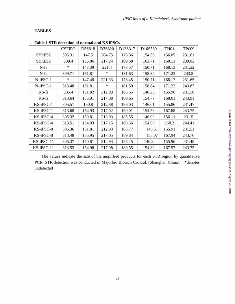
Table 1 STR detection of normal and KS iPSCs CSFIPO D5S818 D7S820 D13S317 D16S539 TH01 TPOX
SHhES2 305.31 147.5 204.75 173.36 154.58 156.05 231.61 SHhES2 309.4 155.86 217.24 189.68 162.71 168.11 239.82
N-fs * 147.39 221.4 173.57 150.71 168.13 231.52 N-fs 309.71 151.81 * 181.63 158.84 171.23 243.8
N-iPSC-1 * 147.48 221.33 173.45 150.71 168.17 231.65 N-iPSC-1 313.48 151.81 * 181.59 158.84 171.22 243.87
KS-fs 305.4 151.82 212.93 185.55 146.23 155.96 231.56 KS-fs 313.64 155.91 217.08 189.65 154.77 168.01 243.91
KS-iPSC-1 305.51 150.8 212.88 186.03 146.01 155.86 231.47 KS-iPSC-1 313.68 154.93 217.02 190.01 154.58 167.88 243.75 KS-iPSC-4 305.32 150.81 213.03 185.55 146.09 156.11 231.5 KS-iPSC-4 313.51 154.93 217.15 189.56 154.68 168.2 244.41 KS-iPSC-8 305.36 151.81 212.93 185.77 146.51 155.91 231.51 KS-iPSC-8 313.48 155.95 217.05 189.84 155.07 167.94 243.76 KS-iPSC-11 305.37 150.81 212.93 185.45 146.3 155.96 231.48 KS-iPSC-11 313.53 154.98 217.08 189.55 154.82 167.97 243.75
The values indicate the size of the amplified products for each STR region by quantitative PCR. STR detection was conducted in Majorbio Biotech Co. Ltd. (Shanghai, China). *denotes undetected
by guest on August 26, 2018
http://ww
w.jbc.org/
Dow
nloaded from
iPSC lines of a Klinefelter’s Syndrome patient
16
Table 2 Information of antibodies. Protein Company VIMENTIN Abcam OCT4 Prepared by our lab SOX2 Prepared by our lab NANOG R & D SSEA4 Millipore TRA-1-60 Millipore TRA-1-81 Chemicon AFP DAKO SOX17 R & D FLK1 Santa Cruz NESTIN Millipore TUJ1 Promega VASA Abcam H3K27me3 Millipore macroH2A1 Active Motif
by guest on August 26, 2018
http://ww
w.jbc.org/
Dow
nloaded from
iPSC lines of a Klinefelter’s Syndrome patient
17
Table 3 information of primers Gene Sequences For quantitative PCR NANOG F GAATGAAATCTAAGAGGTGGCA
R CCTGGTGGTAGGAAGAGTAAAGG VASA F TCGGATAACCATTTAGCACAG
R ATCATCTACTGGATTGGGAGC STRA8 F CAACCCAGAAAACCCAGAGGAGA
R CTTCATCAACGGGAAAGGATGCT BLIMP1 F ACGGCCTTTCAAATGTCAGACTTG
R TCTTGAGATTGCTGGTGCTGCTAA IFITM1 F TCACTCAACACTTCCTTCCCCAAA
R TCGCCAACCATCTTCCTGTCCCTA PELO F AGTGAAGACCGACAACAAACTGCT
R CTCCACCTGCTTGAGTCCATAGAA AMH F TGCAGGCACTGCTGTTCGG
R CGTGAGCGTCTCCAGGAAGG FSHR F CAGCCTCACCTATCCCAGCCATT
R CTGACCCCTAGCCTGAGTCATAT GAPDH F TCCACCCATGGCAAATTCC
R TCGCCCCACTTGATTTTGG XIST F GGCACTCTAGCACTTGAGGAT
R GAGAAACATGGAAATGGGTAA ZXDA F TTCACCACCTCTTACAAGCTCA
R CTCACATTTGAACGAGTTCTCCT For BSP OCT4 (outside) F GTTAAGGTTAGTGGGTGGGATT
R ATCACCTCCACCACCTAAAAA OCT4 (inside) F AGAGAGGGGTTGAGTAGTTTTTT
R ACCTCCACCACCTAAAAAAAAC
by guest on August 26, 2018
http://ww
w.jbc.org/
Dow
nloaded from
iPSC lines of a Klinefelter’s Syndrome patient
18
Figure 1
by guest on August 26, 2018
http://ww
w.jbc.org/
Dow
nloaded from
iPSC lines of a Klinefelter’s Syndrome patient
19
Figure 2
by guest on August 26, 2018
http://ww
w.jbc.org/
Dow
nloaded from
iPSC lines of a Klinefelter’s Syndrome patient
20
Figure 3
by guest on August 26, 2018
http://ww
w.jbc.org/
Dow
nloaded from
iPSC lines of a Klinefelter’s Syndrome patient
21
Figure 4
by guest on August 26, 2018
http://ww
w.jbc.org/
Dow
nloaded from
iPSC lines of a Klinefelter’s Syndrome patient
22
Figure 5
by guest on August 26, 2018
http://ww
w.jbc.org/
Dow
nloaded from
Li and Ying JinYu Ma, Chunliang Li, Junjie Gu, Fan Tang, Chun Li, Peng Li, Ping Ping, Shi Yang, Zheng
a Klinefelter's Syndrome patientAberrant gene expression profiles in pluripotent stem cells induced from fibroblasts of
published online September 27, 2012J. Biol. Chem.
10.1074/jbc.M112.380204Access the most updated version of this article at doi:
Alerts:
When a correction for this article is posted•
When this article is cited•
to choose from all of JBC's e-mail alertsClick here
by guest on August 26, 2018
http://ww
w.jbc.org/
Dow
nloaded from