Embed Size (px)
Citation preview
The Scientific World JournalVolume 2012, Article ID 363296, 8 pagesdoi:10.1100/2012/363296
The cientificWorldJOURNAL
Clinical Study
Association between Levels of IgA Antibodies toTissue Transglutaminase and Gliadin-Related Nonapeptides inDermatitis Herpetiformis
Justyna Gornowicz-Porowska,1 Monika Bowszyc-Dmochowska,1 Agnieszka Seraszek-Jaros,2
Elzbieta Kaczmarek,2 and Marian Dmochowski1
1 Cutaneous Histopathology and Immunopathology Section, Department of Dermatology, Poznan University of Medical Sciences,49 Przybyszewskiego Street, 60-355 Poznan, Poland
2 Department of Bioinformatics and Computational Biology, Poznan University of Medical Sciences, 79 Dabrowskiego Street,60-529 Poznan, Poland
Correspondence should be addressed to Marian Dmochowski, [email protected]
Received 10 October 2011; Accepted 2 November 2011
Academic Editors: B. B. Adams and C. Sitaru
Copyright © 2012 Justyna Gornowicz-Porowska et al. This is an open access article distributed under the Creative CommonsAttribution License, which permits unrestricted use, distribution, and reproduction in any medium, provided the original work isproperly cited.
Dermatitis herpetiformis (DH) is an autoimmunity-driven inflammatory blistering dermatosis associated with a gluten-dependententeropathy. Tissue transglutaminase (tTG) and nonapeptides of gliadin (npG) are considered in its pathomechanism/diagnostics.Here, the diagnostic accuracy of anti-tTG/anti-npG IgA ELISAs in Slavic DH patients with active skin rash was assessed throughcreating receiver operating characteristic (ROC) curves, determining cutoff values, and calculating correlations between levels ofanti-tTG/anti-npG IgA in DH, IgA/neutrophil-mediated non-DH patients and healthy persons. Altogether, sera from 80 Slavicindividuals were examined. There were negligible differences between cutoff points obtained by the ELISAs manufacturer andthose in this study. There were statistically significant correlations between levels of anti-tTG/anti-npG IgA in both DH group andthe group of IgA/neutrophil-mediated non-DH dermatoses. There was no such correlation in healthy controls. It seems that IgAautoantibodies to tTG and npG in the IgA/neutrophil-mediated DH are produced in the coordinated way implying their causalrelationship.
1. Introduction
Dermatitis herpetiformis (DH) is a chronic, IgA-mediated,inflammatory dermatosis with intense itching and polymor-phic eruption undergoing the spatial-temporal evolution[1]. In its pathomechanism, the role is played by bothgenetic and environmental factors. Thus, familial occurrenceis sometimes observed [2]. It is universally thought thatDH is associated with gluten-sensitive enteropathy (GSE),being a cutaneous manifestation of celiac disease (CD) [3].These diseases are caused by an immune reaction to proline-rich gliadin, a prolamin (gluten protein) found in wheat[4]. However, the trigger/triggers of pathological antigliadinautoimmune response in DH and relationship between CDand DH still remain inadequately understood. Some studies
indicated epidemiologic trends of increasing incidence ofCD. DH is also an important medical issue demanding highlyefficient medical and social services. DH is characterizedby cutaneous microgranular IgA deposits in the dermalpapillae (microgranular and fibrillar deposits are sometimesseen there) and/or along the dermal-epidermal junction [1];however, interesting issue is which IgA subclass is dominantin cutaneous deposits. In humans, IgA1 is a predominantsubclass in the sera, and IgA2 prevails in mucosal secre-tions of the colon [1]. Immunofluorescence analysis withmonoclonal antibodies revealed that IgA1 without IgA2 wasfound in the cutaneous deposits in all four patients examinedin an early study [5]. It was therefore speculated that bothIgA1 and IgA2 may be produced in the pathologic gut-associated lymphoid tissue, but only IgA1 is involved in
2 The Scientific World Journal
(a) (b)
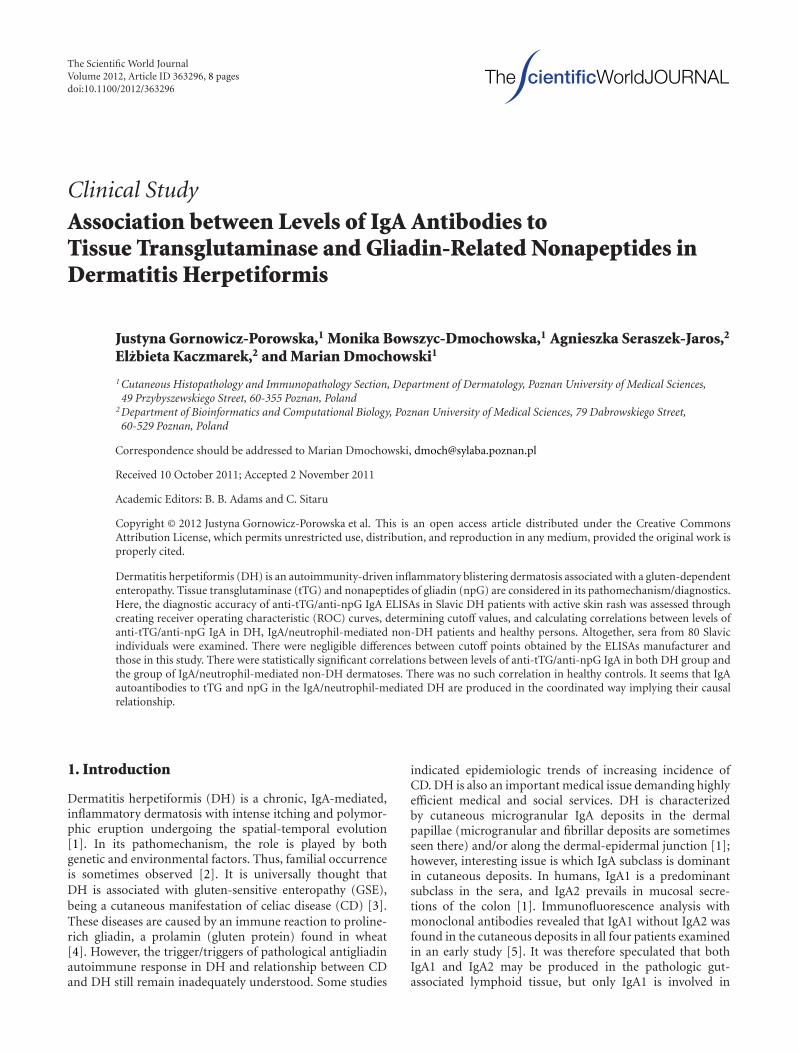
Figure 1: (a) Microgranular and fibrillar IgA1 deposits at dermal papillae in DIF in a young man with DH (original magnification×400). (b)Neutrophil elastase deposits in immunohistochemistry in lesional skin in a middle-aged woman with DH (original magnification, ×200).
the production of cutaneous lesions [5]. Still, there are newerdata that both IgA1 and IgA2 are forming IgA cutaneousdeposits in DH, although IgA1 (Figure 1(a)) predominates[1, 6]. In the development of DH, important is the accumu-lation of activated (neutrophil elastase-secreting) neutrophils(Figure 1(b)) that are forming microabscesses in the dermalpapillae with subsequent formation of microvesicles andfinally subepidermal (intralamina lucida) blisters [7]. Mainautoantigens in DH are enzymes of the transglutaminasefamily [8, 9]: epidermal transglutaminase (eTG) and closelyrelated tissue transglutaminase (tTG). They are consideredto be autoantigens plausibly recognized by principally IgA1autoantibodies in this disease [10]. Recently, the role ofnonapeptides of gliadin (npG) in pathomechanism of DHis considered [11]. Further, there are findings indicatingthat antibodies against deamidated synthetic gliadin-derivedpeptides are the most reliable tool in order to identify glutensensitivity in DH patients [12]. Interestingly, recent data[13] indicated that cross-linking microbial TG (mTG) mayreduce immunoreactivity of milk proteins. Cross-linkingby mTG results in integration of milk proteins epitopesinto newly created protein conglomerates, in such a waythat prevents recognition of those epitopes by specificantibodies [13]. Beneficial effect of TG was also observed onimmunoreactivity modification of cereals proteins. In thisway, it can be used to influence the clinical manifestationof food sensitivity. Watanabe et al. [14] showed that theuse of TG allows to obtain hypoallergenic flour from wheat,which can be consumed by persons with hypersensitivityto wheat. In light of the above, diverse roles of TGs inimmune responses are very intriguing. Poland’s nationaldata indicated that cross-linking by TG caused decreaseof gluten immunoreactivity [15], which raises hopes forTG use to modify nutrition of CD/DH patients. Thus,having knowledge of TGs is essential for understanding thepathogenesis of CD and DH [16], in which the production ofautoantibodies to TGs (as a result of chain of events initiatedby deamidation of glutamine residue in gliadin catalyzed bytTG) might surprisingly be of minor significance comparedwith benefits resulting from TGs-mediated cross-linking ofproteins. Regardless of pathogenetical considerations, direct
immunofluorescence test (DIF) of nonlesional skin remainsdefinitive laboratory test for diagnosing DH [7]. However,due to numerous clinical manifestations of GSE (includingDH), the use of serological techniques becomes helpful inclinical practice lowering the need for performing invasivegut biopsies [17].
Receiver operating characteristic (ROC) curves representa relationship between sensitivity and specificity of a labora-tory test over all possible diagnostic cutoff values. So, usingROC curves in laboratory medicine should be a commonpractice to facilitate clinical decision making.
2. Aim of the Study
The aim of this study was to investigate the diagnosticaccuracy of ELISA tests evaluating serum IgA antibodies totTG and npG in Slavic DH patients through performingthe receiver operating characteristic (ROC) and determiningproper cutoff values. Also, the aim was to assess correlationsbetween levels of anti-tTG and anti-npG IgA antibodiesin IgA/neutrophil-mediated DH and non-DH patients andhealthy individuals.
3. Material and Methods
Altogether, sera from 80 Slavic individuals were tested.Serum samples were obtained from patients with DH (19men and 12 women) with skin rash active enough to promptthem to seek dermatological attention and IgA/neutrophil-mediated non-DH dermatoses (25 patients as positive con-trol involved 17 cases of linear IgA bullous dermatosis, 4cases of IgA pemphigus, 3 cases of epidermolysis bullosaacquisita and 1 case of subcorneal pustular dermatosis)as well as from healthy individuals (negative control, 24donors). The DH group and IgA/neutrophil-mediated non-DH dermatoses group served as mutual control groups.The diagnosis of DH in all cases suspected of having DHat the clinical level was established when cutaneous IgAdeposition in any of seven possible diagnostic patterns wasseen with conventional DIF and corroborated by histologicalpicture with hematoxylin and eosin (H + E) staining.
The Scientific World Journal 3
To distinguish from DH and establish the clinical diagnosesof IgA/neutrophil-mediated non-DH dermatoses, DIF and H+ E staining were also done. Testing of sera with biochemicalmolecular techniques was done in cases requiring broaderdifferential diagnostics. DH serum samples were collectedduring the period from February 2010 through July 2011,and all serum samples were evaluated in the CutaneousHistopathology and Immunopathology Section, Departmentof Dermatology, Poznan University of Medical Sciences,Poland. All healthy controls were not relatives of DH patientsand gave no history of intolerance to gluten.
The levels of serum IgA autoantibodies against thefusion protein containing nonapeptides of gliadin wereevaluated with Antigliadin (GAF-3X) ELISA (Euroimmun,Germany) with the manufacturer’s cutoff value 25 RU/mL.The levels of serum IgA autoantibodies against tTG wereassessed with Anti-tTG ELISA (Euroimmun, Germany) withthe manufacturer’s cutoff value 20 RU/mL. Both tests arerecommended by the producer as useful in DH diagnosis.All measurements were made using a programmable ELISAreader with MikroWin 2000 software by a single operatorfollowing the manufacturer’s instructions. All tests wereperformed with meticulous care to eliminate the risk ofintralaboratory errors.
Antigens which were used in the tests were obtained withmolecular engineering methods. The antigen in Antigliadin(GAF-3X) ELISA is a recombinant fusion protein, which wascreated using DNA sequences encoding gliadin-analogousfusion peptide, GAF. These gliadin-analogous peptides con-sist of three repeated sequences to increase the diagnosticefficacy. The created protein composition involves syntheticnonapeptide which is gliadin analoque and nonapeptide ofdigested gliadin which was deamidated by transglutaminases.Antigen in Anti-tTG ELISA is recombinant tTG. The expres-sion of proper human cDNA was done with baculovirusvector in insect cell systems.
In this study, the ROC analysis was used for deter-mining cutoff points for the best sensitivity and specificityof tTG/npG ELISA tests and evaluating their ability todiscriminate disease-affected from normal cases. Statisticalanalysis was performed by use of MedCalc statistical software(http://www.medcalc.org/). Kruskal-Wallis test with post hocDunn’s test was performed to detect significant differencesbetween three studied groups. Spearman’s rank correlationcoefficient was computed to find out associations betweenlevels of IgA antibodies to tTG and npG in all groups.
4. Results
Results of statistical analysis of three groups of examinedpatients are presented in Table 1. The ROC plots createdfor various combinations of examined groups are shown inFigures 2, 3, and 4.
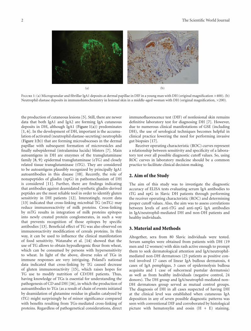
The optimal cut-off for the Anti-tTG IgA ELISAin all combinations (DH patients versus healthy con-trol; DH patients versus IgA/neutrophil-mediated non-DH dermatoses; combined groups) of examined groups is17.199 RU/mL (manufacturer’s cut-off is 20 RU/mL). Theoptimal cut-off for the Antigliadin (GAF-3X) IgA ELISA
is 24.633 RU/mL in DH patients versus IgA/neutrophil-mediated non-DH dermatoses and in combined groups(manufacturer’s cut-off is 25 RU/mL). The optimal cut-offfor the Antigliadin (GAF-3X) IgA ELISA is 24.08 RU/mL inDH patients versus healthy controls (manufacturer’s cut-offis 25 RU/mL). It is noteworthy that in each combination thearea under the ROC curve (AUC) is 1.00.
Regarding values of both anti-tTG and anti-npG IgA,differences between healthy control and DH patients (P <0.001) were significant as well as between IgA/neutrophil-mediated non-DH dermatoses and DH patients (P <0.001), whereas differences between healthy control andIgA/neutrophil-mediated non-DH dermatoses were notrevealed (P > 0.05). The analysis of correlation showedstatistically significant correlations between levels of IgAantibodies to tTG and npG in both DH group (r =0.4149) and the group of IgA/neutrophil-mediated non-DH dermatoses (r = 0.7890), whereas there was no suchcorrelation in healthy controls (r = 0.2231).
The following positive results were obtained in examinedgroups: (i) in case of anti-tTG IgA: 90% of DH patients, 4%of IgA/neutrophil-mediated non-DH dermatoses patients,and 0% in healthy controls; (ii) in case of anti-npG IgA:90% of DH patients, 8% of IgA/neutrophil-mediated non-DH dermatoses patients, and 0% in healthy controls.
5. Discussion
In our Slavic DH patients, the ratio between men and womenwas 1.6 : 1, which is consistent with the reports that DHaffects men slightly more often [18, 19].
In this study, we attempt to assess diagnostic usefulnessand accuracy of tTG/npG ELISA tests in the managementof Slavic DH patients. Curiously, the research literaturedata about DH and studies on its pathomechanism areamazingly scanty in relation to the other autoimmuneblistering dermatoses, while it seems that DH pathogenesisis far more complex. As mentioned above, GSE, bothCD and DH, is a growing medical problem, particularlyamong young people, with nutritional impact on their healthstatus. Thus, specific, reliable, and objective criteria fordiagnosing and monitoring of DH should be established. DIFof nonlesional skin remains a definitive laboratory test fordiagnosing this disease, however its invasiveness is a seriouslimitation for screening. Indirect immunofluorescence is atime-consuming, expensive, and subjective technique; then,ELISA seems to be free from those disadvantages. At present,ELISA is the method of choice for serological screening ofDH. However, there is a problem what kind of ELISA-basedtest is the best. The medical diagnostics market offers awide range of ELISA kits with biotechnologically obtainedeTG, tTG, and npG as the most frequent antigen sourcesfor diagnosing DH. Literature data indicated that thereare discrepancies regarding the specificity and repeatabilityof test results. Some findings showed that 52% of DHpatients have anti-eTG IgA elevated [20], which is consistentwith our previous study [21]. On the other hand, severalcommunications revealed that more than 90% of DHpatients have anti-eTG IgA elevated [19, 22]. Such divergent
4 The Scientific World Journal
Table 1: Statistical analysis of the three groups of Slavic subjects studied.
IgA antibodies evaluated with Examined group Number of patients Mean SD Dunn’s test
Anti- tTG ELISA (RU/mL)Group I 24 2.35 1.58 Group I versus group III P < 0.001
Group II versus group III P < 0.001Group I versus group II P > 0.05
Group II 25 13.57 46.57
Group III 31 429.20 435.23
Antigliadin (GAF-3X) ELISA(RU/mL)
Group I 24 1.43 3.32 Group I versus group III P < 0.001Group II versus group III P < 0.001Group I versus group II P > 0.05
Group II 25 15.10 44.17
Group III 30 271.00 499.25
Explanations: group I: healthy controls, group II: IgA/neutrophil-mediated non-DH dermatoses group, group III: DH patients. SD: standard deviation.
0
20
40
60
80
100
0 20 40 60 80 100
Sensitivity: 100, 0Specificity: 100, 0
tTG
Criterion: > 17, 199
100-specificity
Sen
siti
vity
(a)
GAF
0
0
20
20
40
40
60
60
80
80
100
100
Criterion: > 24, 08
Sensitivity: 100, 0Specificity: 100, 0
100-specificity
Sen
siti
vity
(b)
Figure 2: ROC analysis for DH patients and healthy controls. (a) shows the plot for Anti-tTG IgA ELISA, and (b) shows Antigliadin (GAF-3X) IgA ELISA. The cutoff point marked as criterion (17.199 and 24.08, resp.).
results might be caused by evaluating series of DH patientsdiffering in severity of cutaneous rash. In the light of this,there is an urgent need to find/develop a new or modifiedantigen/epitope that would make the DH diagnostics moreaccurate. Thus, the usefulness of tTG/npG ELISA-based testsshould be considered. Literature associated with this problemdemonstrated that there is also incompatibility about thelevel of anti-tTG IgA. Some investigators [20] obtainedonly 25% of anti-tTG IgA-positive results in DH patients,whereas other researchers achieved about 79% of positiveresults in their patients [19, 23]. Our own experience inthis area is satisfactory: 90% of DH patients examined inthis study had anti-tTG IgA above normal range. Importantissue remains the composition and structural design of theantigen/epitope. Byrne et al. [24] indicated that the use ofnovel mutagenic variant of tTG lacking the catalytic triaddecreases the binding of IgA to the mutant tTG with themean reduction of 58% in DH and even bigger meanreduction of 79% in CD samples. Fernandez et al. [25]analyzed six different human anti-tTG ELISA kits. This
group of researchers showed that there are differences in thesensitivity and specificity of the human tTG ELISA assays.Furthermore, they suggested that diagnostic accuracy of testswas significantly improved by adjusting the cutoff thresholdsaccording to ROC curve analyses, which was done in ourstudy. Manufacturer’s cut-off is not standardized to eachlaboratory conditions, therefore the standardization basedon ROC curve analyses should be recommended to allELISA tests performed for diagnostic purposes. Interestingly,Fernandez et al. [25] demonstrated that the correction ofthe cut-off with the use of the ROC curve analysis modifiesthe decision limit in more than 50% in five of the sixexamined anti-human tTG ELISA kits. Hence, there is theevidence that the way/source of production and furthermodification of antigen have the impact on test accuracy.Researchers and diagnosticians should take it into accountbefore the choice of the appropriate test.
Recently [26, 27], investigators indicated the usefulnessof synthetic deamidated gliadin-derived peptides (GDR) asantigen, which is useful for the detection ofsensitivity to
The Scientific World Journal 5
tTG
0
20
40
60
80
100
0 20 40 60 80 100
100-specificity
Sen
siti
vity
Sensitivity: 100, 0Specificity: 100, 0Criterion: > 17, 199
(a)
GAF
0
20
40
60
80
100
0 20 40 60 80 100
100-specificitySe
nsi
tivi
ty
Sensitivity: 100, 0Specificity: 100, 0Criterion: > 24, 633
(b)
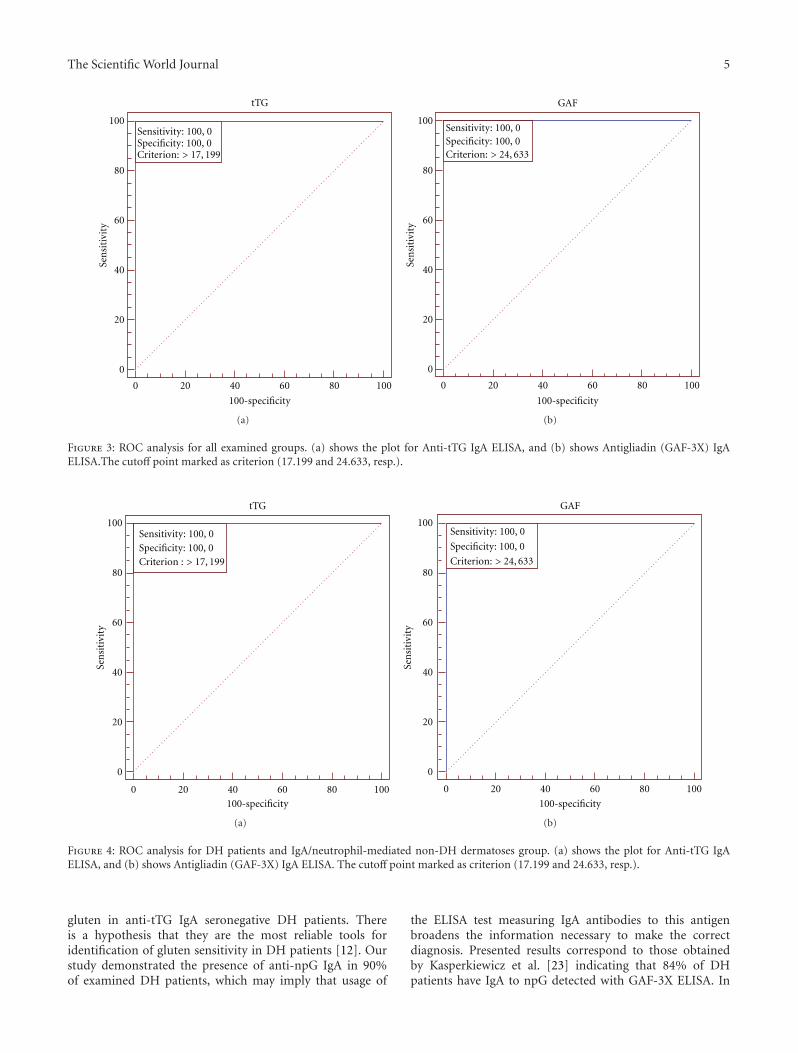
Figure 3: ROC analysis for all examined groups. (a) shows the plot for Anti-tTG IgA ELISA, and (b) shows Antigliadin (GAF-3X) IgAELISA.The cutoff point marked as criterion (17.199 and 24.633, resp.).
tTG
0
20
40
60
80
100
0 20 40 60 80 100
100-specificity
Sen
siti
vity
Sensitivity: 100, 0Specificity: 100, 0Criterion : > 17, 199
(a)
GAF
0
20
40
60
80
100
0 20 40 60 80 100
100-specificity
Sen
siti
vity
Sensitivity: 100, 0
Specificity: 100, 0
Criterion: > 24, 633
(b)
Figure 4: ROC analysis for DH patients and IgA/neutrophil-mediated non-DH dermatoses group. (a) shows the plot for Anti-tTG IgAELISA, and (b) shows Antigliadin (GAF-3X) IgA ELISA. The cutoff point marked as criterion (17.199 and 24.633, resp.).
gluten in anti-tTG IgA seronegative DH patients. Thereis a hypothesis that they are the most reliable tools foridentification of gluten sensitivity in DH patients [12]. Ourstudy demonstrated the presence of anti-npG IgA in 90%of examined DH patients, which may imply that usage of
the ELISA test measuring IgA antibodies to this antigenbroadens the information necessary to make the correctdiagnosis. Presented results correspond to those obtainedby Kasperkiewicz et al. [23] indicating that 84% of DHpatients have IgA to npG detected with GAF-3X ELISA. In

6 The Scientific World Journal
this study, we determined the cutoff value for examinedDH patients in anti-tTG ELISA as the level 17.199 RU/mL(for all examined groups), while the manufacturer’s cut-offis 20 RU/mL. In anti-npG ELISA, the cut-off for examinedpopulation is 24.633 RU/mL (for 2 combinations of groups)and 24.08 RU/mL (for DH and healthy groups), while themanufacturer’s cut-off is 25 RU/mL. Thus, there are negligi-ble differences between them (especially in case of antigliadinGAF-3X ELISA). It can be explained by the fact that kitsused are well standardized in a genetically similar population.It is known that the cutoff point can vary depending onexamined population, for example, different cutoff value ofanti-tTG ELISA for Italian and Spanish populations [28, 29].Interestingly, the cutoff points in our studied groups werealmost identical. It may be due to the fact that diseased group,IgA/neutrophil-mediated non-DH dermatoses, was chosenas pathogenetically most closely related to DH, as far ascutaneous pathology is concerned. Moreover, AUC obtainedin this study was equal 1, which may be due to a perfectseparation of the values of the examined groups. It shouldbe stressed here that we had precise inclusion criteria andeach diagnosis had to be confirmed with the combinationof microscopic and biochemical/molecular techniques. Thus,using the new cutoff values derived from ROC curves, thediagnostic accuracy of the tests is improved (sensitivity andspecificity of 100%). It could be due to fact that the tests wereadapted to our native population.
There were no individuals with positive anti-tTG/npGIgA antibodies in the healthy controls, although we didnot make the biopsy for DIF in order to rule out DH inthese subjects presenting no cutaneous lesions whatsoever. Inaddition, the percentage of cases with positive anti-tTG/npGIgA antibodies in IgA/neutrophil-mediated non-DH der-matoses group has been quite low (4% of anti-tTG ELISAand 8% of anti-npG ELISA). Still, it cannot be excluded thatsome such cases had low-grade GST with no overt clinicalsymptoms in addition to their IgA/neutrophil-mediatednon-DH dermatosis. Interestingly, findings obtained byLudvigsson et al. [30] in a nationwide cohort study indicatedthat individuals with CD were at increased risk of psoriasisboth before and after CD diagnosis. Thus, one should beaware that CD can coexist with other dermatoses, not onlywith DH. In the light of the above, it is suggested thatrecognition of anti-npG IgA alone does not mean that oneis dealing with DH as a cutaneous manifestation of GSE,and DIF of the uninvolved skin still remains crucial fordiagnosing DH.
In our study, we demonstrated that the determination ofanti-tTG/npG IgA by means of ELISA is a precise method tobroaden the body of knowledge about DH patients in Slavicpopulation. However, what was noticed by Fernandez et al.[25] is that it is necessary to select the ELISA kit with thehighest sensitivity and specificity and recalculate the cutoffthreshold using samples from any given native population.This sequence of actions is essential for the laboratorydiagnostician to provide reliable information to the clinicianto facilitate making right decision on the implementation oftroublesome therapy with the risk of potentially dangeroussideeffects.
Statistically significant correlation between levels of IgAantibodies to tTG and npG in DH group (r = 0.4149) foundin this study and no such correlation in healthy controls(r = 0.2231) might suggest that the anti-tTG and anti-npG IgA antibodies in the IgA/neutrophil-mediated DH, butnot in healthy individuals, are produced in the coordinatedway in contrast to healthy individuals. These findings arein agreement with our previous data [31]. This mightcorrespond with a suggestion that, as the catalytic site of tTGseems to be targeted by IgA autoantibodies, intermolecularepitope spreading from the gliadin epitopes to the catalyticsite of tTG does take place in CD [24].
In recent years, the cutaneous immunopathology of DHhas become the area of extensive studies in humans andusing animal models of the disease [21, 32, 33]. We feel thatthe key issue in understanding the blister formation in DHis not simply linking it to the GSE, but evaluating insteadlocal cutaneous factors, conceivably neutrophils’ Fc receptorsinvolvement, at the human skin level.
Nonetheless, it should be kept in mind, as far as theissue of importance of npG in pathogenesis and diagnosticsof GSE is concerned, that within the gliadin peptide theN-terminal proline residue and the C-terminal glutamineresidues were reported to be essential for antibody recogni-tion in addition to the deamidated glutamine residue [34].Finally, our data might suggest that Anti-tTG IgA ELISAis marginally superior to Antigliadin (GAF-3X) IgA ELISAfor differential diagnosis of cutaneous itchy rashes suspectedto be DH at the clinical level. Plausibly, in connectionwith the data that IgA1 deposits predominate in the skinof DH sufferers, modification of the above ELISA tests toenable the determination of IgA1 subclass antibodies to tTGand npG would be even more valuable for differentiatingIgA/neutrophil-mediated dermatoses.
6. Conclusions
IgA antibodies to tTG and npG are detectable in the vastmajority of DH Slavic patients. Tests based on these antigenstailored to native/local populations, for example, Slavicassessed in this study, broaden DH diagnostics, being usefulfor the detection of gluten sensitivity in DH. However, DIFof the uninvolved skin still remains the reference method fordiagnosing DH.
It seems that the anti-tTG and anti-npG IgA antibodiesin the IgA/neutrophil-mediated DH, but not in healthyindividuals, are produced in the coordinated way implyingtheir causal relationship.
Acknowledgments
The authors wish to thank Euroimmun (Lubeck, Germany)for providing the part of commercial kits of Anti-tTGand Antigliadin (GAF-3X) IgA ELISA used in this study.This study was partly funded from grant of the PolishMinistry of Science and Higher Education (N N 401 00 1239). J. Gornowicz-Porowska was supported by VoivodeshipLabour Office in Poznan (scholarship from project called
The Scientific World Journal 7
“Scholarships for PhD students from faculties regardedas strategic for Wielkopolska region development”, subsec-tion 8.2.2. Operational Programme: Human Capital). A partof this study was presented as a poster at the 2011 AnnualSID Meeting (May 4–7, 2011, Phoenix, Arizona, USA) andpublished in the abstract form: [31].
References
[1] M. Dmochowski, “Dermatitis herpetiformis,” in AutoimmuneBlistering Dermatoses, pp. 281–311, Scientific Publishing ofPoznan University of Medical Sciences, Poznan, 2006.
[2] M. Dus, A. Danczak-Pazdrowska, J. Gornowicz, M. Bowszyc-Dmochowska, and M. Dmochowski, “Transient manifestationof dermatitis herpetiformis in a female with familial predispo-sition induced by propafenone,” Advances in Dermatology andAllergology, vol. 26, no. 4, pp. 239–242, 2009.
[3] J. J. Zone, “Skin manifestations of celiac disease,” in Proceed-ings of the NIH Consensus Development Conference on CeliacDisease, pp. 73–75, Bethesda, Md, USA, June 2004.
[4] T. L. Reunala, “Dermatitis herpetiformis,” Clinics in Dermatol-ogy, vol. 19, no. 6, pp. 728–736, 2001.
[5] S. M. Olbricht, T. J. Flotte, B. Collins, C. M. Chapman, and T.J. Harrist, “Dermatitis herpetiformis. Cutaneous deposition ofpolyclonal IgA1,” Archives of Dermatology, vol. 122, no. 4, pp.418–421, 1986.
[6] F. Wojnarowska, D. Delacroix, and P. Gengoux, “CutaneousIgA subclasses in dermatitis herpetiformis and linear IgAdisease,” Journal of Cutaneous Pathology, vol. 15, no. 5, pp.272–275, 1988.
[7] J. Gornowicz, M. Bowszyc-Dmochowska, and M. Dmo-chowski, “IgA antibodies against fusion protein containingmodified nonapeptide of gliadin in dermatitis herpetiformis,”Dermatologia Kliniczna, vol. 12, no. 4, pp. 239–243, 2010.
[8] M. Sardy, S. Karpati, B. Merkl, M. Paulsson, and N. Smyth,“Epidermal transglutaminase (TGase 3) is the autoantigen ofdermatitis herpetiformis,” Journal of Experimental Medicine,vol. 195, no. 6, pp. 747–757, 2002.
[9] W. Dieterich, E. Laag, L. Bruckner-Tuderman et al., “Anti-bodies to tissue transglutaminase as serologic markers inpatients with dermatitis herpetiformis,” Journal of InvestigativeDermatology, vol. 113, no. 1, pp. 133–136, 1999.
[10] J. Gornowicz-Porowska, M. Bowszyc-Dmochowska, and M.Dmochowski, “Autoimmunity-driven enzymatic remodelingof the dermal-epidermal junction in bullous pemphigoid anddermatitis herpetiformis,” Autoimmunity, 2011.
[11] E. Schwertz, F. Kahlenberg, U. Sack et al., “Serologic assaybased on gliadin-related nonapeptides as a highly sensitive andspecific diagnostic aid in celiac disease,” Clinical Chemistry,vol. 50, no. 12, pp. 2370–2375, 2004.
[12] E. Sugai, E. Smecuol, S. Niveloni et al., “Celiac disease serologyin dermatitis herpetiformis. Which is the best option fordetecting gluten sensitivity?” Acta Gastroenterologica Lati-noamericana, vol. 36, no. 4, pp. 197–201, 2006.
[13] B. Wroblewska, A. Kaliszewska, E. Malinowska, and A.Troszynska, “Immunoreactivity of transglutaminase cross-linked milk proteins in fermentem milk product obtainedwith Lactobacillus acidophilus,” Advances in Dermatology andAllergology, vol. 28, no. 4, pp. 261–267, 2011.
[14] M. Watanabe, J. Miyakawa, Z. Ikezawa, Y. Suzuki, andT. Hirao, “Production of hypoallergenic rice by enzymaticdecomposition of constituent proteins,” Journal of Food Sci-ence, vol. 55, pp. 781–783, 2004.
[15] J. Leszczynska, “Immunoreaktywnosc wybranych składnikowzywnosci i sposoby jej zmniejszania. (The methods ofreducing immunoreactivity of the food allergens),” ScientificNotebooks, Dissertations/Technical University of Lodz, vol. Z376, pp. 3–112, 2009.
[16] L. Lorand and R. M. Graham, “Transglutaminases: crosslink-ing enzymes with pleiotropic functions,” Nature ReviewsMolecular Cell Biology, vol. 4, no. 2, pp. 140–156, 2003.
[17] E. Zintzaras and A. E. Germenis, “Performance of antibodiesagainst tissue transglutaminase for the diagnosis of celiacdisease: meta-analysis,” Clinical and Vaccine Immunology, vol.13, no. 2, pp. 187–192, 2006.
[18] O. Braun-Falco and H. H. Plewig, “Dermatitis herpetiformis,”in Dermatology T I, W. Gilinski, H. Wolska, and P. Zaborowski,Eds., pp. 649–652, Czelej Publishing House, Lublin, 2002.
[19] C. Rose, E. B. Brocker, and D. Zillikens, “Clinical, histologicaland immunpathological findings in 32 patients with dermati-tis herpetiformis Duhring,” JDDG, vol. 8, no. 4, pp. 265–270,2010.
[20] C. M. Hull, M. Liddle, N. Hansen et al., “Elevation ofIgA anti-epidermal transglutaminase antibodies in dermatitisherpetiformis,” British Journal of Dermatology, vol. 159, no. 1,pp. 120–124, 2008.
[21] M. Bowszyc-Dmochowska, A. Seraszek, E. Kaczmarek, J.Gornowicz, and M. Dmochowski, “Low strength of correla-tion between the level of serum IgA antibodies to epidermaltransglutaminase and the intensity of neutrophil elastaseexpression in lesional skin in dermatitis herpetiformis,” TheOpen Autoimmunity Journal, vol. 1, pp. 1–4, 2009.
[22] C. Rose, F. P. Armbruster, J. Ruppert, B. W. Igl, D. Zillikens,and I. Shimanovich, “Autoantibodies against epidermal trans-glutaminase are a sensitive diagnostic marker in patients withdermatitis herpetiformis on a normal or gluten-free diet,”Journal of the American Academy of Dermatology, vol. 61, no.1, pp. 39–43, 2009.
[23] M. Kasperkiewicz, C. Dahnrich, C. Probst et al., “Novelassay for detecting celiac disease-associated autoantibodies indermatitis herpetiformis using deamidated gliadin-analogousfusion peptides,” Journal of the American Academy of Derma-tology, 2011.
[24] G. Byrne, F. Ryan, J. Jackson, C. Feighery, and J. Kelly,“Mutagenesis of the catalytic triad of tissue transglutaminaseabrogates coeliac disease serum IgA autoantibody binding,”Gut, vol. 56, no. 3, pp. 336–341, 2007.
[25] E. Fernandez, S. Riestra, L. Rodrigo et al., “Comparison of sixhuman anti-transglutaminase ELISA-tests in the diagnosis ofceliac disease in the Saharawi population,” World Journal ofGastroenterology, vol. 11, no. 24, pp. 3762–3766, 2005.
[26] E. Sugai, H. J. Hwang, H. Vazquez et al., “New serologyassays can detect gluten sensitivity among enteropathy patientsseronegative for anti-tissue transglutaminase,” Clinical Chem-istry, vol. 56, no. 4, pp. 661–665, 2010.
[27] T. D. Jaskowski, M. R. Donaldson, C. M. Hull et al., “Novelscreening assay performance in pediatric celiac disease andadult dermatitis herpetiformis,” Journal of Pediatric Gastroen-terology and Nutrition, vol. 51, no. 1, pp. 19–23, 2010.
[28] S. Martini, G. Mengozzi, G. Aimo, R. Pagni, and C. Sategna-Guidetti, “Diagnostic accuracies for Celiac disease of fourtissue transglutaminase autoantibody tests using human anti-gen,” Clinical Chemistry, vol. 47, no. 9, pp. 1722–1725, 2001.
[29] S. Vivas, J. M. Ruiz de Morales, J. Martınez et al., “Humanrecombinant anti-transglutaminase antibody testing is usefulin the diagnosis of silent coeliac disease in a selected group
8 The Scientific World Journal
of at-risk patients,” European Journal of Gastroenterology andHepatology, vol. 15, no. 5, pp. 479–483, 2003.
[30] J. F. Ludvigsson, B. Lindelof, F. Zingone, and C. Ciamci,“Psoriasis in nationwide cohort study of patients with celiacdisease,” Journal of Investigative Dermatology, vol. 131, pp.2010–2016, 2011.
[31] M. Dmochowski, J. Gornowicz, A. Seraszek et al., “Cutaneousexpression of neutrophil elastase in relation to IgA autoanti-bodies to nonapeptides of gliadin and tissue transglutaminasein dermatitis herpetiformis,” Journal of Investigative Dermatol-ogy, vol. 131, supplement 1, p. S2, 2011.
[32] S. Mihai and C. Sitaru, “Immunopathology and moleculardiagnosis of autoimmune bullous diseases,” Journal of Cellularand Molecular Medicine, vol. 11, no. 3, pp. 462–481, 2007.
[33] J. J. Zone, L. A. Schmidt, T. B. Taylor et al., “Dermatitisherpetiformis sera or goat anti-transglutaminase-3 transferredto human skin-grafted mice mimics dermatitis herpetiformisimmunopathology,” Journal of Immunology, vol. 186, no. 7, pp.4474–4480, 2011.
[34] N. H. Petersen, P. N. Hansen, and G. Houen, “Fast and effi-cient characterization of an anti-gliadin monoclonal antibodyepitope related to celiac disease using resin-bound peptides,”Journal of Immunological Methods, vol. 365, no. 1-2, pp. 174–182, 2011.

Submit your manuscripts athttp://www.hindawi.com
Stem CellsInternational
Hindawi Publishing Corporationhttp://www.hindawi.com Volume 2014
Hindawi Publishing Corporationhttp://www.hindawi.com Volume 2014
MEDIATORSINFLAMMATION
of
Hindawi Publishing Corporationhttp://www.hindawi.com Volume 2014
Behavioural Neurology
EndocrinologyInternational Journal of
Hindawi Publishing Corporationhttp://www.hindawi.com Volume 2014
Hindawi Publishing Corporationhttp://www.hindawi.com Volume 2014
Disease Markers
Hindawi Publishing Corporationhttp://www.hindawi.com Volume 2014
BioMed Research International
OncologyJournal of
Hindawi Publishing Corporationhttp://www.hindawi.com Volume 2014
Hindawi Publishing Corporationhttp://www.hindawi.com Volume 2014
Oxidative Medicine and Cellular Longevity
Hindawi Publishing Corporationhttp://www.hindawi.com Volume 2014
PPAR Research
The Scientific World JournalHindawi Publishing Corporation http://www.hindawi.com Volume 2014
Immunology ResearchHindawi Publishing Corporationhttp://www.hindawi.com Volume 2014
Journal of
ObesityJournal of
Hindawi Publishing Corporationhttp://www.hindawi.com Volume 2014
Hindawi Publishing Corporationhttp://www.hindawi.com Volume 2014
Computational and Mathematical Methods in Medicine
OphthalmologyJournal of
Hindawi Publishing Corporationhttp://www.hindawi.com Volume 2014
Diabetes ResearchJournal of
Hindawi Publishing Corporationhttp://www.hindawi.com Volume 2014
Hindawi Publishing Corporationhttp://www.hindawi.com Volume 2014
Research and TreatmentAIDS
Hindawi Publishing Corporationhttp://www.hindawi.com Volume 2014
Gastroenterology Research and Practice
Hindawi Publishing Corporationhttp://www.hindawi.com Volume 2014
Parkinson’s Disease
Evidence-Based Complementary and Alternative Medicine
Volume 2014Hindawi Publishing Corporationhttp://www.hindawi.com