Embed Size (px)
Citation preview
J Psychiatry Neurosci 2014;39(1) 31
Research Paper
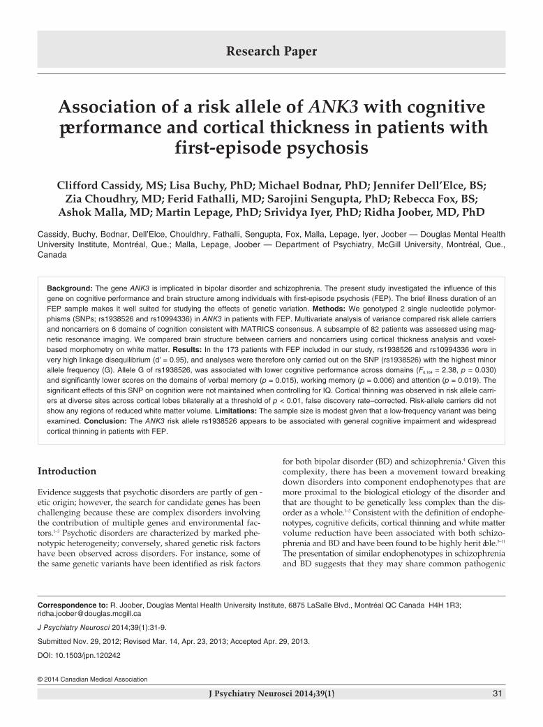
Association of a risk allele of ANK3 with cognitive performance and cortical thickness in patients with
first-episode psychosis
Clifford Cassidy, MS; Lisa Buchy, PhD; Michael Bodnar, PhD; Jennifer Dell’Elce, BS; Zia Choudhry, MD; Ferid Fathalli, MD; Sarojini Sengupta, PhD; Rebecca Fox, BS;
Ashok Malla, MD; Martin Lepage, PhD; Srividya Iyer, PhD; Ridha Joober, MD, PhD
Cassidy, Buchy, Bodnar, Dell’Elce, Chouldhry, Fathalli, Sengupta, Fox, Malla, Lepage, Iyer, Joober — Douglas Mental HealthUniversity Institute, Montréal, Que.; Malla, Lepage, Joober — Department of Psychiatry, McGill University, Montréal, Que.,Canada
Introduction
Evidence suggests that psychotic disorders are partly of gen -etic origin; however, the search for candidate genes has beenchallenging because these are complex disorders involvingthe contribution of multiple genes and environmental fac-tors.1–3 Psychotic disorders are characterized by marked phe-notypic heterogeneity; conversely, shared genetic risk factorshave been observed across disorders. For instance, some ofthe same genetic variants have been identified as risk factors
for both bipolar disorder (BD) and schizophrenia.4 Given thiscomplexity, there has been a movement toward breakingdown disorders into component endophenotypes that aremore proximal to the biological etiology of the disorder andthat are thought to be genetically less complex than the dis -order as a whole.1–3 Consistent with the definition of endophe-notypes, cognitive deficits, cortical thinning and white mattervolume reduction have been associated with both schizo -phrenia and BD and have been found to be highly herit able.5–11
The presentation of similar endophenotypes in schizophreniaand BD suggests that they may share common pathogenic
Correspondence to: R. Joober, Douglas Mental Health University Institute, 6875 LaSalle Blvd., Montréal QC Canada H4H 1R3;[email protected]
J Psychiatry Neurosci 2014;39(1):31-9.
Submitted Nov. 29, 2012; Revised Mar. 14, Apr. 23, 2013; Accepted Apr. 29, 2013.
DOI: 10.1503/jpn.120242
© 2014 Canadian Medical Association
Background: The gene ANK3 is implicated in bipolar disorder and schizophrenia. The present study investigated the influence of thisgene on cognitive performance and brain structure among individuals with first-episode psychosis (FEP). The brief illness duration of anFEP sample makes it well suited for studying the effects of genetic variation. Methods: We genotyped 2 single nucleotide polymor-phisms (SNPs; rs1938526 and rs10994336) in ANK3 in patients with FEP. Multivariate analysis of variance compared risk allele carriersand noncarriers on 6 domains of cognition consistent with MATRICS consensus. A subsample of 82 patients was assessed using mag-netic resonance imaging. We compared brain structure between carriers and noncarriers using cortical thickness analysis and voxel-based morphometry on white matter. Results: In the 173 patients with FEP included in our study, rs1938526 and rs10994336 were invery high linkage disequilibrium (d' = 0.95), and analyses were therefore only carried out on the SNP (rs1938526) with the highest minorallele frequency (G). Allele G of rs1938526, was associated with lower cognitive performance across domains (F6,164 = 2.38, p = 0.030)and significantly lower scores on the domains of verbal memory (p = 0.015), working memory (p = 0.006) and attention (p = 0.019). Thesignificant effects of this SNP on cognition were not maintained when controlling for IQ. Cortical thinning was observed in risk allele carri-ers at diverse sites across cortical lobes bilaterally at a threshold of p < 0.01, false discovery rate–corrected. Risk-allele carriers did notshow any regions of reduced white matter volume. Limitations: The sample size is modest given that a low-frequency variant was beingexamined. Conclusion: The ANK3 risk allele rs1938526 appears to be associated with general cognitive impairment and widespreadcortical thinning in patients with FEP.
pathways. Therefore, researchers often use such measuresrather than diagnosis when studying the effects of geneticvariation.1–3
Numerous variants of the gene ANK3, encoding AnkyrinG, have been associated with BD in genome-wide associationstudies (GWAS)12,13 and a large genome-wide study combin-ing schizophrenia and BD.14 The evidence for an associationwith schizophrenia is not as strong, as no genome-wide studies have found a significant effect. Three studies, how-ever, have reported an association with schizophrenia.15–17
One of them was genome-wide and showed near-significantassociation after correction,15 and another one included ameta-analysis showing significant association of the singlenucleotide polymorphisms (SNPs) rs10761482 andrs10994336 with schizophrenia.17 Furthermore, lower levels ofANK3 expression have been observed in multiple cortical ar-eas in patients with schizophrenia.16,18 The risk-associatedANK3 SNPs are intronic, and rs1938526 and other SNPs havebeen associated with reduced gene expression.16,19
Several studies have examined the role of these geneticvariants on neurocognition. A study on BD found significantassociation between rs10994336 and sustained attention, butno effect on general intelligence, memory or executive func-tioning.20 In healthy controls, 1 study did not report any effectof this SNP on measures of attention, working memory or ex-ecutive functioning,21 whereas another study found an associ-ation between this SNP and set-shifting, risk-taking behav-iour and decreased white matter integrity in the internalcapsule.22 A further study found rs9804190 to be associatedwith poor working memory and executive functioning andhigher prefrontal cortical activity during a working memorytask.16 A familial mutation in ANK3 is associated with intel-lectual disability, and knockdown of this gene in drosophilais associated with memory deficits.23
Ankyrin G is found at axon initial segments (AIS) andnodes of Ranvier (NOR). It links voltage-gated sodium chan-nels to the cytoskeleton and promotes their clustering in con-junction with cell adhesion molecules and oligodendrocytes.24
At the AIS, via neurofascin cell adhesion molecules, AnkyrinG promotes formation of GABAergic synapses.25 The reducedexpression of ANK3 observed in patients with schizophre-nia16,18 is consistent with a large body of research implicatingγ-aminobutyric acid (GABA) synapses at the AIS in peoplewith this condition.18,26 Through actions at the NOR, AnkyrinG facilitates the propagation of action potentials.27 Ankyrin Gcould thus influence the efficiency and synchrony of trans-mission of neuronal impulses and be necessary for long-range γ oscillations between cortical subfields,28 a processlinked to sustained attention.29 In patients with schizo -phrenia, excessive cortical γ oscillations have been observedduring working memory performance.30 A role of Ankyrin Gat the NOR is also consistent with a large body of researchpointing to deficits in white matter and oligodendrocytes inpatients with schizophrenia and BD.31,32
The objective of the present study was to test the associa-tion of allelic variation in ANK3 with performance acrossdifferent domains of cognition and with brain structure inwhite matter and cortical grey matter in a sample of pa-
tients shortly after the onset of a first episode of affective ornonaffective psychosis (FEP). We hypothesized that2 ANK3 alleles previously associated with psychopathol-ogy, rs10994336 (T allele) and rs1938526 (G allele), wouldbe associated with lower performance on measures of at-tention and working memory. We expected ANK3 risk al -leles to be associated with cortical thinning and white mat-ter volume reduction; however, these analyses are moreexploratory given the lack of existing research. We usedvoxel-based morphometry (VBM) to measure white mattervolume, but for grey matter we used cortical thickness, amore appropriate endophenotype than cortical grey mattervolume, which is confounded by distinct genetic influenceson the cortical area.5
Methods
Participants
Participants were treated at the Prevention and Early inter-vention Program for Psychoses (PEPP-Montréal), a special-ized early-intervention service available to all patients withFEP in 1 sector of a Canadian urban centre. The details of thetreatment model have been provided elsewhere.33 Admissioncriteria were age 14–30 years; symptoms consistent withDSM-IV affective (including BD and major depressive disor-der with psychosis) or nonaffective psychotic disorder (in-cluding schizophrenia, schizoaffective disorder, schizo-phreniform disorder, delusional disorder and psychosis nototherwise specified); and no antipsychotic therapy for 1 ormore months before admission. All patients who met criteriafor treatment in the service were eligible to participate in thestudy. All evaluations reported in this study were approvedby the ethics board of the Douglas Institute, and all partici-pants provided written informed consent. All PEPP-Montréalpatients are deemed competent and must also provide writ-ten informed consent to receive treatment at the researchclinic.
Instruments and assessment
Cognitive measuresThe cognitive test battery was administered by a trainedprofessional when patients had reached a stable, but notnecessarily asymptomatic, condition. The test battery wasadministered in either English or French according to themother tongue of the participant; those whose mothertongue was neither English nor French completed the as-sessment in 1 of these 2 languages as well. The 6 domainsof cognitive performance tested were derived from a stan-dardized neuropsychological test battery, representing 6 ofthe 7 cognitive domains suggested by the National Insti-tute of Mental Health Measurement and Treatment Re-search to Improve Cognition in Schizophrenia group.34,35
The 6 domains were verbal memory derived from theLogic al Memory subtests of the Wechsler Memory Scale,3rd Edition36 (WMS-III; immediate recall, delayed recall,recognition); visual memory derived from the Visual
Cassidy et al.
32 J Psychiatry Neurosci 2014;39(1)
ANK3, cognitive performance and cortical thickness in FEP
J Psychiatry Neurosci 2014;39(1) 33
Reproduction subtests of the WMS-III (immediate recall,delayed recall, recognition); working memory from theSpatial Span subtests of the WMS-III and the Digit Spansubtests of the Wechsler Adult Intelligence Scale (WAIS-III);37 speed of processing from the Digit Symbol subtest ofthe WAIS-III and the Trail Making Test A;38 reasoning/problem solving from the Block Design subtest of theWAIS-III and the Trail Making Test B;38 and attention fromthe D2 Test of Attention concentration performance score.39
We measured IQ using the WAIS-III. Standard equivalents(z scores) for all neuropsychological variables were ob-tained. For each participant, we z-transformed the rawscore from each subtest of a given domain using norm -ative data from a group of 34 healthy controls. The z scoreswere then averaged to obtain a mean z score of perform -ance in each domain. The z score for the reasoning andproblem solving domain was transformed to achieve nor-mal distribution by log- transforming all individual taskscores for patients and controls and recalculating z scores.
Clinical and demographic measures
The Structured Clinical Interview for DSM-IV (SCID)40 wasadministered at entry to the program and 1 year later to de-termine primary diagnoses and lifetime substance-use diag-noses (abuse or dependence, excluding nicotine). We re-tained the most recent available SCID diagnosis, anddiagnoses were confirmed at a consensus meeting attendedby a senior research psychiatrist (A.M. or R.J.).
Genetics
We extracted DNA from blood or saliva samples collectedfrom each participant. Genotyping was done at the McGillUniversity and Génome Québec Innovation Centre using Se-quenom iPlex Gold Technology.41 The genotyping successrate was 98.3%. We calculated linkage disequilibrium usingHaploview 4.0.
Image acquisition
Magnetic resonance imaging (MRI) was carried out at theMontreal Neurological Institute (MNI) on a 1.5 T SiemensSonata whole body MRI system. Structural T1 volumes wereacquired for each participant using a 3-dimensional (3D) gra-dient echo pulse sequence with sagittal volume excitation(repetition time 22 ms, echo time 9.2 ms, flip angle 30°,180 contiguous sagittal 1 mm slices). The rectangular field ofview for the images was 256 × 204 mm. For analysis of VBM,T1 MRI scans were subsequently combined into 3D volume(NIfTI file format) using SPM8 (Wellcome Institute).
Statistical analysis
CognitionWe investigated the effect of allelic variation in the ANK3SNP rs1938526 using a dominant model separating the riskallele carriers (GG+ AG) and noncarriers (AA). We used the
χ2 statistic and t tests to compare the 2 genotype groups withregard to clinical and demographic characteristics. We thenperformed multivariate analysis of covariance (MANCOVA)with sex as a covariate given the significant association be-tween female sex and presence of the risk allele. Statisticaltests were 2-tailed and performed on SPSS version 15.0 forWindows.
Cortical thicknessWe submitted MRI scans to the CIVET processing pipelineversion 1.1.9 (http://wiki.bic.mni .mcgill.ca/index .php/CIVET).42,43 Native T1-weighted images were first registeredto the ICBM152 template using linear transformation44,45 andsimultaneously corrected for nonuniformity artifacts usingN3.46 We then segmented the transformed images into greymatter, white matter, cerebrospinal fluid (CSF) and back-ground using a neural net classifier (INSECT).42 Grey andwhite matter surfaces were extracted using CLASP algo-rithm.47–49 We used a spherical-mesh deformation algorithmto produce a surface mesh of 81 920 polygons (40 962 nodesor vertices) for each hemisphere. Nonlinear registration ofboth cortical surfaces to a high-resolution average surfacetemplate generated from the ICBM152 data set was per-formed to establish interparticipant correspondence of ver-tices.50,51 Reverse linear transformation of volumes was per-formed to allow vertex-based corticometric measurements innative space for each participant’s MRI scan.52 The deforma-tion algorithm first fits the white matter surface and then ex-pands to the outer grey matter and CSF intersection. Fromthese surfaces, we computed cortical thickness in nativespace using the t-link method,53 which determines the linkeddistance between the inner and outer cortical surfaces ateach of 40 962 vertices. Each participant’s cortical thicknessmap was subsequently blurred using a 20 mm full-width athalf-maximum (FWHM) surface-based diffusion smoothingkernel.54
Cortical thickness maps of t statistics for effect of group(carrier v. noncarrier) at 40 962 surface points per hemi-sphere were projected onto an average brain template, re-vealing vertices that differed significantly between groups.We did not include total intracranial volume as a covariate,because cortical thickness and brain volume are poorly cor-related;52,55 however, age, sex and handedness were includedas covariates in the analysis. Statistical maps were thresh-olded and multiple comparisons were taken into account using the false discovery rate (FDR) procedure, with q =0.01.56 We considered results to be significant at t = 2.65(p < 0.01, FDR-corrected).
Voxel-based morphometryWe analyzed structural T1 images using VBM57,58 with VBM8software version 414 (http ://dbm .neuro.uni-jena.de/vbm/).First, the T1 images were normalized to a template space using high-dimensional (DARTEL) spatial normalization andthen segmented into grey matter, white matter and CSF. After preprocessing, the resulting modulated images weresmoothed with a 8 mm FWHM Gaussian kernel. We explored white matter differences between carriers and
noncarriers using a t test with total intracranial volume (esti-mated during preprocessing), age at the time of scanning, sexand handedness as covariates.Contrasts were explored using a statistical threshold of
p < 0.05, family-wise error–corrected for multiple compar-isons. Using the MNI coordinates of the peak voxel(s) for sig-nificant clusters, we identified white matter tract(s) based onstructures identified using the automated anatomic labellingtoolbox.59 Finally, we estimated whole brain grey matter,white matter and CSF volumes during preprocessing for eachparticipant, which we summed for an estimation of total in-tracranial volume (TIV). We then compared the 4 volumes be-tween the 2 groups using a 2-way analysis of variance(ANOVA) with genotype and sex as between-subjects factors.
Results
Of the 380 patients accepted in the PEPP-Montréal clinic dur-ing our study period, 173 (26 risk allele carriers and 147 non-
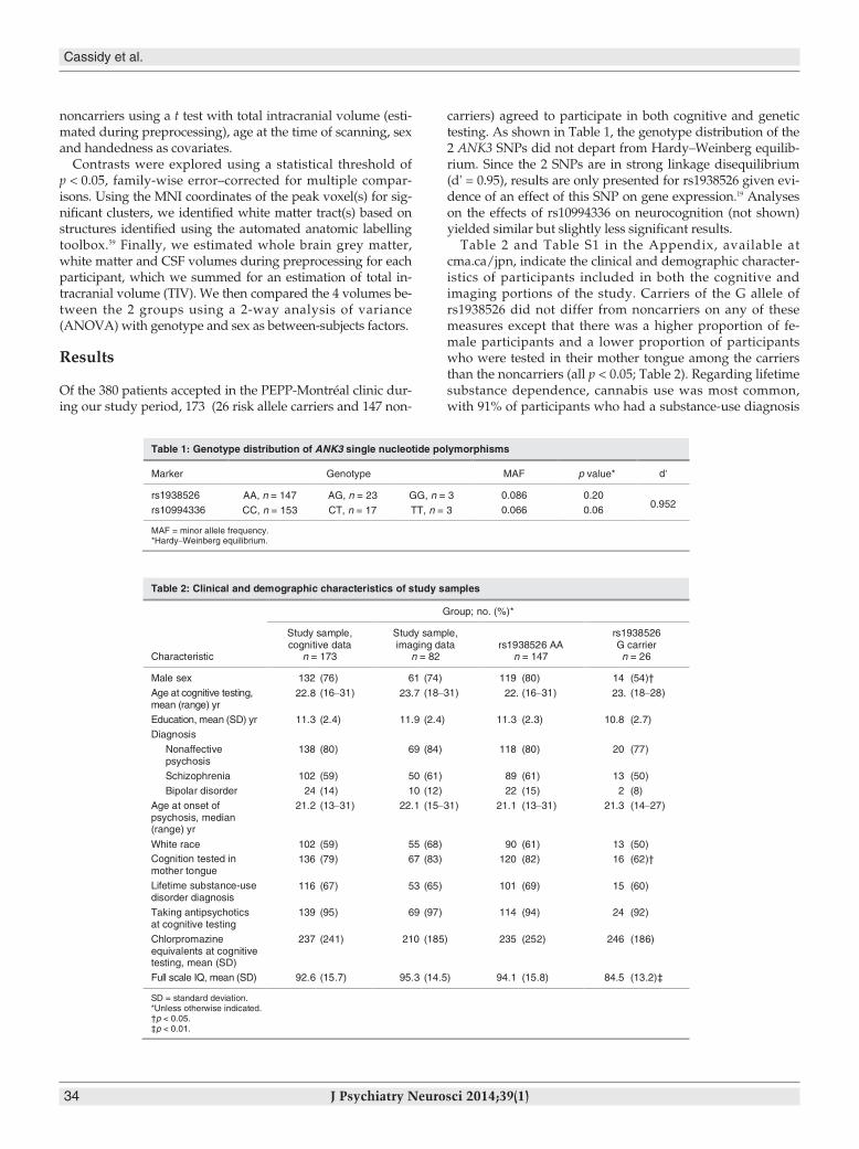
carriers) agreed to participate in both cognitive and genetictesting. As shown in Table 1, the genotype distribution of the2 ANK3 SNPs did not depart from Hardy–Weinberg equilib-rium. Since the 2 SNPs are in strong linkage disequilibrium(d' = 0.95), results are only presented for rs1938526 given evi-dence of an effect of this SNP on gene expression.19 Analyseson the effects of rs10994336 on neurocognition (not shown)yielded similar but slightly less significant results.Table 2 and Table S1 in the Appendix, available at
cma.ca/jpn, indicate the clinical and demographic character-istics of participants included in both the cognitive andimaging portions of the study. Carriers of the G allele ofrs1938526 did not differ from noncarriers on any of thesemeasures except that there was a higher proportion of fe-male participants and a lower proportion of participantswho were tested in their mother tongue among the carriersthan the noncarriers (all p < 0.05; Table 2). Regarding lifetimesubstance dependence, cannabis use was most common,with 91% of participants who had a substance-use diagnosis
Cassidy et al.
34 J Psychiatry Neurosci 2014;39(1)
Table 1: Genotype distribution of ANK3 single nucleotide polymorphisms
FAMepytoneGrekraM p value* d'
rs1938526 AA, n = 147 AG, n = 23 GG, n = 3 0.086 0.20 0.952
rs10994336 CC, n = 153 CT, n = 17 TT, n = 3 0.066 0.06
MAF = minor allele frequency. *Hardy–Weinberg equilibrium.
Table 2: Clinical and demographic characteristics of study samples
Group; no. (%)*
Characteristic
Study sample,cognitive data
n = 173
Study sample,imaging data
n = 82rs1938526 AA
n = 147
rs1938526G carriern = 26
Male sex 132 (76) 61 (74) 119 (80) 14 (54)†
Age at cognitive testing,mean (range) yr
22.8 (16–31) 23.7 (18–31) 22. (16–31) 23. (18–28)
Education, mean (SD) yr 11.3 (2.4) 11.9 (2.4) 11.3 (2.3) 10.8 (2.7)
Diagnosis
Nonaffectivepsychosis
138 (80) 69 (84) 118 (80) 20 (77)
Schizophrenia 102 (59) 50 (61) 89 (61) 13 (50)
Bipolar disorder 24 (14) 10 (12) 22 (15) 2 (8)
Age at onset ofpsychosis, median(range) yr
21.2 (13–31) 22.1 (15–31) 21.1 (13–31) 21.3 (14–27)
White race 102 (59) 55 (68) 90 (61) 13 (50)Cognition tested inmother tongue
136 (79) 67 (83) 120 (82) 16 (62)†
Lifetime substance-usedisorder diagnosis
116 (67) 53 (65) 101 (69) 15 (60)
Taking antipsychoticsat cognitive testing
139 (95) 69 (97) 114 (94) 24 (92)
Chlorpromazineequivalents at cognitivetesting, mean (SD)
237 (241) 210 (185) 235 (252) 246 (186)
Full scale IQ, mean (SD) 92.6 (15.7) 95.3 (14.5) 94.1 (15.8) 84.5 (13.2)‡
SD = standard deviation.*Unless otherwise indicated.†p < 0.05.‡p < 0.01.
ANK3, cognitive performance and cortical thickness in FEP
J Psychiatry Neurosci 2014;39(1) 35
using cannabis. Regarding DNA extraction, blood sampleswere available from most participants (n = 125 samples,72%), and saliva samples were available for the remainingparticipants.Eighty percent of patients (n = 139) were tested within
4 months of program entry. The genotype groups did notdiffer on time of testing after treatment entry (mean 78 d inG allele carriers v. 93 d in noncarriers, t = 0.67, p = 0.46). Allscans were carried out within the first 16 months followingtreatment entry; most occurred within the first 6 months (n= 66, 82%). The genotype groups did not differ on time ofscanning after treatment entry (mean 5.6 mo for G allele car-riers v. 4.9 mo for noncarriers, t = 0.85, p = 0.41).
Cognitive performance
The MANCOVA including sex as a covariate revealed amain effect of the rs1938526 SNP on cognitive performanceacross all domains. The overall performance of G allele carriers was weaker than that of noncarriers (F6,164 = 2.4, par-tial η2 = 0.080, p = 0.030). There were significant differencesbetween the genotype groups on working memory (F1,171 =7.6, partial η2 = 0.043, p = 0.006), verbal memory (F1,171 = 6.0,partial η2 = 0.034, p = 0.015), and attention (F1,171 = 5.5, partialη2 = 0.032, p = 0.019) and a borderline effect on reasoningand problem solving (F1,171 = 2.8, partial η2 = 0.016, p = 0.09).There was no significant effect of genotype on visual mem-ory (partial η2 = 0.011, p = 0.17) or speed of processing (par-tial η2 = 0.002, p = 0.58). In addition, the risk allele carriershad lower full-scale IQ scores than non carriers (ANOVAcontrolling for sex: F1,172 = 8.9, p = 0.003). Adding diagnosis(affective, nonaffective) and mother tongue (English,French, other) as covariates in the MANCOVA yielded anidentical pattern of significant results with minimal reduc-tion in effect size (partial η2 for overall cognition, workingmemory, verbal memory and attention were 0.077, 0.036,0.032 and 0.030, respectively). Adding full-scale IQ as a co-variate eliminated all significant effects of ANK3 on cogni-tion.
Cortical thickness
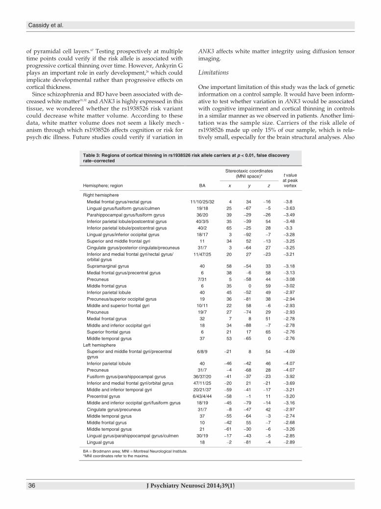
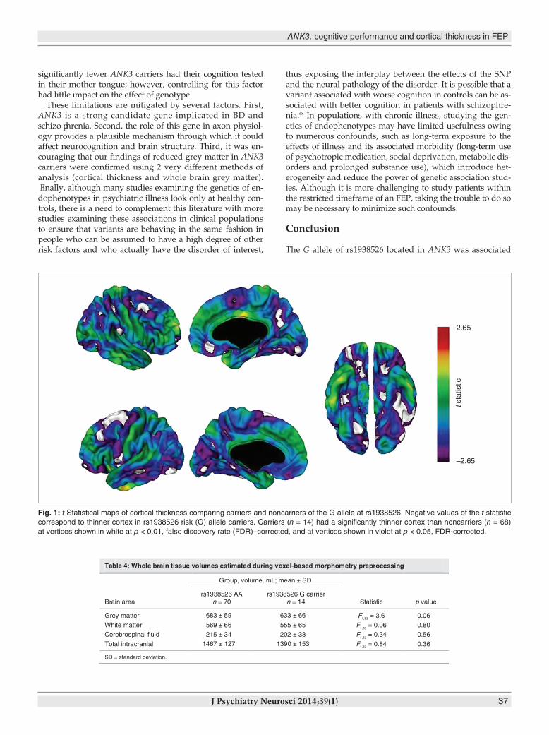
Carriers of the ANK3 rs1938526 risk allele (n = 14) had a sig-nificantly thinner cortex at numerous regions than noncarri-ers (n = 68), as shown in Table 3 and Figure 1. At p < 0.01,FDR-corrected, carriers had a thinner cortex in the righthemisphere at regions with maxima in the medial frontalgyrus, lingual gyrus, parahippocampal gyrus, inferior pari-etal lobule, superior frontal gyrus, cingulate gyrus, inferiorfrontal gyrus, supramarginal gyrus, precuneus, middlefrontal gyrus, middle occipital gyrus, superior frontal gyrusand middle temporal gyrus (descending order of signifi-cance) and in the left hemisphere at regions with maxima inthe superior frontal gyrus, inferior parietal lobule, precuneus,fusiform gyrus, inferior frontal gyrus, middle temporalgyrus, precentral gyrus, middle occipital gyrus, cingulategyrus, middle frontal gyrus and lingual gyrus. Carriers hadno regions of cortical thickening.
Voxel-based morphometry: differences in white matter andwhole brain volumes
The “carrier > noncarrier” and the “noncarrier > carrier” con-trasts revealed no significant white matter differences. Therewas less whole brain grey matter in carriers than noncarriers(p = 0.06); groups did not significantly differ on whole brainwhite matter, TIV, or CSF volumes (Table 4).
Discussion
The present investigation found the G allele of rs1938526 tobe associated with poor performance across 6 domains ofcognition and with widespread cortical thinning and greymatter volume reduction in patients with FEP, but this vari-ant had no significant effect on white matter volume.To our knowledge, this is the first study of the effects of the
rs1938526 SNP on neurocognition. This SNP was associatedwith lower global cognitive performance and there were nosignificant effects of the SNP on any particular cognitive do-main when controlling for IQ. Thus we conclude that it maybe promoting a general intellectual impairment; however, theeffect of genotype was stronger for certain domains, particu-larly working memory, which is consistent with a role ofANK3 in this domain.16 Although there has been a great dealof interest in deficits in specific domains of cognition inpsych otic illness, it is also well established that a general in-tellectual deficit is often comorbid with psychotic disordersand is a risk factor for such disorders.60–62
Our results suggest that the rs1938526 risk variant of ANK3is associated with significant and widespread reduction incortical thickness and grey matter volume across diverse re-gions of the cortex. The thinning was so extensive that we in-creased our FDR-corrected threshold from p < 0.05 to p < 0.01to highlight the regions most affected. Some regions of thin-ning, such as the orbitofrontal cortex, parahippocampalgyrus, dorsolateral prefrontal cortex and cingulate gyrus, arehighly implicated in psychotic disorders.8,10,11,31,63,64 Previousstudies looking specifically at cortical thinning in patientswith schizophrenia have reported widespread thinningacross diverse cortical areas even at the time of a first epi -sode.8–10 Cortical thinning in patients with BD may be morerestricted, with robust findings in prefrontal areas7,8,64 and inthe left ACC.7,64 A recent twin study merging schizophreniaand BD found genetic liability for these disorders to be asso-ciated with thinning of the right parahippocampal gyrus andright orbitofrontal cortex.11
It is not surprising that carriers of the risk allele had bothpoor cognition and cortical thinning, since these characteris-tics have shown strong association.65 The mechanism throughwhich ANK3 could impact cortical thickness is not fully clear.ANK3 promotes the propagation of action potentials,27 andsome researchers have posited that decreased ANK3 expres-sion in patients with schizophrenia could reduce pyramidalcell activation in the cortex.26 Reduced cognitive activation hasbeen associated with cortical thinning in individuals withschizophrenia.66 Furthermore, it has been shown that corticalthinning in the frontal lobes of these patients is due to thinning
of pyramidal cell layers.67 Testing prospectively at multipletime points could verify if the risk allele is associated withprogressive cortical thinning over time. However, Ankyrin Gplays an important role in early development,26 which couldimplicate developmental rather than progressive effects oncortical thickness.Since schizophrenia and BD have been associated with de-
creased white matter31,32 and ANK3 is highly expressed in thistissue, we wondered whether the rs1938526 risk variantcould decrease white matter volume. According to thesedata, white matter volume does not seem a likely mech -anism through which rs1938526 affects cognition or risk forpsych otic illness. Future studies could verify if variation in
ANK3 affects white matter integrity using diffusion tensorimaging.
Limitations
One important limitation of this study was the lack of geneticinformation on a control sample. It would have been inform -ative to test whether variation in ANK3 would be associatedwith cognitive impairment and cortical thinning in controlsin a similar manner as we observed in patients. Another limi-tation was the sample size. Carriers of the risk allele ofrs1938526 made up only 15% of our sample, which is rela-tively small, especially for the brain structural analyses. Also
Cassidy et al.
36 J Psychiatry Neurosci 2014;39(1)
Table 3: Regions of cortical thinning in rs1938526 risk allele carriers at p < 0.01, false discoveryrate–corrected
Hemisphere; region BA
Stereotaxic coordinates(MNI space)* t value
at peakvertexx y z
Right hemisphere
Medial frontal gyrus/rectal gyrus 11/10/25/32 4 34 –16 –3.8
Lingual gyrus/fusiform gyrus/culmen 19/18 25 –67 –5 –3.63
Parahippocampal gyrus/fusiform gyrus 36/20 39 –29 –26 –3.49
Inferior parietal lobule/postcentral gyrus 40/3/5 35 –39 54 –3.48
Inferior parietal lobule/postcentral gyrus 40/2 65 –25 28 -3.3
Lingual gyrus/inferior occipital gyrus 18/17 3 –92 –7 –3.28
Superior and middle frontal gyri 11 34 52 –13 –3.25
Cingulate gyrus/posterior cingulate/precuneus 31/7 3 –64 27 –3.25
Inferior and medial frontal gyri/rectal gyrus/orbital gyrus
11/47/25 20 27 –23 –3.21
Supramarginal gyrus 40 58 –54 33 –3.18
Medial frontal gyrus/precentral gyrus 6 38 –6 58 –3.13
Precuneus 7/31 5 –58 44 –3.08
Middle frontal gyrus 6 35 0 59 –3.02
Inferior parietal lobule 40 45 –52 49 –2.97
Precuneus/superior occipital gyrus 19 36 –81 38 –2.94
Middle and superior frontal gyri 10/11 22 58 –6 –2.93
Precuneus 19/7 27 –74 29 –2.93
Medial frontal gyrus 32 7 8 51 –2.78
Middle and inferior occipital gyri 18 34 –88 –7 –2.78
Superior frontal gyrus 6 21 17 65 –2.76
Middle temporal gyrus 37 53 –65 0 –2.76
Left hemisphereSuperior and middle frontal gyri/precentralgyrus
6/8/9 –21 8 54 –4.09
Inferior parietal lobule 40 –46 –42 46 –4.07
Precuneus 31/7 –4 -68 28 –4.07
Fusiform gyrus/parahippocampal gyrus 36/37/20 –41 –37 –23 –3.92
Inferior and medial frontal gyri/orbital gyrus 47/11/25 –20 21 –21 –3.69
Middle and inferior temporal gyri 20/21/37 –59 –41 –17 –3.21
Precentral gyrus 6/43/4/44 –58 –1 11 –3.20
Middle and inferior occipital gyri/fusiform gyrus 18/19 –45 –79 –14 –3.16
Cingulate gyrus/precuneus 31/7 –8 –47 42 –2.97
Middle temporal gyrus 37 –55 –64 –3 –2.74
Middle frontal gyrus 10 –42 55 –7 –2.68
Middle temporal gyrus 21 –61 –30 –6 –3.26
Lingual gyrus/parahippocampal gyrus/culmen 30/19 –17 –43 –5 –2.85
Lingual gyrus 18 –2 –81 –4 –2.89
BA = Brodmann area; MNI = Montreal Neurological Institute.*MNI coordinates refer to the maxima.
ANK3, cognitive performance and cortical thickness in FEP
J Psychiatry Neurosci 2014;39(1) 37
significantly fewer ANK3 carriers had their cognition testedin their mother tongue; however, controlling for this factorhad little impact on the effect of genotype.These limitations are mitigated by several factors. First,
ANK3 is a strong candidate gene implicated in BD andschizo phrenia. Second, the role of this gene in axon physiol-ogy provides a plausible mechanism through which it couldaffect neurocognition and brain structure. Third, it was en-couraging that our findings of reduced grey matter in ANK3carriers were confirmed using 2 very different methods ofanalysis (cortical thickness and whole brain grey matter). Finally, although many studies examining the genetics of en-dophenotypes in psychiatric illness look only at healthy con-trols, there is a need to complement this literature with morestudies examining these associations in clinical populationsto ensure that variants are behaving in the same fashion inpeople who can be assumed to have a high degree of otherrisk factors and who actually have the disorder of interest,
thus exposing the interplay between the effects of the SNPand the neural pathology of the disorder. It is possible that avariant associated with worse cognition in controls can be as-sociated with better cognition in patients with schizophre-nia.68 In populations with chronic illness, studying the gen -etics of endophenotypes may have limited usefulness owingto numerous confounds, such as long-term exposure to theeffects of illness and its associated morbidity (long-term useof psychotropic medication, social deprivation, metabolic dis-orders and prolonged substance use), which introduce het-erogeneity and reduce the power of genetic association stud-ies. Although it is more challenging to study patients withinthe restricted timeframe of an FEP, taking the trouble to do somay be necessary to minimize such confounds.
Conclusion
The G allele of rs1938526 located in ANK3 was associated
2.65
tsta
tistic
–2.65
Fig. 1: t Statistical maps of cortical thickness comparing carriers and noncarriers of the G allele at rs1938526. Negative values of the t statisticcorrespond to thinner cortex in rs1938526 risk (G) allele carriers. Carriers (n = 14) had a significantly thinner cortex than noncarriers (n = 68)at vertices shown in white at p < 0.01, false discovery rate (FDR)–corrected, and at vertices shown in violet at p < 0.05, FDR-corrected.
Table 4: Whole brain tissue volumes estimated during voxel-based morphometry preprocessing
Group, volume, mL; mean ± SD
Brain arears1938526 AA
n = 70rs1938526 G carrier
n = 14 Statistic p value
Grey matter 683 ± 59 633 ± 66 F1,83 = 3.6 0.06
White matter 569 ± 66 555 ± 65 F1,83
= 0.06 0.80
Cerebrospinal fluid 215 ± 34 202 ± 33 F1,83
= 0.34 0.56
Total intracranial 1467 ± 127 1390 ± 153 F1,83 = 0.84 0.36
SD = standard deviation.
with widespread cortical thinning and reduced cognitive per-formance across domains, but not with decreased white mat-ter in patients with FEP. These findings suggest that cogni-tive deficits and cortical thinning could be mechanismsthrough which ANK3 confers risk for psychotic disorders, asobserved in GWAS.
Acknowledgements: We acknowledge the contributions ofDr. Mallar Chakravarty, Dr. Norbert Schmitz and Dr. AurelieLabbé for statis tical advice and Aldanie Rho, Anastasia Lezos andConnie Lee for assistance with sample collection. This work wassupported by grants from the Canadian Institutes of Health Re-search to A. Malla, M. Lepage and R. Joober. R. Joober has salarysupport from Fonds de la recherche en santé du Québec. The fund-ing sources had no role in the design or conduct of the study, nor inthe collection, interpretation of data, nor in the preparation, review,or approval of the manuscript.
Competing interests:None declared.
Contributors: A. Malla, M. Lepage and R. Joober designed the study.C. Cassidy, M. Bodnar, J. Dell’Elce, F. Ferid, R. Fox, S. Iyer andR. Joober acquired the data, which C. Cassidy, L. Buchy, M. Bodnar,J. Dell’Elce, Z. Choudhry, S. Sengupta, R. Fox, A. Malla, M. Lepageand R. Joober analyzed. C. Cassidy, M. Bodnar, J. Dell’Elce, R. Fox,S. Iyer and R. Joober wrote the article, which all authors reviewedand approved for publication.
References
1. Snitz BE, Macdonald AW III, Carter CS. Cognitive deficits in unaf-fected first-degree relatives of schizophrenia patients: a meta- analytic review of putative endophenotypes. Schizophr Bull 2006;32:179-94.
2. Savitz JB, Solms M, Ramesar RS. Neurocognitive function as an en-dophenotype for genetic studies of bipolar affective disorder. Neuromolecular Med 2005;7:275-86.
3. Kaymaz N, van Os J. Heritability of structural brain traits an en-dophenotype approach to deconstruct schizophrenia. Int Rev Neurobiol 2009;89:85-130.
4. Williams HJ, Craddock N, Russo G, et al. Most genome-wide sig-nificant susceptibility loci for schizophrenia and bipolar disorderreported to date cross-traditional diagnostic boundaries. Hum MolGenet 2011;20:387-91.
5. Panizzon MS, Fennema-Notestine C, Eyler LT, et al. Distinct gen -etic influences on cortical surface area and cortical thickness. CerebCortex 2009;19:2728-35.
6. van Haren NE, Rijsdijk F, Schnack HG, et al. The genetic and en -vironmental determinants of the association between brain abnor-malities and schizophrenia: the schizophrenia twins and relativesconsortium. Biol Psychiatry 2012.71:915-21
7. Lyoo IK, Sung YH, Dager SR, et al. Regional cerebral cortical thin-ning in bipolar disorder. Bipolar Disord 2006;8:65-74.
8. Rimol LM, Hartberg CB, Nesvag R, et al. Cortical thickness andsubcortical volumes in schizophrenia and bipolar disorder. BiolPsychiatry 2010;68:41-50.
9. Schultz CC, Koch K, Wagner G, et al. Reduced cortical thickness infirst episode schizophrenia. Schizophr Res 2010;116:204-9.
10. van Haren NE, Schnack HG, Cahn W, et al. Changes in corticalthickness during the course of illness in schizophrenia. Arch GenPsychiatry 2011;68:871-80.
11. Hulshoff Pol HE, van Baal GC, Schnack HG, et al. Overlapping andsegregating structural brain abnormalities in twins with schizo -phrenia or bipolar disorder. Arch Gen Psychiatry 2012;69:349-59.
12. Ferreira MA, O’Donovan MC, Meng YA, et al. Collaborativegenome-wide association analysis supports a role for ANK3 andCACNA1C in bipolar disorder. Nat Genet 2008;40:1056-8.
13. Smith EN, Bloss CS, Badner JA, et al. Genome-wide associationstudy of bipolar disorder in European American and AfricanAmerican individuals. Mol Psychiatry 2009;14:755-63.
14. Ripke S, Sanders AR, Kendler KS, et al. Genome-wide associationstudy identifies five new schizophrenia loci. Nat Genet 2011;43:969-76.
15. Athanasiu L, Mattingsdal M, Kahler AK, et al. Gene variants associ-ated with schizophrenia in a Norwegian genome-wide study arereplicated in a large European cohort. J Psychiatr Res 2010;44:748-53.
16. Roussos P, Katsel P, Davis KL, et al. Molecular and genetic evi-dence for abnormalities in the nodes of ranvier in schizophrenia.Arch Gen Psychiatry 2012;69:7-15
17. Yuan A, Yi Z, Wang Q, et al. ANK3 as a risk gene for schizophre-nia: new data in han Chinese and meta analysis. Am J Med Genet BNeuropsychiatr Genet 2012;159B:997-1005.
18. Cruz DA, Weaver CL, Lovallo EM, et al. Selective alterations inpostsynaptic markers of chandelier cell inputs to cortical pyramid -al neurons in participants with schizophrenia. Neuropsychopharma-cology 2009;34:2112-24.
19. Rueckert EH, Barker D, Ruderfer D, et al. Cis-acting regulation ofbrain-specific ANK3 gene expression by a genetic variant associ-ated with bipolar disorder. Mol Psychiatry 2012 Jul 31 [Epub aheadof print].
20. Ruberto G, Vassos E, Lewis CM, et al. The cognitive impact of theANK3 risk variant for bipolar disorder: initial evidence of selectiv-ity to signal detection during sustained attention. PLoS ONE 2011;6:e16671.
21. Roussos P, Giakoumaki SG, Georgakopoulos A, et al. TheCACNA1C and ANK3 risk alleles impact on affective personalitytraits and startle reactivity but not on cognition or gating inhealthy males. Bipolar Disord 2011;13:250-9.
22. Linke J, Witt SH, King AV, et al. Genome-wide supported riskvariant for bipolar disorder alters anatomical connectivity in thehuman brain. Neuroimage 2012;59:3288-96.
23. Iqbal Z, Vandeweyer G, van der Voet M, et al. Homozygous andheterozygous disruptions of ANK3: at the crossroads of neuro -developmental and psychiatric disorders. Hum Mol Genet 2013; 22:1960-70.
24. Poliak S, Peles E. The local differentiation of myelinated axons atnodes of Ranvier. Nat Rev Neurosci 2003;4:968-80.
25. Ango F, di Cristo G, Higashiyama H, et al. Ankyrin-based subcel-lular gradient of neurofascin, an immunoglobulin family protein,directs GABAergic innervation at purkinje axon initial segment.Cell 2004;119:257-72.
26. Lewis DA. The chandelier neuron in schizophrenia. Dev Neurobiol2011; 71:118-27.
27. Zhou D, Lambert S, Malen PL, et al. Ankyrin G is required forclustering of voltage-gated Na channels at axon initial segmentsand for normal action potential firing. J Cell Biol 1998;143:1295-304.
28. Nave KA. Myelination and support of axonal integrity by glia. Nature 2010;468:244-52.
29. Gregoriou GG, Gotts SJ, Zhou H, et al. High-frequency, long-rangecoupling between prefrontal and visual cortex during attention.Science 2009;324:1207-10.
30. Barr MS, Farzan F, Tran LC, et al. Evidence for excessive frontalevoked gamma oscillatory activity in schizophrenia during work-ing memory. Schizophr Res 2010;121:146-52.
31. Shepherd AM, Laurens KR, Matheson SL, et al. Systematic
Cassidy et al.
38 J Psychiatry Neurosci 2014;39(1)
ANK3, cognitive performance and cortical thickness in FEP
J Psychiatry Neurosci 2014;39(1) 39
meta-review and quality assessment of the structural brain alter-ations in schizophrenia. Neurosci Biobehav Rev 2012;36:1342-56.
32. Vita A, De Peri L, Sacchetti E. Gray matter, white matter, brain,and intracranial volumes in first-episode bipolar disorder: a meta-analysis of magnetic resonance imaging studies. Bipolar Disord2009; 11:807-14.
33. Malla A, Norman R, McLean T, et al. A Canadian programme forearly intervention in non-affective psychotic disorders. Aust N Z JPsychiatry 2003;37:407-13.
34. Green MF, Nuechterlein KH, Gold JM, et al. Approaching a con-sensus cognitive battery for clinical trials in schizophrenia: theNIMH-MATRICS conference to select cognitive domains and testcriteria. Biol Psychiatry 2004;56:301-7.
35. Nuechterlein KH, Barch DM, Gold JM, et al. Identification of sepa-rable cognitive factors in schizophrenia. Schizophr Res 2004;72:29-39.
36. Wechsler D. Weschler Memory Scale. 3rd ed. Toronto (ON): ThePsycho logical Corporation; 1997.
37. Wechsler D. Weschler Adult Intelligence Scale. 3rd ed. Toronto (ON):The Psychological Corporation; 1997.
38. Reitan R. Trail Making Test: Manual for Administration and Scoring.Tuscon (AZ): Reitan Neuropsychology Laboratory; 1992.
39. Brickencamp RZE. The D2 test of attention. Seattle (WA): Hogrefe &Huber; 1998.
40. First M, Spitzer R, Miriam G, et al. Structured clinical interview forDSM-IV-TR Axis I disorders, research version, patient edition.(SCID-I/P) New York (NY): Biometrics Research, New York StatePsychiatric Institute; 2002.
41. Ehrich M, Bocker S, van den Boom D. Multiplexed discovery of se-quence polymorphisms using base-specific cleavage and MALDI-TOF MS. Nucleic Acids Res 2005;33:e38.
42. Zijdenbos AP, Forghani R, Evans AC. Automatic “pipeline” analy-sis of 3-D MRI data for clinical trials: application to multiple scler -osis. IEEE Trans Med Imaging 2002;21:1280-91.
43. Ad-Dab’bagh YED, Lyttelton O, Muehlboeck JS, et al. The CIVETimage-processing environment: a fully automated comprehensivepipeline for anatomical neuroimaging research [presentation]. 12thannual meeting of the Organization for Human Brain Mapping(OHBM); 2006; Florence, Italy.
44. Collins DP, Peters MT, Evans, TA. An automated 3D non-lineardeformation procedure for determination of gross morphometricvariability in the human brain. In: Robb RA, editor. Visualization inBiomedical Computing; 1994 Oct 4; Rochester (MN): SPIE; 1994. p.180-94.
45. Grabner G, Janke AL, Budge MM, et al. Symmetric atlasing andmodel based segmentation: an application to the hippocampus inolder adults. Med Image Comput Assist Interv 2006;9:58-66.
46. Sled JG, Zijdenbos AP, Evans AC. A nonparametric method forautomatic correction of intensity nonuniformity in MRI data. IEEETrans Med Imaging 1998;17:87-97.
47. Kabani N, Le Goualher G, MacDonald D, et al. Measurement ofcortical thickness using an automated 3-D algorithm: a validationstudy. Neuroimage 2001;13:375-80.
48. Kim JS, Singh V, Lee JK, et al. Automated 3-D extraction andevalu ation of the inner and outer cortical surfaces using a Laplacianmap and partial volume effect classification. Neuroimage 2005; 27:210-21.
49. MacDonald D, Kabani N, Avis D, et al. Automated 3-D extraction ofinner and outer surfaces of cerebral cortex from MRI. Neuroimage2000;12:340-56.
50. Lyttelton O, Boucher M, Robbins S, et al. An unbiased iterative
group registration template for cortical surface analysis. Neuroimage2007;34:1535-44.
51. Robbins SM. Anatomical standardization of the human brain ineuclidean 3-space and on the cortical 2-manifold. Montréal (QC):McGill University; 2004.
52. Ad-Dab’bagh YSV, Robbins S, Lerch J et al. Native space corticalthickness measurement and the absence of correlation to cerebralvolume [presentation]. 11th annual meeting of the Organizationfor Human Brain Mapping (OHBM); 2005; Toronto (ON).
53. Lerch JP, Evans AC. Cortical thickness analysis examined throughpower analysis and a population simulation. Neuroimage 2005;24:163-73.
54. Chung MK, Worsley KJ, Robbins S, et al. Deformation-based sur-face morphometry applied to gray matter deformation. Neuroimage2003; 18:198-213.
55. Sowell ER, Peterson BS, Kan E, et al. Sex differences in corticalthickness mapped in 176 healthy individuals between 7 and87 years of age. Cereb Cortex 2007;17:1550-60.
56. Genovese CR, Lazar NA, Nichols T. Thresholding of statisticalmaps in functional neuroimaging using the false discovery rate.Neuroimage 2002;15:870-8.
57. Ashburner J, Friston KJ. Voxel-based morphometry — the methods.Neuroimage 2000;11:805-21.
58. Good CD, Johnsrude IS, Ashburner J, et al. A voxel-based morphomet-ric study of ageing in 465 normal adult human brains. Neuroimage2001;14:21-36.
59. Tzourio-Mazoyer N, Landeau B, Papathanassiou D, et al. Auto-mated anatomical labeling of activations in SPM using a macro-scopic anatomical parcellation of the MNI MRI single-participantbrain. Neuroimage 2002;15:273-89.
60. Owen MJ. Intellectual disability and major psychiatric disorders: acontinuum of neurodevelopmental causality. Br J Psychiatry 2012;200:268-9.
61. Morgan VA, Croft ML, Valuri GM, et al. Intellectual disability andother neuropsychiatric outcomes in high-risk children of motherswith schizophrenia, bipolar disorder and unipolar major depres-sion. Br J Psychiatry 2012;200:282-9.
62. Fusar-Poli P, Deste G, Smieskova R, et al. Cognitive functioning inprodromal psychosis: a meta-analysis. Arch Gen Psychiatry 2012; 69:562-71.
63. Bodnar M, Malla AK, Joober R, et al. Neural markers of early re-mission in first-episode schizophrenia: a volumetric neuroimagingstudy of the parahippocampus. Psychiatry Res 2012;201:40-7.
64. Foland-Ross LC, Thompson PM, Sugar CA, et al. Investigation ofcortical thickness abnormalities in lithium-free adults with bipolarI disorder using cortical pattern matching. Am J Psychiatry 2011;168: 530-9.
65. Hartberg CB, Lawyer G, Nyman H, et al. Investigating relation-ships between cortical thickness and cognitive performance in pa-tients with schizophrenia and healthy adults. Psychiatry Res 2010;182:123-33.
66. Schultz CC, Koch K, Wagner G, et al. Reduced anterior cingulatecognitive activation is associated with prefrontal-temporal corticalthinning in schizophrenia. Biol Psychiatry 2012;71:146-53.
67. Williams MR, Chaudhry R, Perera S, et al. Changes in corticalthickness in the frontal lobes in schizophrenia are a result of thin-ning of pyramidal cell layers. Eur Arch Psychiatry Clin Neurosci2013; 263:25-39.
68. Zhu X, Gu H, Liu Z, et al. Associations between TCF4 gene poly-morphism and cognitive functions in schizophrenia patients andhealthy controls. Neuropsychopharmacology 2013;38:683-9.


![The Genetic Basis of Addiction - Axónmedia.axon.es/pdf/87033_1.pdf · 2 The Genetic Basis of Addiction 37 [ 12, 13 ] . The Met158 allele is associated with better cognitive performance](https://img.pdfslide.net/doc/110x75/5b8156a77f8b9ae87c8c0f29/the-genetic-basis-of-addiction-axo-2-the-genetic-basis-of-addiction-37-.jpg)