Embed Size (px)
Citation preview
Associations between cardiac fibrosis and permanent atrial fibrillation in
advanced heart failure
Bashar Aldhoon, MD, PhD1, Tomáš Kučera, MD, PhD
2, Natalia Smorodinová, MD
2, Jindřich
Martínek, MD, PhD2, Vojtěch Melenovský, MD, PhD
1, Josef Kautzner MD, PhD
1
1Institute for Clinical and Experimental Medicine-IKEM, Department of Cardiology,
2Charles
University in Prague, The First Faculty of Medicine, Institute of Histology and Embryology,
Prague, Czech Republic.
Corresponding Author:
Bashar Aldhoon, MD, PhD
Department of Cardiology
Institute for Clinical and Experimental Medicine - IKEM
Videnska 1958/9, 140 21, Prague 4, Czech Republic
Telephone: +420 236 055 191
Fax: +420 261 362 985
E-mail: [email protected]
1
Abstract:
Aims: Atrial fibrosis is considered as the basis in the development of long-standing atrial
fibrillation (AF). However, in advanced heart failure (HF), the independent role of fibrosis for
AF development is less clear since HF itself leads to atrial scarring. Our study aimed to
differentiate patients with AF from patients without AF in a population consisting of patients
with advanced HF.
Methods: Myocardial samples from the right atrial and the left ventricular wall were obtained
during heart transplantation from the explanted hearts of 21 male patients with advanced HF.
Long-standing AF was present in 10 of them and the remaining 11 patients served as sinus
rhythm controls. Echocardiographic and hemodynamic measurements were recorded prior to
heart transplantation. Collagen volume fraction (CVF), transforming growth factor-beta
(TGF-β), and connective tissue growth factor (CTGF) expression in myocardial specimens
were assessed histologically and immunohistochemically.
Results: The groups were well matched according to age (51.9±8.8 vs. 51.3±9.3y) and co-
morbidities. The AF group had higher blood pressure in the right atrium (13.6±7.7 vs.
6.0±5.0mmHg; p=0.02), larger left atrium diameter (56.1±7.7 vs. 50±5.1mm; p=0.043),
higher left atrium wall stress (18.1±2.1 vs. 16.1±1.7 kdynes/m2; p=0.04), and longer duration
of HF (5.0±2.9 vs. 2.0±1.6y, p=0.008). There were no significant differences in CVF
(p=0.12), in CTGF (p=0.60), and in TGF-β expression (p=0.66) in the atrial myocardium
between the two study groups.
Conclusions: In advanced HF, atrial fibrosis expressed by CVF is invariably present
regardless of occurrence of AF. In addition to atrial wall fibrosis, increased wall stress might
contribute to AF development in long-standing AF.
Keywords: heart failure; atrial fibrillation; atrial fibrosis; extracellular matrix; transforming
growth factor; connective tissue growth factor
2
Introduction:
Atrial fibrillation (AF) is the most common arrhythmia in clinical practice and a major
cause of morbidity and mortality (Kannel and Benjamin 2009). AF is also very common in
congestive heart failure (HF) patients. In this respect, AF precedes the onset of HF as often as
HF precedes the onset of AF and the one condition advances the other. Development of AF in
HF patients is associated with an increased risk of mortality. Prevalence of AF in HF
increases with NYHA class and reaches up to 50% of patients with advanced HF (Maisel and
Stevenson 2003). Atrial extracellular matrix remodelling and fibrosis are considered as the
main mechanisms for the development and persistence of AF (Polyakova et al. 2004, Aldhoon
et al. 2010). However, the independent role of fibrosis for development of AF in advanced
HF is less clear because alterations of atrial extracellular matrix and structural remodelling
also occur in advanced HF. Few studies have been conducted to elucidate the role of atrial
fibrosis in the onset and persistence of AF in advanced HF (Xu et al. 2004, Mukherjee et al.
2006). Collagen type-I is the major collagenous product of cardiac fibroblasts and accounts
for about 80% of total cardiac collagen content (Lijnen et al. 2000). Atrial fibrosis is mainly
associated with the up-regulation of collagen type-I synthesis. Transforming growth factor-
beta (TGF-β) is a profibrotic cytokine that controls the composition of the extracellular matrix
in many tissues. A previous study has demonstrated elevated levels of serum TGF-β in AF
(Seko et al. 2000). In a recently published study, TGF-β was also over-expressed in atrial
tissue during AF (Gramley et al. 2010). Connective tissue growth factor (CTGF) is another
growth factor that is involved in tissue fibrosis in different organs (Aldhoon et al. 2010). The
role of these growth factors in myocardial fibrosis leading to the onset and persistence of AF
is under investigation. The aim of this study was to differentiate patients with AF from
patients without AF in a population consisting of patients with advanced HF.
3
Methods:
Study subjects:
The study group consisted of 21 male patients (age 51.6 ± 8.9 y; long-standing AF in
10 pts.; sinus rhythm in 11 pts.) with advanced HF who underwent heart transplantation at
Institute for Clinical and Experimental Medicine (IKEM) in Prague. The underlying aetiology
of HF was coronary artery disease (10 pts.), dilated cardiomyopathy (9 pts.), severe valvular
dysfunction (1 pt.) and restrictive cardiomyopathy (1 pt.). Long-standing AF was defined as
AF that has persisted for more than 1 year, either because cardioversion has failed or
cardioversion has not been attempted. ECG records of the study subjects were regularly taken
at 3-month intervals for at least the last 12 months during regular follow up at our institution.
The overall duration of AF (including the follow up period at our institution) as well as
overall HF duration was estimated using the previous medical records from each patient’s
regional cardiologist. Myocardial samples of the anterior free wall of the right atrium (RA)
(sampling of the left atrium was not technically possible because its anatomy was grossly
damaged during the explanation procedure) and the left ventricle were taken during heart
transplantation from all explanted hearts of these 21 patients. Echocardiography was
performed for all patients before transplantation. Right ventricular (RV) and left ventricular
(LV) dimensions, left atrial (LA) size, wall thickness of the interventricular septum and LV
ejection fraction were measured by standard echocardiographic techniques. No systematic
MR imaging before heart transplantation was performed. Hemodynamic measurements were
obtained prior to heart transplantation. Left atrium and left ventricle wall stress (WS) was
calculated by using the formula: WS = 0.334 * P(LAD)/WT(1+WT/LAD), where P = left
atrium/ventricle pressure, which was taken during cardiac catheterisation, LAD = left
atrium/ventricle dimension, and WT = wall thickness (Iwanaga et al. 2006). Based on
previous anatomical and imaging studies (Beinart et al. 2011, Hall et al. 2006), wall thickness
4
of the left atrium used in this formula was estimated as 2 mm in all patients. The posterior
wall thickness of the LV was used to assess WT regardless of regional wall motion
abnormalities.
The ethics committee of our institution approved the study which conformed to the
principles outlined in the Helsinki Declaration. The subjects signed informed consent
documents agreeing to the transplantation procedure and use of the explanted heart for
research purposes.
Histological and immunohistochemical analysis:
Myocardial samples were fixed with paraformaldehyde, embedded into paraffin and
cut to 7μm thick tissue sections. For quantification of collagen volume fraction (CVF), the
sections were stained with the picrosirius staining. The expression of CTGF and TGF-beta
was assessed immunohistochemically. Three-step immunoperoxidase detection was
performed on the paraffin sections. After antigen retrieval with citric buffer (pH=6.0), the
endogenous peroxidase activity and the non-specific antibody binding sites were blocked.
Next, the sections were incubated with a primary antibody - mouse monoclonal anti-human
CTGF/CCN2 C terminal peptide (MAB660; R&D systems, MN, USA) 1:50 or mouse
monoclonal anti-human TGF-beta (MAB1032; Chemicon Int., CA, USA) 1:500 for 60 min at
room temperature. Visualisation of antibody binding was performed using a LSAB+
peroxidase kit (Dako, Glostrup, Denmark). The omission of primary antibody as well as the
application of isotype-matched control antibody in the same concentration as the specific
antibodies yielded negative staining.
Histomorphometry
Images for quantification were collected by systematic uniform random sampling of
tissue sections using the 40x dry objective of a Leica DMLB microscope (Leica Microsystems
GmbH, Wetzlar, Germany). All morphometrical parameters were obtained using interactive
image analysis software (LeicaQWin, Leica Microsystems GmbH, Wetzlar, Germany). CVF
5
was quantified as an area fraction of myocardial tissue section containing collagen fibres
labelled with picrosirius staining. Only endomysial collagen fibres were quantified, while
perimysial connective tissue was omitted.
Immunohistochemical staining (DAB-brown colour) for CTGF and TGF-beta was
quantified as follows. Images were converted to an 8-bit grey scale format and the threshold
was set above the background staining intensity. The average optical intensity above this
threshold level was measured within 0-255 scale (0=white colour, 255=black colour) (A.U. -
arbitrary unit of optical density).
Statistical Analysis:
Continuous variables were expressed as means with standard deviations after testing
for normality of distribution (Shapiro Wilk’s test) and compared with the 2-tailed t-test for
independent samples. Non-normally distributed variables were expressed as medians and
interquartile range and compared by Mann–Whitney U test. Categorical variables were
expressed as percentages and compared by χ2-test. A value of P < 0.05 was considered
significant. Statistics were performed with SPSS statistical software (SPSS Inc., Chicago, IL,
USA, 17.0). Graphs were constructed by Graphpad Prism software (Graphpad software, Inc.,
CA, USA, 5.01).
Results:
Patient characteristics
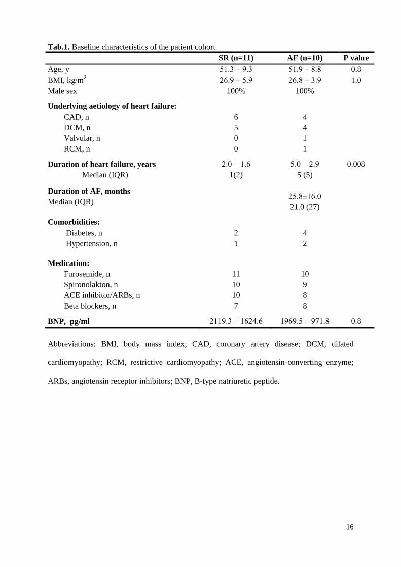
Both study groups were well matched according to age, body mass index, co-
morbidities, medication, and underlying aetiology of HF (Table 1). In the AF group, the
overall mean duration of AF was 25.8 ± 16.0 months (median 21.0; IQR 27). The time course
of heart failure was significantly longer in the AF group (5.0 ± 2.9 vs. 2.0 ± 1.6 years; p =
0.008). LA diameter obtained by echocardiography was significantly greater in the AF group
than in the sinus rhythm group (56.1 ± 7.7 vs. 50.1 ± 5.1 mm; p = 0.043) (Table 2). Also, the
6
AF group presented with significantly higher pressure in the RA (13.6 ± 7.7 vs. 6.0 ± 5.0
mmHg; p = 0.02). LA wall stress was significantly greater in the AF group than in the sinus
rhythm group (18.1 ± 2.1 vs. 16.1 ± 1.7 kdynes/m2; p = 0.04). The receiver operating
characteristic curve analysis showed that the best cut-off LA wall stress, associated with AF
development, was 18.0 kdynes/m2 having an 50% sensitivity, 100% specificity, and 76%
accuracy (area under the curve was 0.76, CI 0.52 – 0.97; p = 0.01). There were no difference
in LV diastolic wall stress between AF and sinus rhythm group (58.1 ± 29.1 vs. 70.8 ± 33.9
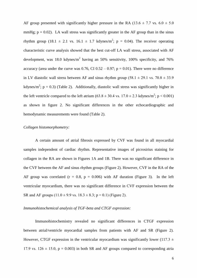
kdynes/m2; p = 0.3) (Table 2). Additionally, diastolic wall stress was significantly higher in
the left ventricle compared to the left atrium (63.8 ± 30.4 vs. 17.0 ± 2.3 kdynes/m2; p < 0.001)
as shown in figure 2. No significant differences in the other echocardiographic and
hemodynamic measurements were found (Table 2).
Collagen histomorphometry:
A certain amount of atrial fibrosis expressed by CVF was found in all myocardial
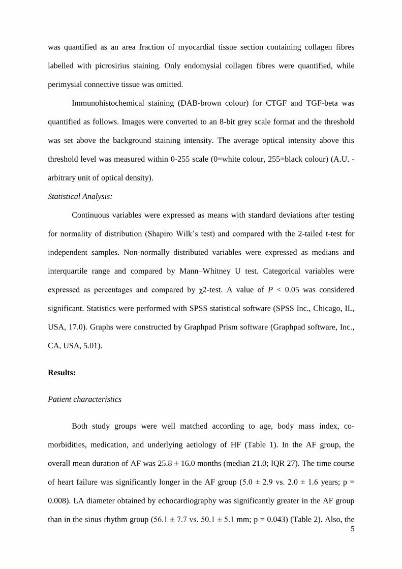
samples independent of cardiac rhythm. Representative images of picrosirius staining for
collagen in the RA are shown in Figures 1A and 1B. There was no significant difference in
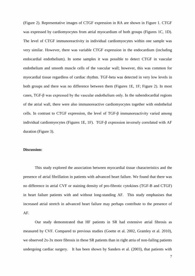
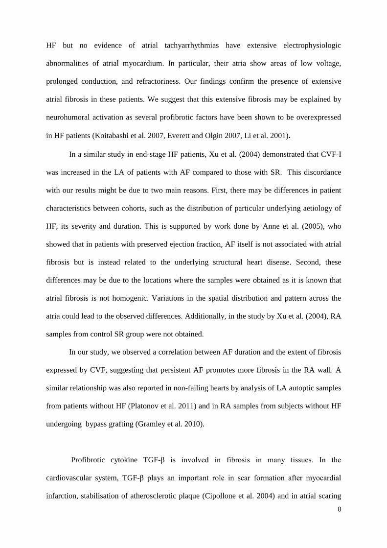
the CVF between the AF and sinus rhythm groups (Figure 2). However, CVF in the RA of the
AF group was correlated (r = 0.8, p = 0.006) with AF duration (Figure 3). In the left
ventricular myocardium, there was no significant difference in CVF expression between the
SR and AF groups (11.0 ± 9.9 vs. 18.3 ± 8.3; p = 0.1) (Figure 2).
Immunohistochemical analysis of TGF-beta and CTGF expression:
Immunohistochemistry revealed no significant differences in CTGF expression
between atrial/ventricle myocardial samples from patients with AF and SR (Figure 2).
However, CTGF expression in the ventricular myocardium was significantly lower (117.3 ±
17.9 vs. 126 ± 15.0, p = 0.003) in both SR and AF groups compared to corresponding atria
7
(Figure 2). Representative images of CTGF expression in RA are shown in Figure 1. CTGF
was expressed by cardiomyocytes from atrial myocardium of both groups (Figures 1C, 1D).
The level of CTGF immunoreactivity in individual cardiomyocytes within one sample was
very similar. However, there was variable CTGF expression in the endocardium (including
endocardial endothelium). In some samples it was possible to detect CTGF in vascular
endothelium and smooth muscle cells of the vascular wall; however, this was common for
myocardial tissue regardless of cardiac rhythm. TGF-beta was detected in very low levels in
both groups and there was no difference between them (Figures 1E, 1F; Figure 2). In most
cases, TGF-β was expressed by the vascular endothelium only. In the subendocardial regions
of the atrial wall, there were also immunoreactive cardiomyocytes together with endothelial
cells. In contrast to CTGF expression, the level of TGF-β immunoreactivity varied among
individual cardiomyocytes (Figures 1E, 1F). TGF-β expression inversely correlated with AF
duration (Figure 3).
Discussion:
This study explored the association between myocardial tissue characteristics and the
presence of atrial fibrillation in patients with advanced heart failure. We found that there was
no difference in atrial CVF or staining density of pro-fibrotic cytokines (TGF-B and CTGF)
in heart failure patients with and without long-standing AF. This study emphasises that
increased atrial stretch in advanced heart failure may perhaps contribute to the presence of
AF.
Our study demonstrated that HF patients in SR had extensive atrial fibrosis as
measured by CVF. Compared to previous studies (Goette et al. 2002, Gramley et al. 2010),
we observed 2x-3x more fibrosis in these SR patients than in right atria of non-failing patients
undergoing cardiac surgery. It has been shown by Sanders et al. (2003), that patients with
8
HF but no evidence of atrial tachyarrhythmias have extensive electrophysiologic
abnormalities of atrial myocardium. In particular, their atria show areas of low voltage,
prolonged conduction, and refractoriness. Our findings confirm the presence of extensive
atrial fibrosis in these patients. We suggest that this extensive fibrosis may be explained by
neurohumoral activation as several profibrotic factors have been shown to be overexpressed
in HF patients (Koitabashi et al. 2007, Everett and Olgin 2007, Li et al. 2001).
In a similar study in end-stage HF patients, Xu et al. (2004) demonstrated that CVF-I
was increased in the LA of patients with AF compared to those with SR. This discordance
with our results might be due to two main reasons. First, there may be differences in patient
characteristics between cohorts, such as the distribution of particular underlying aetiology of
HF, its severity and duration. This is supported by work done by Anne et al. (2005), who
showed that in patients with preserved ejection fraction, AF itself is not associated with atrial
fibrosis but is instead related to the underlying structural heart disease. Second, these
differences may be due to the locations where the samples were obtained as it is known that
atrial fibrosis is not homogenic. Variations in the spatial distribution and pattern across the
atria could lead to the observed differences. Additionally, in the study by Xu et al. (2004), RA
samples from control SR group were not obtained.
In our study, we observed a correlation between AF duration and the extent of fibrosis
expressed by CVF, suggesting that persistent AF promotes more fibrosis in the RA wall. A
similar relationship was also reported in non-failing hearts by analysis of LA autoptic samples
from patients without HF (Platonov et al. 2011) and in RA samples from subjects without HF
undergoing bypass grafting (Gramley et al. 2010).
Profibrotic cytokine TGF-β is involved in fibrosis in many tissues. In the
cardiovascular system, TGF-β plays an important role in scar formation after myocardial
infarction, stabilisation of atherosclerotic plaque (Cipollone et al. 2004) and in atrial scaring
9
due to HF (Sonnylal et al. 2010). Recently, Gramley et al. (2010) examined TGF-β expression
in RA tissue in a large group (n=163) of patients undergoing cardiac surgery. They found that
collagen content and TGF-β expression were higher in patients with chronic AF or SR
compared to paroxysmal AF or SR. They also found that TGF-β proportionally increased with
the duration of AF. However, the expression of downstream TGF-β beta pathway components
(TGFBR, SMAD2) were attenuated with an increased AF burden, suggesting a reduced TGF-
β effect with prolonged AF duration.
Increased atrial tissue levels of TGF-β were implicated in the development of atrial
fibrosis in an experimental model of HF induced in dogs by rapid pacing (Hanna et al. 2004).
Our study showed that atrial and ventricular expression of TGF-β was similar in both the SR
and AF groups. We also showed a negative correlation, although marginally statistically
significant, between TGF-β expression and AF duration. Increased TGF-β expression
therefore appears to play a role in the early response to increased hemodynamic overload and
in the early phase of atrial remodelling. However, its role in advanced atrial fibrosis is
probably less significant. This is in agreement with findings of Gramley et al. (2010), who
described a biphasic pattern of TGF-β expression during the course of AF.
CTGF is a protein involved in wound healing and tissue repair. Enhanced and
prolonged expression of CTGF has been associated with tissue fibrosis (Sonnylal et al. 2010).
To date, no relevant human data on the role of CTGF in AF associated with HF are available.
In patients without HF undergoing cardiac surgery, AF subjects displayed a significantly
increased expression of CTGF in the RA appendage compared to controls in SR (Ko et al.
2011). In our subjects, no significant difference in CTGF expression between the SR and AF
groups was found. TGF-β and CTGF might play a role in atrial fibrosis in advanced HF
because increased expression of both TGF-β and CTGF in the right atrium was shown.
However, TGF-β and CTGF expression is probably less relevant in the development of AF in
10
those patients with advanced HF. Nevertheless, there is the need to emphasise that fibrosis
was measured in the RA.
Increased wall stress represents one of the signals for pro-fibrotic remodelling of the
myocardium. In relation to wall stress, fibrosis and profibrotic cytokine expression is
relatively larger in the atria than in the ventricles. In our study, atrial tissue demonstrated
enhanced CTGF immunostaining compared to ventricles. In an experimental model of dogs
with HF, Burstein and Nattel (2008) showed that atrial fibroblasts behave differently to
ventricular fibroblasts and had increased reactivity and greater fibrotic response.
Since stretch stimulates atrial arrhythmias, we compared atrial stretch between AF and SR. It
has been shown in animal studies that myocardial stretch produced by volume or pressure
overload can modulate the electric activity of the heart (Nazir and Lab 1996). Similar results
were demonstrated by Coronel et al. (2010) in patients with mitral stenosis. These include an
initiation of premature beats that lead to arrhythmia, among other responses. On the other
hand, it is known that fluid congestion is very common in patients with advanced HF. This
volume overloading increases atrial wall stress (or tension). The present study demonstrates
increased wall stress in patients with long-standing AF compared to patients with SR. This
increased wall stress may be due to volume overloading since both higher LA and RA
pressures in the AF group were found. The mechanism for wall stress inducing AF is likely to
be through modulation of electrical activity in the atrium. The 18.0 kdynes/m2 wall stress
value was identified as the best cut-off level for the prediction of AF. The acceptance of the
cut-off value is much dependent on the existence of a causal relation of high wall stress and
increased AF incidence. The present evidence is, however, based on a case-control study, and
not on the result of randomised prospective intervention study showing that increasing wall
stress leads to AF development.
11
Limitations:
This study has several potential limitations. Firstly, we only analysed RA tissue
samples since the remnants of the LA tissue in the explanted heart were damaged and no
standard site for sample collection was obtainable. It is not certain that there is a 1:1
correlation between remodelling in the RA and the LA. Secondly, rhythm verification in the
control group was done based on ECG records during regular 3-month follow up visits. As a
result, we could not exclude incidental paroxysms of AF in the control group. Thirdly, we
used only qualitative immunohistochemistry for TGF-β and CTGF since only fixed tissue was
available for analysis.
Conclusions:
In advanced HF, extensive atrial fibrosis expressed by CVF is invariably present regardless of
the presence or absence of permanent AF. Although this study cannot conclusively prove a
cause-effect relationship between wall stress and atrial fibrillation, it suggests that wall stress
is another mechanism, along with atrial fibrosis, that may perhaps contributes to the
development of AF. Further studies should be done to confirm these results.
Conflict of interest: none declared
Acknowledgments:
This work was supported by grants from the Ministry of Health (MZO-00023001), by the EU
Operational Program Prague - Competitiveness; project "CEVKOON"
(CZ.2.16/3.1.00/22126), by the Grant Agency of the Academy of Sciences of the Czech
Republic (305/09/1390), and by the Research Program of Charles University (PRVOUK-
“METABOLISMUS”).
12
References:
ALDHOON B, MELENOVSKY V, PEICHL P, KAUTZNER J: New insights into
mechanisms of atrial fibrillation. Physiol Res 59: 1-12, 2010.
ANNE W, WILLEMS R, ROSKAMS T, SERGEANT P, HERIJGERS P, HOLEMANS P,
ECTOR H, HEIDBUCHEL H: Matrix metalloproteinases and atrial remodeling in
patients with mitral valve disease and atrial fibrillation. Cardiovasc Res 67: 655-66,
2005.
BEINART R, ABBARA S, BLUM A, FERENCIK M, HEIST K, RUSKIN J, MANSOUR M:
Left atrial wall thickness variability measured by CT scans in patients undergoing
pulmonary vein isolation. J Cardiovasc Electrophysiol 22: 1232-6, 2011.
BURSTEIN B, NATTEL S: Atrial fibrosis: mechanisms and clinical relevance in atrial
fibrillation. J Am Coll Cardiol 51: 802-9, 2008.
CIPOLLONE F, FAZIA M, MINCIONE G, IEZZI A, PINI B, CUCCURULLO C,
UCCHINO S, SPIGONARDO F, DI NISIO M, CUCCURULLO F, MEZZETTI A,
PORRECA E: Increased expression of transforming growth factor-beta1 as a
stabilizing factor in human atherosclerotic plaques. Stroke 35: 2253-7, 2004.
CORONEL R, LANGERVELD J, BOERSMA L V, WEVER E F, BON L, VAN DESSEL P
F, LINNENBANK A C, VAN GILST W H, ERNST S M, OPTHOF T, VAN HEMEL
N M: Left atrial pressure reduction for mitral stenosis reverses left atrial direction-
dependent conduction abnormalities. Cardiovasc Res 85: 711-8, 2010.
EVERETT T H T, OLGIN J E: Atrial fibrosis and the mechanisms of atrial fibrillation. Heart
Rhythm 4: S24-7, 2007.
GOETTE A, JUENEMANN G, PETERS B, KLEIN H U, ROESSNER A, HUTH C,
ROCKEN C: Determinants and consequences of atrial fibrosis in patients undergoing
open heart surgery. Cardiovasc Res 54: 390-6, 2002.
13
GRAMLEY F, LORENZEN J, KOELLENSPERGER E, KETTERING K, WEISS C,
MUNZEL T: Atrial fibrosis and atrial fibrillation: the role of the TGF-beta1 signaling
pathway. Int J Cardiol 143: 405-13, 2010.
HALL B, JEEVANANTHAM V, SIMON R, FILIPPONE J, VOROBIOF G, DAUBERT J:
Variation in left atrial transmural wall thickness at sites commonly targeted for
ablation of atrial fibrillation. J Interv Card Electrophysiol 17: 127-32, 2006.
HANNA N, CARDIN S, LEUNG T K, NATTEL S: Differences in atrial versus ventricular
remodeling in dogs with ventricular tachypacing-induced congestive heart failure.
Cardiovasc Res 63: 236-44, 2004.
IWANAGA Y, NISHI I, FURUICHI S, NOGUCHI T, SASE K, KIHARA Y, GOTO Y,
NONOGI H: B-type natriuretic peptide strongly reflects diastolic wall stress in
patients with chronic heart failure: comparison between systolic and diastolic heart
failure. J Am Coll Cardiol 47: 742-8, 2006.
KANNEL W B, BENJAMIN E J: Current perceptions of the epidemiology of atrial
fibrillation. Cardiol Clin 27: 13-24, 2009.
KO W C, HONG C Y, HOU S M, LIN C H, ONG E T, LEE C F, TSAI C T, LAI L P:
Elevated expression of connective tissue growth factor in human atrial fibrillation and
angiotensin II-treated cardiomyocytes. Circ J 75: 1592-600, 2011.
KOITABASHI N, ARAI M, KOGURE S, NIWANO K, WATANABE A, AOKI Y, MAENO
T, NISHIDA T, KUBOTA S, TAKIGAWA M, KURABAYASHI M: Increased
connective tissue growth factor relative to brain natriuretic peptide as a determinant of
myocardial fibrosis. Hypertension 49: 1120-7, 2007.
LI D, SHINAGAWA K, PANG L, LEUNG T K, CARDIN S, WANG Z, NATTEL S: Effects
of angiotensin-converting enzyme inhibition on the development of the atrial
fibrillation substrate in dogs with ventricular tachypacing-induced congestive heart
failure. Circulation 104: 2608-14, 2001.
14
LIJNEN P J, PETROV V V, FAGARD R H: Induction of cardiac fibrosis by transforming
growth factor-beta(1). Mol Genet Metab 71: 418-35, 2000.
MAISEL W H, STEVENSON L W: Atrial fibrillation in heart failure: epidemiology,
pathophysiology, and rationale for therapy. Am J Cardiol 91: 2D-8D. 2003.
MUKHERJEE R, HERRON A R, LOWRY A S, STROUD R E, STROUD M R, WHARTON
J M, IKONOMIDIS J S, CRUMBLEY A J, 3RD, SPINALE F G, GOLD M R:
Selective induction of matrix metalloproteinases and tissue inhibitor of
metalloproteinases in atrial and ventricular myocardium in patients with atrial
fibrillation. Am J Cardiol 97: 532-7, 2006.
NAZIR S A, LAB M J: Mechanoelectric feedback and atrial arrhythmias. Cardiovasc Res 32:
52-61, 1996.
PLATONOV P G, MITROFANOVA L B, ORSHANSKAYA V, HO S Y: Structural
abnormalities in atrial walls are associated with presence and persistency of atrial
fibrillation but not with age. J Am Coll Cardiol 58: 2225-32, 2011.
POLYAKOVA V, HEIN S, KOSTIN S, ZIEGELHOEFFER T, SCHAPER J: Matrix
metalloproteinases and their tissue inhibitors in pressure-overloaded human
myocardium during heart failure progression. J Am Coll Cardiol 44: 1609-18, 2004.
SANDERS P, MORTON J B, DAVIDSON N C, SPENCE S J, VOHRA J K, SPARKS P B,
KALMAN J M: Electrical remodeling of the atria in congestive heart failure:
electrophysiological and electroanatomic mapping in humans. Circulation 108: 1461-
8, 2003.
SEKO Y, NISHIMURA H, TAKAHASHI N, ASHIDA T, NAGAI R: Serum levels of
vascular endothelial growth factor and transforming growth factor-beta1 in patients
with atrial fibrillation undergoing defibrillation therapy. Jpn Heart J 41: 27-32, 2000.
SONNYLAL S, SHI-WEN X, LEONI P, NAFF K, VAN PELT C S, NAKAMURA H,
LEASK A, ABRAHAM D, BOU-GHARIOS G, DE CROMBRUGGHE B: Selective
15
expression of connective tissue growth factor in fibroblasts in vivo promotes systemic
tissue fibrosis. Arthritis Rheum 62: 1523-32, 2010.
XU J, CUI G, ESMAILIAN F, PLUNKETT M, MARELLI D, ARDEHALI A, ODIM J,
LAKS H, SEN L: Atrial extracellular matrix remodeling and the maintenance of atrial
fibrillation. Circulation 109: 363-8, 2004.
16
Tab.1. Baseline characteristics of the patient cohort
SR (n=11) AF (n=10) P value
Age, y 51.3 ± 9.3 51.9 ± 8.8 0.8
BMI, kg/m2 26.9 ± 5.9 26.8 ± 3.9 1.0
Male sex 100% 100%
Underlying aetiology of heart failure:
CAD, n
DCM, n
Valvular, n
RCM, n
6
5
0
0
4
4
1
1
Duration of heart failure, years
Median (IQR)
2.0 ± 1.6
1(2)
5.0 ± 2.9
5 (5)
0.008
Duration of AF, months
Median (IQR)
25.8±16.0
21.0 (27)
Comorbidities:
Diabetes, n
Hypertension, n
2
1
4
2
Medication:
Furosemide, n
Spironolakton, n
ACE inhibitor/ARBs, n
Beta blockers, n
11
10
10
7
10
9
8
8
BNP, pg/ml 2119.3 ± 1624.6 1969.5 ± 971.8 0.8
Abbreviations: BMI, body mass index; CAD, coronary artery disease; DCM, dilated
cardiomyopathy; RCM, restrictive cardiomyopathy; ACE, angiotensin-converting enzyme;
ARBs, angiotensin receptor inhibitors; BNP, B-type natriuretic peptide.
17
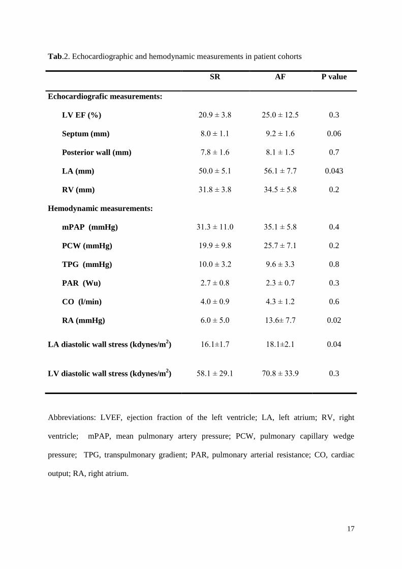
Tab.2. Echocardiographic and hemodynamic measurements in patient cohorts
SR AF P value
Echocardiografic measurements:
LV EF (%)
Septum (mm)
Posterior wall (mm)
LA (mm)
RV (mm)
20.9 ± 3.8
8.0 ± 1.1
7.8 ± 1.6
50.0 ± 5.1
31.8 ± 3.8
25.0 ± 12.5
9.2 ± 1.6
8.1 ± 1.5
56.1 ± 7.7
34.5 ± 5.8
0.3
0.06
0.7
0.043
0.2
Hemodynamic measurements:
mPAP (mmHg)
PCW (mmHg)
TPG (mmHg)
PAR (Wu)
CO (l/min)
RA (mmHg)
31.3 ± 11.0
19.9 ± 9.8
10.0 ± 3.2
2.7 ± 0.8
4.0 ± 0.9
6.0 ± 5.0
35.1 ± 5.8
25.7 ± 7.1
9.6 ± 3.3
2.3 ± 0.7
4.3 ± 1.2
13.6± 7.7
0.4
0.2
0.8
0.3
0.6
0.02
LA diastolic wall stress (kdynes/m2) 16.1±1.7 18.1±2.1 0.04
LV diastolic wall stress (kdynes/m2) 58.1 ± 29.1 70.8 ± 33.9 0.3
Abbreviations: LVEF, ejection fraction of the left ventricle; LA, left atrium; RV, right
ventricle; mPAP, mean pulmonary artery pressure; PCW, pulmonary capillary wedge
pressure; TPG, transpulmonary gradient; PAR, pulmonary arterial resistance; CO, cardiac
output; RA, right atrium.
18
Figure 1.
19
Figure 2.
A.U
.
A.U
.
Collagen volume fraction
Atrium Ventricle 0
10
20
30
%
Diastolic wall stress
Atrium Ventricle 10
20
90
140 p<0.001
p=0.04
TGF- immunoreactivity
Atrium Ventricle 50
100
150 CTGF immunoreactivity
Atrium Ventricle 100
120
140
160 p=0.003
sinus rhythm atrial fibrillation
Kd
yne
s/m
2
20
Figure 3.
0 20 40 60 800
10
20
30 r = 0.8p = 0.006
AF Duration (months)
Atr
ial C
VF
(%
)
0 20 40 60 800
50
100
150
200
r = -0.6p = 0.09
AF Duration (months)
TG
F-
(A
.U.)
0 20 40 60 800
50
100
150
200
r = -0.6p = 0.09
AF Duration (months)
TG
F-
(A
.U.)
21
Figures legends:
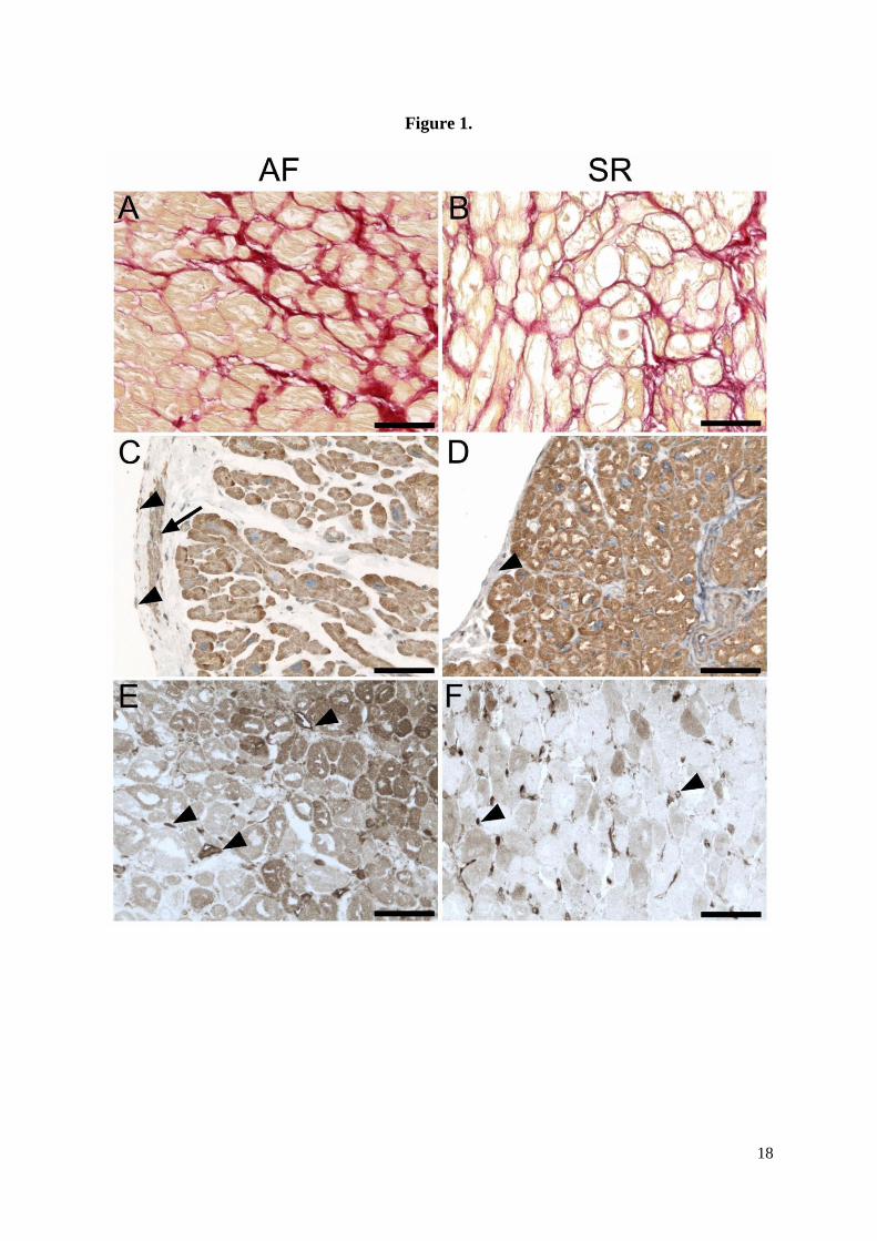
Figure 1. Morphological analysis of right atrial tissue from patients with atrial fibrillation (A,
C, E) and sinus rhythm (B, D, F).
(A, B) Picrosirius staining of tissue sections of atrial myocardium shows collagen fibres in
endomysium (red colour). (C, D) Immunohistochemical detection of CTGF using
immunoperoxidase method (DAB-brown precipitate). (C) Sample from a patient with AF
showing myocardium and endocardium. CTGF immunoreactivity is visible in
cardiomyocytes, endocardial smooth muscle cells (arrow) and endocardial endothelium
(arrowheads). The nuclei are counterstained with hematoxylin. (D) Sample from a patient
with SR showing myocardium and endocardium. CTGF immunoreactivity is visible in
cardiomyocytes, while endocardium stains only weakly (arrowheads). (E, F)
Immunohistochemical detection of TGF-beta using immunoperoxidase method (DAB-brown
precipitate). (E) Sample from a patient with AF showing myocardial tissue close to
endocardium. The immunoreactivity for TGF-beta is localised in the vascular endothelium
(arrowheads) and variable amount of reaction product is found in cardiomyocytes. (F) Sample
from a patient with SR showing myocardial tissue close to endocardium. Capillary
endothelium displays the highest level of TGF-beta immunoreactivity in this region
(arrowheads) compared to rather moderate or low expression in cardiomyocytes. Scale bar in
A-F, 100μm.
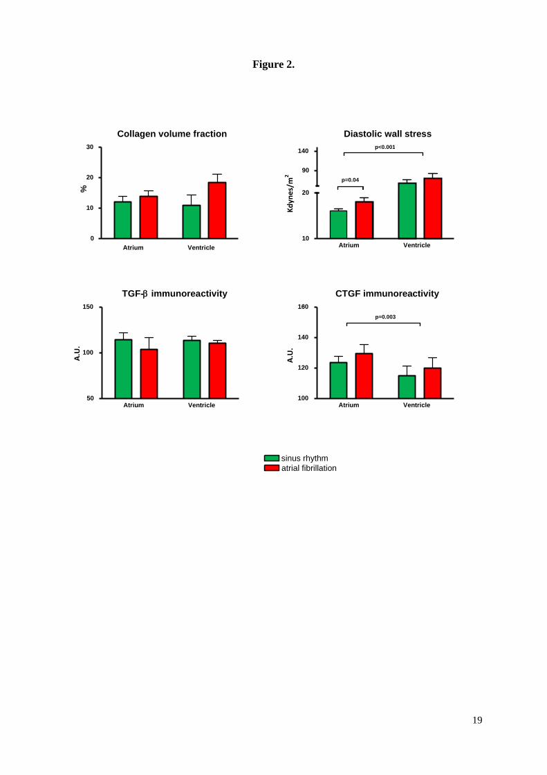
Figure 2. CVF, CTGF, and TGF-β in right atrium and left ventricle of explanted hearts with
or without AF (left and right lower bar graph). Diastolic wall stress of right atrium and left
ventricle in patients of advanced heart failure with and without AF (right upper bar graph).
22
Immunoreactivity of CTGF and TGF-β is expressed by mean optical density arbitrary units
(A.U.). Diastolic wall stress is expressed by kdynes/m2.
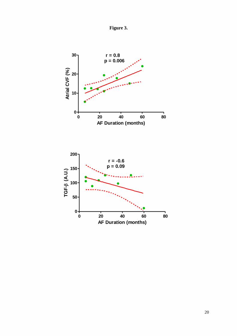
Figure 3. Left panel: correlation between AF duration and atrial fibrosis expressed by
collagen volume fraction. Right panel: correlation between AF duration and TGF-β
expression.