Embed Size (px)
DESCRIPTION
A description of susceptibility testing methods.Part of the Microbiology Technical Workshop, 2013.
Citation preview

Broth and Agar testing methods
Automated susceptibility testing
Microbiology Technical Workshop
Dr Tan Yen Ee
Registrar
Singapore General Hospital
25th Sept 2013

Outline
• Introduction
• Broth testing methods
- Macrodilution
- Microdilution
• Agar dilution
• Automated Antimicrobial Susceptibility Testing (AST) systems
- Vitek-2
- Phoenix
- Microscan
- Sensititre
• Considerations in evaluating AST systems

Introduction

Aims of antimicrobial susceptibility testing:
1. Confirm the susceptibility to chosen
empirical therapy
2. Detect resistance

Various methods of antibiotic susceptibility
testing are:
1. Quantitative Methods
2. Qualitative Methods
3. Automated Susceptibility Tests
4. Newer Non-Automated Susceptibility Tests
5. Molecular Techniques
Broth testing methods

Reference methods for in vitro susceptibility testing
1. Macrobroth dilution (Tube dilution)
2. Microbroth dilution
CLSI, BSAC, EUCAST guidelines

• Broth preparation
• Antimicrobial agent
- Stock solution
- Working solutions
• Preparation of tubes/ plates
• Inoculum preparation
• Inoculation of tubes/ plates
• Incubation of tubes/ plates
• Reading results
• Quality control

Broth preparation
• Mueller–Hinton broth (MHB)
- General purpose medium (non fastidious)
- Cation- adjusted
- Optimum pH

Antimicrobial agent
�Stock solution
-Potency (from manufacturer)
-Appropriate solvent for dilution (CLSI M100 Table 5A)
-Stock solutions may be stored at -60°C for more than
6 months for most antimicrobial agents
-Avoid repeated freeze thaw
Wt of powder (mg) = Vol of solvent (mL) x Concentration (mg/L)
Potency of powder (mg/g)

�Working solution
- Serial doubling dilutions
- Dilution scheme available in CLSI M100
Tubes/ Plates preparation
• Store at appropriate temperature

Inoculum preparation
• Direct colony suspension method
- Most convenient
- Fresh colonies; 0.5 McFarland standard
- Fastidious organisms (eg: Neisseria
gonorrhoeae; Haemophilus spp)
• Growth method
- When smooth suspension cannot be made
- Non- fastidious organisms

Inoculation of tubes/ plates
https://www.boundless.com/image/measuring-minimal- inhibitory-
concentration-via-the-microbroth-dilution-method/
http://archive.ispub.com/journal/the- internet-journal-of-herbal-and-plant-
medicine/volume-1-number-1/the-antibacterial-or-antifungal-effects-of-
eurycoma-longifolia-root-extract.html

Incubation of tubes/ plates
-Incubate inoculated macrodilution tubes/
microdilution trays at 35 +/- 2°C for 16- 20 hours
in ambient air incubator
-Within 15 minutes of adding inoculum
-Do not stack trays >4 high
-Seal each tray
-CLSI M7A9 Appendix C

Reading results
• QC passed; GC wells +
• The MIC is the lowest concentration of the
agent that completely inhibits visible growth
as judged by the naked eye


Quality control
• To include at least 1 control organism (ATCC)
with each batch of testing
• Test values for the control strains should be
within the published range

Limitations
• Labour intensive
• Strict adherence to protocol is required
• The MIC value is not the sole predictor for
clinical outcome

Agar dilution method

• Antimicrobial agent
- Stock solution
- Working solutions
• Preparation of agar and plates
• Inoculum preparation
• Inoculation of plates
• Incubation of plates
• Reading results
• Quality control

Agar and Plates preparation
• Mueller–Hinton agar is considered the reference
medium
• One concentration of antibiotic/ plate
• Include a drug free control

• Allow the sterilized agar to cool to 50°C in a
water-bath, before adding antibiotics
• Set at room temperature. Do not overdry.
• Store plates at 4-8⁰C

Inoculation of plates
• Mark the plates for orientation
• Use an inoculum- replicating apparatus to
transfer the inocula to the series of agar plates
• Allow the inoculum spots to dry at room
temperature before inverting the plates for
incubation.


Reading results

Limitations
• Labour intensive
• Strict adherence to protocol is required
• The MIC value is not the sole predictor for
clinical outcome

Automated Susceptibility Testing

• Several automated systems for antimicrobial
susceptibility testing are commercially
available
• Examples:
- Vitek 2 System (BioMérieux)
- Phoenix Automated Microbiology System
(SD Diagnostic System)
- MicroScan Walkaway System
(Siemens Healthcare Diagnostics)
- Sensititre Aris 2X (Trek Diagnostic System)

General advantages
• Quantitative results (MIC va lues)
• Reproducibility
• Cost- effective for laboratories with high throughput
• Reduction in labour
• Ease of performance
• Faster reporting of susceptibility results
• Convenient interface with the laboratory information system (LIS)
• “Expert systems” software to interpret susceptibility results involving
atypical patterns and unusual resistance phenotypes

General limitations• Space
• Cost
• Regular maintenance required with upgrading of computer
software
• Lag time in upgrading of new breakpoints in software
• Manual preparation of inoculum
• Limited range of organisms
• Limited accuracy in certain organism-antimicrobial
combinations
• Limited flexibility in antibiotic panels
• Testing space on the antibiotic susceptibility cards is not
infinite, and therefore not a ll MICs can be tested (egTest range
MIC ≤ 2 μg/ml)
• No mixed culture

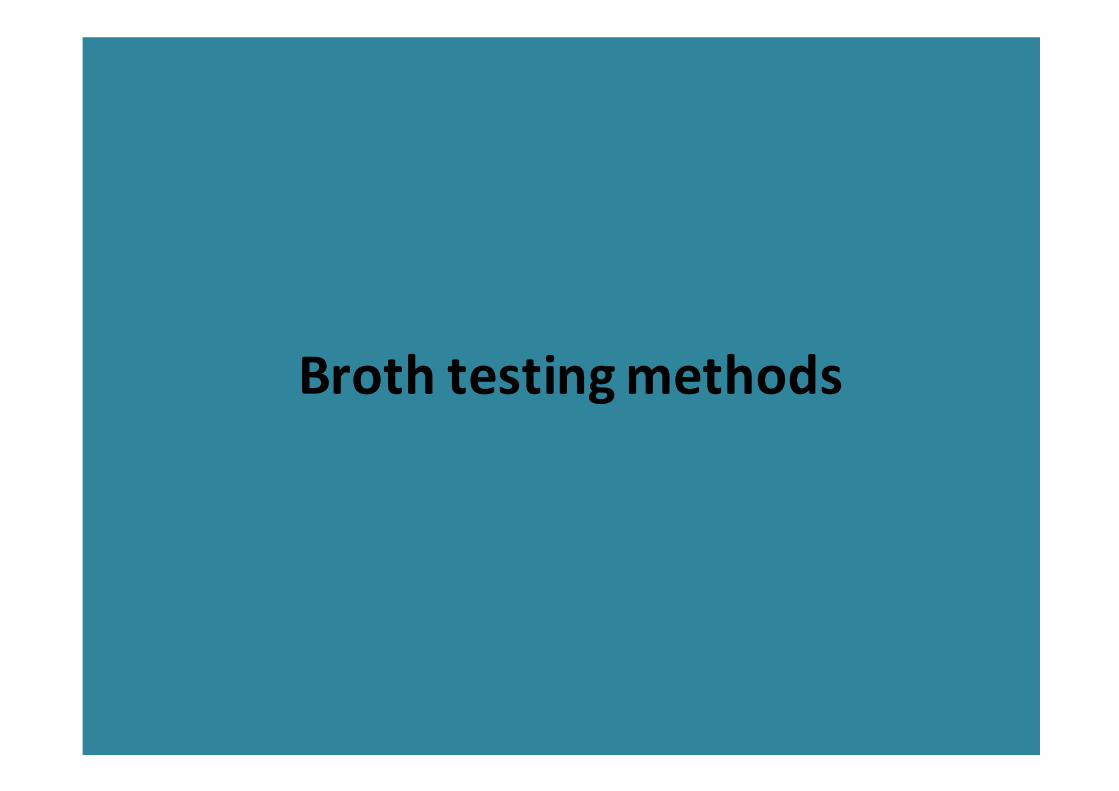
Vitek 2
Principle: Utilized growth-based technology
Uses compact colorimetric reagent
cards that are incubated and
interpreted automatically

http://www.biomerieux-industry.com/servlet/srt/bio/industry-
microbiology/dynPage?open=NDY_IND_BPA_PRD&doc=NDY_BPA_PRD_G_PRD_NDY_10&pubparams.sform=4&lang=en

Phoenix
Principle:
This system uses an optimized colorimetric
oxidation-reduction indicator to detect organism
growth in the presence of an antimicrobial agent
for susceptibility testing


MicroScan WalkAway
Principle:
Large self-contained incubator/reader device
that can incubate and analyze 40-96 microdilution
trays with a photometer/ fluorometer to
determine growth development


Sensititre Aris 2X
Principle:
Automated, overnight, incubator/reader device
that can incubate and analyze up to 64
microdilution trays.
Growth is determined by fluorescence
measurement after 18–24 h of incubation.


Considerations in evaluating
AST systems
• Performance
• Cost
• Practical considerations (Eg: Throughput; Space)
• Software
• Manufacturer’s support
• Technical considerations
• Workflow considerations

Thank you

Vitek 2 http://www.youtube.com/watch?v=1bVIcY30YU0
Sensititre Arix X 2 http://www.youtube.com/watch?v=l21VMzHLhqQ
?Direct inoculums


![METHODS FOR TESTING AND EVALUATING … - Day Three - Methods for Testing...METHODS FOR TESTING AND EVALUATING SURVEY QUESTIONS ... [i.e., Gallup] and Fortune [i.e ... The pivotal development](https://img.pdfslide.net/doc/110x75/5aa744617f8b9a294b8bdb20/methods-for-testing-and-evaluating-day-three-methods-for-testingmethods.jpg)