Embed Size (px)
Citation preview
Asterosplcularia laurae, n. gen. et n. sp .,the Type ~f a New Family of Alcyonarians with Stellate Spicules
HUZIO UTINOMIl
AMONG THOSE SPECIMENS of Alcyonaria obtained during my trip to Formosa in 1938,there is an interesting form which cannot bereferred to any known genus of the group.It is represented only by a single specimenobtained on the coral reef at Daijubo,southernmost promontory of Formo sa, onJune 14, 1938. Superficially it shows a resemblance to the genus Capnella in the modeof branchin g and in the appearance of polyps .Closer examination of the total specimen and .sectioned preparations, however , has revealed that the coenenchyma as well as thepolyp wall has a honeycombed texture filledwith numerous stellate spicules and that thetent acles bear no trace of pinnules. Suchcharacters are entirely unknown in the wholegroup of Alcyonaria, so I propose to institutefor this form a new genus and even a newfamily.
The following description .is based on aperfect specimen which was taken as the type.
EXTERNAL APPEARANCE
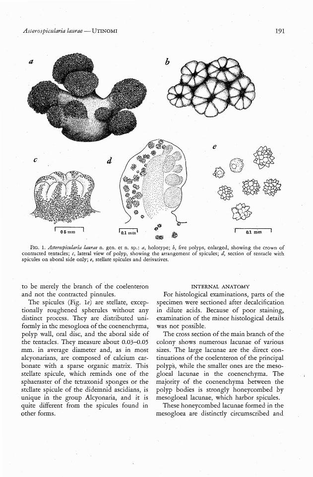
The specimen (Fig. l a) is in the form of asmall colony of bushy growth and has ayellowish white colour in alcohol. The baseof the colony is broadened, flattened, andabout 12 mm. in longest diameter . From theflattened upper surface of the short columnarstem arise nine large and small polyp-bearinglappets . Each of the lappets is mushroomshaped ; each consists of a polyp-bearingrounded capitulum and a short wide sterile
'S ew M arine Biolog ical Lab or atory, Sirah ama,Wakayama-Ke n, J apan. Manuscript received September 6, 1950.
stalk, the former being marked off sharplyfrom the latter. In the largest lappet, thecapitulum is about p mm . in diameter and5 mm . in height, and the stalk is about 8 mm .in diamete r and 5 mm . in height. The totalheight of the colony is about 13-17 mm .
No polyp dimorphism is found . The autozooids are closely set and rather large,beingup to 1 mm. in diameter and 1.5 mm . inheight. They are apparently capable of considerable contrac tion , and they look likepapillae provided with a rather large centralmouth surrounded by eight-lobed tentacles(Fig. Ib ). The whole surface of the trunk ofthe zooid is thickly covered with stellatespicules of sub-equal size which are continuedonto the dorsal surface of the tentacl es. Thespicules are evenly scattered as in the Xeniidaeand do not form any densely packed row orridge along the intermesenteric area (Fig. Ie).
The tentacles, in the contracted state, appear oval in side view and measure about0.5 mm. long and 0.37 mm . wide (Fig. Id).The pinnules are missing. Careful microscopic examination of serial sections of thetentacle reveals tha t from 8 to 10 opaquerounded bodies are imbedded slightly beneath the oral surface of the tentacles (Fig .2e). These bodies are scattered here and therein the peripheral layer, but are closely unitedwith one another in the deeper layer; they arecontinuous with the coelenteric cavity belowthe mouth. The interior is filled with numerous zooxanthellae and endoderm cells, andthere is no connection between this regionand the epidermis . Here the epidermis showsno sinus or indentation suggesting the presence of pinnules. Thus I consider this body
II
[190 ]
A sterospicularia laurae - UTINOMI 191
c
Oli mm
d
FI G. I. Asterosplcularia laurae n. gen. er n. sp .: a, holo rype; b, five polyps, enlarged , showin g the crown ofcontracted tentacles; c, lateral view of polyp, showing the arrangement of spicules ; d, section of tentacle withspicules on aboral side only; e, stell ate spicules and derivatives.
to be merely the branch of the coelenteronand not the contracted pinnules .
The spicules (Fig. Ie) are stellate, exceptionall y roughened spherules without anydistinct process . They are distributed uniformly in the mesogloea of the coenenchyma,polyp wall, oral disc, and the aboral side ofthe tentacles. They measure about 0.03-0.05mm. in average diameter and , as in mostalcyonarians, are composed of calcium carbonate with a sparse organic matrix. Thisstellate spicule, which reminds one of thesphaeraster of the tetraxonid sponges or thestellate spicule of the didemnid ascidians , isunique in the group Alcyonaria, and it isquite different from the spicules found inother forms.
INTERNAL ANATOMY
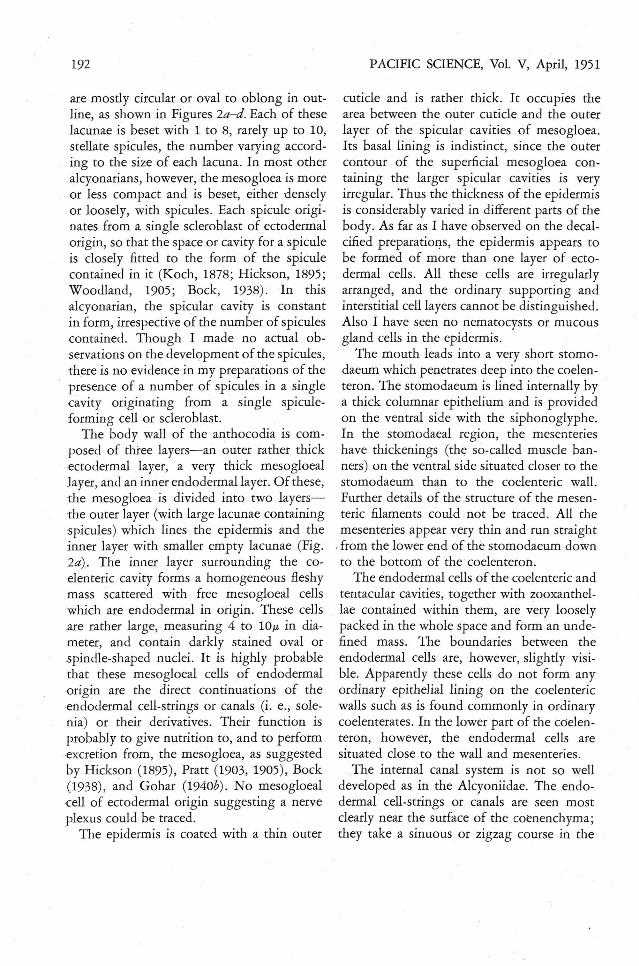
For histo logical examinations , parts of thespecimen were sectioned after decalcificationin dilute acids. Because of poor staining ,examination of the minor histological detailswas not possib le.
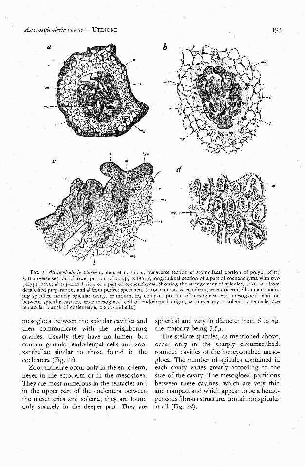
The cross section of the main branch of thecolon y shows numerous lacunae of varioussizes. The large lacunae are the direct continuations of the coelenteron of the principalpolyps, while the smaller ones are the mesogloeal lacunae in the coenenchyma. Themajority of the coenenchyma between thepolyp bodies is strongly honeycombed bymesogloeal lacunae, which harbor spicules.
These honeycombed lacunae formed-in themesogloea are distinctly circumscribed and
192
are mostly circular or oval to oblong in outline, as shown in Figures 2a-d. Each of theselacunae is beset with 1 to 8, rarely up to 10,stellate spicules, the number varying according to the size of each lacuna. In most otheralcyonarians, however, the mesogloea is moreor less compact and is beset, either denselyor loosely, with spicules. Each spicule originates from a single scleroblast of ectodermalorigin, so that the space or cavity for a spiculeis closely fitted to the form of the spiculecontained in it (Koch, 1878; Hickson, 1895;Woodland, 1905; Bock, 1938). In thisalcyonarian, the spicular cavity is constantin form, irrespective of the number of spiculescontained. Though I made no actual observations on the development of the spicules,there is no evidence in my preparations of thepresence of a number of spicules in a singlecavity originating from a single spiculeformin g cell or scleroblast .
The body wall of the anthocodia is composed of three layers-an outer rather thickectodermal layer, a very thick mesogloeallayer , and an inner endodermallayer. Of these,the mesogloea is divided into two layersthe outer layer (with large lacunae containingspicules ) which lines the epidermis and theinner layer with smaller empty lacunae (Fig .2a). The inner layer surrounding the coelenteric cavity forms a homogeneous fleshymass scattered wirh free mesogloeal cellswhich are endodermal in origin. These cellsare rather large, measuring 4 to lOt! in diameter, and contain darkly stained oval orspindle-shaped nuclei. It is highly probablethat these mesogloeal cells of endodermalorigin are the direct continuations of theendodermal cell-strings or canals (i. e., solenia) or their derivatives. Their function isprobably to give nutrition to, and to performexcretion from, the mesogloea, as suggestedby Hickson (1895), Pratt (1903, 1905), Bock(1938), and Gohar (1940b). No mesogloealcell of ectodermal origin suggesting a nerveple xus could be traced.. The epidermis is coated with a thin outer
PACIFIC SCIENCE, ·Vol. V, April, 1951
cuticle and is rather thick. It occupies thearea between the outer cuticle and the outerlayer of the spicular cavities of mesogloea.Its basal lining is indistinct, since the outercontour of the superficial mesogloea containing the larger spicular cavities is veryirregular. Thus the thickness of the epidermisis considerably varied in different parts of thebody. As far as I have observed on the decalcified preparations, the epidermis appears tobe formed of more than one layer of ectodermal cells. All these cells are irregularlyarranged, and the ordinary supporting andinterstitial cell layers cannot be distinguished.Also I have seen no nematocysts or mucousgland cells in the epidermis.
The mouth leads into a very short stomodaeum which penetrates deep into the coelenteron . The stomodaeum is lined internally by .a thick columnar epithelium and is providedon the ventral side with the siphonoglyphe.In the stomodaeal region, the mesenterieshave thickenings (the so-called muscle banners) on the ventral side situated closer to thestomodaeum than to the coelenteric wall.Further details of the structure of the mesenteric filaments could not be traced. All themesenteries appear very thin and run straight
.from the lower end of the stomodaeum downto the bottom of the coelenteron.
The endodermal cells of the coelenteric andtentacular cavities, together with zooxanrhellae contained within them, are very looselypacked in the whole space and form an undefined mass. The boundaries between theendodermal cells are, however, slightly visible. Apparently these cells do not form anyordinary epithelial lining on the coelentericwalls such as is found commonly in ordinarycoelenterates . In the lower part of the coelenteron, however, the endodermal cells aresituated close to the wall and mesenteries .
The internal canal system is not so welldeveloped as in the Alcyoniidae. The endodermal cell-strings or canals are seen mostclearly near the surface of the coenenchyrna;they take a sinuous or zigzag course in the
Asterospicularia laurae - UTINOMI
Len
193
FIG. 2. Asterospicularia laurae n. gen . et n. sp .: a , tra nsverse section of stomodaeal portion of polyp , X85 ;b, trans verse section of lower portion of polyp, X 135; c, longitudinal section of a part of coenenchyma with twopolyps, X 50; d, superficial view of a part of coenenchyma, showing the arrangement of spicules, X 70. a-c fromdecalcified preparations and d from perfect specimen. (c coelenteron, ec ectoderm, en endoderm, I lacuna containing spicules, namely sp icular cavity, m mouth, mg compact portion of mesogloea, mg.s mesogloeal partitionbetween sp icular cavities, m.en mesogloeal cell of endodermal origin, ms mesentery, s solenia, t tentacle, t .ententacular branch of coelenteron, z zooxanthella.)
mesogloea between the spicular cavities andthen communicate with the neighboringcavities. Usually they have no lumen, butcontain granular endodermal cells and zooxanthellae similar to those found in thecoelentera (Fig. 2c).
Zooxanthellae occur only in the endoderm,never in the ectoderm or in the mesogloea .They are most numerous in the tentacles andin the upper part of the coelentera between
. the mesenteries and solenia; they are foundonly sparsely in the deeper part. They are
spherical and vary in diameter from 6 to 8}J.,the majority being 7.5}J. .
The stellate spicules, as mentioned above,occur only in the sharply circumscribed,rounded cavities of the honeycombed mesogloea. The number of spicules contained ineach cavity varies greatly according to thesize of the cavity. The mesogloeal partitionsbetween these cavities, which are very thinand compact and which appear to be a homogeneous fibrous structure, contain no spiculesat all (Fig. u:
194
REMARKS ON SYSTEMATIC POSITION
Before entering into a consideration of thesystematic position of this remarkable alcyonarian, some historical remarks may not beamiss. It is interesting to note that there isgood evidence that another specimen evidently referable to this form had been obtained and observed by two famous Englishworkers on Alcyonaria 20 years ago. In anelaborate work on the alcyonarians collectedby the Siboga Expedition (Siboga-Expeditie,XIIId, p. 219, pl . 21, fig. 8) issued in 1931,we find a short account by Sir Arthur J.Thomson and Miss Laura M. 1. Dean (Mrs.1. M.l. Macfadyen) entitled; "DeceptiveFragments ," as follows:
Stat. 60. Haingsisi Reef. 1 Ex.Stat. 81. Sebangkatan, Borneo-Bank. 34M.
Coral bottom and Lithothamnion. Several Ex.
Several badly preserved fragments of a ,pronounced white colour, with a marked suggestion of shrunken Alcyonium or Lobulariacolonies, and showing on some of the zooidseight tentacles (non -pinnate however), turnout to be compound Tunicates, not far removed from Sarcodidemnoides. The calcareousspicules, minute tuberculate spherules, aremore refractive than those of Alcyonarians;as an instance of deceptive convergence wehave figured a few.
This account and a sketch of spicules showquite decidedly that the so-called "deceptivefragment" was none other than a specimen ofthe present alcyonarian. It is small wonderthat both authorities took it for a didernnidlike tunicate because of the peculiar shape ofits spicules alone. In the group Alcyonaria,the form and arrangement of spicules constitute one of the most important charactersof the genera and, often, of the species. Theyare usually represented by discs, rods, clubs,spindles, or capstans , all of which are simple,warty, or more complicatedly tuberculated orramified. However, such distinctly starshaped spicules resembling those in certainsponges and tunicates, as found in this
PACIFIC SCIENCE, Vol. V, April, 1951
Asterospicularia, are yet unknown in theAlcyonaria. In this respect , Hickson's opinion (1930: 230) that "The characters of thespicules are of great value in distinguishingthe genera, and often of the species, but theyare of little value in the division of theAlcyonaria into groups of higher rank ," isnot applicable to this case.
In this alcyonarian, the spicules are remarkably constant in form and size and areuniformly distributedIn the mesogloea, except on the oral side of the tentacles , and donot form any special polyp armature. In thisrespect this species resembles more closelymembers of the Xeniidae than of any otherfamily. However, it may be distinguishedfrom the Xeniidae by the presence of spiculesin the mesogloeallacunae; for in the Xeniidaethe spicules are confined to the ectodermallayer. The mesogloeal lacunae containingspicules are better developed thari in theAlcyoniidae and Nephthyidae and are constant in form; moreover, the spicules in eachlacuna are generally more than one in number.In the latter families, each lacuna is rather illdefined and contains usually a single spiculealmost similar to the outer contour of thelacuna .
In the mode of branching and in havingnumerous spicules scattered in the thickcoenenchyma between the polyp cavities, thisalcyonarian seems to be more closely relatedto the family Alcyoniidae and the genusCapnella of the Nephthyidae than to anyother form, except that the mesogloeaof thecoenenchyma shows an unusually honeycombed structure as in that of the polyp body.
The coelenteron, which has scattered endodermal cells and zooxanthellae withoutformin g any ordinary epithelial lining, seemsto be rather peculiar. But the endodermalcanal system seems to differ little from that ofthe Alcyoniidae .
The absence of pinnules in the tentacles is 'abo characteristic , and is certainly noteworthy, since the pinnate tentacle is one of

Asterospicularia laurae - UTINOMI
the most important diagnostic characters ofthe group Aleyonaria, with the one exceptionof Acrossota liposclera (cf. Kiikenthal, 1924).A remarkable pinnule-Iess alcyonarian, first recorded from Amboina by Burchardt (1898) asClavularia amboinensis, was, in fact, anAnthelia. A second species, Acrossota liposcleradescribed by Bourne (1914) as a new genusand new species, is, according to Th oms onand Dean (1931), probably identical with Burchardt's, C. amboinensis. In some aleyonarians-e.g ., Pachyclavularia erecta, Cespitularia stolonifera, and others-examined by early authorsand also by me, there are often foundsome highly contracted tentacles seeminglydevoid of pinnules (cf. Thomson and Dean1931: 20; Gohar 1940a : 5). It is possible thatthis condition is the result of bad preservation and contraction. The present A sterospicularia is obviously different from Pachyclavularia, Cespitularia, and other xeniid speci-mens, "as mentioned above. "
Taking into consideration all of the uniquefeature s of the specimen described above, it "seems certain that it represents a new type ofaleyonarian which shows a close affinity withthe Aleyon iidae as well as with the Nephthyidae; but it cannot to be included in eitherfamily. M oreover, it also demands a modification of the definition of the group Aleyonaria or Octocorallia generally used in allhandbooks and textbooks of zoology. ThusI think it necessary to erect a separate family,ASTEROSPICULARIIDAE, for this specializedform, which I propose to call Asterospicularialaurae,2 n. gen . et n. sp.
The characteristics of the new family maybe put down as follows:
ASTEROSPICULARIIDAE n. fam.Fleshy Aleyonaria of bushy growth type,
with mushroom-like, polyp-bearing lobes anda sterile common stalk. Polyps of similar sizenon-retractile and protected by uniformspiculation continued from coenench yma.
'The specific name is chosen in honor of Mrs. LauraM. 1. Macfadyen, who first recorded this form.
195
Coenenchyma rather thick berween polypcavities and formed of highl y honeycombedmesogloea which conta ins one or more stellate spicules in each cavity. Polyp cavitiesunited together by means of well-developedendoderm al cell-strings or solenia running inmesogloeal partitions berween spicular cavities. No dimorphism of polyps . Tentacleshighly contractile , with no trace of pinnules.
TYPE: A sterospicularia laurae, n. gen . etn. sp .
REFERENCES
ASHWORTH, J. H . 1899. The structure ofXenia hicksoni, ~ov. sp., with some observations on Heteroxenia elizabethae, Kolliker,Micros. Sci., Quart. J our. 42(2) : 245-304,pls, 23-27.
BOCK, S. 1938. The aleyonarian genus Bathyaleyon. Kungl. Svenska Vetensk. Handl.,Tredj e Ser., 16(5): 1-54, pls. 1-2. "
BOURNE, G . C. 1914. On Acrossota liposcler«,a new genus and species of aleyonarianwith simple tentacles. M icros. Sci., Quart .J our. 60(2): 261-272, pl. 22.
BURCHARDT, E. 1898. Aleyonaceen vonThursday Island (Torres Strasse) und vonAmboina. Semon's Zool. Forschungsreise Australia 5: 431-442 , 2 pls .
GOHAR, H . A. F. 1940a. A revision of somegenera of the Stolonifera. M ar. BioI. Sta.Ghardaqa (Red Sea), Pub. No. 3: 1--':23, pl . 1.
--- 1940b. The development of someXeniidae (Aleyonaria). Mar. BioI. Sta.Ghardaqa (Red Sea), Pub. N o. 3: 25-79,pls . 1-5 .
HICKSON, S. J. 1895. The anatomy of Alcyonium digitatum. Micros. Sci., Quart. Jour.37(3) : 343-388,4 pls,
--- 1930. On the classification of theAlcyonaria, Zool. Soc. L ondon, Proc. 1930,No. 15: 229-252. "
KOCH, G. VON. 1878. Mittheilungen iiberCoelenreraten, (D) Das Skelet der Aleyonarien . Morph. J ahrb. 4: 447-473, pls .22-23.
196
KUKENTHAL, W. 1924. Octocorallia. In:Kiikenthal-Krumbach 's Handbuch der Zoologie, I: 690-769.
PRATT, E. M. 1903. The mesogloeal cells ofAlcyonium. (Prelim . acct .) Zool. Anz. , 25:545-548.
--- 1905. The digestive organs of theAlcyonaria and their relation to the mesogloeal cell plexus . Micros. Sci., Quart.Jour.49(2) : 327-362, pls. 20-22.
PACIFIC SCIENCE, Vol. V, April, 1951
THOMSON, J. ARTHUR, and LAURA M . 1.DEAN. 1931. The Alcyonacea of the SibogaExpedition, with an appendum to theGorgonacea. Siboga-Expeditie, mono 13d,Iivr. 115, 227 pp ., 28 pls.
WOODLAND, W. 1905. Studies in spicule formation. Pt. II. Spicule formation in Alcyonium digitatum, with remarks on the histology. Micros. Sci., Quart. Jour. 49(2):283-304, pls. 16-17.