Embed Size (px)
Citation preview

8/8/2019 Astrocytes Modulate Distribution and Neuronal Signaling of Leptin - Pan Et Al 2010
http://slidepdf.com/reader/full/astrocytes-modulate-distribution-and-neuronal-signaling-of-leptin-pan-et 1/7
Astrocytes Modulate Distribution and Neuronal Signaling
of Leptin in the Hypothalamus of Obese Avy
Mice
Weihong Pan & Hung Hsuchou & Changlei Xu &
Xiaojun Wu & Sebastien G. Bouret & Abba J. Kastin
Received: 17 October 2010 /Accepted: 28 October 2010# Springer Science+Business Media, LLC 2010
Abstract We tested the hypothesis that astrocytic activity
modulates neuronal uptake and signaling of leptin in theadult-onset obese agouti viable yellow ( Avy ) mouse. In the
immunohistochemical study, Avy mice were pretreated with
the astrocyte metabolic inhibitor fluorocitrate or phosphate-
buffered saline (PBS) vehicle intracerebroventricularly (icv)
followed 1 h later by Alexa568-leptin. Confocal microsco-
py showed that fluorocitrate pretreatment reduced astrocytic
uptake of Alexa568-leptin 30 min after icv while increasing
neuronal uptake in the arcuate nucleus and dorsomedial
hypothalamus. Fluorocitrate also induced mild astrogliosis
and moderately increased pSTAT3 immunopositive neurons
in response to Alexa568-leptin in the dorsomedial hypo-
thalamus. In the Western blotting study, Avy mice were
pretreated with either PBS or fluorocitrate, and received
PBS or leptin 1 h later followed by determination of
pSTAT3 and GFAP expression an additional 30 min
afterward. The results show that fluorocitrate induced a
mild pSTAT3 activation but attenuated leptin-induced
pSTAT3 activation and decreased GFAP expression inde-
pendently of leptin treatment. We conclude that inhibition
of astrocytic activity resulted in enhanced neuronal leptin
uptake and signaling. This suggests opposite roles of
astrocytes and neurons in leptin’s actions in the Avy mouse
with adult-onset obesity.
Keywords Leptin . Astrocytes . Neuron . Hypothalamus .
ObR . pSTAT3 . Obesity
Introduction
We tested the hypothesis that astrocytes control the fate and
signaling patterns of leptin after intracerebroventricular (icv)
injection. Adult-onset obesity is associated with increased
astrocytic leptin receptor (ObR) in selective brain regions.
This is seen in agouti viable yellow ( Avy ) mice, particularly
in the arcuate nucleus and dorsomedial hypothalamus (Pan et
al. 2008), as well as in normal C57 mice with diet-induced
obesity (DIO) (Hsuchou et al. 2009). In both models,
neuronal ObR expression did not show a parallel increase,
but rather decreased or remained unchanged. Microglial ObR
was not significantly changed, and the extrahypothalamic
region also did not show apparent ObR regulation at the
protein level measured by immunohistochemistry (Pan et al.,
unpublished observations).
Central administration of leptin results in reduction of
food intake although obesity is associated with leptin
resistance at multiple levels, including saturation of its
transport and signaling pathways (Pan and Kastin 2007a ).
While neurons have been the focus of study for most
neuroscientists in the ever expanding leptin field, Avy and
DIO mice show a reduction of ObR-positive neurons and a
corresponding increase of ObR-positive astrocytes in the
hypothalamus (Pan et al. 2008;Hsuchou et al. 2009). These
regulatory changes suggest that astrocytes play an essential
role in leptin distribution and signaling. Using a metabolic
inhibitor against astrocytes in Avy mice, we determined
whether astrocytic activity affects the distribution and
pSTAT3 signaling induced by leptin after its icv adminis-
tration into Avy mice with adult-onset obesity.
W. Pan (*) : H. Hsuchou : C. Xu : X. Wu : A. J. Kastin
Blood – Brain Barrier Group,
Pennington Biomedical Research Center,
6400 Perkins Road,
Baton Rouge, LA 70808, USA
e-mail: [email protected]
S. G. Bouret
Children’s Hospital, University of Southern California,
Los Angeles, CA, USA
J Mol Neurosci
DOI 10.1007/s12031-010-9470-6
8/8/2019 Astrocytes Modulate Distribution and Neuronal Signaling of Leptin - Pan Et Al 2010
http://slidepdf.com/reader/full/astrocytes-modulate-distribution-and-neuronal-signaling-of-leptin-pan-et 2/7
Materials and Methods
The animal protocol was approved by the Institutional
Animal Care and Use Committee. Avy mice were originally
obtained from Jackson Laboratory (Bar Harbor, ME) and
maintained in the animal care facility for several gener-
ations. The mice were housed with a 12/12 h light – dark
cycle with free access to water and food (Lab rodent diet 5001). Obesity is evident after 2 months and correlates with
increased fat composition (Pan and Kastin 2007b). Obese
mice with a solid yellow coat (resulting from the MC1R
mutation) were used at body weights of 43.1 – 48.8 g. We
used only male mice to avoid interference from the estrous
cycle, and only Avy mice to address the effects of
fluorocitrate on astrogliosis since age-matched normal B6
controls do not show hypothalamic astrogliosis (Pan et al.
2008). Carrier-free recombinant murine leptin (R & D
Systems, Minneapolis, MN) was conjugated with Alexa568
with a protein labeling kit from Invitrogen (Molecular
Probes, Eugene, Oregon) and purified on a BioGel P10column (Bio-Rad Laboratories, Hercules, CA, USA) as
described previously (Tu et al. 2010).
For immunohistochemistry (IHC), 4-month-old male Avy
mice were used. After anesthesia, the experimental group
received stereotaxically guided injection of fluorocitrate
(FC), 1 nmole in 1 μ l of phosphate-buffered saline (PBS;
n=3/group) through a 30 gauge needle into the left lateral
ventricle whereas the control group received PBS only. The
coordinates in relation to the bregma were: 1 mm lateral,
0.2 mm posterior, and 2.5 mm below the skull. The injection
was driven by a syringe pump connected to PE10 tubing and
delivered at a speed of 0.5 μ l/min. The needle remained for
an additional 2 min before being withdrawn. The mouse was
maintained on a 37°C heating pad. One hour later,
Alexa568-leptin (1 μ g in 1 μ l for each mouse) was delivered
through the opposite (right) lateral ventricle with the
corresponding coordinates and same delivery speed. The
preparation of FC solution was described by Paulsen et al.
(Paulsen et al. 1987), and the conjugation and purification of
Alexa568-leptin was described by us previously (Tu et al.
2010). Thirty minutes after Alexa568-leptin injection, the
mice were perfused intracardially with PBS followed by 2%
paraformaldehyde (PFA). The brain was post-fixed in 2%
PFA at 4°C for 1 h and cryoprotected in sucrose. Coronal
sections of 30 μ m thickness were permeabilized with 0.3%
Triton X-100, blocked with 10% normal donkey serum, and
incubated with anti-pSTAT3 (Tyr 705) (1:100; Cell Signal-
ing, Danvers, MA) or anti-GFAP (1:500; Sigma) at 4°C for
48 h. After thorough washes with Tris-buffered saline, the
sections were incubated with Alexa488-conjugated second-
ary antibody. An Olympus FV1000 laser confocal micro-
scope was used to acquire images with ×20 and ×60 oil
objectives.
For Western blotting (WB), 7-month-old Avy mice were
used (n=2/group) in four study groups: (1) pretreatment
with PBS icv in the left lateral ventricle, treatment with
PBS icv in the right lateral ventricle 1 h later, and
decapitation an additional 30 min later; (2) pretreatment
with PBS and treatment with leptin (1 μ g i n 1 μ l); (3)
pretreatment with FC (1 nmole in 1 μ l) and treatment with
PBS; and (4) pretreatment with FC (1 nmole in 1 μ l) andtreatment with leptin. The hypothalamus was dissected and
processed for WB for pSTAT3, GFAP, and the housekeep-
ing gene β-actin as described previously (He et al. 2009;
Pan et al. 2009; 2010).
Results
Effects of FC on the Distribution of Alexa568-leptin
in the Arcuate Nucleus
In the control group, Alexa568-leptin was seen both inastrocytes and neurons. In the astrocytes, Alexa568 showed
colocalization with GFAP (+) processes. In the neurons,
Alexa568 was mainly present in vesicular form whereas fewer
neurons showed diffuse cytoplasmic staining. In the FC
group, the fluorescent intensity of Alexa568 in astrocytes was
reduced but that in neurons was increased. In the dorsal part of
the arcuate nucleus, there was an apparent increase of neurons
with diffuse cytoplasmic Alexa568-leptin (Fig. 1a , lower
panel, yellow arrowheads). In the ventromedial part along
the third ventricular recess bordering the median eminence,
there was an increase of Alexa568 fluorescence in β1
Fig. 1 Distribution of Alexa568-leptin (red ) in the arcuate nucleus
30 min after icv administration, and partial colocalization with GFAP
immunopositive cells ( green). FC treatment increased the neuronal
distribution of Alexa568-leptin. a In a representative brain section
from a mouse receiving the control PBS, Alexa568-leptin is present in
GFAP-positive cells (white arrows) and GFAP-negative cells that
show neuronal morphology ( yellow arrows). The Alexa dye shows
either a vesicular distribution (white arrowheads) or a homogeneous
pattern in the cytoplasm ( yellow arrowheads). In a representative brain
section from an FC pretreated mouse, there is a reduction of Alexa568
intensity in the GFAP-positive cells but an increase of Alexa568 (+)
neurons that show a diffuse cytoplasmic staining pattern. FC
pretreatment also induced subtle gliotic morphology in the astrocytes
and increased the permeation of Alexa568-leptin along β1 tanycytes(oval arrows). 3V ventral third ventricle. Scale bars 30 μ m. b From
higher magnification confocal z -stacking sections in an enlarged area
in the arcuate nucleus, Alexa568-leptin is seen both in astrocytes
(homogeneous, corresponding to GFAP immmoreactivity) and neu-
rons (vesicular pattern of cytoplasmic fluorescence). In the FC
pretreated group, there is a reduction of Alexa568-leptin intensity in
GFAP (+) cells, an increase in neurons adjacent to the GFAP (+) cells
(arrows), and a ruffled morphology of GFAP (+) processes in contrast
to the more extended filaments in the PBS pretreated group. c Overlay
images with z -lines showing the presence of Alexa568-leptin in GFAP
(+) cells (arrowheads) in the PBS pretreated group and lack of
colocalization in the FC pretreated group. Scale bars 10 μ m
J Mol Neurosci
8/8/2019 Astrocytes Modulate Distribution and Neuronal Signaling of Leptin - Pan Et Al 2010
http://slidepdf.com/reader/full/astrocytes-modulate-distribution-and-neuronal-signaling-of-leptin-pan-et 3/7
tanycytes. The vesicular pattern of Alexa568-leptin in
neurons was also increased by FC treatment. The astrocytic
endocytosis of Alexa568-leptin seen in the PBS treated
group was scarcely present in the FC-treated mice (Fig. 1b –
c). Furthermore, the GFAP (+) processes showed asymmetry
and partial retraction in FC-treated mice, indicating mild
astrogliosis. Overall, FC treatment suppressed astrocytic
uptake of Alexa568-leptin and induced a shift of its cellular
J Mol Neurosci

8/8/2019 Astrocytes Modulate Distribution and Neuronal Signaling of Leptin - Pan Et Al 2010
http://slidepdf.com/reader/full/astrocytes-modulate-distribution-and-neuronal-signaling-of-leptin-pan-et 4/7
distribution from astrocytes to neurons. An exception,
however, was seen in the specialized β1 tanycytes that
appeared to accumulate more Alexa568-leptin.
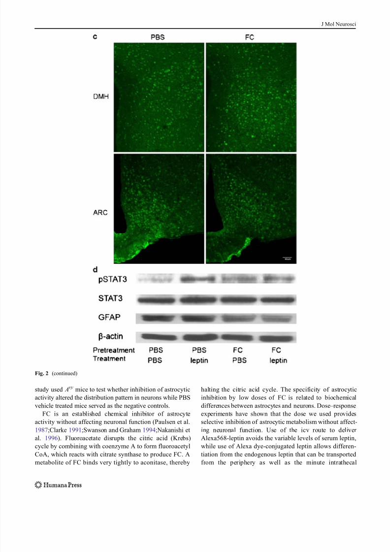
Effects of FC on the Distribution of Alexa568-leptin
and pSTAT3 Activation in the Dorsal MedialHypothalamus
Although astrocytes are abundant in the dorsal medial
hypothalamus (DMH), only very few endocytosed a
substantial amount of Alexa568-leptin 30 min after its icv
delivery. Most of the Alexa568 (+) cells resembled
neuronal morphology and showed a punctate distribution
of the fluorescence. The α 1 an d α 2 tanycytes also
endocytosed Alexa568-leptin. In mice receiving FC pre-
treatment, there was an increase in the fluorescent intensity
of Alexa568-leptin, mainly in neurons (Fig. 2a ). This was
better seen in the higher magnification images. FC also
induced mild astrogliosis, shown by the higher fluorescent
intensity of GFAP and more tortuous and condensed
processes (Fig. 2b).
In mice pretreated with PBS vehicle before icv injection
of Alexa568-leptin, there were more pSTAT3 (+) cells in
the arcuate nucleus than in the DMH. Most of the cells
showed neuronal morphology, although there were sparse
astrocytic processes in the arcuate nucleus adjacent to the
median eminence that were also pSTAT3 (+). In mice
receiving FC pretreatment, there were increased numbers of
pSTAT3 (+) cells, more apparent in the DMH than in the
arcuate nucleus (Fig. 2c).
In hypothalamic homogenates from the Avy mice, the basal
level of pSTAT3 signal was low though GFAP expression
was high. Leptin treatment induced a robust increase of
pSTAT3 at 30 min. FC pretreatment also induced a mild
increase of pSTAT3 at 1.5 h after pretreatment, but the GFAP
signal was reduced. Pretreatment of mice with FC and later
leptin caused a reduction of pSTAT3. The reduction of GFAP
in the FC+leptin treated mice was similar to that in the mice
without leptin treatment (Fig. 2d).
Discussion
Avy mice provide a unique model of adult-onset obesity as a
result of a gain-of-function mutation resulting from consti-
tutive activation of the promoter driving the expression of
agouti signaling peptide. This leads to ectopic expression of agouti-related protein that serves as a reverse antagonist
against melanocortin receptors, resulting in an agouti coat
color by MC1R blockade, defective hypothalamic MC3R
and MC4R signaling, and eventually an obesity phenotype
(Wolff 1965). In Avy mice, we have shown a greater
reduction of influx rate of leptin across the blood – brain
barrier (BBB) in middle-aged adult mice than older adult
mice, mainly explained by changes in blood concentrations
of leptin (Pan et al. 2008). Although the BBB transport
systems for leptin (Banks et al. 1996) and mahogany
peptide (Kastin and Akerstrom 2000;Pan and Kastin 2007b)
persist in the Avy mouse, leptin processing within the brain
Fig. 1 (continued)
Fig. 2 Distribution of Alexa568-leptin (red ) in the DMH 30 min after
icv administration, its partial colocalization with GFAP immunopositive
astrocytic cells ( green), and activation of STAT3. FC treatment
increased the neuronal distribution of Alexa568-leptin and the number
of pSTAT3 immunopositive cells. a In the PBS pretreated group, there
are fewer GFAP cells that internalized Alexa568-leptin (arrows) in the
DMH than those in the arcuate nucleus. In neurons, the endocytosed
Alexa568-leptin is present in perinuclear regions (arrowheads). In the
FC pretreated group, tanycytes at the left border of the image are also
Alexa568-leptin positive (oval arrow). The third ventricle is at the left
border of each image. Scale bars 30 μ m. b In higher magnification
images with z -stacking, Alexa568-leptin has a vesicular distribution in
the cytoplasm of neurons. There is an increase of Alexa568-leptinfluorescent intensity in neurons adjacent to the GFAP astrocytes in the
FC pretreatment group (arrows). FC also induced an altered morphol-
ogy, as the GFAP (+) processes appear more compact. Scale bars
10 μ m. c Regional difference and effects of FC on pSTAT3 distribution
in mice 30 min after Alexa568-leptin icv. There are more pSTAT3 (+)
cells in the arcuate nucleus ( ARC ) than DMH. FC treatment increased
the amount of pSTAT3 (+) cells in the DMH whereas that in the ARC
did not show apparent change. Scale bars 50 μ m. d pSTAT3 activation
by leptin was seen by WB and reduced by FC pretreatment. In mice
receiving FC, leptin did not show an additional effect on the level of
pSTAT3 activation but GFAP expression was reduced in comparison
with the internal control β-actin
J Mol Neurosci

8/8/2019 Astrocytes Modulate Distribution and Neuronal Signaling of Leptin - Pan Et Al 2010
http://slidepdf.com/reader/full/astrocytes-modulate-distribution-and-neuronal-signaling-of-leptin-pan-et 5/7
is greatly modulated by astrocytic activity. This suggests
that astrocytes provide a second barrier and probably
contribute to leptin resistance in obesity. This is strongly
supported by the present observation that FC altered the
distribution and signaling of Alexa568-leptin. Mild astro-
gliosis is not seen in lean controls but only in mice with
adult-onset obesity, as we have shown in previous studies
(Pan et al. 2008;Hsuchou et al. 2009). Therefore, the present
J Mol Neurosci
8/8/2019 Astrocytes Modulate Distribution and Neuronal Signaling of Leptin - Pan Et Al 2010
http://slidepdf.com/reader/full/astrocytes-modulate-distribution-and-neuronal-signaling-of-leptin-pan-et 6/7
study used Avy mice to test whether inhibition of astrocytic
activity altered the distribution pattern in neurons while PBS
vehicle treated mice served as the negative controls.
FC is an established chemical inhibitor of astrocyte
activity without affecting neuronal function (Paulsen et al.
1987;Clarke 1991;Swanson and Graham 1994;Nakanishi et
al. 1996). Fluoroacetate disrupts the citric acid (Krebs)
cycle by combining with coenzyme A to form fluoroacetyl
CoA, which reacts with citrate synthase to produce FC. A
metabolite of FC binds very tightly to aconitase, thereby
halting the citric acid cycle. The specificity of astrocytic
inhibition by low doses of FC is related to biochemical
differences between astrocytes and neurons. Dose – response
experiments have shown that the dose we used provides
selective inhibition of astrocytic metabolism without affect-
ing neuronal function. Use of the icv route to deliver
Alexa568-leptin avoids the variable levels of serum leptin,
while use of Alexa dye-conjugated leptin allows differen-
tiation from the endogenous leptin that can be transported
from the periphery as well as the minute intrathecal
Fig. 2 (continued)
J Mol Neurosci
8/8/2019 Astrocytes Modulate Distribution and Neuronal Signaling of Leptin - Pan Et Al 2010
http://slidepdf.com/reader/full/astrocytes-modulate-distribution-and-neuronal-signaling-of-leptin-pan-et 7/7
production in the pituitary and cerebral cortex detected by
RT-PCR (Morash et al. 1999).
The transmigration of Alexa568-leptin from CSF to
medial hypothalamus probably involves both volume
transmission along the extracellular space (Johanson
1995) and specific transport by tanycytes along the walls
of the third ventricle (Blazquez and Rodriguez 2010). The
α 1 tanycytes adjacent to the DMH and the α 2 tanycytesnear the arcuate nucleus do not form as tight a junction as
do the β1 tanycytes at the junction of the arcuate nucleus
and median eminence recess, yet transport along tanycyte
processes was seen. It has been clearly established that the
arcuate nucleus lies within the BBB and is separated from
the median eminence by well-formed junctional structures
(Blazquez and Rodriguez 2010). The β1 tanycytes provid-
ed a barrier to Alexa-leptin diffusion, but this seems to be
weakened in the FC-treated group since stronger fluores-
cence was seen along the intracellular space as well as
within the tanycytes. The increased accumulation of
Alexa568-leptin in β1 tanycytes probably represents a decrease of the transport process. The distribution of
Alexa568-leptin along the border of the median eminence
and arcuate nucleus resembles that of Cdk5/P35, signaling
proteins that can be activated by leptin and in turn modulate
the actions of leptin (He et al. 2009).
In the Avy mice, more than half the neurons in the arcuate
nucleus and some in the DMH showed robust pSTAT3
immunoreactivity. FC treatment further increased the
number of pSTAT3 (+) neurons. The results indicate that
inhibition of astrocytic metabolic activity enhances neuro-
nal leptin signaling. This is consistent with the finding of
increased Alexa568-leptin distribution in neurons in FC-
treated mice. Although we did not detect strong ObR
immunoreactivity in DMH neurons (Hsuchou et al. 2009),
it remains possible that leptin directly activates these
neurons despite their low level of ObR expression.
Nonetheless, WB analysis of hypothalamic homogenates
of the Avy mice showed that the overall pSTAT3 signal was
reduced by FC in comparison with the group receiving PBS
pretreatment and leptin treatment, although the sample size
was too small for statistical analysis. When both groups
with FC treatment were compared, leptin did not induce a
significant increase of pSTAT3 in the total hypothalamus.
This indicates a dilutional effect as the increase of pSTAT3
in IHC was seen mainly in the DMH, an effector region of
the hypothalamus.
In summary, Avy mice showed region-specific distribu-
tion of icv Alexa568-leptin and pSTAT3 signaling. In these
mice we showed for the first time that inhibition of
astrocytic activity alters the fate of leptin after icv delivery.
The increase of neuronal leptin uptake and enhanced
pSTAT3 signaling in the DMH suggest that the astrocytic
leptin system provides a counter-regulatory role, opposite
to that seen in neuronal leptin signaling. This important
principle may be a missing link in leptin resistance.
Acknowledgement This study was supported by NIH (DK54880 to
AJK and NS62291 to WP).
Disclosures/Conflicts of Interest The authors have no conflict of
interest and nothing to disclose.
References
Banks WA, Kastin AJ, Huang W, Jaspan JB, Maness LM (1996)
Leptin enters the brain by a saturable system independent of
insulin. Peptides 17:305 – 311
Blazquez JL, Rodriguez EM (2010) The design of barriers in the
hypothalamus allows the median eminence and the arcuate nucleus
to enjoy private milieus: the former opens to the portal blood and
the latter to the cerebrospinal fluid. Peptides 31:757 – 776
Clarke DD (1991) Fluoroacetate and fluorocitrate: mechanism of
action. Neurochem Res 16:1055 – 1058
He Y, Kastin AJ, Hsuchou H, Pan W (2009) The Cdk5/p35 kinases
modulate leptin-induced STAT3 signaling. J Mol Neurosci
39:49 – 58
Hsuchou H, He Y, Kastin AJ, Tu H, Markadakis EN, Rogers RC,
Fossier PB, Pan W (2009) Obesity induces functional astrocytic
leptin receptors in hypothalamus. Brain 132:889 – 902
Johanson CE (1995) Ventricles and cerebrospinal fluid. In: Conn PM (ed)
Neuroscience in Medicine. Lippincott, Philadelphia, pp 171 – 196
Kastin AJ, Akerstrom V (2000) Mahogany (1377 – 1428) enters brainby a
saturable transport system. J Pharmacol Exp Ther 294:633 – 636
Morash B, Li A, Murphy PR, Wilkinson M, Ur E (1999) Leptin gene
expression in the brain and pituitary gland. Endocrinology
140:5995 – 5998
Nakanishi H, Kawachi A, Okada M, Fujiwara M, Yamamoto K (1996)
Protective effect of MK-801 on the anoxia-aglycemia induceddamage in the fluorocitrate-treated hippocampal slice of the rat.
Brain Res 732:232 – 236
Pan W, Kastin AJ (2007a) Adipokines and the blood-brain barrier.
Peptides 28:1317 – 1330
Pan W, Kastin AJ (2007b) Mahogany, blood-brain barrier, and fat
mass surge in Avy mice. Intl J Obes 31:1030 – 1032
Pan W, Hsuchou H, He Y, Sakharkar A, Cain C, Yu C, Kastin AJ
(2008) Astrocyte leptin receptor (ObR) and leptin transport in
adult-onset obese mice. Endocrinology 149:2798 – 2806
Pan W, Yu C, Hsuhou H, Khan RS, Kastin AJ (2009) Cerebral
microvascular IL15 is a novel mediator of TNF action. J
Neurochem 111:819 – 827
Pan W, Wu X, Kastin AJ, Zhang Y, Hsuchou H, Halberg F, Chatu F,
Khan RS, Robert B, Kastin AJ, Cornelissen-Guillaume GG
(2010) Potential protective role of IL15R α during inflammation.J Mol Neurosci. PMID 20981579
Paulsen RE, Contestabile A, Villani L, Fonnum F (1987) An in vivo
model for studying function of brain tissue temporarily devoid of
glial cell metabolism: the use of fluorocitrate. J Neurochem
48:1377 – 1385
Swanson RA, Graham SH (1994) Fluorocitrate and fluoroacetate
effects on astrocyte metabolism in vitro. Brain Res 664:94 – 100
Tu H, Hsuchou H, Kastin AJ, Wu X, Pan W (2010) Unique leptin
trafficking by a tailless receptor. FASEB J 24:2281 – 2291
Wolff GL (1965) Body composition and coat color correlation in
different phenotypes of "viable yellow" mice. Science 147:1145 –
1147
J Mol Neurosci