Embed Size (px)
Citation preview

Trisomy 13 Syndrome
In 1960, Patau et al. first recognized the relation of
trisomy 13 to a clinical syndrome. Incidence is esti-
mated to be 1/4,000–1/10,000 live births.
Synonyms and Related Disorders
Patau syndrome
Genetics/Basic Defects
1. Trisomy 13
a. Mechanism: due to meiotic nondisjunction
i. Maternal origin of the extra chromosome
(90%)
ii. Stage of nondisjunction: mostly maternal
meiosis I (vs. meiosis II in trisomy 18)
iii. Paternal origin of the extra chromosome
(10%): The majority is primarily postzygotic
mitotic errors.
b. Frequency: 75% of cases
2. Translocation trisomy 13
a. Mechanism: de novo (75%) or familial transmis-
sion (25%)
b. Frequency: 20% of cases
c. Trisomy 13 due to t(13;13): The structural abnor-
malities are usually isochromosomes originating
in mitosis.
3. Mosaic trisomy 13
a. Mechanism: due to postzygotic (postferti-
lization) mitotic nondisjunction
b. Frequency: 5% of cases
c. Variable phenotype from full trisomy to near
normal
d. Variable mental retardation with longer
survival
e. Trisomy 13/triploidy mosaicism: a rare event
4. Partial trisomy 13
a. Partial trisomy of proximal segment with
nonspecific clinical features and little similarity
to full trisomy 13
b. Partial trisomy of distal segment with specific
clinical features
Clinical Features
1. General
a. Low birth weight
b. Thrombocytopenia
2. CNS
a. Severe mental retardation
b. Holoprosencephaly
c. Seizures
d. Central apnea
3. Craniofacial abnormalities
a. Microcephaly
b. Wide sagittal sutures
c. Wide fontanels
d. Scalp defect (aplasia cutis congenita, 50%)
e. Capillary hemangioma of the forehead
f. Ocular abnormalities
i. Microphthalmia/anophthalmia
ii. Colobomas
iii. Retinal dysplasia
g. Cleft lip/palate
h. Abnormal auricles
i. Low-set ears
j. Sensorineural and conductive deafness
H. Chen, Atlas of Genetic Diagnosis and Counseling, DOI 10.1007/978-1-4614-1037-9_235,# Springer Science+Business Media, LLC 2012
2057
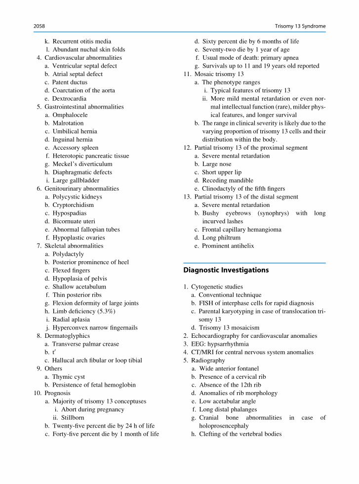
k. Recurrent otitis media
l. Abundant nuchal skin folds
4. Cardiovascular abnormalities
a. Ventricular septal defect
b. Atrial septal defect
c. Patent ductus
d. Coarctation of the aorta
e. Dextrocardia
5. Gastrointestinal abnormalities
a. Omphalocele
b. Malrotation
c. Umbilical hernia
d. Inguinal hernia
e. Accessory spleen
f. Heterotopic pancreatic tissue
g. Meckel’s diverticulum
h. Diaphragmatic defects
i. Large gallbladder
6. Genitourinary abnormalities
a. Polycystic kidneys
b. Cryptorchidism
c. Hypospadias
d. Bicornuate uteri
e. Abnormal fallopian tubes
f. Hypoplastic ovaries
7. Skeletal abnormalities
a. Polydactyly
b. Posterior prominence of heel
c. Flexed fingers
d. Hypoplasia of pelvis
e. Shallow acetabulum
f. Thin posterior ribs
g. Flexion deformity of large joints
h. Limb deficiency (5.3%)
i. Radial aplasia
j. Hyperconvex narrow fingernails
8. Dermatoglyphics
a. Transverse palmar crease
b. t0
c. Hallucal arch fibular or loop tibial
9. Others
a. Thymic cyst
b. Persistence of fetal hemoglobin
10. Prognosis
a. Majority of trisomy 13 conceptuses
i. Abort during pregnancy
ii. Stillborn
b. Twenty-five percent die by 24 h of life
c. Forty-five percent die by 1 month of life
d. Sixty percent die by 6 months of life
e. Seventy-two die by 1 year of age
f. Usual mode of death: primary apnea
g. Survivals up to 11 and 19 years old reported
11. Mosaic trisomy 13
a. The phenotype ranges
i. Typical features of trisomy 13
ii. More mild mental retardation or even nor-
mal intellectual function (rare), milder phys-
ical features, and longer survival
b. The range in clinical severity is likely due to the
varying proportion of trisomy 13 cells and their
distribution within the body.
12. Partial trisomy 13 of the proximal segment
a. Severe mental retardation
b. Large nose
c. Short upper lip
d. Receding mandible
e. Clinodactyly of the fifth fingers
13. Partial trisomy 13 of the distal segment
a. Severe mental retardation
b. Bushy eyebrows (synophrys) with long
incurved lashes
c. Frontal capillary hemangioma
d. Long philtrum
e. Prominent antihelix
Diagnostic Investigations
1. Cytogenetic studies
a. Conventional technique
b. FISH of interphase cells for rapid diagnosis
c. Parental karyotyping in case of translocation tri-
somy 13
d. Trisomy 13 mosaicism
2. Echocardiography for cardiovascular anomalies
3. EEG: hypsarrhythmia
4. CT/MRI for central nervous system anomalies
5. Radiography
a. Wide anterior fontanel
b. Presence of a cervical rib
c. Absence of the 12th rib
d. Anomalies of rib morphology
e. Low acetabular angle
f. Long distal phalanges
g. Cranial bone abnormalities in case of
holoprosencephaly
h. Clefting of the vertebral bodies
2058 Trisomy 13 Syndrome
i. Abnormal postsphenoid component of the
sphenoid bone
j. Agenesis of the nasal bones
6. Placenta: Partial molar change of the placenta may
rarely occur in trisomy 13 (Has et al. 2002).
Genetic Counseling
1. Recurrence risk
a. Patient’s sib
i. Trisomy 13: about 1 in 4,000
ii. De novo translocation: about 1 in 4,000
iii. Familial translocation: 5–15%
iv. Mosaicism: 1 in 4,000
b. Patient’s offspring: not surviving to reproductive
age
2. Prenatal diagnosis
a. Prenatal ultrasonography: prevalence of ultra-
sound abnormalities (91%)
i. General
a) IUGR (48%)
b) Single umbilical artery
c) Polyhydramnios (15%)
d) Oligohydramnios (12%)
ii. Cranium and CNS abnormalities (58%)
a) Holoprosencephaly (39%)
b) Neural tube defects
c) Lateral ventricular dilatation without
holoprosencephaly (9%)
d) Enlarged cisterna magna or Dandy-
Walker variant (15%)
e) Microcephaly (12%)
f) Linear branching echogenicity of the
thalamus or basal ganglia (representing
vasculopathy)
g) Choroid plexus cyst
iii. Facial anomalies
a) Cyclopia
b) Proboscis
c) Hypotelorism
d) Hypoplastic midface
e) Cleft lip/palate (36%)
f) Micrognathia
iv. Neck anomalies
a) Nuchal thickening
b) Cystic hygroma
c) Hydrops
d) Lymphangiectasia
v. Chest/cardiac abnormalities
a) Diaphragmatic hernia
b) Ventricular septal defect
c) Hypoplastic left heart
d) Echogenic chorda tendineae (30%)
vi. Renal abnormalities (33%)
a) Echogenic kidneys
b) Pyelectasis
c) Enlarged kidneys
d) Hydronephrosis
vii. Abdominal abnormalities
a) Omphalocele
b) Echogenic bowel (6%)
c) Bladder exstrophy
viii. Limb abnormalities (33%)
a) Clenched and overlapping digits
b) Polydactyly
c) Radial aplasia
d) Short femur length
e) Talipes equinovarus
f) Rocker bottom feet
b. Chromosome analyses
i. Amniocentesis
ii. CVS, followed by amniocentesis
iii. Fetal cells isolated from maternal
blood using either flow sorting or magnetic
sorting
c. Dilemma for genetic counseling with trisomy 13
mosaicism
i. Infrequent occurrence
ii. Single-cell pseudomosaicism (3.3%)
iii. Multiple-cell pseudomosaicism (4%)
iv. Often represents pseudomosaicism or con-
fined placental mosaicism
v. True fetal mosaicism (in the context of low-
level single-digit percentage mosaicism):
not necessarily associated with congenital
defects and/or mental abnormalities
vi. An optimistic approach in case of normal
ultrasonography and absence of trisomy 13
cells in the fetal blood
vii. Possibility of adverse phenotype and intel-
lectual function in case of true low-level
fetal mosaicism
3. Management
a. Feedings
i. Nasal tube feeding
ii. Oral gastric tube feeding
iii. Gastrostomy feeding
Trisomy 13 Syndrome 2059
b. Nissan fundoplication for gastroesophageal
reflux
c. Risk for anesthesia
d. Early intervention programs
e. Seizure control
f. Monitor apneic spells
g. Treat infections
h. Symptomatic treatment for heart failure
i. Cardiac operation rarely performed
References
Alizad, A., & Seward, J. B. (2000). Echocardiographic features
of genetic diseases: Part 7. Complex genetic disorders.
Journal of the American Society of Echocardiography, 13,707–714.
Baty, B. J., Blackburn, B. L., & Carey, J. C. (1994). Natural
history of trisomy 18 and trisomy 13: I. Growth physical
assessment, medical histories, survival, and recurrence risk.
American Journal of Medical Genetics, 49, 175–188.Baty, B. J., Jorde, L. B., & Blackburn, B. L. (1994). Natural
history of trisomy 18 and trisomy 13: II. Psychomotor devel-
opment. American Journal of Medical Genetics, 49, 189–194.Benacerraf, B. R., Miller, W. A., & Frigoletto, F. D., Jr. (1988).
Sonographic detection of fetuses with trisomies 13 and 18:
Accuracy and limitations. American Journal of Obstetricsand Gynecology, 158, 404–409.
Brewer,C.M.,Holloway, S.H., Stone,D.H., et al. (2002). Survival
in trisomy 13 and trisomy 18 cases ascertained from popula-
tion based registers. Journal of Medical Genetics, 39, e54.Chabra, S., Kriss, V. M., Pauly, T. H., et al. (1997).
Neurosonographic diagnosis of thalamic/basal ganglia
vasculopathy in trisomy 13-An important diagnostic aid.
American Journal of Medical Genetics, 72, 291–293.Colacino, S. C., & Pettersen, J. C. (1978). Analysis of the gross
anatomical variations found in four cases of trisomy 13.
American Journal of Medical Genetics, 2, 31–50.Curtin, W. M., Marcotte, M. P., Myers, L. L., et al. (2001).
Trisomy 13 appearing as a mimic of a triploid partial mole.
Journal of Ultrasound in Medicine, 20, 1137–1139.Delatycki, M., & Gardner, R. J. (1997). Three cases of trisomy 13
mosaicism and a review of the literature. Clinical Genetics,51, 403–407.
Delatycki, M. B., Pertile, M. D., & Gardner, R. J. (1998).
Trisomy 13 mosaicism at prenatal diagnosis: Dilemmas in
interpretation. Prenatal Diagnosis, 18, 45–50.Eubanks, S. R., Kuller, J. A., Amjadi, D., et al. (1998). Prenatal
diagnosis of mosaic trisomy 13: A case report. PrenatalDiagnosis, 18, 971–974.
Goldstein, H., & Nielsen, K. G. (1988). Rates and survival of
individuals with trisomy 13 and 18. Data from a 10-year
period in Denmark. Clinical Genetics, 34, 366–372.Hahnemann, J. M., & Vejerslev, L. O. (1997). European collab-
orative research on mosaicism in CVS (EUCROMIC)–fetal
and extrafetal cell lineages in 192 gestations with CVS
mosaicism involving single autosomal trisomy. AmericanJournal of Medical Genetics, 70, 179–187.
Hansen, C. B., Fergestad, J. M., Barnes, A., et al. (2000). An
analysis of heart surgery in children with trisomy 18, 13. TheJournal of Medical Investigation, 48, 47A.
Has, R., Ibrahimoglu, L., Ergene, H., et al. (2002). Partial molar
appearance of the placenta in trisomy 13. Fetal Diagnosisand Therapy, 17, 205–208.
Hodes, M. E., et al. (1978). Clinical experience with trisomies 18
and 13. Journal of Medical Genetics, 15, 48–60.Janiaux, E., Halder, A., & Partington, C. (1998). A case of partial
mole associated with trisomy 13. Ultrasound in Obstetrics &Gynecology, 11, 62–64.
Kjaer, I., Keeling, J. W., & Hansen, B. F. (1997). Pattern of
malformations in the axial skeleton in human trisomy 13
fetuses. American Journal of Medical Genetics, 70, 421–426.Lehman, C. D., Nyberg, D. A., Winter, T. C., III, et al. (1995).
Trisomy 13 syndrome: Prenatal US findings in a review of 33
cases. Radiology, 194, 217–222.Martınez-Frıas, M. L., Villa, A., de Pablo, R. A., et al. (2000).
Limb deficiencies in infants with trisomy 13. AmericanJournal of Medical Genetics, 93, 339–341.
Moerman, P., Fryns, J. P., van der Steen, K., et al. (1988). The
pathology of trisomy 13 syndrome: A study of 12 cases.
Human Genetics, 80, 349–356.Nyberg, D. A., & Souter, V. L. (2001). Sonographic markers of
fetal trisomies. Journal of Ultrasound in Medicine, 20,655–674.
Oosterwijk, J. C., Mesker, W. E., Ouwerkerk-van Velzen,
M. C. M., et al. (1998). Prenatal diagnosis of trisomy 13 on
fetal cells obtained from maternal blood after minor enrich-
ment. Prenatal Diagnosis, 18, 1082–1085.Patau, K., Therman, D. G., Cameron, A. H., et al. (1960). A new
trisomic syndrome. Lancet, 1, 787–789.Pettersen, J. C., et al. (1979). An examination of the spectrum of
anatomic defects and variations found in eight cases of trisomy
13. American Journal of Medical Genetics, 3, 183–210.Phelan, M. C., Rogers, R. C., Michaelis, R. C., et al. (2001).
Prenatal diagnosis of mosaicism for triploidy and trisomy 13.
Prenatal Diagnosis, 21, 457–460.Redheendran, R., Neu, R. L., & Bannerman, R. M. (1981). Long
survival in trisomy 13 syndrome: 21 cases including
prolonged survival in two patients 11 and 19 years old.
American Journal of Medical Genetics, 8, 167–172.Robinson, W. P., Bernasconi, F., Dutly, F., et al. (1996). Molec-
ular studies of translocations and trisomy involving chromo-
some 13. American Journal of Medical Genetics, 61,158–163.
Rogers, J. F. (1984). Clinical delineation of proximal and distal
partial 13q trisomy. Clinical Genetics, 25, 221–229.Schinzel, A., et al. (1976). Further delineation of the clinical
picture of trisomy for the distal segment of chromosome 13.
Human Genetics, 32, 1–12.Smith, K., Lowther, G., Maher, E., et al. (1999). The predictive
value of findings of the common aneuploidies, trisomies 13,
18 and 21, and numerical sex chromosome abnormalities at
CVS: Experience from the ACC U.K. Collaborative Study.
Association of Clinical Cytogeneticists Prenatal Diagnosis
Working Party. Prenatal Diagnosis, 19, 817–826.Snijders, R. J., Sebire, N. J., Nayar, R., et al. (1999). Increased
nuchal translucency in trisomy 13 fetuses at 10–14 weeks of
gestation. American Journal of Medical Genetics, 86,205–207.
2060 Trisomy 13 Syndrome
Taylor, A. I. (1968). Autosomal trisomy syndromes: A detailed
study of 27 cases of Edwards’ syndrome and 27 cases of
Patau’s syndrome. Journal of Medical Genetics, 5, 227–241.Tharapel, S. A., Lewadowski, R. C., Tharapel, A. T., et al.
(1986). Phenotype-karyotype correlation in patients trisomic
for various segments of chromosome 13. Journal of MedicalGenetics, 23, 310–315.
Tongson, T., Sirichotiyakul, S., Wanapirak, C., et al. (2002).
Sonographic features of trisomy 13 at midpregnancy. Inter-national Journal of Gynecology and Obstetrics, 76, 143–148.
Tuohy, J. F., & James, D. K. (1994). Pre-eclampsia and trisomy
13. British Journal of Obstetrics and Gynaecology, 99,891–894.
Wallerstein, R., Yu, M.-T., Neu, R. L., et al. (2000). Common
trisomy mosaicism diagnosed in amniocytes involving chro-
mosomes 13, 18, 20 and 21: Karyotype-phenotype correla-
tions. Prenatal Diagnosis, 20, 103–122.Warkany, J., et al. (1966). Congenital malformations in autoso-
mal trisomy syndromes. American Journal of Diseases ofChildren, 112, 502–517.
Wyllie, J. P., Wright, M. J., Burn, J., et al. (1994). Natural history
of trisomy 13. Archives of Disease in Childhood, 71,343–345.
Zoll, B., Wolf, J., Lensing-Hebben, D., et al. (1993). Trisomy 13
(Patau syndrome) with an 11-year survival. Clinical Genetics,43, 46–50.
Trisomy 13 Syndrome 2061

a bFig. 1 (a, b) An infant with
trisomy 13 showing
microcephaly,
microphthalmia, forehead
hemangioma, cleft lip/palate,
and postaxial polydactyly
baFig. 2 (a, b) An infant with
trisomy 13 showing
microcephaly,
microphthalmia, cleft lip/
palate, and omphalocele
2062 Trisomy 13 Syndrome
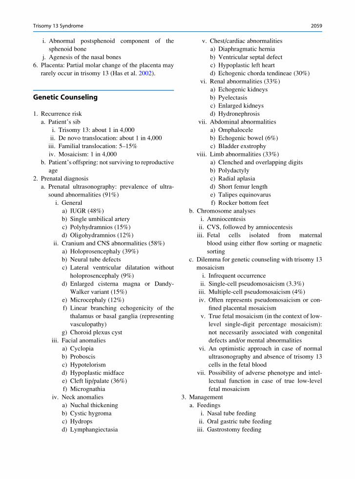
a bFig. 3 (a, b) Postaxialpolydactyly of the hand and
the feet in an infant with
trisomy 13
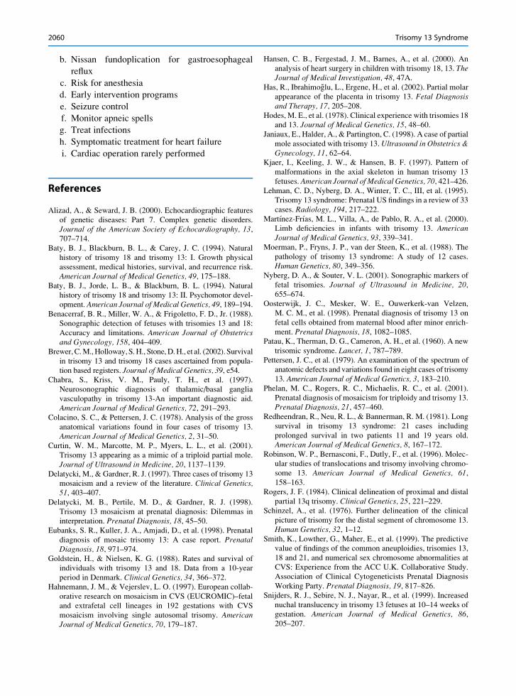
a bFig. 4 (a, b) An infant with
trisomy 13 showing
microcephaly, forehead
hemangioma, upslanted
palpebral fissures, cleft lip/
palate, and short neck
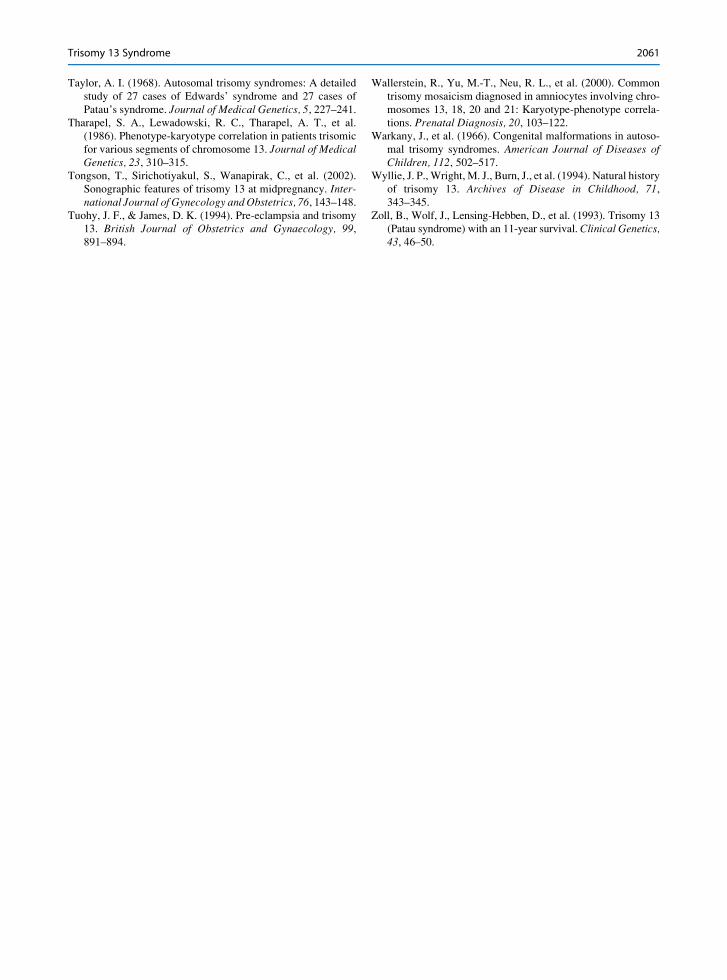
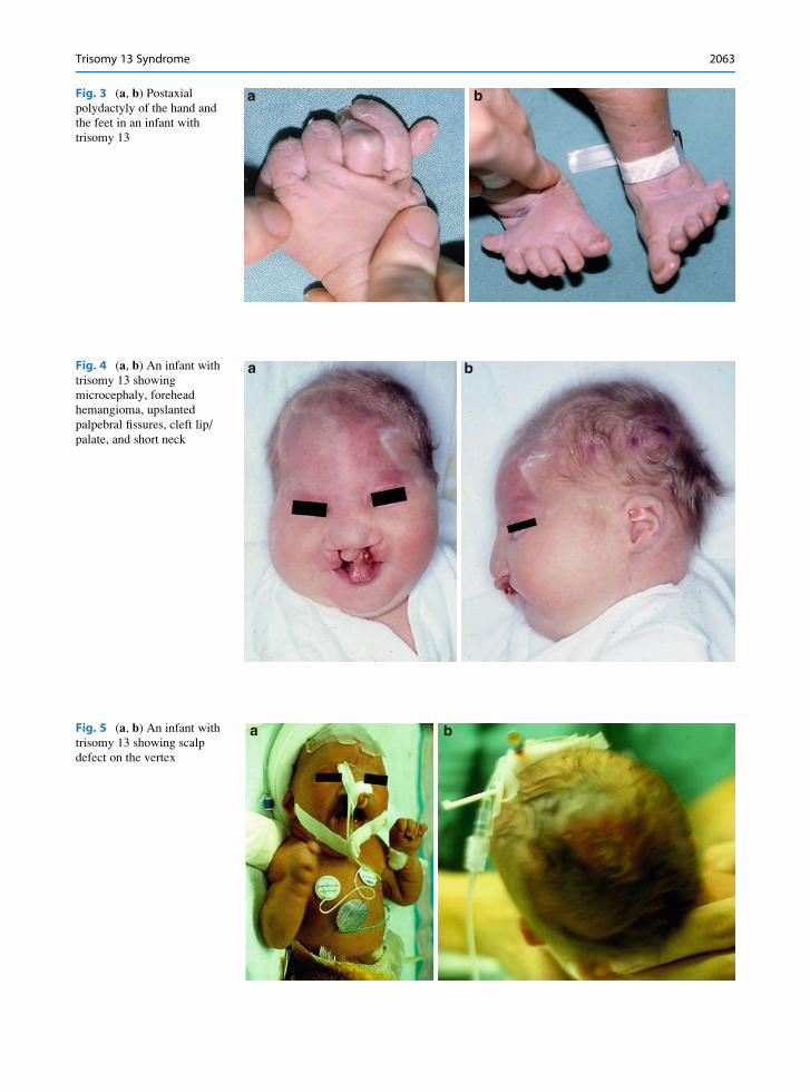
a bFig. 5 (a, b) An infant with
trisomy 13 showing scalp
defect on the vertex
Trisomy 13 Syndrome 2063
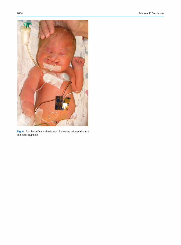
Fig. 6 Another infant with trisomy 13 showing microphthalmia
and cleft lip/palate
2064 Trisomy 13 Syndrome
a b
c
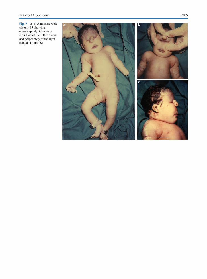
Fig. 7 (a–c) A neonate with
trisomy 13 showing
ethmocephaly, transverse
reduction of the left forearm,
and polydactyly of the right
hand and both feet
Trisomy 13 Syndrome 2065
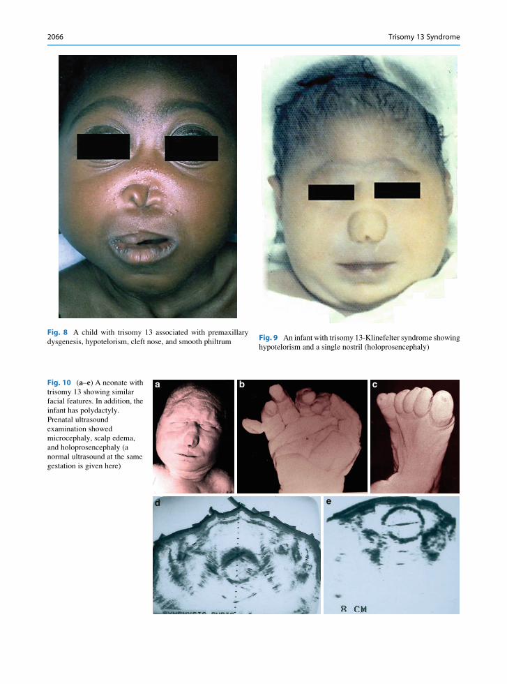
Fig. 9 An infant with trisomy 13-Klinefelter syndrome showing
hypotelorism and a single nostril (holoprosencephaly)
Fig. 8 A child with trisomy 13 associated with premaxillary
dysgenesis, hypotelorism, cleft nose, and smooth philtrum
b c
ed
aFig. 10 (a–e) A neonate with
trisomy 13 showing similar
facial features. In addition, the
infant has polydactyly.
Prenatal ultrasound
examination showed
microcephaly, scalp edema,
and holoprosencephaly (a
normal ultrasound at the same
gestation is given here)
2066 Trisomy 13 Syndrome
1
6 7 8 9 10 11 12
181716151413
19 20 21 22 X Y
2 3 4 5
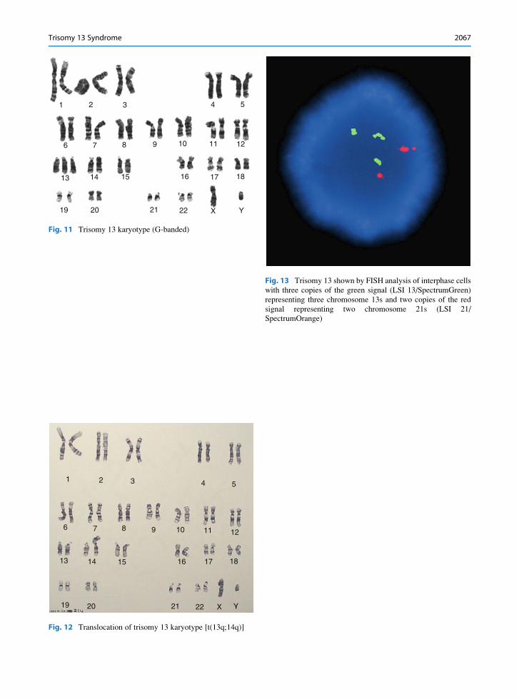
Fig. 11 Trisomy 13 karyotype (G-banded)
1
6 7 8 9 10 11 12
181716151413
19 20 21 22 X Y
2 3 4 5
Fig. 12 Translocation of trisomy 13 karyotype [t(13q;14q)]
Fig. 13 Trisomy 13 shown by FISH analysis of interphase cells
with three copies of the green signal (LSI 13/SpectrumGreen)
representing three chromosome 13s and two copies of the red
signal representing two chromosome 21s (LSI 21/
SpectrumOrange)
Trisomy 13 Syndrome 2067