Embed Size (px)
Citation preview
4007Development 125, 4007-4017 (1998)Printed in Great Britain © The Company of Biologists Limited 1998DEV4974
Atm deficiency results in severe meiotic disruption as early as leptonema of
prophase I
Carrolee Barlow 1,*,‡, Marek Liyanage 2,*,§, Peter B. Moens 3, Madalina Tarsounas 3, Kunio Nagashima 4,Kevin Brown 5, Scott Rottinghaus 6, Stephen P. Jackson 6, Danilo Tagle 5, Thomas Ried 2
and Anthony Wynshaw-Boris 1,¶
1Genetic Disease Research Branch, 2Genome Technology Branch and 5Molecular Genetics and Biology Branch, National HumanGenome Research Institute, National Institutes of Health, Bethesda, MD 20892, USA4Laboratory of Cell and Molecular Structure, SAIC Frederick, NCI-Frederick Cancer Research and Development Center, Frederick,MD 21702, USA3Department of Biology, York University, Downsview, Ontario, M3J 1P3, Canada6Wellcome/CRC Institute, Tennis Court Road, Cambridge CB2 1QR, UK*The first two authors contributed equally‡Present address: The Salk Institute for Biological Studies, 10010 North Torrey Pines Road, La Jolla, CA 92037, USA§Present address: Aurora Biosciences Corporation, 11010 Torreyana Road, San Diego, CA 92121, USA¶Author for correspondence (e-mail: [email protected])
Accepted 29 July; published on WWW 14 September 1998
Infertility is a common feature of the human disorderataxia-telangiectasia and Atm-deficient mice are completelyinfertile. To gain further insight into the role of ATM inmeiosis, we examined meiotic cells in Atm-deficient miceduring development. Spermatocyte degeneration beginsbetween postnatal days 8 and 16.5, soon after entry intoprophase I of meiosis, while oocytes degenerate late inembryogenesis prior to dictyate arrest. Using electronmicroscopy and immunolocalization of meiotic proteins inmutant adult spermatocytes, we found that male and femalegametogenesis is severely disrupted in Atm-deficient mice asearly as leptonema of prophase I, resulting in apoptoticdegeneration. A small number of mutant cells progress intolater stages of meiosis, but no cells proceed beyond prophaseI. ATR, a protein related to ATM, DMC1, a RAD51 family
member, and RAD51 are mislocalized to chromatin andhave reduced localization to developing synaptonemalcomplexes in spermatocytes from Atm-deficient mice,suggesting dysregulation of the orderly progression ofmeiotic events. ATM protein is normally present at highlevels primarily in ova cytoplasm of developing ovarianfollicles, and in the nucleus of spermatogonia and to a lesserextent in spermatoctyes, but without localization to thesynaptonemal complex. We propose a model in which ATMacts to monitor meiosis by participation in the regulation orsurveillance of meiotic progression, similar to its role as amonitor of mitotic cell cycle progression.
Key words: ATM, Meiosis, Prophase I, ATR, DMC1, Electronmicroscopy, Immunolocalization, Mouse
SUMMARY
dne
5;or
er
),
etuslls,e ofry
INTRODUCTION
Reduction from the diploid to haploid state is accomplishedpairing, synapsis, recombination and segregation homologous chromosomes during meiotic prophase ameiosis I division (reviewed in Kleckner, 1996; Koehler et a1996; Roeder, 1997). Interference with one or more of thfunctions can lead to lethality of reproductive cells or formation of defective meiotic products, a point that has bewell demonstrated in several Saccharomyces cerevisiaeandDrosophila melanogastermutants (reviewed in Orr-Weaver1995; Roeder, 1995).
In addition, targeted inactivation of several mammaligenes has resulted in infertility phenotypes, implicating thegenes in processes important in meiosis. For example,(Barlow et al., 1996) and others (Elson et al., 1996; Xu et
byofndl.,eseinen
,
anse
weal.,
1996) described the phenotype of mice with a targetedisruption of Atm, the mouse homologue of the human ge(ATM) that is mutated in ataxia-telangiectasia (AT). Atm-deficient mice recapitulate the phenotype of AT (Boder, 197Sedgewick and Boder, 1991), providing an excellent model fthis disease. The pleiotropic effects of mutations in ATM andAtm indicate that these genes are involved in the propfunctioning of a variety of mitotic, postmitotic and meioticallyactive cells.
Most AT patients (Boder, 1975; Sedgewick and Boder, 1991and all Atm-deficient mice, of both sexes are infertile due tocomplete absence of mature gametes in adult gonads (Barlowal., 1996; Elson et al., 1996; Xu et al., 1996). The seminiferotubules of mutant males contain spermatogonia and Sertoli cebut no normal spermatocytes, and display a complete absencspermatids or mature sperm. In female mutants, the adult ova
4008
cewontes0d
lr,0
y
rthere
yed
orndal
edndedith
yryn
yo
ch
-or
nootan-
nt
e.,s
es,y,
C. Barlow and others
is devoid of maturing follicles, primordial follicles and oocyteThe severe nature of these defects in both sexes suggestAtm is important during an early stage in meiosis I, possibduring meiotic prophase. This hypothesis is consistent with meiotic recombination phenotypes of the Atm homologuesMEC1 (ESR1)in Saccharomyces cerevisiae (Kato and Ogaw1994) and mei41 in Drosophila melanogaster (Baker andCarpenter, 1972), and the meiotic checkpoint function of MEC1(Lydall et al., 1996). In addition, surface spread spermatocyfrom Atm-deficient mice show abnormalities that suggest arrin zygonema/pachynema of prophase I, with chromosofragmentation (Xu et al., 1996; Plug et al., 1997). These resare consistent with an important role for Atm in prophase I ofmeiosis.
In support of the hypothesis that Atmplays an important rolein meiotic prophase, we have recently demonstrated that RADfoci are not assembled properly on unpaired axial elementleptotene of spermatocytes from Atm-deficient mice, and p53,p21 and BAX are elevated in mutant testes (Barlow et 1997a). In Atm/p53or Atm/p21double mutants, spermatogenesprogresses further into pachytene stages, but not to diplotAssembly of Rad51 foci on axial elements remains defectand p53, p21 and BAX remain elevated unless geneticeliminated. These results demonstrate that Atm is important forproper RAD51 assembly onto the axial elements, as well assuppressing p53, p21 and BAX levels in testes.
We have examined the infertility phenotype of Atm-deficientmice in detail in order to understand the role of Atm in meiosisand provide insight into the general function of this genecellular processes in vivo. We have analyzed the developmof the germ cell defects in male and female gametogenesis,performed morphological and fluorescent immunostainianalyses of meiosis in the adult testis using sections surface spreads of spermatocytes. Our results indicate a crrole for Atm as early as leptonema of prophase I durinmeiosis.
MATERIALS AND METHODS
Mating and genotyping miceThe construction of the Atm-deficient mice (allele designationAtmins5790neo) was previously described (Barlow et al., 1996). Micfrom heterozygous crosses were genotyped by Southern blottindescribed (Barlow et al., 1996), using EcoRV-digested DNA andgenomic probe surrounding the targeted exon, or by PCR (develoby Mai-Jing Liao and Terry Van Dyke). PCR primers are: ATM-FGAC TTC TGT CAG ATG TTG CTG CC; ATM-B: CGA ATT TGCAGG AGT TGC TGA G; ATM-NEO: GGG TGG GAT TAG ATAAAT GCC TG. The PCR products are 440 bp for the mutant alland 162 bp for the wild-type allele. Heterozygotes were derived frmating in a completely inbred 129SvEv background, or in 129SvEv×NIH Black Swiss mixed background. No significandifferences were observed between backgrounds (data not show
Histological analysisTestes and ovaries were isolated and fixed in 20 volumes of 1buffered formalin. Fixed tissues were embedded in paraffin or plassectioned and stained using standard methods (Luna, 1992). Plsections of 1.8 µm in thickness were stained with toluidine blueSections were examined and photographed with light microscopy.TUNEL assays, paraffin-embedded sections were dewaxed analyzed using the TACS in situ kit (Trevigen).
s.s thatlythe
a,
tesestmeults
51s in
al.,isene.ive,ally
for
inent andnganducialg
eg as aped:
eleoma
tn).
0%tic,astic.
Forand
Preparation of male meiotic prophase spermatocytesSpreads of cells in meiotic prophase were prepared by the surfamicrospreading technique (Dresser and Moses, 1979) to allosequential analysis of microspreads by light and transmission electrmicroscopy. In brief, a single cell suspension was prepared from tesof 2-month-old male mice. Cells were lysed in hypotonic salt (14mM, pH 8.0) and nuclei attached to glass- or plastic-coatemicroscope slides. Nuclei were fixed for 6 minutes (2%paraformaldehyde, with or without 0.03% SDS, pH 8.2). After severawashes with Photoflo (0.4% Kodak Photoflo 600 in distilled watepH 8.2), slides were dried. Slides were stained for approximately 9minutes at 60°C with a silver staining solution (50% silver nitrate,0.03% formalin in distilled water). After destaining in distilled water,slides were air dried. Photographic images at the light microscoplevel were acquired using a 100× objective.
For electron microscopy, slides were immersed in 30-50% silvenitrate, covered with nylon mesh and incubated in a moisenvironment for 10-30 minutes. Slides were then washed witPhotoflo and the film containing the nuclei was floated off of thmicroscope slides onto the surface of distilled water. Grids werandomly placed on the film and the film with grids were lifted fromthe water surface by Parafilm. Isolated grids were examined btransmission electron microscopy and electron micrographs acquirat magnifications between 3,000 and 15,000×.
ImmunohistochemistryParaffin-embedded tissues were prepared and used fimmunohistochemistry according to standard techniques (Harlow aLane, 1988), using the Renaissance TSA (Tyramide SignAmplification, DuPont NEN, Boston, MA) kit for the enhancementof the fluorescent signal.
Fluorescence immunostaining of spread meiotic nucleiImmunostaining of surface spreads of spermatocytes was performas previously described (Dobson et al., 1994). Mouse anti-Cor1 amouse anti-Syn1 antibodies were used as previously describ(Dobson et al., 1994). Secondary antibodies were conjugated wFITC or rhodamine, and purchased commercially (Pierce).
Anti-ATM, ATR, DMC1 and RAD51 antibodiesEight different antibodies to human and murine ATM, produced bseveral different laboratories, were used for immunohistochemistand fluorescence immunostaining: two different lots of the anti-humaATM peptide (amino acids 1353-1366) antibody ATM-1 (or DH-1)from Oncogene Research (Cambridge, MA), which reportedllocalized to the synaptonemal complexes (Keegan et al., 1996); twother anti-human ATM peptide antibodies from Oncogene Resear(ATM-3: amino acids 13-24; and ATM-6: amino acids 819-844); twopolyclonal anti-human ATM antibodies, generated from histidinetagged fusion proteins spanning amino acids 1391-1693 (Atm.B) amino acids 1980-1338 (Atm.V), which detect mouse ATM byimmunoblot analysis (Lakin et al., 1996); a monoclonal anti-humaATM antibody (2C6), generated from a GST-fusion protein to aminacids 2577-3056, that also recognizes mouse ATM by immunoblanalysis (Barlow et al., 1997b; Chen and Lee, 1996); and pAb354, anti-human ATM GST fusion protein antibody to amino acids 22872572 (K. B. and D. T., unpublished). Specific ATM localization wasconfirmed by comparing immunolocalization signals in wild-type andAtm−/− spermatocytes, since the 350 kDa ATM protein is not presein Atm−/− mouse tissues (Barlow et al., 1997b).
Two anti-human ATR antibodies were used for fluorescencimmunolocalization studies: ATR-A and ATR-C (S. R. and S. P. Junpublished data). Neither preimmune serum produceimmunofluorescent foci on surface spermatocytes but ATR-Apreimmune serum has a weak reaction with synaptonemal complexwhich is common for rabbit serum. The anti-mouse DMC1 antibod17RTB is a mouse polyclonal antiserum (P. B. M. and M. T.
4009ATM and meiosis in mice
not to
osisall We
cem
iesticse
for inny.r to
rty1
alesofofy a
esInlesg
tes.
he
le3).
Aafter
ild-
al
eh
16ytes.al
nttesllsesey.
in
s
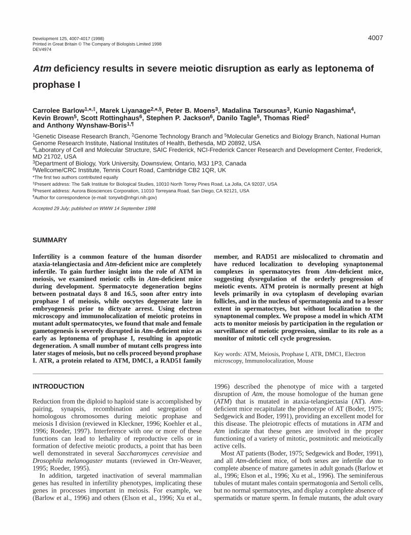
Fig. 1.Abnormal oogenesis in Atm-deficient mice. Embryonic day17.5 ovaries from wild-type (A) and mutant (B) littermates stainedwith hematoxylin and eosin. Note the pycnotic cells in the ovaryfrom mutant mice (B, inset, arrowheads). Wild-type (C) and mutant(D) ovaries were examined for apoptotic cells by the TUNEL in situassay and positive cells were only detected in the mutant ovary (D,inset, arrowheads). (C,D) Adjacent sections to A and B, respectively.Hematoxylin and eosin stained sections of ovaries from 11-day-oldwild-type (E) and mutant (F) mice. The arrowheads indicate primaryoocytes in preantral follicles. Magnifications were 20×(E,F), 40×(A-D) and 60× (insets).
unpublished) against His-tagged DMC1 that was expressed in bacfrom full-length mouse cDNA (Yoshida et al., 1998). All mouspreimmune sera were negative for meiotic prophase proteins. Beinjection, the protein was purified on a Ni-NTA column. To eliminacross-hybridization with RAD51, 17RTB was passed twice overcolumn containing RAD51 protein so that the elutriate recognizonly DMC1 and not RAD51 by western blotting (data not shownThe anti-mouse RAD51 antibody was used as described (Dobsoal., 1994; Barlow et al., 1997).
RESULTS
Development of gametogenesis defects in Atm -deficient miceWe analyzed the developmental onset of the gametogendefects in Atm-deficient mice of both sexes. Primordial germcells migrate from the allantois to the genital ridges by 11days postconception (d.p.c.) (Hogan et al., 1994). Embryfrom heterozygous crosses were fixed at 12.5 d.p.c., staiwith alkaline phosphatase to detect germ cells in the genridges and genotyped by Southern analysis. Embryos ofthree genotypes had equivalent numbers of alkalphosphatase-positive germ cells in the genital ridges (datashown), demonstrating that germ cell survival and migrationthe ridges were unaffected in mutant mice.
Oocytes arrest in the postpachytene dictyate stage of meiI beginning late in gestation, and by 5 days after birth, oocytes are arrested at this stage (Hogan et al., 1994).examined the ovaries from wild-type and Atm-deficientembryos at embryonic day 16.5. Ovaries from wild-type mihad several primordial oocytes (Fig. 1A), as did ovaries fromutant mice (Fig. 1B). However, several oocytes in ovarfrom mutant mice appeared to be undergoing apoptodegeneration and had pycnotic nuclei (Fig. 1B, inset). Theobservations were confirmed by the TUNEL in situ assay apoptosis. TUNEL-positive oocytes were rarely observedovaries from wild-type mice (Fig. 1C), but there were maTUNEL-positive oocytes in ovaries from mutant mice (Fig1D, and inset) demonstrating that oocytes degenerated priobirth in Atm-deficient mice. After birth, in wild-type mice,oocytes continue to mature until recruited at and after pubefor progression to mature follicles. Wild-type ovaries at 1days of age (Fig. 1E) were filled with oocytes, primordifollicles and maturing follicles (see inset). However, ovarifrom Atm-deficient mice at 11 days (Fig. 1F) were devoid oocytes and follicles (Fig. 1F, inset). Only one ovary out eight examined had any follicles at all, and in this case onlsingle abnormal follicle was observed (data not shown).
As we previously showed (Barlow et al., 1996), adult ovarifrom mutant mice were barren, without any oocytes. addition, attempted superovulation of several mutant femadid not lead to follicle formation (data not shown), supportinthe observation that mutant ovaries contained no oocyThese results demonstrate that the defect in oogenesis in Atm-deficient mice is complete and occurs prior to or during tdictyate stage of prophase I arrest.
Soon after arriving in the gonadal ridge, maprospermatogonia undergo arrest (reviewed in Bellvé, 199After birth, the spermatogonia divide mitotically. These typespermatogonia populate the seminiferous tubules and therereside adjacent to the basement membrane. Testes from w
teriaeforete aed).n et
esis
.5os
nedital alline
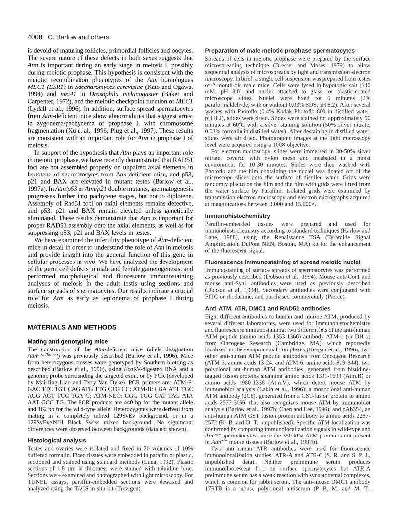
type (Fig. 2A) and Atm-deficient mice (Fig. 2B) wereindistinguishable at 8 days of age, demonstrating that normdifferentiation and mitotic division of spermatogonia occur inthe absence of Atm. Between 8 and 10 days of age, some typA spermatogonia differentiate to type B spermatogonia, whicthen progress into meiotic prophase as spermatocytes. Bydays of age, leptotene, zygotene and pachytene spermatoccan be identified. Testis from 16-day-old wild-type mice (Fig2C) contained all of these cell types. In contrast, normspermatogenesis was completely disrupted in Atm-deficientmice (Fig. 2D). Spermatogonia and Sertoli cells were presealong the basement membrane, but no normal spermatocycould be identified. Instead, there were smaller, pycnotic cethat appeared to be undergoing apoptotic degeneration. Thobservations were confirmed by the TUNEL in situ assaSeminiferous tubules from wild-type controls had only lownumbers of TUNEL-positive cells (Fig. 2E). However, therewas a four-fold increase in apoptotic, degenerating cells tubules from Atm-deficient littermates (Fig. 2F).
We previously showed (Barlow et al., 1996) that sectionfrom adult testes of Atm-deficient mice contained only
4010
vedins,dds
des,esedrele
edbeatain
reald
iate noice
of I,sis.
in
res.ticleIn6),desghto-tly
R
in).ghternd
esis
er in
C. Barlow and others
Fig. 2.Abnormal spermatocyte development in Atm-deficient mice.Hematoxylin and eosin stained sections of mouse testes from 8-dold wild-type (A) and mutant (B) mice are similar. By day 16.5 ofage, toluidine blue staining of testes sections demonstrates thepresence of disrupted spermatogenesis and degenerating cells inmutant (D) as compared to the normal mouse (C). Arrowheadsindicate normal pachytene spermatocytes present in wild-type (Cbut absent in mutant (D) mice. TUNEL assay demonstrates anincrease in apoptotic cells in the 16.5-day-old mutant (F) ascompared to the normal mouse (E). A-D are 100×magnification; E,Fare 40× magnification.
spermatogonia, abnormal spermatocytes and degeneracells, as well as Sertoli and Leydig cells (data not shown). have never seen any form of spermatid or maturispermatozoa, demonstrating that the defect in meiosiscomplete and profound. The developmental defects of boogenesis and spermatogenesis observed in Atm-deficient micedemonstrate that there is a total disruption of meiosis durprophase I. Neither oocytes nor normal spermatocytes wpresent during any stage of development, demonstrating Atmplays an essential role in prophase of meiosis I.
Electron microscopic analysis of Atm -deficientspermatocytesTo identify meiotic defects in cells from Atm-deficient mice,we examined silver-stained microspreads from wild-type amutant mice by light (data not shown) and electromicroscopy. In wild-type mice, all stages of prophase I we
tingWeng isoth
ingerethat
ndnre
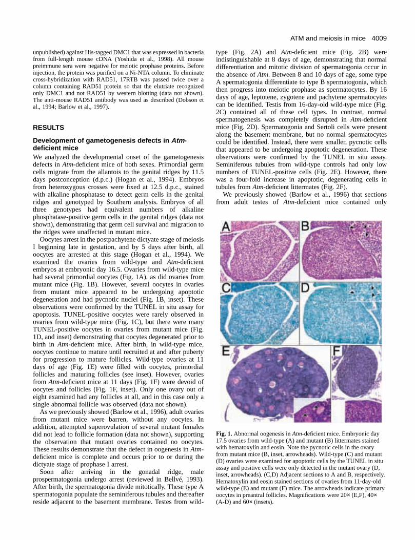
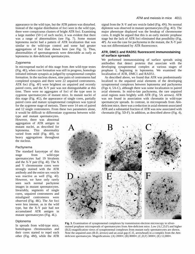
observed (data not shown). In microspreads from Atm-deficientmice, however, normal prophase I stages were rarely obser(Fig. 3). Defects in the axial elements were widespread mutant spreads (Fig. 3A,B), synaptonemal complexedisplayed abnormal, non-homologous pairing (Fig. 3C,Darrow), and there were small, paired and unpairesynaptonemal complex fragments observed in many sprea(Fig. 3B-D, arrows).
At higher magnification, normal chromosomal pairing ansynapsis occurred in some regions of synaptonemal complexbut extensive pairing abnormalities with discontinuities werobserved in several complexes (Fig. 3E, arrowhead). Theaxial gaps, occurring on a single axis of the pairesynaptonemal complex, were often found and weapproximately 10-25% of the length of the complex. Axiagaps were found located in the middle or end of thsynaptonemal complex. In microspreads that containcomplexes with axial gaps, fragments of what appeared to a single axis and fragmented termini were often observed (dnot shown). These abnormalities were not observed spermatocytes from wild-type mice (data not shown).
In many spreads from mutant mice, fragmentation was moextensive, resulting in complete disruption of all synaptonemcomplexes (Fig. 3F). It is not clear whether the fragmentecomplexes represented apoptotic cells, or a stage intermedbetween axial gap formation and apoptosis. In either case,normal spermatocyte spreads were observed in the mutant mafter axial pairing. These results demonstrate that the lossAtm results in a severe and complete disruption of meiosisand the end result is chromosomal fragmentation and apopto
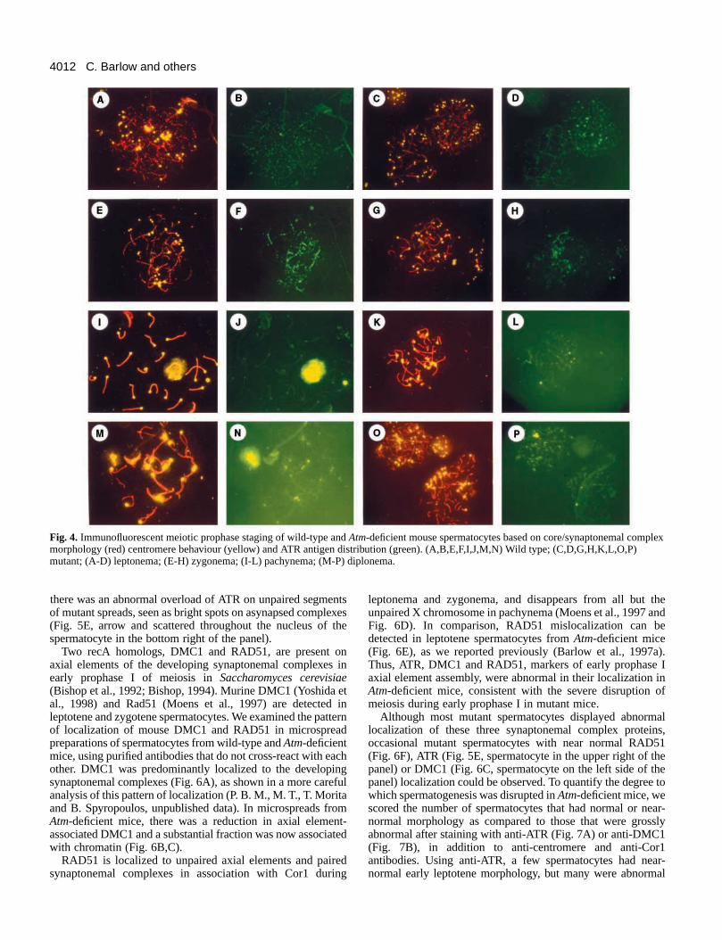
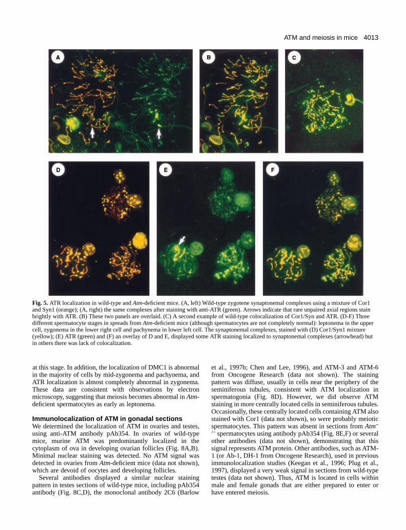
Prophase I disruption in Atm -deficientspermatocytesTo define the meiotic defect in Atm-deficient mice moreprecisely, we compared meiotic prophase stages microspreads of spermatocytes from wild-type and Atm mutantlittermates immunostained for chromosome cores, centromeand ATR protein, a closely related family member of ATM (Fig4). Staging was based on three criteria: traditional meiochromosome morphology; centromere configuration (singversus paired); and ATR immunofluorescent foci patterns. agreement with previously published data (Keegan et al., 199we found that ATR was predominantly localized to the unpaireaxial elements of the developing synaptonemal complexbetween leptonema and pachynema (Figs 4, 5A-C), althouthere was some localization to paired axial elements. In mid-late pachynema, the rare unpaired axial regions stain brighwith ATR (Fig. 5A, arrows). ATR was not found in associationwith chromatin in wild-type spermatocyte spreads. Thus, ATis a marker of early prophase I axial element assembly.
LeptonemaShort segments of chromosome cores have started to formmicrospread nuclei of this stage from wild-type testes (Fig. 4AThere was no evidence for synapsis and there were 40 brifluorescent centromeres. ATR foci were evenly distributed ovthe chromosome core segments (Fig. 4B). The sperm tails anuclei in the background serve as evidence that spermatogenwas efficient in this wild-type mouse. In leptotene nuclei froman Atm-deficient littermate (Fig. 4C,D), the short chromosomcore segments and the 40 single centromeres were simila
ay-
the
),
4011ATM and meiosis in mice
lhee
se.air
ge
ofe
lygmad
ed
tedth),
ptonemal complexes by transmission electron microscopy in silver-ads of spermatocytes from Atm-deficient mice. Low (A,C,D,F) and higher
f synaptonemal complexes from mutant early spermatocytes are shown.D, arrows) and an axial gap (C-E, arrowhead) in a complex from the Atm-gnifications: (A) 2000×; (B) 8000×; (C,D,F) 3000×; (E) 12,000×.
appearance to the wild type, but the ATR pattern was disturbInstead of the regular distribution of foci seen in the wild typthere were conspicuous clusters of bright ATR foci. Examinia large number (50+) of such nuclei, it was evident that thwas a range of abnormalities (see Fig. 7). Some mutleptonema nuclei had a pattern of ATR localization that wsimilar to the wild-type control and some had greataggregations of foci than shown here (see Fig. 5). Thabnormalities of spermatogenesis were detectable as earlleptonema in Atm-deficient spermatocytes.
ZygonemaIn microspread nuclei of this stage from Atm wild-type testes(Fig. 4E), when core formation was still in progress, homoloinitiated intimate synapsis as judged by synaptonemal compformation. In the nucleus shown, nine pairs of centromeres hcompleted synapsis and there were 22 unpaired centromeATR foci (Fig. 4F) were brightest on unpaired and recentpaired cores, and the X-Y pair was not distinguishable at ttime. There were no aggregates of foci of the type seenzygotene spermatocytes of mutant mice. In mutant nucleithis stage (Fig. 4G), the appearance of single cores, partipaired cores and mature synaptonemal complexes was typfor the zygotene stage of meiosis. There were 14 sets of paand 12 single centromeres. From these two parameters alit would be difficult to differentiate zygonema between wildtype and mutant spermatocytes.However, there was abnormalaggregation of ATR antigen inmutant nuclei, as described forleptonema. This abnormalityvaried from mild (Fig. 4H), toheavy aggregations throughoutthe nucleus.
PachynemaThe standard karyotype of thisstage from wild-typespermatocytes had 19 bivalentsand the X-Y pair (Fig. 4I). The Xand Y chromosome cores werestrongly stained with the ATRantibody and the entire sex vesiclewas reactive as well (Fig. 4J).However, we have only rarelyseen such normal pachyteneimages in mutant spermatocytes.Invariably, segments of singlecores, unpaired centromeres andmisaligned centromeres wereobserved (Fig. 4K). The Atr fociwere less intense, as in the wildtype, but the X-Y pair had notaccumulated ATR antigen inmutant spermatocytes (Fig. 4L).
DiplonemaIn spreads from wild-type mice,homologous chromosomes andtheir cores started to repel eachother (Fig. 4M), while the ATR
Fig. 3.Examination of synastained prophase microspre(B,E) magnification views oNote the unpaired axes (B-deficient spermatocyte. Ma
ed.e,ngereantaserus,y as
gslexadres.lyhis in ofallyicaliredone,-
signal from the X-Y and sex vesicle faded (Fig. 4N). No normadiplotene was observed in mutant spermatocytes (Fig. 4O). Tmajor phenotype displayed was the breakup of chromosomcores. It might be argued that this is an early meiotic prophastage but the lack of ATR foci eliminated that possibility (Fig4P). As was the case for pachynema in the mutant, the X-Y pwas not differentiated by ATR fluorescence.
ATR, DMC1 and RAD51 fluorescent immunostainingof surface spreadsWe performed immunostaining of surface spreads usinantibodies that detect proteins that associate with thdeveloping synaptonemal complex at various stages prophase I, beginning in leptonema. We examined thlocalization of ATR, DMC1 and RAD51.
As described above, we found that ATR was predominantlocalized to the unpaired axial elements of the developinsynaptonemal complexes between leptonema and pachyne(Figs 4, 5A-C), although there was some localization to paireaxial elements. In mid-to-late pachynema, the rare unpairaxial regions stain brightly with ATR (Fig. 5A arrows). ATRwas not found in association with chromatin in wild-typespermatocyte spreads. In contrast, in microspreads from Atm-deficient mice, there was a reduction in axial element-associaATR and a substantial fraction of ATR was now associated wichromatin (Fig. 5D-F). In addition, as described above (Fig. 4
4012
thend
e
). I inof
als,
51e
he to
wear-
sly
1ar-al
C. Barlow and others
Fig. 4. Immunofluorescent meiotic prophase staging of wild-type and Atm-deficient mouse spermatocytes based on core/synaptonemal complexmorphology (red) centromere behaviour (yellow) and ATR antigen distribution (green). (A,B,E,F,I,J,M,N) Wild type; (C,D,G,H,K,L,O,P)mutant; (A-D) leptonema; (E-H) zygonema; (I-L) pachynema; (M-P) diplonema.
there was an abnormal overload of ATR on unpaired segmeof mutant spreads, seen as bright spots on asynapsed comp(Fig. 5E, arrow and scattered throughout the nucleus of spermatocyte in the bottom right of the panel).
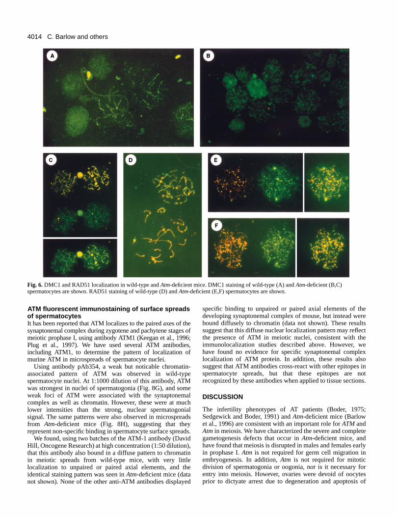
Two recA homologs, DMC1 and RAD51, are present oaxial elements of the developing synaptonemal complexesearly prophase I of meiosis in Saccharomyces cerevisiae(Bishop et al., 1992; Bishop, 1994). Murine DMC1 (Yoshida al., 1998) and Rad51 (Moens et al., 1997) are detectedleptotene and zygotene spermatocytes. We examined the paof localization of mouse DMC1 and RAD51 in microspreapreparations of spermatocytes from wild-type and Atm-deficientmice, using purified antibodies that do not cross-react with eother. DMC1 was predominantly localized to the developinsynaptonemal complexes (Fig. 6A), as shown in a more caranalysis of this pattern of localization (P. B. M., M. T., T. Moritand B. Spyropoulos, unpublished data). In microspreads frAtm-deficient mice, there was a reduction in axial elemeassociated DMC1 and a substantial fraction was now associwith chromatin (Fig. 6B,C).
RAD51 is localized to unpaired axial elements and pairsynaptonemal complexes in association with Cor1 duri
ntslexesthe
n in
et intternd
achg
efulaomnt-ated
edng
leptonema and zygonema, and disappears from all but unpaired X chromosome in pachynema (Moens et al., 1997 aFig. 6D). In comparison, RAD51 mislocalization can bdetected in leptotene spermatocytes from Atm-deficient mice(Fig. 6E), as we reported previously (Barlow et al., 1997aThus, ATR, DMC1 and RAD51, markers of early prophaseaxial element assembly, were abnormal in their localizationAtm-deficient mice, consistent with the severe disruption meiosis during early prophase I in mutant mice.
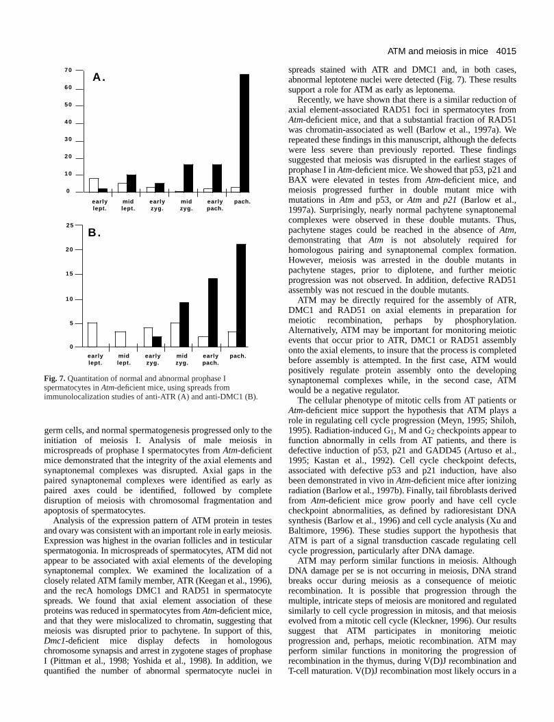
Although most mutant spermatocytes displayed abnormlocalization of these three synaptonemal complex proteinoccasional mutant spermatocytes with near normal RAD(Fig. 6F), ATR (Fig. 5E, spermatocyte in the upper right of thpanel) or DMC1 (Fig. 6C, spermatocyte on the left side of tpanel) localization could be observed. To quantify the degreewhich spermatogenesis was disrupted in Atm-deficient mice,scored the number of spermatocytes that had normal or nenormal morphology as compared to those that were grosabnormal after staining with anti-ATR (Fig. 7A) or anti-DMC1(Fig. 7B), in addition to anti-centromere and anti-Corantibodies. Using anti-ATR, a few spermatocytes had nenormal early leptotene morphology, but many were abnorm
4013ATM and meiosis in mice
ge
.
c
lis-s.,
or
Fig. 5.ATR localization in wild-type and Atm-deficient mice. (A, left) Wild-type zygotene synaptonemal complexes using a mixture of Cor1and Syn1 (orange); (A, right) the same complexes after staining with anti-ATR (green). Arrows indicate that rare unpaired axial regions stainbrightly with ATR. (B) These two panels are overlaid. (C) A second example of wild-type colocalization of Cor1/Syn and ATR. (D-F) Threedifferent spermatocyte stages in spreads from Atm-deficient mice (although spermatocytes are not completely normal): leptonema in the uppercell, zygonema in the lower right cell and pachynema in lower left cell. The synaptonemal complexes, stained with (D) Cor1/Syn1 mixture(yellow); (E) ATR (green) and (F) an overlay of D and E, displayed some ATR staining localized to synaptonemal complexes (arrowhead) butin others there was lack of colocalization.
at this stage. In addition, the localization of DMC1 is abnormin the majority of cells by mid-zygonema and pachynema, aATR localization is almost completely abnormal in zygonemThese data are consistent with observations by electmicroscopy, suggesting that meiosis becomes abnormal in Atm-deficient spermatocytes as early as leptonema.
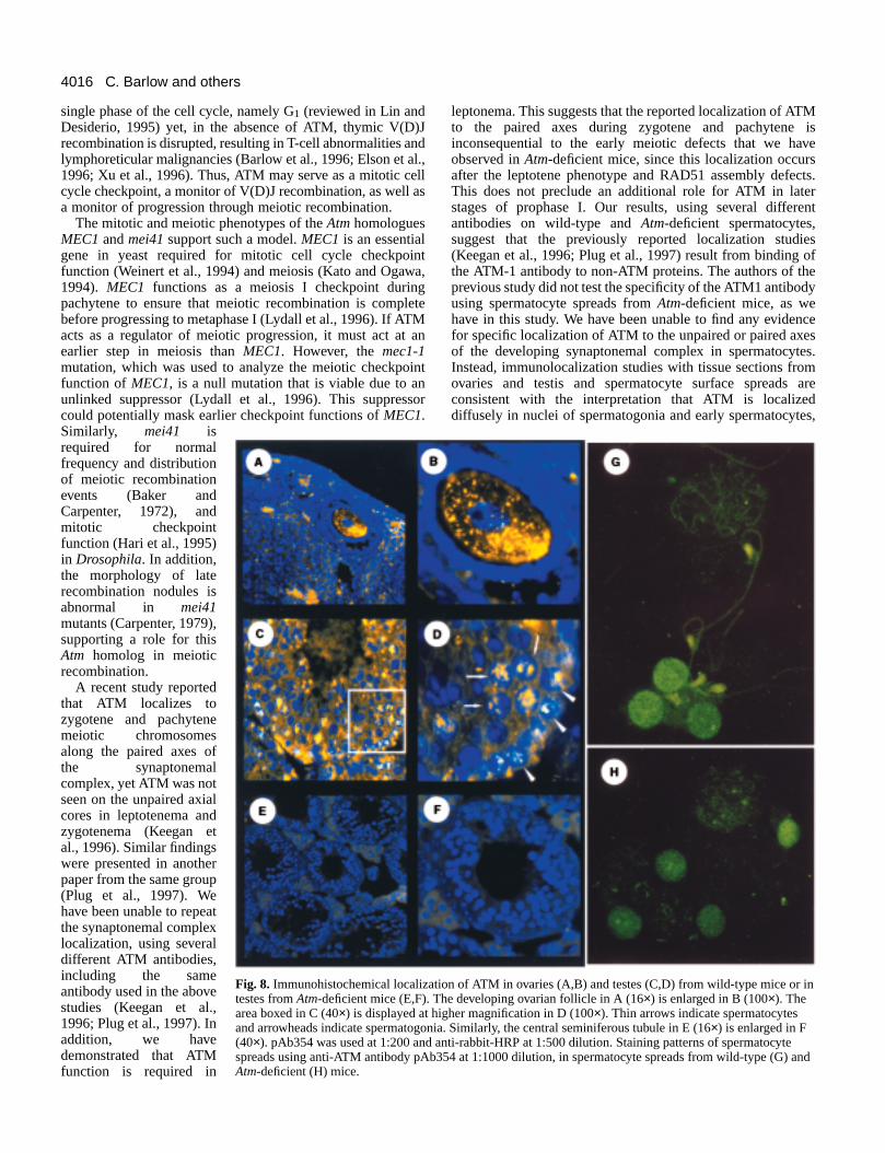
Immunolocalization of ATM in gonadal sectionsWe determined the localization of ATM in ovaries and testeusing anti-ATM antibody pAb354. In ovaries of wild-typemice, murine ATM was predominantly localized in thcytoplasm of ova in developing ovarian follicles (Fig. 8A,BMinimal nuclear staining was detected. No ATM signal wadetected in ovaries from Atm-deficient mice (data not shown)which are devoid of oocytes and developing follicles.
Several antibodies displayed a similar nuclear stainipattern in testes sections of wild-type mice, including pAb3antibody (Fig. 8C,D), the monoclonal antibody 2C6 (Barlo
alnda.ron
s,
e).s
,
ng54w
et al., 1997b; Chen and Lee, 1996), and ATM-3 and ATM-6from Oncogene Research (data not shown). The staininpattern was diffuse, usually in cells near the periphery of thseminiferous tubules, consistent with ATM localization inspermatogonia (Fig. 8D). However, we did observe ATMstaining in more centrally located cells in seminiferous tubulesOccasionally, these centrally located cells containing ATM alsostained with Cor1 (data not shown), so were probably meiotispermatocytes. This pattern was absent in sections from Atm−/− spermatocytes using antibody pAb354 (Fig. 8E,F) or severaother antibodies (data not shown), demonstrating that thsignal represents ATM protein. Other antibodies, such as ATM1 (or Ab-1, DH-1 from Oncogene Research), used in previouimmunolocalization studies (Keegan et al., 1996; Plug et al1997), displayed a very weak signal in sections from wild-typetestes (data not shown). Thus, ATM is located in cells withinmale and female gonads that are either prepared to enter have entered meiosis.
4014
eretsct
ex
inot
ns.
te
rly
rsof
C. Barlow and others
Fig. 6.DMC1 and RAD51 localization in wild-type and Atm-deficient mice. DMC1 staining of wild-type (A) and Atm-deficient (B,C)spermatocytes are shown. RAD51 staining of wild-type (D) and Atm-deficient (E,F) spermatocytes are shown.
ATM fluorescent immunostaining of surface spreadsof spermatocytesIt has been reported that ATM localizes to the paired axes ofsynaptonemal complex during zygotene and pachytene stagmeiotic prophase I, using antibody ATM1 (Keegan et al., 199Plug et al., 1997). We have used several ATM antibodiincluding ATM1, to determine the pattern of localization omurine ATM in microspreads of spermatocyte nuclei.
Using antibody pAb354, a weak but noticable chromatassociated pattern of ATM was observed in wild-typspermatocyte nuclei. At 1:1000 dilution of this antibody, ATMwas strongest in nuclei of spermatogonia (Fig. 8G), and soweak foci of ATM were associated with the synaptonemcomplex as well as chromatin. However, these were at mlower intensities than the strong, nuclear spermatogonsignal. The same patterns were also observed in microsprfrom Atm-deficient mice (Fig. 8H), suggesting that therepresent non-specific binding in spermatocyte surface spre
We found, using two batches of the ATM-1 antibody (DavHill, Oncogene Research) at high concentration (1:50 dilutiothat this antibody also bound in a diffuse pattern to chromain meiotic spreads from wild-type mice, with very littlelocalization to unpaired or paired axial elements, and identical staining pattern was seen in Atm-deficient mice (datanot shown). None of the other anti-ATM antibodies display
thees of6;
es,f
in-e
meal
uchial
eadsyads.idn),tin
the
ed
specific binding to unpaired or paired axial elements of thdeveloping synaptonemal complex of mouse, but instead webound diffusely to chromatin (data not shown). These resulsuggest that this diffuse nuclear localization pattern may reflethe presence of ATM in meiotic nuclei, consistent with theimmunolocalization studies described above. However, whave found no evidence for specific synaptonemal complelocalization of ATM protein. In addition, these results alsosuggest that ATM antibodies cross-react with other epitopes spermatocyte spreads, but that these epitopes are nrecognized by these antibodies when applied to tissue sectio
DISCUSSION
The infertility phenotypes of AT patients (Boder, 1975;Sedgewick and Boder, 1991) and Atm-deficient mice (Barlowet al., 1996) are consistent with an important role for ATM andAtmin meiosis. We have characterized the severe and complegametogenesis defects that occur in Atm-deficient mice, andhave found that meiosis is disrupted in males and females eain prophase I. Atm is not required for germ cell migration inembryogenesis. In addition, Atm is not required for mitoticdivision of spermatogonia or oogonia, nor is it necessary foentry into meiosis. However, ovaries were devoid of oocyteprior to dictyate arrest due to degeneration and apoptosis
4015ATM and meiosis in mice
s,ults
ofm1e
ctsgs
s ofd
h
alus,
n.in
tic1
,r.
ytedldgM
ra,
s.,s,so
d
Adatll
dtice
ediss
yfda
25
20
15
10
5
0
earlylept.
midlept.
earlyzyg.
midzyg.
earlypach.
pach.
earlylept.
midlept.
earlyzyg.
midzyg.
earlypach.
pach.
70
60
50
40
30
20
10
0
A .
B.
Fig. 7.Quantitation of normal and abnormal prophase Ispermatocytes in Atm-deficient mice, using spreads fromimmunolocalization studies of anti-ATR (A) and anti-DMC1 (B).
germ cells, and normal spermatogenesis progressed only toinitiation of meiosis I. Analysis of male meiosis inmicrospreads of prophase I spermatocytes from Atm-deficientmice demonstrated that the integrity of the axial elements asynaptonemal complexes was disrupted. Axial gaps in paired synaptonemal complexes were identified as earlypaired axes could be identified, followed by compledisruption of meiosis with chromosomal fragmentation aapoptosis of spermatocytes.
Analysis of the expression pattern of ATM protein in testand ovary was consistent with an important role in early meioExpression was highest in the ovarian follicles and in testicuspermatogonia. In microspreads of spermatocytes, ATM did appear to be associated with axial elements of the developsynaptonemal complex. We examined the localization ofclosely related ATM family member, ATR (Keegan et al., 1996and the recA homologs DMC1 and RAD51 in spermatocyspreads. We found that axial element association of thproteins was reduced in spermatocytes from Atm-deficient mice,and that they were mislocalized to chromatin, suggesting tmeiosis was disrupted prior to pachytene. In support of thDmc1-deficient mice display defects in homologouchromosome synapsis and arrest in zygotene stages of propI (Pittman et al., 1998; Yoshida et al., 1998). In addition, wquantified the number of abnormal spermatocyte nuclei
the
ndthe astend
essis.larnoting
a),teese
hatis,shaseein
spreads stained with ATR and DMC1 and, in both caseabnormal leptotene nuclei were detected (Fig. 7). These ressupport a role for ATM as early as leptonema.
Recently, we have shown that there is a similar reduction axial element-associated RAD51 foci in spermatocytes froAtm-deficient mice, and that a substantial fraction of RAD5was chromatin-associated as well (Barlow et al., 1997a). Wrepeated these findings in this manuscript, although the defewere less severe than previously reported. These findinsuggested that meiosis was disrupted in the earliest stageprophase I in Atm-deficient mice. We showed that p53, p21 anBAX were elevated in testes from Atm-deficient mice, andmeiosis progressed further in double mutant mice witmutations in Atmand p53, or Atmand p21 (Barlow et al.,1997a). Surprisingly, nearly normal pachytene synaptonemcomplexes were observed in these double mutants. Thpachytene stages could be reached in the absence of Atm,demonstrating that Atmis not absolutely required forhomologous pairing and synaptonemal complex formatioHowever, meiosis was arrested in the double mutants pachytene stages, prior to diplotene, and further meioprogression was not observed. In addition, defective RAD5assembly was not rescued in the double mutants.
ATM may be directly required for the assembly of ATRDMC1 and RAD51 on axial elements in preparation fomeiotic recombination, perhaps by phosphorylationAlternatively, ATM may be important for monitoring meioticevents that occur prior to ATR, DMC1 or RAD51 assemblonto the axial elements, to insure that the process is complebefore assembly is attempted. In the first case, ATM woupositively regulate protein assembly onto the developinsynaptonemal complexes while, in the second case, ATwould be a negative regulator.
The cellular phenotype of mitotic cells from AT patients oAtm-deficient mice support the hypothesis that ATM plays role in regulating cell cycle progression (Meyn, 1995; Shiloh1995). Radiation-induced G1, M and G2 checkpoints appear tofunction abnormally in cells from AT patients, and there idefective induction of p53, p21 and GADD45 (Artuso et al1995; Kastan et al., 1992). Cell cycle checkpoint defectassociated with defective p53 and p21 induction, have albeen demonstrated in vivo in Atm-deficient mice after ionizingradiation (Barlow et al., 1997b). Finally, tail fibroblasts derivefrom Atm-deficient mice grow poorly and have cell cyclecheckpoint abnormalities, as defined by radioresistant DNsynthesis (Barlow et al., 1996) and cell cycle analysis (Xu anBaltimore, 1996). These studies support the hypothesis thATM is part of a signal transduction cascade regulating cecycle progression, particularly after DNA damage.
ATM may perform similar functions in meiosis. AlthoughDNA damage per se is not occurring in meiosis, DNA stranbreaks occur during meiosis as a consequence of meiorecombination. It is possible that progression through thmultiple, intricate steps of meiosis are monitored and regulatsimilarly to cell cycle progression in mitosis, and that meiosevolved from a mitotic cell cycle (Kleckner, 1996). Our resultsuggest that ATM participates in monitoring meioticprogression and, perhaps, meiotic recombination. ATM maperform similar functions in monitoring the progression orecombination in the thymus, during V(D)J recombination anT-cell maturation. V(D)J recombination most likely occurs in
4016
Misests.
nt
sf
ce
s.
are
s,
C. Barlow and others
ation of ATM in ovaries (A,B) and testes (C,D) from wild-type mice or in The developing ovarian follicle in A (16×) is enlarged in B (100×). Thehigher magnification in D (100×). Thin arrows indicate spermatocytesonia. Similarly, the central seminiferous tubule in E (16×) is enlarged in Fd anti-rabbit-HRP at 1:500 dilution. Staining patterns of spermatocyteb354 at 1:1000 dilution, in spermatocyte spreads from wild-type (G) and
single phase of the cell cycle, namely G1 (reviewed in Lin andDesiderio, 1995) yet, in the absence of ATM, thymic V(Drecombination is disrupted, resulting in T-cell abnormalities alymphoreticular malignancies (Barlow et al., 1996; Elson et a1996; Xu et al., 1996). Thus, ATM may serve as a mitotic ccycle checkpoint, a monitor of V(D)J recombination, as well a monitor of progression through meiotic recombination.
The mitotic and meiotic phenotypes of the AtmhomologuesMEC1and mei41support such a model. MEC1is an essentialgene in yeast required for mitotic cell cycle checkpoifunction (Weinert et al., 1994) and meiosis (Kato and Ogaw1994). MEC1 functions as a meiosis I checkpoint durinpachytene to ensure that meiotic recombination is complbefore progressing to metaphase I (Lydall et al., 1996). If ATacts as a regulator of meiotic progression, it must act atearlier step in meiosis than MEC1. However, the mec1-1mutation, which was used to analyze the meiotic checkpofunction of MEC1, is a null mutation that is viable due to aunlinked suppressor (Lydall et al., 1996). This suppresscould potentially mask earlier checkpoint functions of MEC1.Similarly, mei41 isrequired for normalfrequency and distributionof meiotic recombinationevents (Baker andCarpenter, 1972), andmitotic checkpointfunction (Hari et al., 1995)in Drosophila. In addition,the morphology of laterecombination nodules isabnormal in mei41mutants (Carpenter, 1979),supporting a role for thisAtm homolog in meioticrecombination.
A recent study reportedthat ATM localizes tozygotene and pachytenemeiotic chromosomesalong the paired axes ofthe synaptonemalcomplex, yet ATM was notseen on the unpaired axialcores in leptotenema andzygotenema (Keegan etal., 1996). Similar findingswere presented in anotherpaper from the same group(Plug et al., 1997). Wehave been unable to repeatthe synaptonemal complexlocalization, using severaldifferent ATM antibodies,including the sameantibody used in the abovestudies (Keegan et al.,1996; Plug et al., 1997). Inaddition, we havedemonstrated that ATMfunction is required in
Fig. 8. Immunohistochemical localiztestes from Atm-deficient mice (E,F).area boxed in C (40×) is displayed at and arrowheads indicate spermatog(40×). pAb354 was used at 1:200 anspreads using anti-ATM antibody pAAtm-deficient (H) mice.
)Jndl.,
ellas
nta,
geteM an
intnor
leptonema. This suggests that the reported localization of ATto the paired axes during zygotene and pachytene inconsequential to the early meiotic defects that we havobserved in Atm-deficient mice, since this localization occurafter the leptotene phenotype and RAD51 assembly defecThis does not preclude an additional role for ATM in laterstages of prophase I. Our results, using several differeantibodies on wild-type and Atm-deficient spermatocytes,suggest that the previously reported localization studie(Keegan et al., 1996; Plug et al., 1997) result from binding othe ATM-1 antibody to non-ATM proteins. The authors of theprevious study did not test the specificity of the ATM1 antibodyusing spermatocyte spreads from Atm-deficient mice, as wehave in this study. We have been unable to find any evidenfor specific localization of ATM to the unpaired or paired axesof the developing synaptonemal complex in spermatocyteInstead, immunolocalization studies with tissue sections fromovaries and testis and spermatocyte surface spreads consistent with the interpretation that ATM is localizeddiffusely in nuclei of spermatogonia and early spermatocyte
4017ATM and meiosis in mice
l
s
s
g
l
A
a
v.
.
r.
ion
ts,
r
similar to the pattern observed in mitotic cells (Brown et a1997; Chen and Lee, 1996; Lakin et al., 1996).
The understanding of the mechanisms that control the eaevents of mammalian meiosis, especially the early stagesaxial core formation, synaptonemal complex pairing anmeiotic recombination, will likely result from the geneticanalysis of mutants with phenotypes during these early eveThe severe, early and profound defects in meiosis that we hdescribed for the Atm-deficient mouse demonstrate that Atmplays a crucial role during prophase I. Thus, the Atm-deficientmice provide an excellent tool that can be used to further dissearly events in meiotic prophase I and meiotic recombinatio
We would like to thank Matthew Gonda for assistance with eaelectron microscopy experiments, Doug Bishop for insight inmeiosis and comments on the manuscript, Mike Dresser for helpdiscussions about EM analysis of meiosis, Akira Shinohara for Rad51 antibody, Darryl Leja for assistance in figure preparatioMona Schröder, Denise Larson, and Lisa Garrett for excelletechnical assistance, and Robert Nussbaum for generous supporB. is a Clinical Endocrine Fellow supported by the National Instituof Diabetes, Digestive, and Kidney Diseases, and P. B. M. wfinancially supported by the National Research Council of Canada
REFERENCES
Artuso, M., Esteve, A., Bresil, H., Vuillaume, M. and Hall, J.(1995). Therole of the Ataxia telangiectasia gene in the p53, WAF1/CIP1(p21)- aGADD45-mediated response to DNA damage produced by ionisiradiation. Oncogene11, 1427-1435.
Baker, B. S. and Carpenter, A. T. C.(1972). Genetic analysis of sexchromosomal meiotic mutants in Drosophila melanogaster. Genetics90,255-286.
Barlow, C., Hirotsune, S., Paylor, R., Liyanage, M., Eckhaus, M., Collins,F., Shiloh, Y., Crawley, J. N., Ried, T., Tagle, D. and Wynshaw-Boris, A.(1996). Atm-deficient mice: a paradigm of ataxia telangiectasia. Cell 86,159-171.
Barlow, C., Liyanage, M., Moens, P. B., Deng, C.-X., Ried, T. and Wynshaw-Boris, A. (1997a). Partial rescue of the severe prophase I defects of Adeficient mice by p53 and p21 null alleles. Nature Genet.17, 462-466.
Barlow, C., Brown, K. D., Deng, C.-X., Tagle, D. A. and Wynshaw-Boris,A. (1997b). Atm selectively regulates distinct p53-dependent cell cyccheckpoint and apoptotic pathways. Nature Genet.17, 453-456.
Bellvé, A. R. (1993) Purification, culture and fractionation of spermatogencells. Meth. Enzymol. 225, 84-113.
Bishop, D., K,, Park, D., Xu, L. and Kleckner, N.(1992). DMC1: a meiosis-specific yeast homolog of E.coli recA required for recombination,synaptonemal complex formation, and cell cycle progression. Cell 69, 439-456.
Bishop, D. K. (1994). RecA homologs Dmc1 and Rad51 interact to formultiple nuclear complexes prior to meiotic chromosome synapsis. Cell 79,1081-1092.
Boder, E. (1975). Ataxia-telangiectasia: some historic, clinical and pathologobservations. Birth Defects11, 255-270.
Brown, K. D., Ziv, Y., Sadanandan, S. N., Chessa, L., Collins, F. S., Shiloh,Y. and Tagle, D. A. (1997). The ataxia-telangiectasia gene product, constitutively expressed nuclear protein that is not upregulated followigenome damage. Proc. Natl. Acad. Sci., USA94, 1840-1845.
Carpenter, A. T. C. (1979). Recombination nodules and synaptonemcomplex in recombination-defective females of Drosophila melanogasChromosoma75, 259-292.
Chen, G. and Lee, E. Y. H.-P.(1996). The product of the ATM gene is a 370kDa nuclear phosphoprotein. J. Biol. Chem.271, 33693-7.
Dobson, M. J., Pearlman, R. E., Karaiskakis, A., Spyropoulos, B. andMoens, P. B.(1994). Synaptonemal complex proteins: occurrence, epitomapping and chromosome disjunction. J. Cell Sci.107, 2749-2760.
Dresser, M. E. and Moses, M. J.(1979). Silver staining of synaptonemalcomplexes in surface spreads for light and electron microscopy. Exp. CellRes.121, 416-419.
Elson, A., Wang, Y., Daugherty, C. J., Morton, C. C., Zhou, F., Campos-
l.,
rly ofd
nts.ave
ectn.
rlytoful
then,ntt. C.teas.
ndng
tm-
le
ic
m
ic
ang
alter.
-
pe
Torres, J. and Leder, P.(1996). Pleiotropic defects in ataxia-telangiectasiaprotein-deficient mice. Proc. Natl. Acad. Sci., USA93, 13084-13089.
Hari, K. L., Santerre, A., Sekelsky, J. J., McKim, K., Boyd, J. B. andHawley, R. S.(1995). The mei-41gene of D. melanogaster is a structuraand functional homolog of the human ataxia telangiectasia gene. Cell 82,815-821.
Harlow, E. and Lane, D. (1988). Antibodies: A Laboratory Manual. ColdSpring Harbor: Cold Spring Harbor Laboratory Press.
Hogan, B., Beddington, R., Costantini, F. and Lacy, E.(1994).Manipulating the MouseEmbryo: a Laboratory Manual.Second Edition.Cold Spring Harbor: Cold Spring Harbor Laboratory Press.
Kastan, M. B., Zhan, Q., el-Deiry, W. S., Carrier, F., Jacks, T., Walsh, W.V., Plunkett, B. S., Vogelstein, B. and Fornace, A. J., Jr.(1992). Amammalian cell cycle checkpoint pathway utilizing p53 and GADD45 idefective in ataxia-telangiectasia. Cell 71, 587-97.
Kato, R. and Ogawa, H.(1994). An essential gene, ESR1, is required formitotic growth, DNA repair and meiotic recombination in Saccharomycecerevisiae. Nucl. Acids Res.22, 3104-3112.
Keegan, K. S., Holtzman, D. A., Plug, A. W., Christenson, E. R., Brainerd,E. E., Flaggs, G., Bentley, N. J., Taylor, E. M., Meyn, M. S., Moss, S. B.,Carr, A. M., Ashley, T. and Hoekstra, M. F. (1996). The Atr and Atmprotein kinases associate with different sites along meiotically pairinchromosomes. Genes Dev.10, 2423-2437.
Kleckner, N. (1996). Meiosis: how could it work? Proc. Natl. Acad. Sci., USA93, 8167-8174.
Koehler, K. E., Hawley, R. S., Sherman, S. and Hassold, T.(1996).Recombination and non-disjunction in humans and flies. Human Mol.Genet. 5, 1495-1504.
Lakin, N. D., Weber, P., Stankovic, T., Rottinghaus, S. T., Taylor, A. M.and Jackson, S. P.(1996). Analysis of the ATM protein in wild-type andataxia telangiectasia cells. Oncogene13, 2707-16.
Lin, W.-C. and Desiderio, S.(1995). V(D)J recombination and the cell cycle.Immunol. Today16, 279-289.
Luna, L. G. (1992). Histopathological Methods and Color Atlas of SpeciaStains and Tissue. Gaithersburg: American Histolabs, Inc. PublicationsDivision.
Lydall, D., Nikolsky, Y., Bishop, D. K. and Weinert, T. (1996). A meioticrecombination checkpoint controlled by mitotic checkpoint genes. Nature383, 840-843.
Meyn, M. S. (1995). Ataxia-telangiectasia and cellular responses to DNdamage. Cancer Res55, 5991-6001.
Moens, P. B., Chen, D. J., Shen, Z., Kolas, N., Tarsounas, M., Heng, H. H.Q. and Spyropoulos, B.(1997) Rad51 immunocytology in rat and mousespermatocytes and oocytes. Chromosoma105, 207-215.
Orr-Weaver, T. L. (1995). Meiosis in Drosophila: seeing is believing. Proc.Natl. Acad. Sci., USA92, 10443-10449.
Pittman, D. L., Cobb, J., Schimenti, K. J., Wilson, L. A., Cooper, D. M.,Brignull, E., Handel, M. A. and Schimenti, J. C.(1998) Meiotic prophasearrest with failure of chromosome synapsis in mice deficient for Dmc1,germline-specific RecA homolog. Mol. Cell 1, 697-705.
Plug, A. W., Peters, A., Xu., Y., Keegan, K. S., Hoekstra, M. F., Baltimore,D., deBoer, P. and Ashley, T.(1997) ATM and RPA in meiotic chromosomesynapsis and recombination. Nat. Genet.17, 457-461.
Roeder, G. S.(1995). Sex and the single cell: meiosis in yeast. Proc. Natl.Acad. Sci., USA92, 10450-10456.
Roeder, G. S.(1997). Meiotic chromosomes: it takes two to tango. Genes De92, 10450-10456.
Sedgewick, R. and Boder, E. (1991). Ataxia-telangiectasia. In Handbook ofClinical Neurology, (ed. P. Vinken, G. Bruyn and H. Klawans) pp. 347-423New York: Elsevier Scientific Publishers.
Shiloh, Y. (1995). Ataxia-telangiectasia: closer to unraveling the mystery. EuJ. Hum. Genet. 3, 116-38.
Weinert, T. A., Kiser, G. L. and Hartwell, L. H. (1994). Mitotic checkpointgenes in budding yeast and the dependence of mitosis on DNA replicatand repair. Genes Dev.8, 652-665.
Xu, Y., Ashley, T., Brainerd, E. E., Bronson, R. T., Meyn, M. S. andBaltimore, D. (1996). Targeted disruption of ATM leads to growthretardation, chromosomal fragmentation during meiosis, immune defecand thymic lymphoma. Genes Dev.10, 2411-2422.
Xu, Y. and Baltimore, D. (1996). Dual roles of ATM in the cellular responseto irradiation and in cell growth. Genes Dev.10, 2401-2410.
Yoshida, K., Kondoh, G., Matsuda, Y., Habu, T., Nishimune, Y. andMorita, T. (1998) The mouse RecA-like gene Dmc1 is required fohomologous chromosome synapsis during meiosis. Mol. Cell. 1, 707-718.