Embed Size (px)
Citation preview
Atomic Force Microscopy-
Basics and Applications
Summer School June 2006„Complex Materials: Cooperative Projects of the
Natural, Engineering and Biosciences“
Astrid KronenbergerSchool of Engineering and Science
Outline
• Scanning Probe Microscopy
• Atomic Force Microscopy– General set-up & operation modes– Sample preparation
• Applications in life science– Imaging mode– Force-distance mode
• Conclusion

Scanning Probe Microscopy(SPM)
~1600 Light Microscope
1938: Transmission Electron Microscope
1964: Scanning Electron Microscope
1982: Scanning Tunneling Microscope
1984: Scanning Near-fieldOptical Microscope
1986: Atomic Force Microscope- magnetic force, lateral force, chemical force...
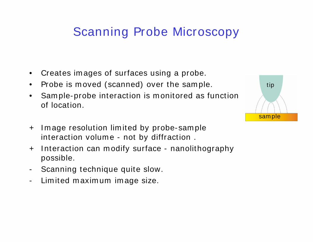
Scanning Probe Microscopy
• Creates images of surfaces using a probe.• Probe is moved (scanned) over the sample.• Sample-probe interaction is monitored as function
of location.
+ Image resolution limited by probe-sampleinteraction volume - not by diffraction .
+ Interaction can modify surface - nanolithographypossible.
- Scanning technique quite slow.- Limited maximum image size.
sample
tip
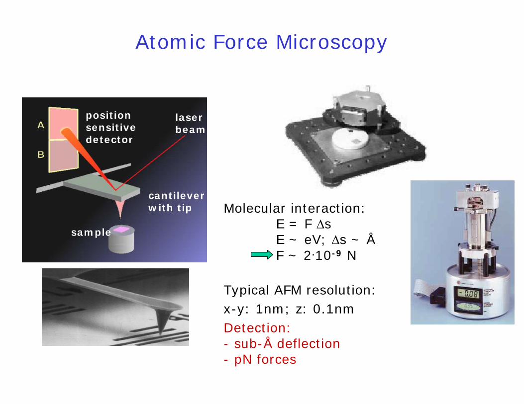
Atomic Force Microscopy
laser beam
positionsensitivedetector
sample
cantileverwith tip Molecular interaction:
E = F ΔsE ~ eV; Δs ~ ÅF ~ 2.10-9 N
Typical AFM resolution: x-y: 1nm; z: 0.1nmDetection:- sub-Å deflection - pN forces
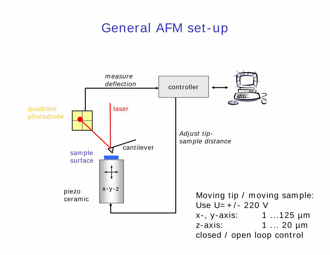
General AFM set-up
controller
piezoceramic
samplesurface
laser
cantilever
quadrantphotodiode
Moving tip / moving sample:Use U=+/- 220 V x-, y-axis: 1 ...125 µmz-axis: 1 ... 20 µm closed / open loop control
x-y-z
measuredeflection
Adjust tip-sample distance
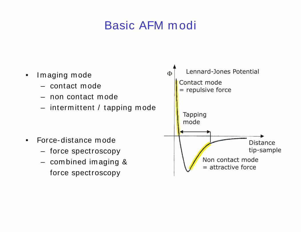
Basic AFM modi
• Imaging mode– contact mode– non contact mode– intermittent / tapping mode
• Force-distance mode– force spectroscopy– combined imaging &
force spectroscopy

• Contact mode: – tip in continuous contact with sample– preferably used for hard samples– imaging in air and liquid– high resolutiondetect: deflection
• Force spectroscopy mode: – consecutive cycles of tip approach and
retract– interaction forces between tip and sample
are recorded
F = - kspring. Δx
Static AFM modi
http://www.jpk.com/tutorial/tutorial1.htm

Dynamic AFM modi
• Intermittent/tapping mode: – oscillating cantilever, tip touching surface
gently and frequently– often used for biological samples– imaging in air and liquid– good resolution
• Non contact mode:– oscillating cantilever, tip not in contact with
sample– used for soft samples– imaging in vacuum– distance range 50Å - 150Å
detect: amplitudephasedeflection
http://www.jpk.com/tutorial/tutorial1.htm
effmkω =
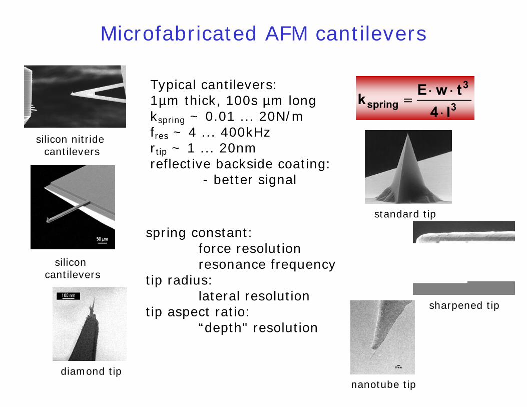
Microfabricated AFM cantilevers
standard tip
diamond tipnanotube tip
sharpened tip
silicon nitridecantilevers
siliconcantilevers
spring constant:force resolutionresonance frequency
tip radius:lateral resolution
tip aspect ratio:“depth" resolution
Typical cantilevers:1µm thick, 100s µm longkspring ~ 0.01 ... 20N/mfres ~ 4 ... 400kHzrtip ~ 1 ... 20nmreflective backside coating:
- better signal
3
3
spring l4twEk
⋅⋅⋅
=
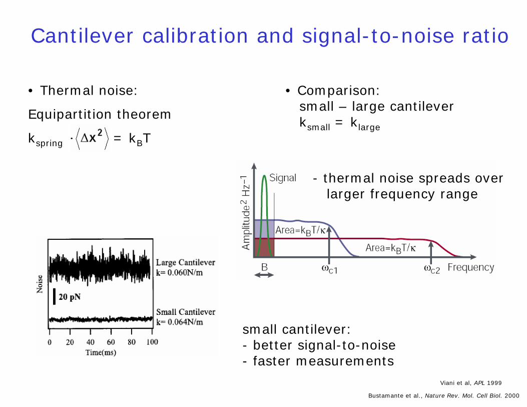
• Thermal noise:
Equipartition theorem
kspring = kBT
Cantilever calibration and signal-to-noise ratio
Bustamante et al., Nature Rev. Mol. Cell Biol. 2000
• Comparison:small – large cantileverksmall = klarge
- thermal noise spreads overlarger frequency range
Viani et al, APL 1999
small cantilever: - better signal-to-noise- faster measurements
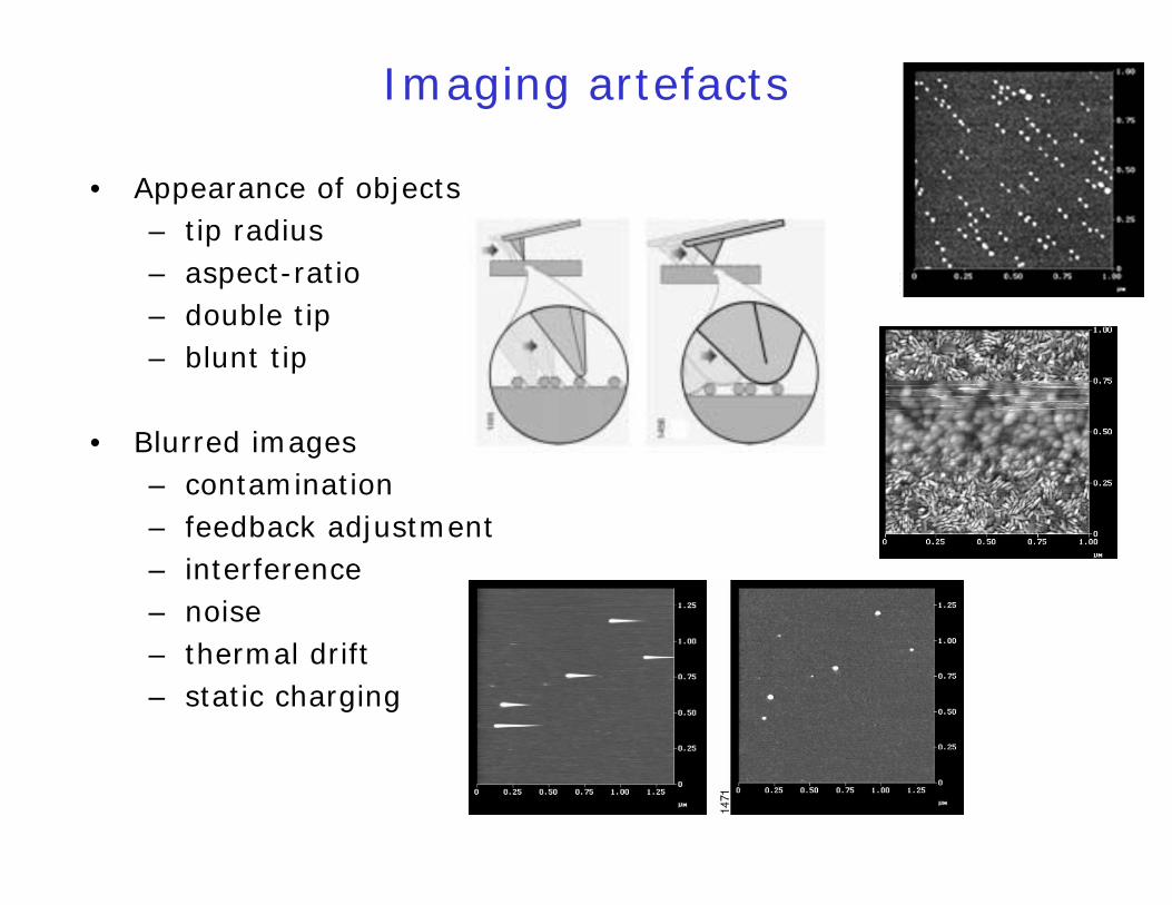
Imaging artefacts
• Appearance of objects– tip radius– aspect-ratio– double tip– blunt tip
• Blurred images– contamination– feedback adjustment– interference– noise– thermal drift– static charging
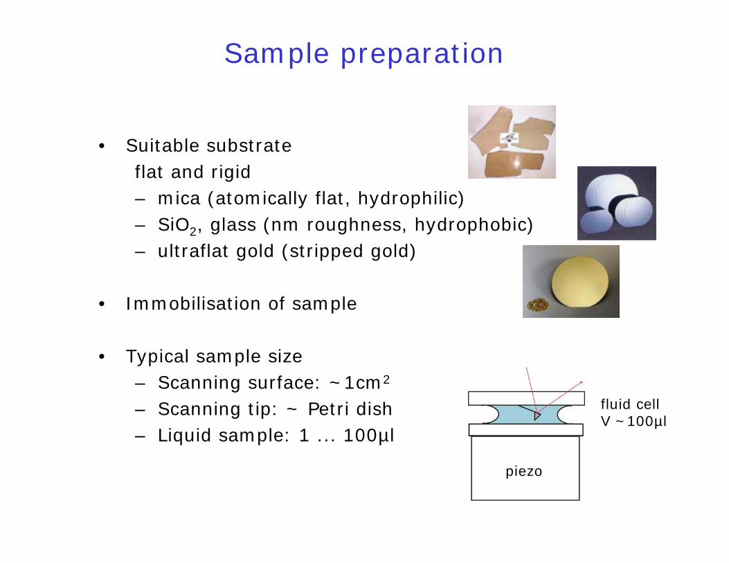
Sample preparation
• Suitable substrateflat and rigid– mica (atomically flat, hydrophilic)– SiO2, glass (nm roughness, hydrophobic)– ultraflat gold (stripped gold)
• Immobilisation of sample
• Typical sample size– Scanning surface: ~1cm2
– Scanning tip: ~ Petri dish– Liquid sample: 1 ... 100µl
piezo
fluid cellV ~100µl
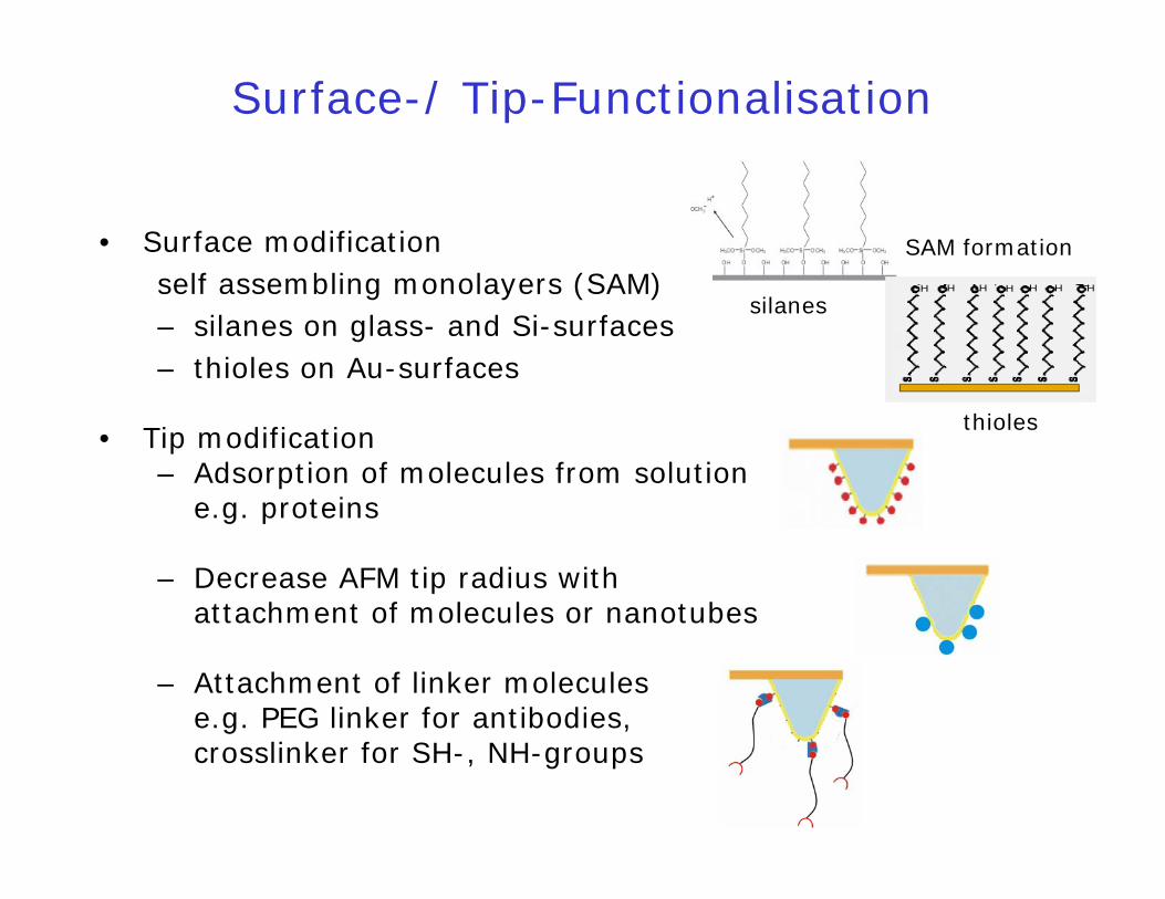
• Surface modificationself assembling monolayers (SAM)– silanes on glass- and Si-surfaces– thioles on Au-surfaces
• Tip modification– Adsorption of molecules from solution
e.g. proteins
– Decrease AFM tip radius withattachment of molecules or nanotubes
– Attachment of linker moleculese.g. PEG linker for antibodies, crosslinker for SH-, NH-groups
Surface-/ Tip-Functionalisation
H H H H H H H
SAM formation
silanes
thioles
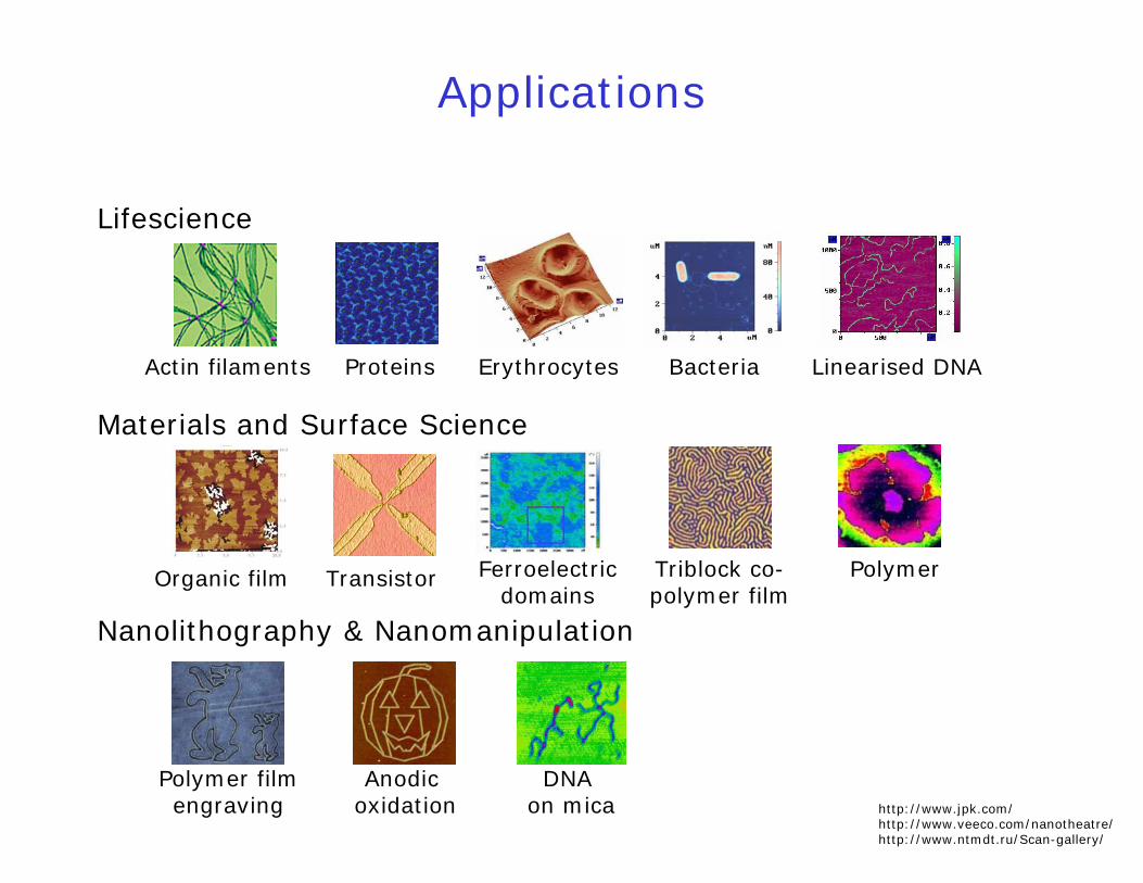
Applications
Lifescience
Materials and Surface Science
Nanolithography & Nanomanipulation
Polymer filmengraving
DNA on mica
ProteinsActin filaments Linearised DNABacteriaErythrocytes
PolymerOrganic film Transistor Ferroelectricdomains
Triblock co-polymer film
Anodicoxidation http://www.jpk.com/
http://www.veeco.com/nanotheatre/http://www.ntmdt.ru/Scan-gallery/
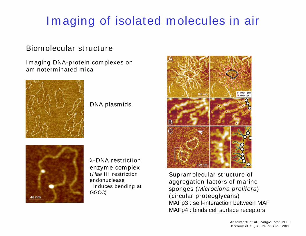
Imaging of isolated molecules in air
Biomolecular structure
Supramolecular structure ofaggregation factors of marinesponges (Microciona prolifera)(circular proteoglycans)MAFp3 : self-interaction between MAFMAFp4 : binds cell surface receptors
Imaging DNA-protein complexes on aminoterminated mica
λ-DNA restrictionenzyme complex(Hae III restrictionendonuclease
induces bending at GGCC)
DNA plasmids
Anselmetti et al., Single. Mol. 2000Jarchow et al., J. Struct. Biol. 2000
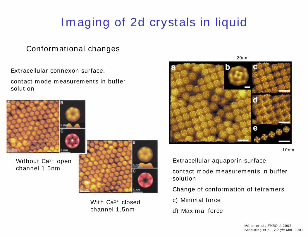
Imaging of 2d crystals in liquid
Conformational changes
10nm
20nm
Extracellular connexon surface.
contact mode measurements in buffersolution
Müller et al., EMBO J. 2002Scheuring et al., Single Mol. 2001
Extracellular aquaporin surface.
contact mode measurements in buffersolution
Change of conformation of tetramers
c) Minimal force
d) Maximal force
Without Ca2+ openchannel 1.5nm
With Ca2+ closedchannel 1.5nm

Imaging of cells and long term processes
Goldsbury et al., J. Mol. Biol. 1999
Time-lapse AFM for imaging growth of amyloid fibrils (synthetic human amylin)
Measuring the heartbeat ofsingle cells(chicken cardiomyocytes)
Radmacher, Uni Bremen

Forces in molecular biology
motor proteins
RNA polymerase
active forces
extractinglipids
DNA B-S transition
unfolding titindextranbond flip
extractingbacteriorhodopsin
elasticities
extracting forces
typ.at 0.1 - 5 nN/sec
10
50
1
100
500
1000
pN
selectins
biotin - avidin
covalent bond
antigen -antibody
rupture forces
unzipping DNAGC -AT -
opticaltweezers
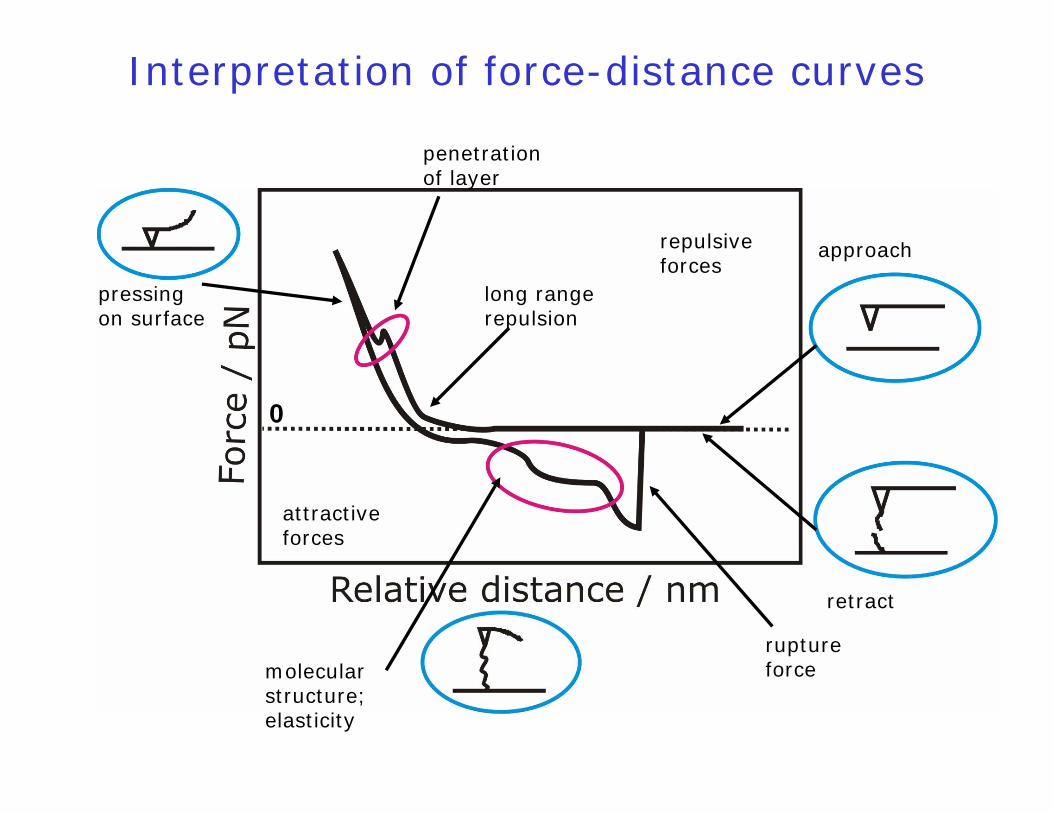
Interpretation of force-distance curves
long rangerepulsion
approach
retract
penetrationof layer
pressingon surface
repulsiveforces
molecularstructure;elasticity
attractiveforces
ruptureforce
0
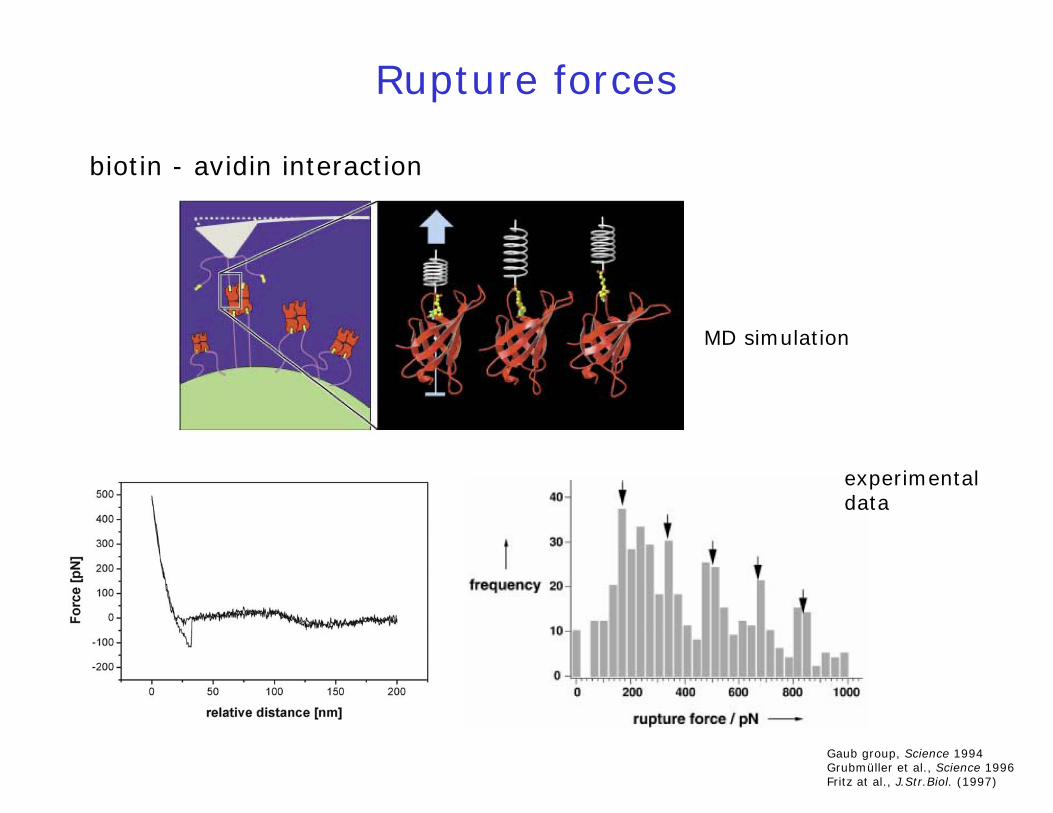
Rupture forces
Gaub group, Science 1994Grubmüller et al., Science 1996Fritz at al., J.Str.Biol. (1997)
biotin - avidin interaction
MD simulation
experimental data
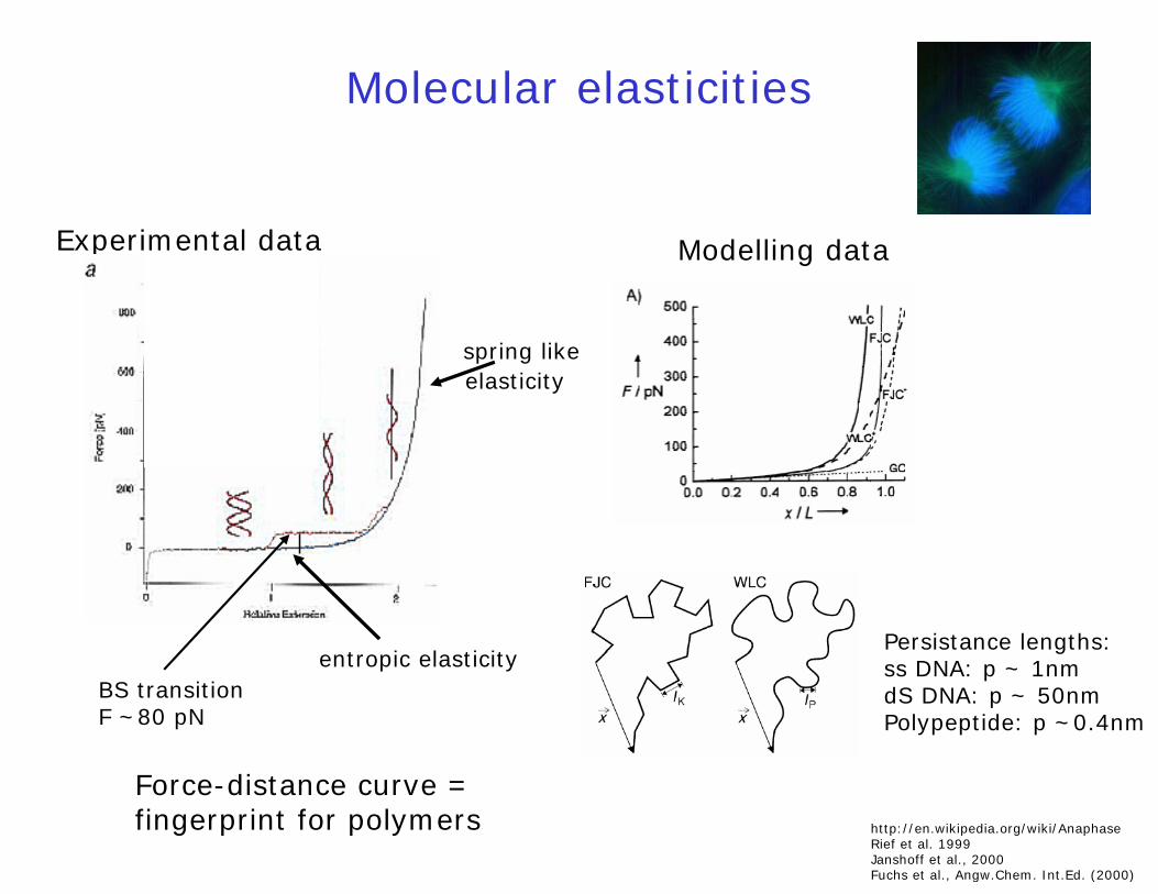
Molecular elasticities
Experimental data
ss DNA
entropic elasticity
spring like
Modelling data
http://en.wikipedia.org/wiki/AnaphaseRief et al. 1999Janshoff et al., 2000Fuchs et al., Angw.Chem. Int.Ed. (2000)
elasticity
Persistance lengths:ss DNA: p ~ 1nmdS DNA: p ~ 50nmPolypeptide: p ~0.4nm
BS transitionF ~80 pN
Force-distance curve = fingerprint for polymers
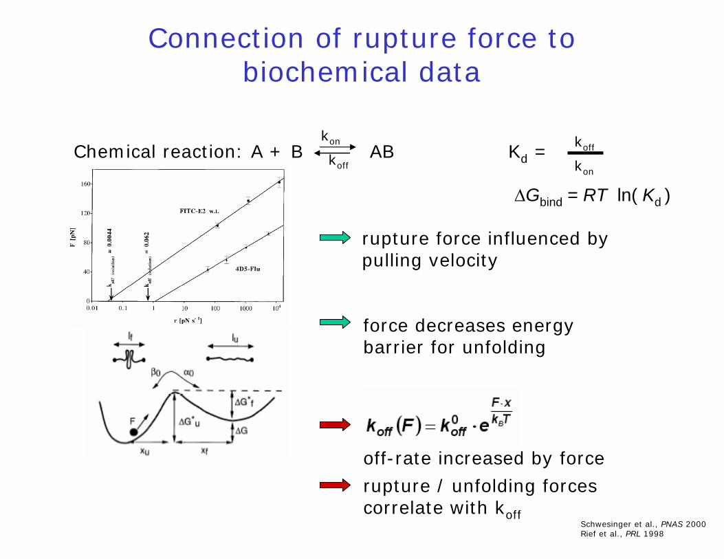
Chemical reaction: A + B AB Kd =
ΔGbind = RT ln( Kd )
Connection of rupture force to biochemical data
Schwesinger et al., PNAS 2000Rief et al., PRL 1998
off-rate increased by force
kon
koff
koff
kon
rupture force influenced by pulling velocity
force decreases energybarrier for unfolding
rupture / unfolding forcescorrelate with koff
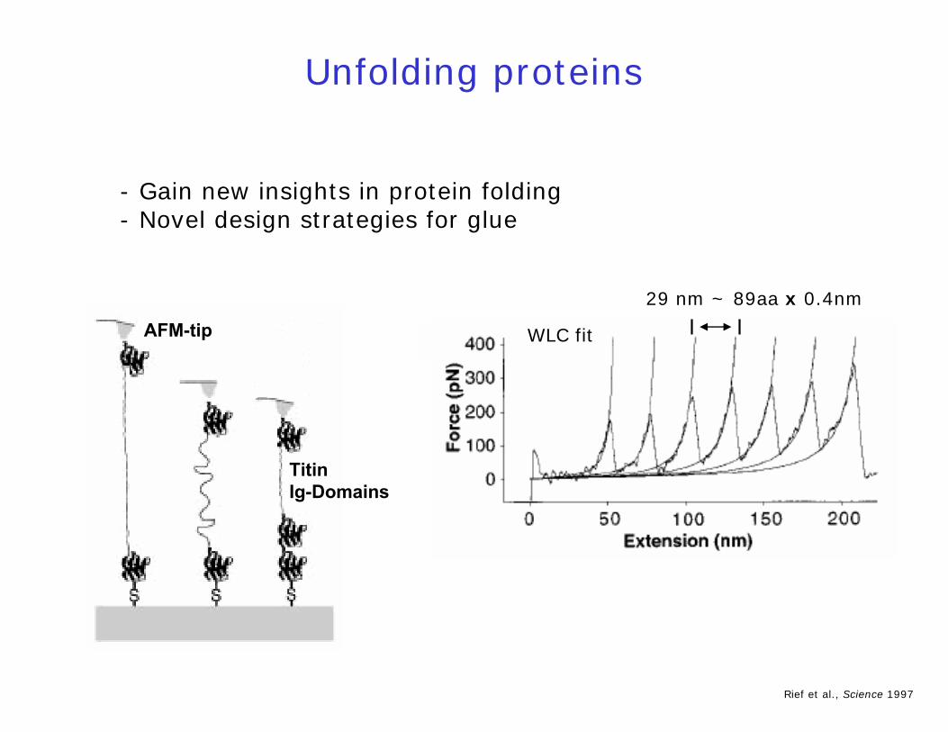
Unfolding proteins
AFM-tip
TitinIg-Domains
29 nm ~ 89aa x 0.4nm
WLC fit
- Gain new insights in protein folding- Novel design strategies for glue
Rief et al., Science 1997
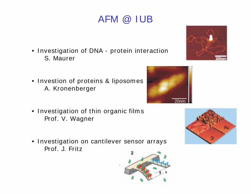
AFM @ IUB
• Investigation of DNA - protein interactionS. Maurer
• Investion of proteins & liposomesA. Kronenberger
• Investigation of thin organic filmsProf. V. Wagner
• Investigation on cantilever sensor arraysProf. J. Fritz
20nm

Conclusion
AFM is a versatile tool to investigate
- topography of surfaces- properties of surfaces- properties of single molecules- forces within molecules
But: always consider experimental conditions and artefacts on measurements!

Thanks!