Embed Size (px)
Citation preview
101
23 (;regory, R. B. (1995) in Protein-Solvem lnteractiom (Grego~', R. B., ed.), pp. 191-264, Marcel Dekker
24 Pal:fleck, R., Haynes, C. A. and Clark, D. S. (1992) Proc..N'atl. AcM. Sci. (r. S. A. 89, 5167 517(I
25 Zaks, A. and Klibanov, A. M. (1985) Pro, ,M~tl. Acad. Sci. U. S. A. 82, 3192-3196
26 Bell, G., Hailing, P.J., Moore, B. 1).. Partndge. J. and Rees, 1). G. (1995) Trotds Biotechnol. 13, 468-473
27 Kitaguchi, H. and Klibanov, A. M. (1989) j . Am. (3win. S0c 1 I1. c~272-9273
28 [;lackwood, A. D., Curran. L. J., Moore, B. 1). and Hailing, P. J. (1994) Biochim. Biophys. Acta 1206, 161-165
29 )[u, K. and Klibanov, A. M. (1996)J. Am. (ghent. Soc. 118. 9815 9819 30 I)abulis, K. and Klibanov, A. M. (1993) Biotedmot. Bioe,(e. 41. 566-571
31 Khmdnitsky, Y. L., Welch, S. H., Clark, D. S. and Dordick, J. S. (1994)J. Am Chcm Soc. 116, 2647-2648
32 Russell, A. J. and Klibanov, A. M. (1987).I. Biol. Chem. 263, 11624 11626
33 Broos, J.. Sakodinskaya, 1. K., Engbersen, J. F. J., Verboon, W. and R.einhoudt, D. N. (1995).1. Chem. &,c., Chem. Commlm. 255-256
34 Okahata, Y. and ljiro, K. (1992) Bull. Chef,,. Soc. Jpn. 65, 2411-2420
35 Paradkar, V. M. and Dordick, J. S. (1994)J. Am. Chem. Soc. 116, 5009-5111()
36 Ahnarsson, O. and Klibanov, A. M. (1996) Bio.'dmoL Bioen, q. 49, 87-92
37 Mingan-o, i.. Gonz~ilez-Navarro, H. and Braco, L. (1996) Biochemistry 35, 9935-9944
Atomic force microscopy in analytical biotechnology
Stephanie Allen, Martyn C. Davies, Clive J. Roberts, Saul J. B. Tendler and Philip M. Williams
The past decade has seen the atomic force microscope evolve not only as a high
resolution imaging tool, but also as an instrument capable of measuring forces
between surfaces and the material properties of samples. Here, the use of atomic
force microscopy for surface force measurement is reviewed, highlighting the
considerable progress that has recently been made in the area of biotechnology.
Particular emphasis is placed on how the instrument can be used to probe directly
biomolecular interactions, illustrating the potential of the technique to impact on
our fundamental understanding of molecular structure and processes.
Since its invention in 1986, the atomic force nficro- scope (AFM) 1 has evolved as a valuable imaging tec]mique with resolution in the micrometre to sub- nanometre range. The ability of the AFM to inaage both insulating and conducting surfaces in a varie D" of environments has facilitated molecular resolution images of a wide range ofbiomolecules, including pro- teir:.s e, lipids 3 and D N A (R.ef. 4). In addition to its imaging abilities, the AFM can also be emplwed to probe spatial variations in surface properties, such as adhesiveness and elasticity 5. The use of the AFM as an imaging technique in the study of biomolecules has frec:uently been the subject of review <7. The aim of this article is to outline the use of an AFM as an instru- ment for surface force measurements, with a particu- lar emphasis on how the technique can be employed in the areas ofbiotechnolog3/and biological science.
S. ~:lle~ ([email protected]), 31. C. Davies, C.J . Roberts, S. J B. Temtler a~d P. M. HTlliams are at the L.,lbomtor}, of Bio- plly~ics and Surface Analysis, Department of l)harmaceutical Sciemcs, The Universit), qf NottiuA~ham, [hlil,ersit), Park, .\\ntinA~hanl, UK N G 7 2RD.
T h e A F M as a force sens ing i n s t r u m e n t Intermolecular forces govern the fundamental prop-
ertics of solids, liquids and gases, the properties of col- loids and the organization of biological structures s. Nmnerous biophysical methods have been employed to investigate specific and non-specific molecular interactions, including optical trapping 9, magnetic force experiments >, pipette suction1' and the surface force apparatus~L Atomic force nficroscopy comple- ments these techniques by uniquely combining an extended force range with high spatial resolution. The AFM has a theoretical force sensitivity of 10 1~ N (Ref. 13), and employs probes with a tip apex of 10-50 nm radius 14, to produce contact areas as small as 10 nm2; therefore providing a means to overcome the limi- tations of other techniques.
By recording force measurements between different probe-sample combinations in various ionic media, it was first illustrated that the AFM could be used to measure forces, such as van der Waals and electrosta- tic forces 15. Ducker and colleagues 1(~ also employed the technique to measure colloidal forces, by attaching spherical particles to the apex of AFM cantilevers. The
Copyright © 1997, Elsevier Science Ltd. All rights reserved. 0167 7799/97/$17.06. PII: S0167-7799(97)01015-9 TIBTECH MARCH 1997 IVOL 15)
1 (i)2
reviews
ability of the instrument to measure discrete adhesive forces of 10 pN was first highlighted by Hoh et al. '7, who attributed the force to that required to rupture a single hydrogen bond. Using these applications of the instrument, various groups then began to analyse the forces controlling more complex systems, including those between polymer-coated surfaces 1~.1~) and protein-protein interactions >--~.
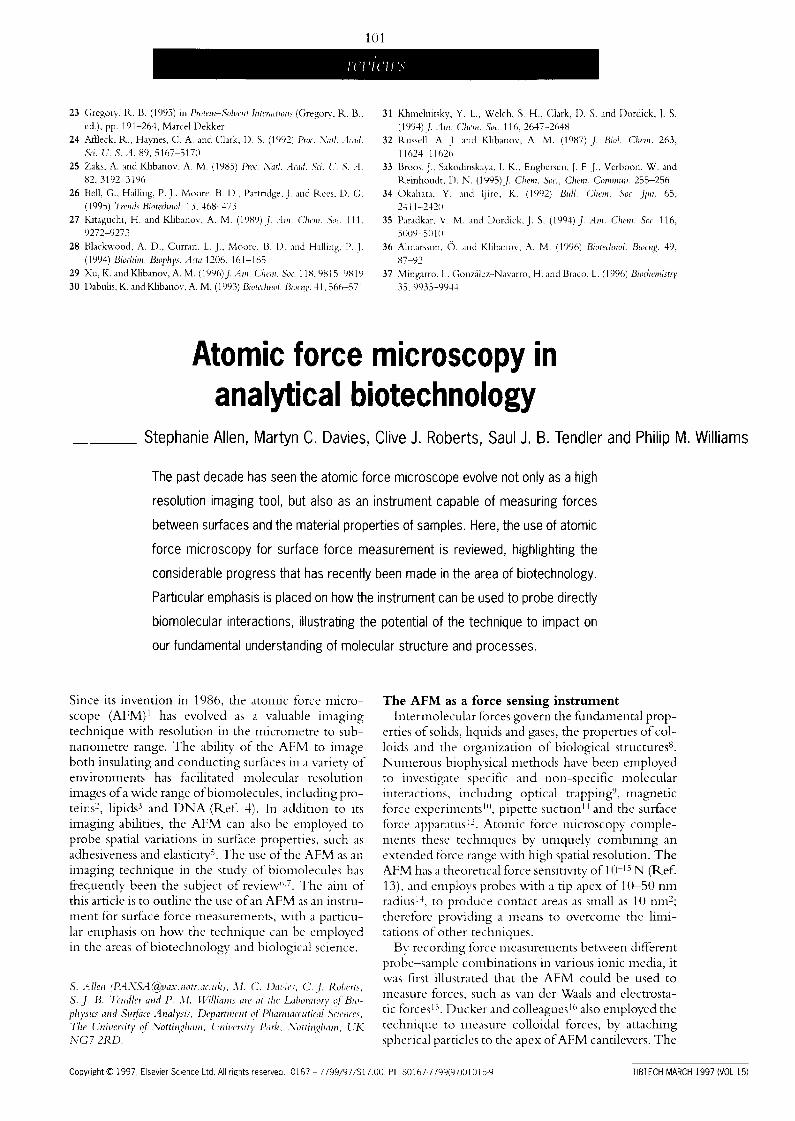
Principle of operation In the AFM a sharp probe is attached to a cantilever
spring, which is scanned in close proximity to a sample surface. During scanning or 'imaging' the cantilever is deflected by forces acting between the sur- face and the probe (Fig. 1). To produce an 'image' the deflections of the cantilever are monitored with respect to its lateral position on the sample (reviewed in R, efs 6,7). However, the cantilever deflections may also be used to directly measure the forces acting between the probe and sample.
When using the AFM probe-cantilever system to detect forces, the deflections of the cantilever are monitored as the probe and sample are brought into contact (approach trace) at constant velocity, and then separated (retract trace). The resultant plot of cantilever deflection versus distance moved by the piezo con- trolling the height of the probe or sample (depending on instrument design) is shown in Fig. 2.
As the probe-sample separation is reduced, the can- tilever is deflected from its rest position by forces act- ing on the probe: attractive tBrces making the can-
Four-segment photodiode (deflection detector)
~ ~ " ~ N Mirr°r /
_ ~ / /~aserbeam
ever
Z I ~ ' ~ ~-Piezoscanner i r~be Z . I - - ~-path of
Figure 1 Contact mode imaging using an atomic force microscope. Typically, the probe is raster scanned (see path of tip) while touching the sample surface. To produce a topographic map of the sample surface, the deflec- tions of the cantilever are monitored using an optical lever. The lateral and vertical positions of the probe (or sample depending on instrument design) during scanning are controlled using piezoceramics.
tilever bend towards the sainple and repulsive forces deflecting the probe away" from the surface. Forces that can be detected in the non-contact region of the force cycle include van der Waals (attractive) and electro- static (attractive or repulsive) forces. Once in contact with the sample surface any further forward motion pro- duces a repulsive force, as the electron orbitals of the atoms in the probe and sample begin to overlap (Born repulsion). In addition to the bending of the cantilever in the contact region of the force curve, the probe and sample may undergo elastic (reversible) or plastic (irre- versible) deformations as the load is increased.
After the point of maximum applied load is reached, the direction of motion is reversed and the probe and sample are separated. During the retraction phase of the measurement the probe often sticks to the surface due to interactions between the probe and sample. As the probe-sample separation is increased further the probe is pulled away from the sample surface and the cantilever deflection returns to its original rest position.
The distance d the cantilever is deflected from its rest position is directly proportional to the force F acting between the probe and surface, as stated by Hooke's Law (F = - k d ) . The constant k in this case represents the spring constant of the cantilever spring. The spring constants of commercially available AFM cantilevers have been found to vary significantly from the value stated by the manufacturer 32. For the accurate measurement of probe-sample interaction forces the calibration of the cantilever spring constant is, there- fore, essential. The most common calibration methods rely on the addition of small tungsten beads (mass approx. 10 ~ g) to the ends of cantilevers [see Fig. 2 (inset)]. The spring constants can then be calculated from the change in the cantilever resonant frequency on the addition of the tungsten bead 32, or from the cantilever deflection caused by the gravitational force acting on the bead 33.
During the imaging of biological samples in air the sample may be irreversibly damaged owing to strong adhesional forces between the probe and sample. These forces are due to a thin layer of water, which covers the surface of most samples. At small probe sample separations a neck of water is formed between the probe and sample, thus generating large probe-sample adhesive forces. Weisenhorn et al. 34 noted that these 'capillary' forces could be reduced by innnersing the sample in liquid. Similarly, most force measurements are performed in liquid so that the large capillary forces do not mask the forces under study.
The study of biomolecular interactions using the AFM
Biological and biochemical processes are governed by a multitude of molecular interactions. Examples of processes mediated by molecular recognition are enzyme-substrate interactions, antigen-antibody interactions and many drug-target associations. This dependence on specific interactions is also true for an increasing number of modern biosensors and chemi- cally or biologically engineered biomaterials. Specific
TI3TECH MARCH 1997 (VOL 15)
1 O3
reviews
mo]ecular interactions, such as those between receptor mo]ecules and their specific ligand molecules, have bond energies that lie between those typical of an ionic bond (e.g. sodium chloride) and van der Waals inter- actions (e.g. between atoms of a noble gas) >. Lee et aJ'. 2{I estimated that a force of approximately 10 9 N was required to break a receptor-ligand complex.
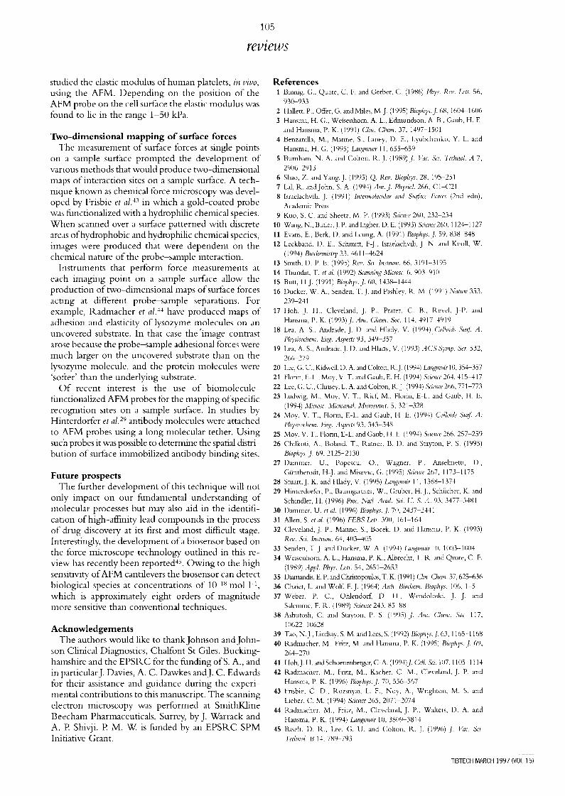
T:) achieve force measurements between individual receptor-ligand complexes, the AFM probe is typi- cally functionalized with a biomolecule of interest and its complementary molecule immobilized onto the sample surface (Fig. 3). Force measurements are then recorded between the functionalized surfaces. During the approach trace of the force measurement the rec(ptor and ligand molecules are brought into con- tact, allowing receptor-ligand complexes to form. The 'sticking force' (Fig. 2) observed as the probe and sur- face' are separated corresponds to the force required to separate the receptor-ligand coinplexes. Because the AFM probes used in such experiments typically have an apex radius of 10-50 nm (Ref. 14), the number of receptor-ligand complexes formed during a force cycle can be as low as one or two molecular pairs.
-1-,3 determine exactly the number of interacting molecules during each force measurement a prior knowledge of the surface density of the immobilized mo]ecules, probe geometry and probe-sample contact area is required. Protein surface coverage can be assessed relatively easily using methods such as radiolabelling > and fluorescence techniques > . However, an exact determination of probe geometry and probe-sample contact area is more problematical. In an attempt to overcome such problems, various groups 2°,22,2~ have attached smooth micrometre-sized spheres (radius of 2-10 t,tm) to the ends of cantilevers and employed them as probes with controlled geometry; this allows a greater control of probe-sample contact area.
In experiments employing spherical probes, the probe-sample contact areas are larger than those in similar experiments employing functionalized AFM probes. Consequently, many receptor-ligand com- plexes can be formed on contact, and hence the adhe- sive forces in such experiments may correspond to the rupture of a number of receptor-ligand bonds. The rupture force for a single receptor-ligand interaction ma F be obtained by blocking out the majority of inter- action sites on one of the surfaces 21, by the addition of known or unknown quantities of receptor or ligand to the experimental system.
~[he streptavidin-biotin 3s system was the initial receptor-ligand interaction to be studied using this technique. Streptavidin, a 66-75 kDa protein, coin- prises four identical subunits each containing a single bioun binding site3% The high specificity and at:finity (binding constant, K d = 10 -I~ tool 1 <) of the complex, and the availability of structuraP v and thermody- namic 38 data made it an ideal model system for the study ofreceptor-ligand interactions. Lee et al. > used biodn-functionalized glass beads attached to AFM cantilevers and streptavidin-coated mica surfaces, and estimated the strength of a single streptavidin-biotin
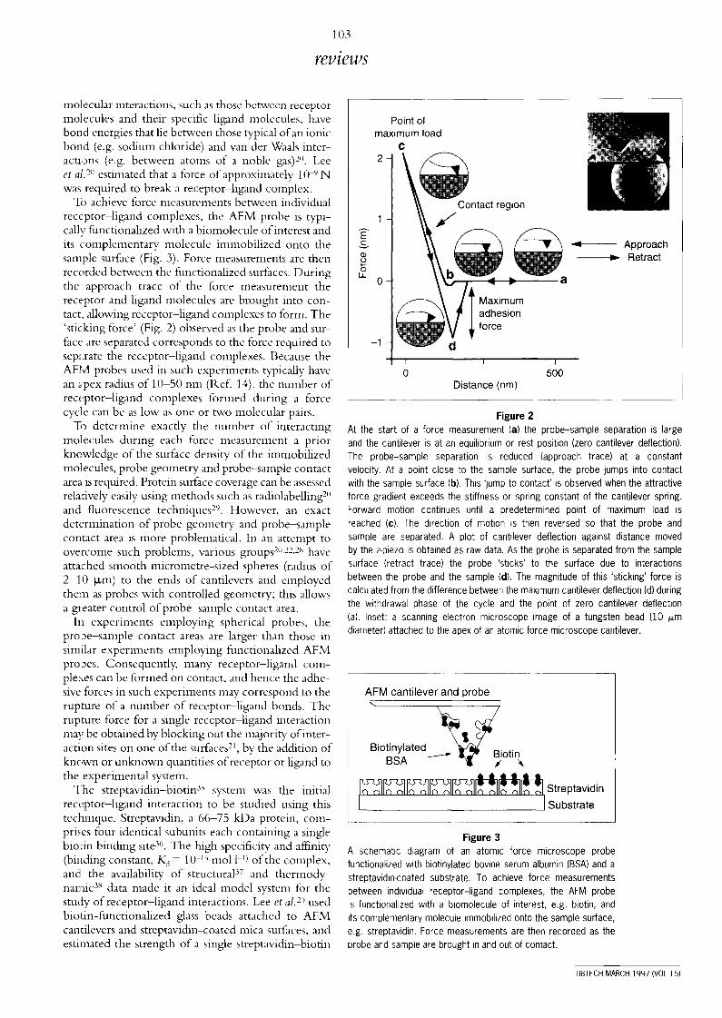
Point of maximum load
C 2 -
1 -
~ Approach o ~ ~ Retract
2_ O-
- 1 -
I I I
0 500 Distance (nm)
Figure 2 At the start of a force measurement (a) the probe-sample separation is large and the cantilever is at an equilibrium or rest position (zero cantilever deflection). The probe-sample separation is reduced (approach trace) at a constant velocity. At a point close to the sample surface, the probe jumps into contact with the sample surface (b). This 'jump to contact' is observed when the attractive force gradient exceeds the stiffness or spring constant of the cantilever spring. Forward motion continues until a predetermined point of maximum load is reached (c). The direction of motion is then reversed so that the probe and sample are separated. A plot of cantilever deflection against distance moved by the z-piezo is obtained as raw data. As the probe is separated from the sample surface (retract trace) the probe 'sticks' to the surface due to interactions between the probe and the sample (d). The magnitude of this 'sticking' force is calculated from the difference between the maximum cantilever deflection (d} during the withdrawal phase of the cycle and the point of zero cantilever deflection (a). Inset: a scanning electron microscope image of a tungsten bead (10 i~m diameter) attached to the apex of an atomic force microscope cantilever.
AFM canti lever and probe
B i°t i n y l a t e d - - - ~ ~ ~ i ° t i n~ BSA
i[~] ~ ] [~][z~'~ ~'~ [~H~ [ ~ ] [ ~ i Streptavidins ubst rate
Figure 3 A schematic diagram of an atomic force microscope probe functionalized with biotinylated bovine serum albumin (BSA) and a streptavidin-coated substrate. To achieve force measurements between individual receptor-ligand complexes, the AFM probe is functionalized with a biomolecule of interest, e.g. biotin, and its complementary molecule immobilized onto the sample surface, e.g. streptavidin. Force measurements are then recorded as the probe and sample are brought in and out of contact.
TIBTECH MARCH 1997 (VOL 15}
104
reviews
E" £- v
8 L--
,9
b
Z i -
v
0 LL
3.5
3
2.5
2
1.5
1
0.5
0
-0.5 5O
5 ¸
~ \ _ Retract
Approach
I I I i I
1 O0 150 200 250 300 Dis tance moved by the cantilever (nm)
4
2 Approach
0
5'0 ' ' ' - 1 0 100 150 200 Dis tance moved by the cantilever (nm)
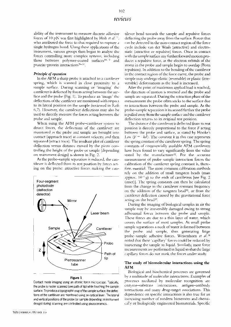
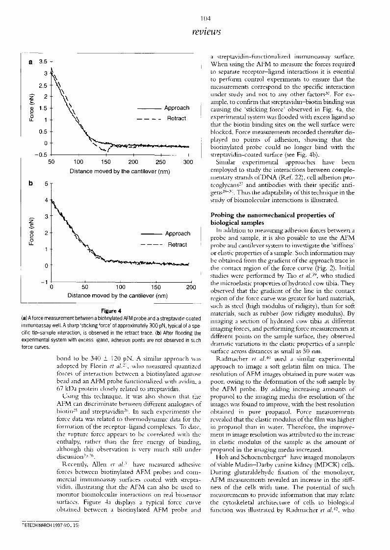
Figure 4
(a) A force measurement between a biotinylated AFM probe and a streptavidin-coated irnmunoassay well. A sharp 'sticking force' of approximately 300 pN, typical of a spe- cific tip-sample interaction, is observed in the retract trace. (b) After flooding the experimental system with excess ligand, adhesion points are not observed in such force curves.
bond to be 340 _+ 120 pN. A similar approach was adopted by Florin et al. 21, who measured quantized forces of interaction between a biotinylated agarose bead and an AFM probe functionalized with avidin, a 67 kDa protein closely related to streptavidin.
Using this technique, it was also shown that the AFM can discriminate between different analogues of biotin 2~ and streptavidin 2('. In such experiments the force data was related to thermodynamic data for the formation of the receptor-ligand complexes. To date, the rupture force appears to be correlated with the enthalpy, rather than the free ener~, of binding, although this observation is very much still under discussion2S.26.
Recently, Allen et al. 31 have measured adhesive forces between biotinylated AFM probes and com- mercial immunoassay surfaces coated with strepta- vidin, illustrating that the AFM can also be used to monitor biomolecular interactions on real biosensor surfaces. Figure 4a displays a typical force curve obtained between a biotinylated AFM probe and
a streptavidin-functionalized immunoassay surface. When using the AFM to measure the forces required to separate receptor-ligand interactions it is essential to perform control experiments to ensure that the measurements correspond to the specific interaction under study and not to any other factors 3°. For ex- ample, to confirm that streptavidin-biotin binding was causing the 'sticking force' observed in Fig. 4a, the experimental system was flooded with excess ligand so that the biotin binding sites on the well surface were blocked. Force measurements recorded thereafter dis- played no points of adhesion, showing that the biotinylated probe could no longer bind with the streptavidin-coated surface (see Fig. 4b).
Similar experimental approaches have been employed to study the interactions between comple- mentary strands o f D N A (Ref. 22), cell adhesion pro- teoglycans 27 and antibodies with their specific anti- dens 2s-3°. Thus the adaptability of this technique in the study ofbiomolecular interactions is illustrated.
Probing the n a n o m e c h a n i c a l properties o f b io logical samples
In addition to measuring adhesion forces between a probe and sample, it is also possible to use the AFM probe and cantilever system to investigate the 'stiffness' or elastic properties of a sample. Such information may be obtained from the gradient of the approach trace in the contact region of the force curve (Fig. 2). Initial studies were performed by Tao eta[. 39, who studied the microelastic properties of hydrated cow tibia. They observed that the gradient of the line in the contact region of the force curve was greater for hard materials, such as steel (high modulus of ridigity), than for soft materials, such as rubber (low ridigity modulus). By imaging a section of hydrated cow tibia at different imaging forces, and performing force measurements at different points on the sample surface, they observed dramatic variations in the elastic properties of a sample surface across distances as small as 50 nm.
Kadmacher et al. 4" used a similar experimental approach to image a soft gelatin film on mica. The resolution of AFM images obtained in pure water was poor, owing to the deformation of the soft sample by the AFM probe. By adding increasing amounts of propanol to the imaging media the resolution of the images was found to improve, with the best resolution obtained in pure propanol. Force measurements revealed that the elastic modulus of the film was higher in,propanol than in water. Therefore, the improve- ment in image resolution was attributed to the increase in elastic modulus of the sample as the amount of propanol in the imaging media'increased.
Hoh and Schoenenberger 41 have imaged monolayers of viable Madin-Darby canine kidney (MDCK) ceils. During glutaraldehyde fixation of the monolayer, AFM measurements revealed an increase in the stiff- ness of the cells with time. The potential of such measurements to provide information that may relate the cytoskeletal architecture of ceils to biological fhnction was illustrated by R, admacher et a/. 42, who
TI[!ITECH MARCH 1997 (VOL 15)
105
reviews
studied the elastic modulus of human platelets, in v ivo ,
using the AFM. Depending on the position of the AFM probe on the cell surface the elastic modulus was found to lie in the range 1-50 kPa.
Two-dimensional mapping o f surface forces The measurement of surface forces at single points
on a sample surface prompted the development of various methods that would produce two-dimensional maps of interaction sites on a sample surface. A tech- nique known as chemical force microscopy was devel- opt:d by Frisbie et al. 43 in which a gold-coated probe wa.,; functionalized with a hydrophilic chemical species. When scanned over a surface patterned with discrete areas ofhydrophobic and hydrophilic chemical species, images were produced that were dependent on the ch(mical nature of the probe-sample interaction.
I:astruments that perform force measurements at each imaging point on a sample surface allow the production of two-dimensional maps of surface forces acting at different probe-sample separations. For example, Radmacher et al. 44 have produced maps of adhesion and elasticity of lysozyme molecules on an uncovered substrate. In that case the'image contrast arose because the probe-sample adhesional forces were much larger on the uncovered substrate than on the lysozyme molecule, and the protein molecules were 'solier' than the underlying substrate.
O f recent interest is the use of biomolecule- functionalized AFM probes for the mapping of specific recognition sites on a sample surface. In studies by Hinterdorfer et al. 29 antibody molecules were attached to RFM probes using a long molecular tether. Using such probes it was possible to determine the spatial distri- bution of surface immobilized antibody binding sites.
Future prospects The further development of this technique will not
only impact on our fundanmntal understanding of molecular processes but may also aid in the identifi- cat:Lon of high-affinity lead compounds in the process of drug discovery at its first and most difficult stage. Interestingly, the development ofa biosensor based on the force microscope technology, outlined in this re- view has recently been reported 45. Owing to the high sensitivity, of AFM cantilevers the biosensor can detect biological species at concentrations of 10 18 tool 1-1, which is approximately eight orders of magnitude mc:,re sensitive than conventional techniques.
Acknowledgements The authors would like to thank Johnson andJohn-
sor:~ Clinical Diagnostics, Chalfont St Giles, Bucking- hamshire and the EPSP, C for the funding ofS. A., and in particularJ. Davies, A. C. Dawkes andJ. C. Edwards for their assistance and guidance during the experi- mental contributions to this manuscript. The scanning electron microscopy was performed at SmithKline Beecham Pharmaceuticals, Surrey, by J. Warrack and A. E Shivji. E M. W. is funded by an EPSR.C SPM Initiative Grant.
References 1 Binnig, G., Quate, C. F. and Gerber, C. (1986) Pkys. Ret,. L*'tt. 56,
930-933 2 Hallett, P., Ot~br, G. and Miles, M.J. (1995) Biophys.ff. 68, 1604 1606 3 Hansma, H. G., Weisenhorn, A. L., Edmundson, A. B., Gaub, H. E.
and Hansma, P. K. (1991) Clin. Chem. 37, 1497-1501 4 Benzanilla, M., Manne, S., Laney, D. E., Lyubchenko, Y. L. and
Hansma, H. G. (1995) Lan2muir 11,655-659 5 Burnham, N. A. and Colton, R.J. (1989)./. l,'ac. Sd. Tedmol. A 7,
29062913 4 Shao, Z. and Yang, J. (1995) Q. Rev. Biophys. 28, 195-251 7 Lal, R. and John, S. A. (1994) Am.J. Ph}~siol. 266, C1-C21 8 Israelachvili, J. (1991) hltermolecular and Su~ace Forces (2nd edn),
Academic Press 9 Kuo, S. C. and Sheetz, M. P. (1993) Science 260, 232 234
10 Wang, N., Butler,J. P. and lngber, D. E. (1993) &ieme260, 1124-1127 11 Evans, E., Berk, 1). and Leung, A. (1991) Biophys. J. 59, 838-848 12 Leckband, 1). E., Schmitt, F-J., Israelachvili, J. N. and Knoll, W.
(1994) Biochemistry 33,4611-4624 13 Smith, 1). P. E. (1995) Rev. &i. lnstmm. 66, 3191-3195 14 Thundat. T. et al. (1992) Scanning 3licrosc. 6. 903-910 15 Butt, H-J. (1991) Biophys. d. 60, 1438-1444 16 Ducker, W. A., Sen&n, T.J. and Pashley, R. M. (1991) Nature 353,
239-241 17 Hoh, J. H., Cleveland, J. P., Prater, C. B., Revel. J-P. and
Hansma, P. K. (1993)J. Anl. Chem. S0c. 114, 4917-4919 18 Lea, A. S., Andrade, J. D. and Hlady, V. (1994) Colloids Surf A:
Phl,sicodlem. Eng. Aspects 93, 349-357 19 Lea. A. S., Andrade, J. D. and Hlady, V. (1993) ACS Syrup. Ser. 532,
266-279 20 Lee, G. U., Kidwell, D. A. and Colton, R.J. (1994) kqql~milir 10,354-.357 21 Florin. E L., Moy, V. T. and Gaub, E. H. (1994) Science 264, 415-417 22 Lee, G. U., Chrisey, L A. and Colton, R.J. (1994) &ience266, 771-773 23 Ludwig, M., Moy, V. T., Rie£ M., Flurin, E L. and Gaub, H. E.
(1994) Microsc Microanal. Mlcrostr, ct. 5, 321-328 24 Moy, V. T., Florin, E-L. and Gaub, H E. (1994) Colloids Surf A:
Physieochem. Enq. Aspects 93, 343-348 25 Moy, V. T., Florin, E-L. and Ganb, H. E. (1994) Science 266,257 259 26 Chilkoti, A., Boland, T., Ramer, B. 1). and 8tayton, P. S. (1995)
Biophys.J. 6% 2125-213(I 27 I)ammer, U., Popescu, O., Wagner, P., Anselmetti, 1).,
GO.ntherodt, H-J. and Misevic, G. (1995) Scie~a' 267, I173-1175 28 Stuart, J. K. and Hlady, V. (1995) Lat~2muir I1, 1368-1374 29 Hinterdorfer, P., Baumgartner, W., Gruber, H.J., Schilcher, K. and
Schindler, H. (1996) Proc. Nad. Acad. Sd. U. S. A. 93. 3477 3481 30 Dammer, U. et al. (1996) Biophys. j . 7(i, 2437-2441 31 Allen, S. et al. (1996) FEBS Lett. 390, 161-164 32 Cleveland, J. P., Manne; S., Bocek, D. and Hansma, P. K. (1993)
Rev. Sci. lnstn~m. 64, 403-405 33 Senden. T. J. and Ducker, W. A. (1994) Lanqnmir 10, 1(1!13-1004 34 Weisenhorn. A. L., Hansma, P. K., Albrecht, T. R. and Quate, C. F.
(1989) Appl. Phys. La~It. 54, 2651 2653 35 Diamandis, E. P. mid Chrismpoulos, T. K. (1991) Clin. Chem. 37, 625-636 36 Chalet, L. and WolE F.J. (1964) Arch. Biod~em. Biophys. 106, 1-5 37 Weber, P. C., OhlendoK D. H., Wendoloski, J. J. and
Salemnle, F. R. (1989) Sciet~ce 243, 85 88 38 Ashutosh, C. and Stayton, P. S. (1995)]. Am. Chem. Soc. 117,
1 I)622-10628 39 Tao, N.J., Lindsay, S. M. and Lees, S. (1992) BiophI,s.]. 63, 1165-1168 40 Radmacher, M., Fritz, M. and Hansma, P. K. (1995) Biophys._l. 69,
264-270 41 Hoh,J. H. and Schoenenberger, C-A. (1994)./. Cell. &i. 107.1105 1114 42 Radmacher, M., Fritz, M., Kacher, C. M., Cleveland, J. P. and
Hansma, P. K. (1996) Biophi,s..J. 7{1, 556-567 43 Frisbie, C. D., Rozsnyai, L. F., No b A., Wnghton, M. S. and
Lieber. C. M. (1994) Scieme 265, 2071-2074 44 Radmacher, M., Fritz, M., Cleveland, J. P., Wakers, D. A. and
Hansma, P. K. (1994) Langmuir 10, 3809-3814 45 Baselt, D. R.., Lee, G. U. and Colton, R. J. (1996) J. Vac. Sci.
Tedmol. B 14, 789-793
TIBTECH MARCH 1997 (VOL 15)