Embed Size (px)
Citation preview

Comptes Rendus
Biologies
Gabor Papai, Alexandre Frechard, Olga Kolesnikova, CorinneCrucifix, Patrick Schultz and Adam Ben-Shem
Atomic structure of the SAGA complex and it’s interaction with TBPVolume 343, issue 3 (2020), p. 247-255.
<https://doi.org/10.5802/crbiol.31>
© Académie des sciences, Paris and the authors, 2020.Some rights reserved.
This article is licensed under theCreative Commons Attribution 4.0 International License.http://creativecommons.org/licenses/by/4.0/
Les Comptes Rendus. Biologies sont membres duCentre Mersenne pour l’édition scientifique ouverte
www.centre-mersenne.org

Comptes RendusBiologies2020, 343, n 3, p. 247-255https://doi.org/10.5802/crbiol.31
Articles / Reviews / Articles / Revues
Atomic structure of the SAGA complex and it’s
interaction with TBP
Structure atomique du complexe SAGA et son interaction avec
TBP
Gabor Papai∗, a, b, c , d, Alexandre Frecharda, b, c , d, Olga Kolesnikovaa, b, c , d,Corinne Crucifixa, b, c , d, Patrick Schultza, b, c , d and Adam Ben-Shema, b, c , d
a Institut de Génétique et de Biologie Moléculaire et Cellulaire, Integrated StructuralBiology Department, Équipe labellisée Ligue Contre le Cancer, Illkirch, Franceb Centre National de la Recherche Scientifique, UMR7104, Illkirch, France
c Institut National de la Santé et de la Recherche Médicale, U1258, Illkirch, France
d Université de Strasbourg, Illkirch, France
E-mails: [email protected] (G. Papai), [email protected] (A. Frechard),[email protected] (O. Kolesnikova), [email protected] (C. Crucifix),[email protected] (P. Schultz), adam.ben-shem.igbmc.fr (A. Ben-Shem)
Gabor Papai was the award winner of the 2020 prize for Great Advances in Biologyfrom the Académie des sciences / Gabor Papai a été lauréat du prix 2020 desGrandes Avancées en Biologie de l’Académie des sciences
Abstract. The transcription of eukaryotic protein genes is controlled by a plethora of proteins whichact together in multi-component complexes to facilitate the DNA dependent RNA polymerase II(Pol II) enzyme to bind to the transcription start site and to generate messenger RNA from the gene’scoding sequence. The protein that guides the transcription machinery to the exact transcription startsite is called TATA-box Binding Protein, or TBP. TBP is part of two large protein complexes involvedin Pol II transcription, TFIID and SAGA. The two complexes share several subunits implicated in theinteraction with TBP and contain proteins with structural elements highly homologous to nucleoso-mal histones. Despite the intensive study of transcription initiation, the mode of interaction of TBPwith these complexes and its release upon DNA binding was elusive. In this study we demonstrate thequasi-atomic model of SAGA in complex with TBP. The structure reveals the intricate network of in-teractions that coordinate the different functional domains of SAGA and resolves a deformed octamerof histone-fold domains at the core of SAGA. This deformed octamer is precisely tuned to establish aperipheral site for TBP binding, where it is protected by steric hindrance against the binding of spuri-ous DNA. Complementary biochemical analysis points to a mechanism for TBP delivery and releasefrom SAGA that requires the general transcription factor TFIIA and whose efficiency correlates withthe affinity of DNA to TBP.
∗Corresponding author.
ISSN (electronic) : 1768-3238 https://comptes-rendus.academie-sciences.fr/biologies/

248 Gabor Papai et al.
As the TBP binding machinery is highly similar in TFIID and SAGA, we demonstrated a universalmechanism of how TBP is delivered to gene promoters during transcription initiation.
Résumé. La transcription des gènes des protéines eucaryotes est contrôlée par une pléthore de pro-téines agissant de concert sous forme de complexes multi-composants pour faciliter la liaison de l’en-zyme ARN polymérase II ADN-dépendante (Pol II) au site d’initiation de la transcription et pour gé-nérer un ARN messager à partir de la séquence codante du gène. La protéine qui guide la machi-nerie de transcription vers le site d’initiation de la transcription est appelée protéine de liaison à laboîte TATA, ou TBP. TBP fait partie de deux complexes protéiques impliqués dans la transcription parla Pol II, TFIID et SAGA. Les deux complexes partagent plusieurs sous-unités impliquées dans l’in-teraction avec TBP et comportent des protéines présentant des éléments structuraux hautement ho-mologues aux histones nucléosomiques. Malgré l’étude intensive de l’initiation de la transcription,le mode d’interaction de TBP avec ces complexes ainsi que sa libération lors de sa liaison de l’ADNétaient évasifs. Dans cette étude, nous avons déterminé un modèle quasi-atomique de SAGA en com-plexe avec TBP. La structure révèle le réseau d’interactions qui coordonnent les différents domainesfonctionnels de SAGA et résout un octamère déformé des domaines homologues aux histones au cœurde SAGA. Cet octamère déformé est précisément adapté pour établir un site périphérique de liaison àTBP, où ce dernier est protégé par une inhibition stérique contre la fixation d’un ADN parasite. L’ana-lyse biochimique complémentaire a mis en évidence un mécanisme de libération de TBP de SAGA quinécessite le facteur de transcription général TFIIA et dont l’efficacité corrèle avec l’affinité de l’ADNpour TBP.
Comme le mécanisme de liaison de TBP est très similaire dans TFIID et SAGA, nous avons mis enévidence un mécanisme universel décrivant la manière dont TBP est délivré aux promoteurs de gèneslors de l’initiation de la transcription.
Keywords. Genes, SGA, TBP, ARN messager, Transcription.
Mots-clés. Gènes, SGA, TBP, ARN messager, Transcription.
Manuscript received and accepted 28th October 2020.
1. Introduction
Transcription of protein-coding genes begins withthe formation of a pre-initiation complex (PIC) com-posed of RNA polymerase II and several generaltranscription factors (TFs) [1]. PIC assembly is nu-cleated by the recruitment of TATA-binding protein(TBP) to promoter DNA, a focal point for regulatedgene expression [2].
Transcription co-activator complexes are requiredfor activated transcription in a chromatin environ-ment. Two multi-protein co-activator complexes,TFIID and SAGA, are globally required for gene ex-pression in yeast [3, 4] and can deliver TBP to thegene promoter [5]. The relative contribution of eachof these two complexes to PIC formation at a par-ticular gene, is dictated by gene-specific featuresincluding promoter sequence, histone modificationsand transcription activator binding sites [6]. TFIIDand SAGA have a homologous histone octamer likemodule, which was reported to be responsible forTBP delivery. They share five TBP Associated Factors,Taf5 and four Tafs containing a histone fold (HF)motifs (Taf6, 9, 10, and 12). SAGA also harbors three
additional subunits in this module containing one(Ada1 and Spt7) or two (Spt3) HF domains. Impor-tant progress was reported in determining the archi-tecture of TFIID, but the machinery that binds anddelivers TBP could not be depicted in detail [7, 8].
The 1.6 MDa Spt-Ada-Gcn5 acetyltransferase(SAGA) complex is composed of 19 subunits orga-nized in four modules with distinct functions: a his-tone acetyl transferase (HAT) module, a histone deu-biquitinase (DUB) module, the 430 KDa Tra1 subunitwhich serves as a docking platform for sequence-specific activators, and a central module physicallyconnected to all other modules and responsible forbinding and delivery of the TATA-box binding pro-tein (TBP) [9]. The functional diversity of SAGA al-lows it, in addition to PIC formation, to modulatetranscription at different steps [10, 11] and to play animportant role in DNA repair signaling [12].
Atomic structures of several individual isolatedcomponents of SAGA were obtained by X-ray crystal-lography. Cryo-EM studies positioned Tra1 in a mapof holo-SAGA but all other subunit appeared togetherin one main lobe that remained poorly resolved [13].
C. R. Biologies, 2020, 343, n 3, 247-255
Gabor Papai et al. 249
2. High resolution structure of SAGA
The dynamic nature of SAGA hampered efforts to re-solve its structure at high resolution, hindering at-tempts to place atomic models of subunits or do-mains within the main lobe. We posited that by im-proving our purification scheme, as well as by in-troducing SAGA natural ligands, the conformationof SAGA may be stabilized. These advances com-bined with local structure refinement resulted in astructure at a resolution which allowed us to positionand model the majority of SAGA subunits [14].
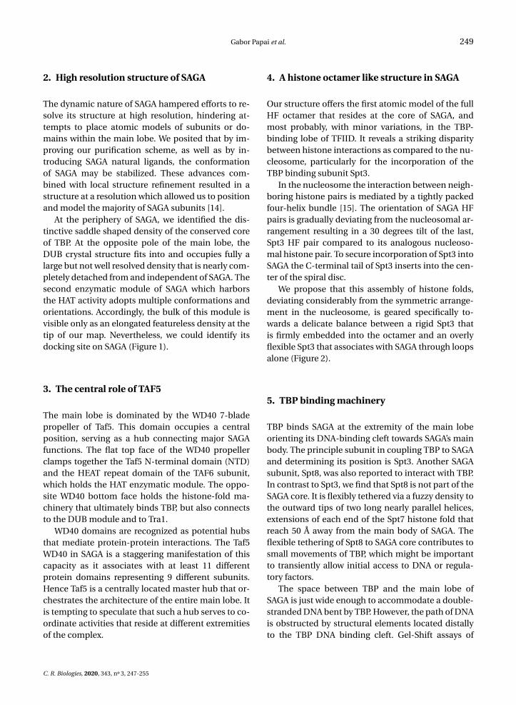
At the periphery of SAGA, we identified the dis-tinctive saddle shaped density of the conserved coreof TBP. At the opposite pole of the main lobe, theDUB crystal structure fits into and occupies fully alarge but not well resolved density that is nearly com-pletely detached from and independent of SAGA. Thesecond enzymatic module of SAGA which harborsthe HAT activity adopts multiple conformations andorientations. Accordingly, the bulk of this module isvisible only as an elongated featureless density at thetip of our map. Nevertheless, we could identify itsdocking site on SAGA (Figure 1).
3. The central role of TAF5
The main lobe is dominated by the WD40 7-bladepropeller of Taf5. This domain occupies a centralposition, serving as a hub connecting major SAGAfunctions. The flat top face of the WD40 propellerclamps together the Taf5 N-terminal domain (NTD)and the HEAT repeat domain of the TAF6 subunit,which holds the HAT enzymatic module. The oppo-site WD40 bottom face holds the histone-fold ma-chinery that ultimately binds TBP, but also connectsto the DUB module and to Tra1.
WD40 domains are recognized as potential hubsthat mediate protein-protein interactions. The Taf5WD40 in SAGA is a staggering manifestation of thiscapacity as it associates with at least 11 differentprotein domains representing 9 different subunits.Hence Taf5 is a centrally located master hub that or-chestrates the architecture of the entire main lobe. Itis tempting to speculate that such a hub serves to co-ordinate activities that reside at different extremitiesof the complex.
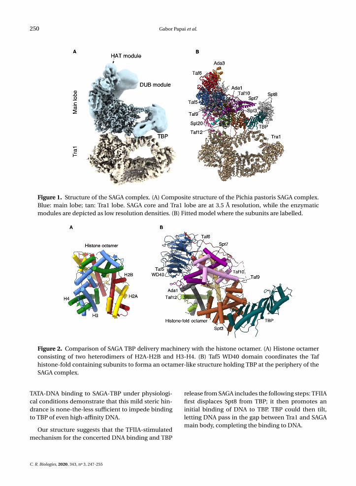
4. A histone octamer like structure in SAGA
Our structure offers the first atomic model of the fullHF octamer that resides at the core of SAGA, andmost probably, with minor variations, in the TBP-binding lobe of TFIID. It reveals a striking disparitybetween histone interactions as compared to the nu-cleosome, particularly for the incorporation of theTBP binding subunit Spt3.
In the nucleosome the interaction between neigh-boring histone pairs is mediated by a tightly packedfour-helix bundle [15]. The orientation of SAGA HFpairs is gradually deviating from the nucleosomal ar-rangement resulting in a 30 degrees tilt of the last,Spt3 HF pair compared to its analogous nucleoso-mal histone pair. To secure incorporation of Spt3 intoSAGA the C-terminal tail of Spt3 inserts into the cen-ter of the spiral disc.
We propose that this assembly of histone folds,deviating considerably from the symmetric arrange-ment in the nucleosome, is geared specifically to-wards a delicate balance between a rigid Spt3 thatis firmly embedded into the octamer and an overlyflexible Spt3 that associates with SAGA through loopsalone (Figure 2).
5. TBP binding machinery
TBP binds SAGA at the extremity of the main lobeorienting its DNA-binding cleft towards SAGA’s mainbody. The principle subunit in coupling TBP to SAGAand determining its position is Spt3. Another SAGAsubunit, Spt8, was also reported to interact with TBP.In contrast to Spt3, we find that Spt8 is not part of theSAGA core. It is flexibly tethered via a fuzzy density tothe outward tips of two long nearly parallel helices,extensions of each end of the Spt7 histone fold thatreach 50 Å away from the main body of SAGA. Theflexible tethering of Spt8 to SAGA core contributes tosmall movements of TBP, which might be importantto transiently allow initial access to DNA or regula-tory factors.
The space between TBP and the main lobe ofSAGA is just wide enough to accommodate a double-stranded DNA bent by TBP. However, the path of DNAis obstructed by structural elements located distallyto the TBP DNA binding cleft. Gel-Shift assays of
C. R. Biologies, 2020, 343, n 3, 247-255
250 Gabor Papai et al.
Figure 1. Structure of the SAGA complex. (A) Composite structure of the Pichia pastoris SAGA complex.Blue: main lobe; tan: Tra1 lobe. SAGA core and Tra1 lobe are at 3.5 Å resolution, while the enzymaticmodules are depicted as low resolution densities. (B) Fitted model where the subunits are labelled.
Figure 2. Comparison of SAGA TBP delivery machinery with the histone octamer. (A) Histone octamerconsisting of two heterodimers of H2A-H2B and H3-H4. (B) Taf5 WD40 domain coordinates the Tafhistone-fold containing subunits to forma an octamer-like structure holding TBP at the periphery of theSAGA complex.
TATA-DNA binding to SAGA-TBP under physiologi-cal conditions demonstrate that this mild steric hin-drance is none-the-less sufficient to impede bindingto TBP of even high-affinity DNA.
Our structure suggests that the TFIIA-stimulatedmechanism for the concerted DNA binding and TBP
release from SAGA includes the following steps: TFIIAfirst displaces Spt8 from TBP; it then promotes aninitial binding of DNA to TBP. TBP could then tilt,letting DNA pass in the gap between Tra1 and SAGAmain body, completing the binding to DNA.
C. R. Biologies, 2020, 343, n 3, 247-255
Gabor Papai et al. 251
6. Flexible enzymatic modules
The DUB and HAT modules are highly dynamic, per-mitting the exploration of a large conformationalspace in search of their chromatin substrates and thestructure shows how these modules maintain robustbinding to SAGA.
SAGA DUB module consists of the catalytic sub-unit Ubp8 as well as Sgf11, Sus1, and the Sgf73 N-terminal end. Two helices in the C-terminal part ofSgf73 are embedded within SAGA core. The DUBmodule is tethered to SAGA’s core through the 140aalinker connecting the anchoring domain to the N-terminal end of Sgf73 which is part of the DUB. Wefind little trace of this linker in the structure exceptfor a short stretch contacting Spt20 and Taf5.
In contrast to the DUB, no parts of the HAT mod-ule are embedded within SAGA core. The HAT densi-ties emerge, harboring two distinct helical domains,at the surface of the Taf6 HEAT repeats.
7. Intricate interaction network
Three distinct bridges serve to physically and possi-bly functionally couple Tra1 with the main lobe. The
first bridge is established by a highly dispersed andextended domain of Spt20 touching Taf5, Taf9, Taf12,Ada1 and Spt7 as it traverses the surface of SAGAcore on its way to Tra1. An exceptionally long loopthat precedes the histone fold of Taf12 is responsiblefor establishing the second bridge. This loop formsa lasso shaped thread at the surface of the Tra1. Thethird bridge is formed by the loop connecting the sec-ond and third helices of cSpt3-HF as its infiltrates be-tween two helices of Tra1.
8. Perspectives
Our work provides the architecture of the multi-subunit yeast SAGA complex and sheds light into itsrole in eukaryotic transcription initiation. The struc-ture paves the way towards better understanding ofhow the TFIID and SAGA complexes, designed todeliver TBP, may share workload on different genepromoters. TBP is required for each transcriptionevent in eukaryotes and interacts with a large num-ber of partners with overlapping binding interfaces.Our results illustrate how TBP is hold with SAGA thusenlarging the regulatory repertoire controlling tran-scription initiation.
French version
1. Introduction
La transcription des gènes codant pour les pro-téines débute par la formation d’un complexe de pré-initiation (PIC) composé de l’ARN polymérase II et deplusieurs facteurs de transcription (TF) généraux [1].L’assemblage du PIC est nucléé par le recrutementde la protéine de liaison à la boîte TATA (TBP) àl’ADN promoteur, un point focal pour la régulationde l’expression des gènes [2].
Des complexes co-activateurs de transcriptionsont nécessaires pour la transcription modulée pardes activateurs de transcription dans un environ-nement chromatinien. Deux complexes multi pro-téiques de co-activation, TFIID et SAGA, sont req-uis pour l’expression génique dans la levure [3, 4]et peuvent déposer TBP sur le promoteur de laséquence codante du gène [5]. La contribution rel-ative de chaque complexe à la formation de PIC estdictée par les caractéristiques spécifiques du gène,
notamment la séquence du promoteur, les modi-fications post-traductionnelles des histones et lessites de liaison des activateurs de transcription [6].TFIID et SAGA comportent tous deux un module pro-téique homologue semblable à l’octamère d’histone,qui serait responsable de l’interaction avec TBP. Ilspartagent cinq facteurs associés à TBP (TAF), Taf5et quatre Tafs supplémentaires contenant un motifde repliement protéique commun aux histones (HF)(Taf6, 9, 10 et 12). SAGA abrite dans ce module troissous-unités supplémentaires contenant un (Ada1 etSpt7) ou deux (Spt3) domaines HF. Des progrès im-portants ont été réalisés dans la détermination del’architecture de TFIID, mais les détails moléculairesde liaison à TBP ainsi que les mécanismes de libéra-tion de TBP n’ont pas été décrits à l’échelle atom-ique [7, 8].
Le complexe Spt-Ada-Gcn5 acétyltransférase(SAGA) est composé de 19 sous-unités organisées en
C. R. Biologies, 2020, 343, n 3, 247-255

252 Gabor Papai et al.
quatre modules aux fonctions distinctes : un mod-ule d’histone acétyltransférase (HAT), un moduled’histone désubiquitinase (DUB), la sous-unité Tra1d’une masse moléculaire de 430 KDa qui sert deplate-forme d’interaction pour les activateurs tran-scriptionnels, et un module central physiquementconnecté aux autres modules et responsable de laliaison et de la délivrance de la protéine TBP [9]. Au-delà de la formation du PIC, la diversité fonctionnellede SAGA permet, de moduler la transcription à dif-férentes étapes [10, 11] mais également de jouer unrôle important dans la signalisation de la réparationde l’ADN [12].
Les structures atomiques de plusieurs com-posants individuels de SAGA ont été obtenues parcristallographie aux rayons X. Les études par cryo-microscopie électronique (cryo-EM) de moléculesuniques ont récemment positionné et déterminéun modèle atomique la sous-unité Tra1 dans unestructure tri-dimensionnelle de SAGA mais toutes lesautres sous-unités, regroupées dans le lobe principal,sont restées mal résolues [13].
2. Structure haute résolution de SAGA
La nature dynamique de SAGA a entravé les effortsvisant à résoudre sa structure à haute résolution, re-streignant les tentatives de placer des modèles atom-iques de sous-unités ou de domaines dans le lobeprincipal. Nous avons stabilisé la conformation deSAGA en améliorant notre schéma de purification,ainsi qu’en introduisant des ligands naturels de SAGAtel que TBP. Ces progrès, combinés à un affinementlocal de la structure, ont permis d’obtenir une struc-ture à une résolution qui nous a permis de position-ner et de modéliser la majorité des sous-unités deSAGA [14].
À la périphérie de SAGA, nous avons identifié ladensité caractéristique en forme de selle de la partieconservée de TBP. Au pôle opposé du lobe principal,la structure cristalline du DUB s’intègre et occupepleinement une densité importante mais mal résoluequi est presque complètement détachée et indépen-dante de SAGA. Le deuxième module enzymatiquede SAGA qui abrite l’activité HAT, adopte de multi-ples conformations et orientations. En conséquence,la majeure partie de ce module n’est visible que sousla forme d’une densité allongée et sans caractéris-tiques à l’extrémité de la structure. Néanmoins, nous
avons pu identifier son site d’ancrage sur SAGA (Fig-ure 1).
3. Le rôle central du TAF5
L’organisation du lobe principal est dominée parl’hélice WD40 à 7 pales de la sous-unité Taf5. Cedomaine occupe une position centrale et sert decharnière reliant les principales fonctions de SAGA.La face supérieure plate de l’hélice WD40 associe ledomaine N-terminal (NTD) de Taf5 et le domainerépété HEAT de la sous-unité Taf6, qui interagit avecle module enzymatique HAT. La face opposée del’hélice WD40 contient la machinerie de repliementd’histones qui non seulement s’associe à TBP, maisse connecte également au module DUB et à la sous-unité Tra1.
Les domaines WD40 sont reconnus commedes plateformes d’interactions protéine-protéine,et le domaine WD40 de Taf5 dans SAGA est unemanifestation stupéfiante de cette capacité car ils’associe à au moins 11 domaines protéiques dif-férents représentant 9 sous-unités distinctes. Ainsi,Taf5 est une plaque tournante centrale qui orchestrel’architecture de l’ensemble du lobe principal. Il esttentant de supposer qu’un tel centre permet de coor-donner les activités qui se déploient aux différentesextrémités du complexe.
4. Une structure de type octamère d’histonedans SAGA
Notre structure offre le premier modèle atomique del’octamère de domaines de type histone qui réside aucœur du SAGA, et très probablement, avec des vari-ations mineures, dans le module de liaison à TBPprésent dans TFIID. Elle révèle une disparité frap-pante avec les interactions des histones du nucléo-some, en particulier pour l’incorporation de la sous-unité Spt3 qui interagit directement avec TBP.
Dans le nucléosome, l’interaction entre les pairesd’histones voisines est médiée par un groupe dequatre hélices très serrées [15]. L’orientation despaires d’HF SAGA s’écarte progressivement del’arrangement nucléosomal, ce qui entraîne uneinclinaison de 30 degrés de la dernière paire d’HFSpt3 par rapport à la paire d’histones nucléosomaleshomologues. Pour consolider l’incorporation de Spt3
C. R. Biologies, 2020, 343, n 3, 247-255

Gabor Papai et al. 253
Figure 1. Structure du complexe SAGA. (A) Structure composite du complexe SAGA de la levure Pichiapastoris. Bleu : lobe principal ; brun : Lobe Tra1. Le cœur de SAGA et le lobe Tra1 ont une résolution de3,5 Å, tandis que les modules enzymatiques flexibles sont représentés sous forme de densités à plus faiblerésolution. (B) Modèle atomique de SAGA où les sous-unités sont assignées.
dans SAGA, l’extrémité C-terminale de Spt3 s’insèreau centre du disque formé par la spirale des HF, unespace vide dans le nucléosome.
Nous proposons que cet assemblage derepliements d’histones, qui s’écarte considérable-ment de la disposition symétrique dans le nucléo-some, soit spécifiquement orienté vers un équilibredélicat entre un Spt3 rigide qui est fermement en-castré dans l’octamère et un Spt3 trop mobile quine s’associe à SAGA que par des boucles flexibles(Figure 2).
5. Mécanisme de liaison des TBP
TBP lie SAGA à l’extrémité du lobe principal, orien-tant son domaine de liaison à l’ADN vers le corpsprincipal de SAGA. La principale sous-unité qui per-met de coupler TBP à SAGA et de déterminer son ori-entation est Spt3. Une autre sous-unité de SAGA,Spt8, a également été reconnue comme interagissantavec TBP. Contrairement à Spt3, nous constatonsque le Spt8 ne fait pas partie du cœur de SAGA, maisest attaché de manière flexible aux extrémités dedeux longues hélices presque parallèles. Ces hélicess’étendent à chaque extrémité du domaine histone
de Spt7 et tiennent Spt8 à une distance de 50 Å àl’écart du corps principal de SAGA. La fixation flexi-ble de Spt8 au cœur de SAGA contribue aux mouve-ments de faible amplitude de TBP, qui peuvent jouerun rôle important pour donner une porte d’accèsinitiale à l’ADN ou à des facteurs de régulation.
L’espace entre TBP et le lobe principal de SAGAest suffisamment assez grand pour accueillir un ADNdouble brin courbé par TBP. Cependant, le cheminde l’ADN est obstrué par des éléments structurauxplacés à distance du domaine de liaison à l’ADNde TBP. Les expériences de retard sur gel de l’ADNcomportant une séquence TATA se liant à la SAGA-TBP dans des conditions physiologiques démontrentque ce léger obstacle stérique est suffisant pour em-pêcher la liaison à la TBP de l’ADN, même de hauteaffinité.
Notre structure suggère que le mécanisme stim-ulé par le facteur TFIIA pour permettre la liaisonconcertée de l’ADN et le détachement de TBP parSAGA comprend les étapes suivantes : TFIIA déplaced’abord Spt8 de TBP ; elle favorise ensuite une liaisoninitiale de l’ADN à TBP. TBP pourrait alors basculer,laissant l’ADN passer dans l’espace entre Tra1 et le
C. R. Biologies, 2020, 343, n 3, 247-255

254 Gabor Papai et al.
Figure 2. Comparaison du module TBP de SAGA avec l’octamère d’histone. (A) Octamère d’histonecomposé de deux hétérodimères de H2A-H2B et H3-H4. (B) Le domaine WD40 de Taf5 coordonne lessous-unités Taf, contenant un repliement histone, pour former une structure octamérique maintenantTBP à la périphérie du complexe SAGA.
corps principal de SAGA, permettant une interactioncomplète avec l’ADN.
6. Modules enzymatiques flexibles
Les modules DUB et HAT sont très mobiles ce quipermet l’exploration d’un grand espace conforma-tionnel à la recherche de leurs substrats chroma-tiniens et la structure montre comment ces modulesentretiennent une liaison robuste avec SAGA.
Le module DUB de SAGA se compose de la sous-unité catalytique Ubp8 ainsi que de Sgf11, Sus1 etde l’extrémité N-terminale Sgf73. Deux hélices de lapartie C-terminale de Sgf73 sont incorporées dansle cœur de SAGA par une connexion protéique de140 résidus reliant le domaine d’ancrage à l’extrémitéN-terminale de Sgf73 qui fait partie du DUB. Noustrouvons peu de traces de ce lien dans la structure, àl’exception d’un court tronçon en contact avec Spt20et Taf5.
Contrairement au domaine DUB, aucune partiedu module HAT n’est intégrée dans le cœur de SAGA.Les densités de HAT émergent à la surface des répéti-tions HEAT de Taf6 sous la forme de deux domaineshélicoïdaux distincts.
7. Un réseau d’interaction complexe
Trois ponts protéiques servent à coupler physique-ment, et éventuellement fonctionnellement, la sous-
unité Tra1 avec le lobe principal. La première con-nexion est établie par un domaine très dispersé etétendu de Spt20 interagissant avec Taf5, Taf9, Taf12,Ada1 et Spt7 en traversant la surface du cœur deSAGA en direction de Tra1. Une boucle exception-nellement longue qui précède le repliement histonede Taf12 est responsable de l’établissement du sec-ond pont. Cette boucle forme une connexion enforme de lasso à la surface de Tra1. Le troisièmepont est formé par la boucle reliant les deuxième ettroisième hélices du domaine histone C-terminal deSpt3 lorsque celle-ci s’infiltre entre deux hélices deTra1.
8. Perspectives
Nos travaux ont permis de déterminer l’architecturedu complexe multi-protéique SAGA de levure et met-tent en lumière son rôle dans l’initiation de la tran-scription chez les organismes eucaryotes. La struc-ture ouvre la voie à une meilleure compréhensionde la manière dont les complexes TFIID et SAGA,conçus pour délivrer TBP, peuvent partager la chargede travail sur différents promoteurs de gènes. TBPest nécessaire pour chaque événement de transcrip-tion et interagit avec un grand nombre de partenairesavec des interfaces de liaison qui se chevauchent.
C. R. Biologies, 2020, 343, n 3, 247-255

Gabor Papai et al. 255
Nos résultats illustrent la manière dont TBP est main-tenu dans SAGA, élargissant ainsi le répertoire de mé-canismes de régulation contrôlant l’initiation de latranscription.
References
[1] R. G. Roeder, “The role of general initiation factors in tran-scription by RNA polymerase II”, Trends. Biochem. Sci. 21(1996), p. 327-335.
[2] S. Hahn, E. T. Young, “Transcriptional regulation in Saccha-romyces cerevisiae: transcription factor regulation and func-tion, mechanisms of initiation, and roles of activators andcoactivators”, Genetics 189 (2011), p. 705-736.
[3] T. Baptista, S. Grunberg, N. Minoungou, M. J. E. Koster,H. T. M. Timmers, S. Hahn, D. Devys, L. Tora, “SAGA is a gen-eral cofactor for RNA polymerase II transcription”, Mol. Cell.68 (2017), p. 130-143.
[4] L. Warfield, S. Ramachandran, T. Baptista, D. Devys, L. Tora,S. Hahn, “Transcription of nearly all yeast RNA polymeraseII-transcribed genes is dependent on transcription factorTFIID”, Mol. Cell. 68 (2017), p. 118-129.
[5] E. Larschan, F. Winston, “The S. cerevisiae SAGA complexfunctions in vivo as a coactivator for transcriptional activationby Gal4”, Genes Dev. 15 (2001), p. 1946-1956.
[6] V. Fischer, K. Schumacher, L. Tora, D. Devys, “Global role forcoactivator complexes in RNA polymerase II transcription”,Transcription 10 (2019), p. 29-36.
[7] O. Kolesnikova, A. Ben-Shem, J. Luo, J. Ranish, P. Schultz,G. Papai, “Molecular structure of promoter-bound yeastTFIID”, Nat. Commun. 9 (2018), article no. 4666.
[8] A. B. Patel, R. K. Louder, B. J. Greber, S. Grunberg, J. Luo,J. Fang, Y. Liu, J. Ranish, S. Hahn, E. Nogales, “Structure of
human TFIID and mechanism of TBP loading onto promoterDNA”, Science 362 (2018), no. 6421, article no. eaau8872.
[9] Y. Han, J. Luo, J. Ranish, S. Hahn, “Architecture of the Saccha-romyces cerevisiae SAGA transcription coactivator complex”,EMBO J. 33 (2014), p. 2534-2546.
[10] A. Wyce, T. Xiao, K. A. Whelan, C. Kosman, W. Walter, D. Eick,T. R. Hughes, N. J. Krogan, B. D. Strahl, S. L. Berger, “H2B ubiq-uitylation acts as a barrier to Ctk1 nucleosomal recruitmentprior to removal by Ubp8 within a SAGA-related complex”,Mol. Cell. 27 (2007), p. 275-288.
[11] G. G. Cabal, A. Genovesio, S. Rodriguez-Navarro, C. Zimmer,O. Gadal, A. Lesne, H. Buc, F. Feuerbach-Fournier, J. C. Olivo-Marin, E. C. Hurt, U. Nehrbass, “SAGA interacting factors con-fine sub-diffusion of transcribed genes to the nuclear enve-lope”, Nature 441 (2006), p. 770-773.
[12] T. Clouaire, V. Rocher, A. Lashgari, C. Arnould, M. Aguir-rebengoa, A. Biernacka, M. Skrzypczak, F. Aymard, B. Fon-gang, N. Dojer, J. Iacovoni, M. Rowicka, K. Ginalski, J. Cote,G. Legube, “Comprehensive mapping of histone modifica-tions at DNA double-strand breaks deciphers repair pathwaychromatin signatures”, Mol. Cell. 72 (2018), p. 250-262.
[13] G. Sharov, K. Voltz, A. Durand, O. Kolesnikova, G. Papai, A. G.Myasnikov, A. Dejaegere, A. Ben Shem, P. Schultz, “Structureof the transcription activator target Tra1 within the chromatin
modifying complex SAGA”, Nat. Commun. 8 (2017), articleno. 1556.
[14] G. Papai, A. Frechard, O. Kolesnikova, C. Crucifix, P. Schultz,A. Ben-Shem, “Structure of SAGA and mechanism of TBPdeposition on gene promoters”, Nature 577 (2020), p. 711-716.
[15] K. Luger, A. W. Mader, R. K. Richmond, D. F. Sargent, T. J.Richmond, “Crystal structure of the nucleosome core particleat 2.8 A resolution”, Nature 389 (1997), p. 251-260.
C. R. Biologies, 2020, 343, n 3, 247-255


![Marvel Saga Adventure - Saga Rules - Pages of Doom - [1999]](https://img.pdfslide.net/doc/110x75/577ccd331a28ab9e788bc499/marvel-saga-adventure-saga-rules-pages-of-doom-1999.jpg)


![The saga of Saga Hill [by] Theodore C. Blegen](https://img.pdfslide.net/doc/110x75/61a59bc0e6a2046cbe681bd9/the-saga-of-saga-hill-by-theodore-c-blegen.jpg)