Embed Size (px)
Citation preview

*For correspondence: fabrizio.
[email protected] (FM); jose.
[email protected] (JDF-G)
Competing interests: The
authors declare that no
competing interests exist.
Funding: See page 9
Received: 30 November 2016
Accepted: 09 February 2017
Published: 10 February 2017
Reviewing editor: Yibing Shan,
DE Shaw Research, United States
This is an open-access article,
free of all copyright, and may be
freely reproduced, distributed,
transmitted, modified, built
upon, or otherwise used by
anyone for any lawful purpose.
The work is made available under
the Creative Commons CC0
public domain dedication.
Atomistic simulations indicate thec-subunit ring of the F1Fo ATP synthase isnot the mitochondrial permeabilitytransition poreWenchang Zhou, Fabrizio Marinelli*, Corrine Nief, Jose D Faraldo-Gomez*
Theoretical Molecular Biophysics Section, National Heart, Lung and Blood Institute,National Institutes of Health, Bethesda, United States
Abstract Pathological metabolic conditions such as ischemia induce the rupture of the
mitochondrial envelope and the release of pro-apoptotic proteins, leading to cell death. At the
onset of this process, the inner mitochondrial membrane becomes depolarized and permeable to
osmolytes, proposedly due to the opening of a non-selective protein channel of unknown molecular
identity. A recent study purports that this channel, referred to as Mitochondrial Permeability
Transition Pore (MPTP), is formed within the c-subunit ring of the ATP synthase, upon its
dissociation from the catalytic domain of the enzyme. Here, we examine this claim for two c-rings
of different lumen width, through calculations of their ion conductance and selectivity based on all-
atom molecular dynamics simulations. We also quantify the likelihood that the lumen of these
c-rings is in a hydrated, potentially conducting state rather than empty or blocked by lipid
molecules. These calculations demonstrate that the structure and biophysical properties of a
correctly assembled c-ring are inconsistent with those attributed to the MPTP.
DOI: 10.7554/eLife.23781.001
IntroductionUnder certain pathological conditions mitochondria cease ATP production and instead trigger apo-
ptosis. At the onset of this process is the depolarization of the inner mitochondrial membrane, which
under normal conditions sustains the proton-motive-force powering the ATP synthase. It has long
been hypothesized that this depolarization is mediated by a channel protein referred to as the Mito-
chondrial Permeability Transition Pore (MPTP) (Szabo and Zoratti, 1992; Bernardi and Forte,
2007; Halestrap, 2009; Azzolin et al., 2010). The hallmark properties of this putative channel are:
very large ionic conductance (up to 1300 pS); high permeability to small molecules, including osmo-
lytes (less than 1.5 kDa); and no solute specificity (aside from size-limit) or ion selectivity. Thus, open-
ing of the MPTP, thought to be induced, for example, by excessive amounts of Ca2+ or ROS in the
mitochondrial matrix, is catastrophic for the cell: not only does it short-circuit the respiratory chain,
causing the ATP synthase to reverse its activity and deplete cellular ATP, but it also induces the
osmotic swelling of the matrix, rupturing the outer membrane and causing the release of pro-apo-
ptotic proteins (Halestrap, 2009).
The molecular identity of the MPTP remains to be established. Seemingly promising studies have
implicated mitochondrial proteins as diverse as the outer-membrane Voltage-Dependent Anion
Channel (VDAC), the inner-membrane Adenine Nucleotide Translocase (ANT), and the non-trans-
membrane cyclophilin D; ultimately, however, none of these proteins was found to be actually essen-
tial (Bernardi and Forte, 2007; Halestrap, 2009; Azzolin et al., 2010; Bernardi et al., 2015). More
recently, Jonas and co-workers have proposed that a sub-complex of the mitochondrial F1Fo ATP
Zhou et al. eLife 2017;6:e23781. DOI: 10.7554/eLife.23781 1 of 11
SHORT REPORT
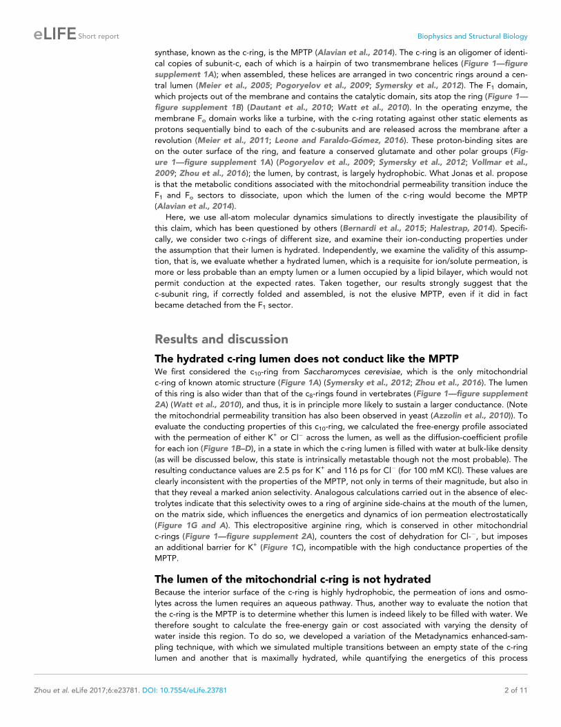
synthase, known as the c-ring, is the MPTP (Alavian et al., 2014). The c-ring is an oligomer of identi-
cal copies of subunit-c, each of which is a hairpin of two transmembrane helices (Figure 1—figure
supplement 1A); when assembled, these helices are arranged in two concentric rings around a cen-
tral lumen (Meier et al., 2005; Pogoryelov et al., 2009; Symersky et al., 2012). The F1 domain,
which projects out of the membrane and contains the catalytic domain, sits atop the ring (Figure 1—
figure supplement 1B) (Dautant et al., 2010; Watt et al., 2010). In the operating enzyme, the
membrane Fo domain works like a turbine, with the c-ring rotating against other static elements as
protons sequentially bind to each of the c-subunits and are released across the membrane after a
revolution (Meier et al., 2011; Leone and Faraldo-Gomez, 2016). These proton-binding sites are
on the outer surface of the ring, and feature a conserved glutamate and other polar groups (Fig-
ure 1—figure supplement 1A) (Pogoryelov et al., 2009; Symersky et al., 2012; Vollmar et al.,
2009; Zhou et al., 2016); the lumen, by contrast, is largely hydrophobic. What Jonas et al. propose
is that the metabolic conditions associated with the mitochondrial permeability transition induce the
F1 and Fo sectors to dissociate, upon which the lumen of the c-ring would become the MPTP
(Alavian et al., 2014).
Here, we use all-atom molecular dynamics simulations to directly investigate the plausibility of
this claim, which has been questioned by others (Bernardi et al., 2015; Halestrap, 2014). Specifi-
cally, we consider two c-rings of different size, and examine their ion-conducting properties under
the assumption that their lumen is hydrated. Independently, we examine the validity of this assump-
tion, that is, we evaluate whether a hydrated lumen, which is a requisite for ion/solute permeation, is
more or less probable than an empty lumen or a lumen occupied by a lipid bilayer, which would not
permit conduction at the expected rates. Taken together, our results strongly suggest that the
c-subunit ring, if correctly folded and assembled, is not the elusive MPTP, even if it did in fact
became detached from the F1 sector.
Results and discussion
The hydrated c-ring lumen does not conduct like the MPTPWe first considered the c10-ring from Saccharomyces cerevisiae, which is the only mitochondrial
c-ring of known atomic structure (Figure 1A) (Symersky et al., 2012; Zhou et al., 2016). The lumen
of this ring is also wider than that of the c8-rings found in vertebrates (Figure 1—figure supplement
2A) (Watt et al., 2010), and thus, it is in principle more likely to sustain a larger conductance. (Note
the mitochondrial permeability transition has also been observed in yeast (Azzolin et al., 2010)). To
evaluate the conducting properties of this c10-ring, we calculated the free-energy profile associated
with the permeation of either K+ or Cl� across the lumen, as well as the diffusion-coefficient profile
for each ion (Figure 1B–D), in a state in which the c-ring lumen is filled with water at bulk-like density
(as will be discussed below, this state is intrinsically metastable though not the most probable). The
resulting conductance values are 2.5 ps for K+ and 116 ps for Cl� (for 100 mM KCl). These values are
clearly inconsistent with the properties of the MPTP, not only in terms of their magnitude, but also in
that they reveal a marked anion selectivity. Analogous calculations carried out in the absence of elec-
trolytes indicate that this selectivity owes to a ring of arginine side-chains at the mouth of the lumen,
on the matrix side, which influences the energetics and dynamics of ion permeation electrostatically
(Figure 1G and A). This electropositive arginine ring, which is conserved in other mitochondrial
c-rings (Figure 1—figure supplement 2A), counters the cost of dehydration for Cl-�, but imposes
an additional barrier for K+ (Figure 1C), incompatible with the high conductance properties of the
MPTP.
The lumen of the mitochondrial c-ring is not hydratedBecause the interior surface of the c-ring is highly hydrophobic, the permeation of ions and osmo-
lytes across the lumen requires an aqueous pathway. Thus, another way to evaluate the notion that
the c-ring is the MPTP is to determine whether this lumen is indeed likely to be filled with water. We
therefore sought to calculate the free-energy gain or cost associated with varying the density of
water inside this region. To do so, we developed a variation of the Metadynamics enhanced-sam-
pling technique, with which we simulated multiple transitions between an empty state of the c-ring
lumen and another that is maximally hydrated, while quantifying the energetics of this process
Zhou et al. eLife 2017;6:e23781. DOI: 10.7554/eLife.23781 2 of 11
Short report Biophysics and Structural Biology

(Materials and methods). The resulting free-energy profile (Figure 2A) reveals that a metastable
state does exist in which the lumen of the mitochondrial c-ring is filled with water, at bulk-like density
(Figure 2B). However, this water-filled state is approximately 15 kcal/mol less favorable than a sec-
ond state in which most of the lumen of the c-ring is void of any water (Figure 2B). That is, the prob-
ability that the c-ring lumen exists in a ‘conducting state’ is negligible – a dehydrated, non-
conducting state is much more probable, by multiple orders of magnitude.
The lumen of the mitochondrial c-ring is plugged by lipid moleculesIrrespective of the question of hydration, the notion that the c-ring is the MPTP implicitly assumes
that lipid molecules are somehow excluded from its lumen as c-subunits gradually assemble to form
a ring. There is, however, no evidence that this is the case; to the contrary, detergent molecules are
often observed in the lumen in X-ray structures of c-rings (Pogoryelov et al., 2010; Schulz et al.,
Figure 1. Evaluation of the ion-conducting properties of the c-ring lumen, assuming a hydrated state. (A) Cross-section of the c10-ring of the
mitochondrial ATP synthase from S. cerevisiae. The surface of the protein is colored as follows: Lys and Arg, blue; Asp and Glu, red; other polar
residues (and protonated Glu), green; other residues, grey. The ring is oriented such that the interface with the F1 domain, inside the mitochondrial
matrix, is up. (B) Molecular simulation system employed to study the properties of the c10-ring, shown again in cross-section (blue). The ring is
embedded in a model phospholipid bilayer, in 100 mM KCl. K+ and Cl- ions are shown as yellow and green spheres, respectively. Note the c-ring
lumen is hydrated. (C, D) Potential-of-mean-force (G(z), PMF) and diffusion-coefficient profiles for the permeation of either K+ or Cl- across the lumen of
the c10-ring (Materials and methods). The lack of binding sites for K+ and the 3 kcal/mol free-energy barrier explain the modest K+ conductance;
permeation by Cl-, by contrast, is strongly favored electrostatically. (E, F) Same as (A, B), for a variant of the c13-ring from Bacillus pseudofirmus
(Figure 1—figure supplement 2B,C), whose lumen is wider than that of the c10-ring. (G) Free-energy of selectivity for Cl- and against K+ by the c10-ring
lumen, examined with and without electrolyte. The selectivity profile, DG(z), was calculated by subtracting the individual PMF profiles, G(z), in each case.
The marked increase in Cl- selectivity toward the lumen entrance, on the matrix side, confirms the strong electrostatic influence of a ring of arginine
residues. (H) Same as (G), for the variant of the c13-ring from B. pseudofirmus. Despite its wider lumen, this ring is also markedly anion selective, unlike
the MPTP.
DOI: 10.7554/eLife.23781.002
The following figure supplements are available for figure 1:
Figure supplement 1. Structure of the F1c10-ring subcomplex of the yeast mitochondrial ATP synthase.
DOI: 10.7554/eLife.23781.003
Figure supplement 2. Comparison of c-rings.
DOI: 10.7554/eLife.23781.004
Zhou et al. eLife 2017;6:e23781. DOI: 10.7554/eLife.23781 3 of 11
Short report Biophysics and Structural Biology

2013; Matthies et al., 2014), and AFM images of two-dimensional ring arrays also indicate the
lumen might be occluded (Meier et al., 2001). To conclusively evaluate this question, we carried out
three independent simulations in which the mitochondrial c-ring lacked two of the c-subunits (Fig-
ure 3). This open c-ring resembles the kind of assembly intermediate that has been observed in
AFM micrographs of prokaryotic c-rings (Muller et al., 2001; Pogoryelov et al., 2005). Initially, we
assumed the lumen of the c-ring to be hydrated (Figure 2B), as this is the necessary feature of the
hypothesized conducting form. As shown in Figure 3A–C, however, the three simulations consis-
tently demonstrate that all this water is quickly displaced by lipid molecules gradually entering the
c-ring lumen; ultimately, these lipid molecules occupy the totality of the pore, in a bilayer-like
arrangement akin to that outside the c-ring (Figure 3C). There is no reason to assume that the incor-
poration of the two missing c-subunits would alter the lipid occupancy of the lumen, which would
therefore remain in a non-conducting state when the c-ring assembles completely.
Enlargement of c-ring lumen does not explain properties of MPTPOne of the elements in the proposal that the mitochondrial c-ring is the MPTP is that the size of
the lumen might expand upon dissociation of the F1 sector (Alavian et al., 2014). This expansion is
not entirely implausible; indeed, subtle variations in the amino-acid sequence of the c-subunits can
Figure 2. Evaluation of the likelihood of hydration of the c-ring lumen. (A) Free energy as a function of the water
density inside the lumen of the mitochondrial c10-ring, calculated with a variant of the Metadynamics technique
(Materials and methods), for a simulation system analogous to that shown in Figure 1B. The density value for bulk
water (for the CHARMM36 forcefield) is indicated in red. Error bars reflect the differences between two profiles
calculated using different halves of all simulation data. (B) Snapshots of the c-ring lumen in the two metastable
minima detected in the free-energy profile shown in panel (A), i.e. a non-conducting, de-wetted state (left), and a
water-filled, putatively-conducting state (right), whose properties are characterized in Figure 1. For clarity, several
c-subunits in the c-ring are omitted, as are the lipid bilayer and the solvent outside the lumen. Hydrogen atoms in
water (red spheres) are also omitted. Note that in the non-conducting state (left), the two regions at the entrance
of the hydrophobic portion of the lumen are hydrated; hence, the density value for this state in free-energy profile
in panel (A) is not zero. (C, D) Same as (A, B), for the variant of the prokaryotic c13-ring from the B. pseudofirmus
ATP synthase.
DOI: 10.7554/eLife.23781.005
The following figure supplement is available for figure 2:
Figure supplement 1. Graphical definition of the density collective-variable with cylindrical geometry.
DOI: 10.7554/eLife.23781.006
Zhou et al. eLife 2017;6:e23781. DOI: 10.7554/eLife.23781 4 of 11
Short report Biophysics and Structural Biology

destabilize the native state of a c-ring, and favor the assembly of larger c-rings with a slightly greater
number of c-subunits (Pogoryelov et al., 2012). With this in mind, we repeated all calculations pre-
sented thus far for a variant of the c13-ring from the bacterium Bacillus pseudofirmus (Figure 1E–F).
Among the ATP synthase c-rings of known structure, this specific c13-ring features the widest lumen
(Preiss et al., 2010). Thus, although no process akin to the permeability transition has been
described in bacteria, we reasoned this ring would be a suitable system to assess whether an
expanded mitochondrial c-ring might be the MPTP. For a better comparison, two of the residues lin-
ing the lumen of the ring were mutated, to make it more similar to the mitochondrial c-ring (Fig-
ure 1—figure supplement 2B,C). The results from this new set of calculations, however, confirm the
conclusions drawn above. Because the lumen is wider, the permeability of the hydrated state is sig-
nificantly larger, but this state remains highly anion-selective (Figure 1H), unlike the MPTP. More-
over, this water-filled state is still disfavored, relative to the empty lumen, albeit to a lesser degree
than in the c10-ring (Figure 2C,D). Importantly, though, independent simulations of an assembly
intermediate lacking two c-subunits demonstrate that the preferred state is again one in which lipid
molecules occlude the lumen, in a bilayer arrangement (Figure 3D–F).
ConclusionsThis study demonstrates that the characteristics of the lumen of the mitochondrial c-ring are incom-
patible with the conducting properties of the MPTP. Consistent with the fact that the F1 domain
Figure 3. Evidence that lipid molecules block the lumen of the c-ring. Three independent molecular dynamics simulations were carried out of assembly
intermediates of the c10- and c13-rings lacking two of the c-subunits. Initially, the lumen of these c-rings was hydrated at bulk-like density. (A) Time-
series of the number of non-hydrogen atoms from either water (dashed lines) or lipid molecules (solid lines) inside the lumen of the mitochondrial
c-ring. The volume consider for this count is a cylinder of radius 7 A and height 32 A, approximately centered in the middle of the membrane. Running
averages are shown for each of the three simulation, colored in black, red and blue, respectively, with the raw data shown in the background in grey.
The number of atoms equivalent to 4 POPC lipid molecules is indicated, for reference. (B) Snapshots of the molecular system, at the beginning (left)
and at the end (right) of the simulation. Water molecules (red) are progressively displaced by lipid molecules (shown in yellow) entering the lumen
laterally. (C) Side-view of the open c-ring at the end of the one of the simulations. The lipid molecules inside the lumen preserve a bilayer arrangement,
and adapt to the specific features of the protein surface. (D, E, F) Same as (A, B, C), for the variant of the c13-ring from B. pseudofirmus. The volume
considered in panel (D) is a cylinder of radius 14 A and height 34 A.
DOI: 10.7554/eLife.23781.007
Zhou et al. eLife 2017;6:e23781. DOI: 10.7554/eLife.23781 5 of 11
Short report Biophysics and Structural Biology

does not actually seal the entrance to the lumen of the c-ring in the fully assembled, operating
enzyme (Figure 1—figure supplement 1C), the interior of the ring is not water-filled, but occupied
by lipid molecules; that is, it is in a non-conducting state, thereby precluding the dissipation of the
ion-motive-force that powers ATP synthesis. Our results also show that even if the c-ring became dis-
sociated from the F1 domain (Alavian et al., 2014), despite the nanomolar affinity of this interaction
(Pogoryelov et al., 2008), and even if its lumen was to become hydrated, under some hypothetical
in vitro conditions, the conductance levels sustained by the c-ring would be unlike those of the
MPTP.
Admittedly, this study does not rule out that an as-yet-unknown molecular structure consisting of
c-subunits, distinct from that of the c-ring, matches the properties of the MPTP. However, it is also
worth noting that no evidence of such alternative structure exists, to our knowledge. Structural stud-
ies of the c-subunit and its oligomers date back two decades; these studies include two- and three-
dimensional crystallography, liquid and solid-state NMR spectroscopy, atomic-force microscopy, and
more recently, single-particle cryo-electron microscopy (Meier et al., 2011; Leone and Faraldo-
Gomez, 2016; von Ballmoos et al., 2009). Across a wide variety of species and experimental condi-
tions, the consistent conclusion from these studies is that the c-subunits assemble in ring-like oligom-
ers such as those analyzed here. This conclusion holds true for wild-type, mutagenized and inhibited
forms of the c-ring, and for a variety of expression systems (including cell-free). Moreover, to our
knowledge no apparent discrepancy exists between this set of structural data and the even larger
collection of biochemical and functional measurements gathered for this enzyme or its constituent
elements. Indeed, in the instances where clear thermodynamic data has been attained, for example,
on Na+/H+ binding to isolated c-rings, it was found to be not only quantitatively consistent with
kinetic measurements for the complete enzyme functioning in membranes, but also explicable based
on existing structures and theoretical considerations (Murata et al., 2008; Leone et al., 2015). In
absence of concrete experimental evidence of the existence of an alternative architecture, the logi-
cal conclusion from this body of work and the present study is therefore that the c-ring from the
mitochondrial ATP synthase is not the MPTP.
Materials and methods
Simulation systems and general specificationsThe molecular dynamics (MD) simulation systems used to study the mitochondrial c10-ring from S.
cerevisiae were adapted from a system used in earlier studies of the proton-binding sites in this
structure (Symersky et al., 2012; Zhou et al., 2016). Briefly, the c10-ring had been embedded in a
hydrated (and pre-equilibrated) palmitoyl-oleoyl-phosphatydyl-choline (POPC) lipid bilayer, using
GRIFFIN (Staritzbichler et al., 2011), and equilibrated extensively through a series of restrained and
unrestrained MD simulations. Here, the four lipid molecules occupying the center of the c-ring were
removed, and instead the lumen was initially filled with water molecules. Several alternative systems
were prepared: for the complete ring, one system included 100 mM KCl, while the other included
counter-ions only; a simulation system was also prepared in which the c-ring lacks two c-subunits,
thus resembling an assembly intermediate. After making these changes, energy minimizations,
restrained and unrestrained molecular dynamics simulations were used to equilibrate these systems.
The simulation systems for the c13-ring from B. pseudofirmus OF4 were also adapted from a sys-
tem used in a previous study of its proton-binding sites (Leone et al., 2010). The central lumen of
this ring is about two-fold wider than that of the mitochondrial c10 ring, but the amino-acid composi-
tion of the protein surface lining this lumen differs. Thus, to make the c13-ring more similar, we intro-
duced two mutations (K26L and E30Q) at the entrance of the lumen, on the side that would be
exposed to the mitochondrial interior or matrix. In addition, the POPC lipid molecules inside the
central lumen were also replaced by water. The resulting systems (with and without added 100 mM
KCl, for the complete c13-ring, plus a system missing two c-subunits) were equilibrated analogously
to the c10-ring systems.
All MD simulations were performed with NAMD2 (Phillips et al., 2005), using the CHARMM36
forcefield (MacKerell et al., 1998; Best et al., 2012; Klauda et al., 2010), at constant temperature
(298 K) and pressure (1 atm) and with periodic boundary conditions in all directions. Long-range
electrostatic interactions were calculated using the Particle-Mesh-Ewald algorithm, with a real-space
Zhou et al. eLife 2017;6:e23781. DOI: 10.7554/eLife.23781 6 of 11
Short report Biophysics and Structural Biology

cut-off of 12 A. Van der Waals interactions were modeled with a Lennard-Jones potential, cut-off at
12 A using a smooth switching function taking effect at a distance of 10 A.
Ion conductance calculationsFollowing Hummer and co-workers (Zhu and Hummer, 2012), the conductance g of the c10-ring
lumen was calculated using the expression:
g¼q2C S
kBT
Z
Z2
Z1
expG zð Þ
kBT
� �
1
D zð ÞdZ
2
4
3
5
�1
where q is the charge of the permeant ion, C is the bulk ion concentration (100 mM), S is the effec-
tive cross-section area of the lumen (201 A2 for c10-ring and 616 A2 for c13-ring), kB is the Boltzmann
constant and T the temperature. G zð Þ is the one-dimensional free-energy landscape reflecting the
translocation of the ion across the protein, and D zð Þ is the position-dependent diffusion coefficient
of the ion along the lumen; both G zð Þ and D zð Þ were calculated using MD simulations, as described
below. It is important to note that this computational framework has been shown to produce con-
ductance values that are in good agreement with experimental measurements (Zhu and Hummer,
2012).
Free-energy profiles for K+ and Cl� permeationThe Adaptive-Biasing-Force (ABF) method (Henin et al., 2010; Fiorin et al., 2013) was used to cal-
culate the potential-of-mean-force associated with the translocation of either K+ and Cl+ along the
lumen of the c-ring, or G zð Þ, for each of the simulation systems described above. The lumen of the
c-ring was filled with water (at bulk-like density) throughout these calculations. (It was not required
to impose this condition, as this state is metastable and sufficiently long-lived to observe tens of per-
meation events). The reaction coordinate was the Z-coordinate of one K+ or one Cl- ion, defined rel-
atively to the Z-coordinate of the center-of-mass of a ring of Ca atoms in the protein (from
residues A22 in the c10-ring, and from A18 in the c13-ring). The permeating ion was confined to a
cylindrical volume (through flat-bottom restraining potentials) whose center and axis coincide with
the center and axis of the lumen (defined by two additional rings of Ca atoms), and whose radius is
slightly larger, namely 8 A for the c10-ring, and 14 A for the c13-ring. The length of the cylinder along
the Z-coordinate was 100 A, therefore projecting into the bulk solution on both sides of the
membrane.
For each ion type and system, two ABF simulations of 300 ns each were carried out, with the per-
meant ion starting on opposite sides of the membrane; in the course of each of these trajectories,
the ions traverse the lumen multiple times, in both directions. The range in the Z-coordinate to be
sampled was divided up in 0.2 A bins, and 1000 unbiased samples were collected for each bin
before the estimated biasing force was applied and updated. A third simulation was then conducted
similarly for 100 ns to combine and refine the bias potentials calculated from the first two simula-
tions. The free-energy profiles shown in Figure 1 reflect the outcome of these final calculations,
while the error bands reflect the differences between the two initial profiles (averaged over the
length of the profile).
Diffusion profiles for K+ and Cl� across the lumenTo calculate the diffusion coefficient of the permeant ion as a function of its position along the c-ring
lumen, or D zð Þ, 51 independent simulations of 10 ns each were carried out for each system. In each
of these simulations, the permeant ion was confined (through flat-bottom restraining potentials) in a
disk of 2 A in length (and radius as specified above), whose center varies in the Z-direction from �50
to 50 A (relative to the protein, as described above). The starting configurations for each of these
simulations were obtained from the ABF trajectories. The position-dependent diffusion coefficient
was calculated a posteriori from the expression (Zhu and Hummer, 2012):
D zð Þ ¼var zð Þ
t zð Þ
Zhou et al. eLife 2017;6:e23781. DOI: 10.7554/eLife.23781 7 of 11
Short report Biophysics and Structural Biology

where the numerator is the variance of the Z-coordinate of the ion (relative to the protein), i.e.
var zð Þ ¼ z2 �
� zh i2, and t zð Þ is the characteristic time of its autocorrelation function, i.e.:
tðzÞ ¼
Z
¥
0
dzð0Þ dzðtÞh i dt = varðzÞ
where dz tð Þ ¼ z tð Þ� zh i, and t is the so-called lag time. In our simulations, the values of t zð Þ are all
well converged for t > 80–90 ps. The diffusion profiles shown in Figure 1 are (cubic-spline) interpola-
tions of the 51 values of D zð Þ obtained using this approach; the error bars reflect the differences
between values obtained from different halves of the simulations.
Density-biased multiple-walker metadynamicsThe Metadynamics method was used to induce the wetting and de-wetting of the lumen of the c10and c13-rings reversibly, and to calculate the associated free-energy changes. The collective variable
biased in these simulations is the water density in the volume of the lumen. Following Feig and co-
workers, we express this density as (Mirjalili and Feig, 2015):
�¼
P
i � Xið Þ
V
where 0� � Xið Þ � 1 is a weight function of the Cartesian coordinates of a given atom i at a given
time, whose value depends on whether that atom is inside (� Xið Þ~1) or outside (� Xið Þ~0) the volume
V considered. Given the geometry of the lumen of the c-rings, we used a weight function � Xð Þ that
defines a cylinder of radius R, whose axis and center coincide with the those of the lumen, and which
extends from �Z to þZ. Importantly, the boundaries of this cylinder are not abrupt, but rather
smoothened over an interval DR and DZ around R and �Z (Figure 2—figure supplement 1A). More
specifically:
�ðXÞ ¼ �zðzÞ �RðrÞ ¼1 if r < R�DR=2 and zj j < Z�DZ=20 if r > RþDR=2 or zj j > ZþDZ=2
�ZðzÞ �RðrÞ otherwise
8
<
:
where �S sð Þ (s denotes z or r and S denotes R or Z) is a switching function whose value varies
smoothly from 1 to 0 in the intervals S�DS=2� s� SþDS=2, constructed so that its derivative is 0 at
the outer boundaries of the cylinder (Figure 2—figure supplement 1B):
�S sð Þ ¼SþDS=2ð Þ2�s2
h i2
SþDS=2ð Þ2þ2s2 � 3 S�DS=2ð Þ2h i
SþDS=2ð Þ2� S�DS=2ð Þ2h i3
Lastly, the effective volume V of this cylinder is:
V ¼
Z
� Xð Þ dX¼
Z
ZþDZ=2
� ZþDZ=2ð Þ
�Z zð Þ dz
Z
RþDR=2
0
2pr �R rð Þ dr
As in standard Metadynamics, a time-dependent biasing potential Ub acting on the collective-
coordinate � was adaptively constructed throughout the simulation; at convergence, the free-energy
profile is the negative of this potential. At a given time, the forces acting on a given atom i due to
this biasing potential can be derived using the chain rule, that is:
Fbi ¼�rUb �ð Þ ¼�
dUb �ð Þ
d�
q�
qxi;q�
qyi;q�
qzi
� �
where xi, yi and zi are the coordinates of atom i. Note that our definition of the weight function � Xð Þ
is such that Fbi is non-zero only if atom i is found within the smooth boundaries of the cylinder,
that is, in the region where the switching functions �Z zið Þ and �R rið Þ are non-zero.
Zhou et al. eLife 2017;6:e23781. DOI: 10.7554/eLife.23781 8 of 11
Short report Biophysics and Structural Biology

The collective variable � defined above is a function of all the atoms considered in the density cal-
culation. In our case, these atoms are all the water oxygen atoms in the simulation system, which in
principle would make the calculation of the density collective variable and related biasing forces pro-
hibitively slow. However, because at a given time the value of the weight function (and associated
forces) is non-zero only for the atoms found inside the target volume, we can speed up the calcula-
tions by introducing a ‘neighbor search’ in the algorithm, whereby a list of water molecules within
and in the vicinity of that volume is created and updated regularly, but not at every simulation step
(Figure 2—figure supplement 1A). In addition, we note that unlike earlier work (Mirjalili and Feig,
2015), our implementation defines � Xð Þ in reference to the protein coordinates, that is, not in abso-
lute Cartesian space, and therefore the tumbling and diffusion of the ring in the membrane is unre-
stricted during the simulations.
The geometric definitions of the target volumes considered for quantification of the free-energies
of hydration of the c10 and c13-ring lumens are indicated in Figure 2—figure supplement 1C,D. The
Metadynamics simulations were carried out using eight concurrent replicas, or walkers, which update
and share a common bias potential but sample different configurations. The biasing potential
applied to the density variable � consisted of a series of Gaussian functions of width 0.0005 A�3,
added in 4-ps intervals. The height of the Gaussians was gradually diminished throughout the simula-
tions, ultimately reaching a value of 0.0035 kcal/mol; the free-energy profiles were obtained by time-
averaging the biasing potential after this time-point (Marinelli et al., 2009). The total simulation
times per walker were 140 ns for the c10-ring (using the last 50 ns for analysis), and 120 ns for the
c13-ring (using the last 80 ns for analysis). The ‘neighbor search’ comprised a region that is 4 A larger
than the target volume in all directions, and the list of atoms therein was updated every 200 simula-
tion steps, resulting in a ~ 1000-fold speed-up of the calculations.
AcknowledgementsThis work was funded by the Division of Intramural Research of the National Heart, Lung and Blood
Institute, National Institutes of Health.
Additional information
Funding
Funder Author
National Heart, Lung, andBlood Institute
Wenchang ZhouFabrizio MarinelliJose D Faraldo-Gomez
The funders had no role in study design, data collection and interpretation, or thedecision to submit the work for publication.
Author contributions
WZ, Formal analysis, Investigation, Writing—original draft, Writing—review and editing; FM, Soft-
ware, Formal analysis, Investigation, Methodology, Writing—original draft, Writing—review and edit-
ing; CN, Formal analysis, Investigation, Writing—original draft; JDF-G, Conceptualization, Formal
analysis, Supervision, Investigation, Writing—original draft, Writing—review and editing
Author ORCIDs
Wenchang Zhou, http://orcid.org/0000-0003-0397-1032
Jose D Faraldo-Gomez, http://orcid.org/0000-0001-7224-7676
ReferencesAlavian KN, Beutner G, Lazrove E, Sacchetti S, Park HA, Licznerski P, Li H, Nabili P, Hockensmith K, Graham M,Porter GA, Jonas EA. 2014. An uncoupling channel within the c-subunit ring of the F1FO ATP synthase is themitochondrial permeability transition pore. PNAS 111:10580–10585. doi: 10.1073/pnas.1401591111, PMID: 24979777
Zhou et al. eLife 2017;6:e23781. DOI: 10.7554/eLife.23781 9 of 11
Short report Biophysics and Structural Biology
Azzolin L, von Stockum S, Basso E, Petronilli V, Forte MA, Bernardi P. 2010. The mitochondrial permeabilitytransition from yeast to mammals. FEBS Letters 584:2504–2509. doi: 10.1016/j.febslet.2010.04.023, PMID: 20398660
Bernardi P, Forte M. 2007. The mitochondrial permeability transition pore. Novartis Foundation Symposium 287:157–164. PMID: 18074637
Bernardi P, Rasola A, Forte M, Lippe G. 2015. The mitochondrial permeability transition pore: channel formationby F-ATP synthase, integration in signal transduction, and role in pathophysiology. Physiological Reviews 95:1111–1155. doi: 10.1152/physrev.00001.2015, PMID: 26269524
Best RB, Zhu X, Shim J, Lopes PE, Mittal J, Feig M, Mackerell AD. 2012. Optimization of the additive CHARMMall-atom protein force field targeting improved sampling of the backbone j, y and side-chain c(1) and c(2)dihedral angles. Journal of Chemical Theory and Computation 8:3257–3273. doi: 10.1021/ct300400x,PMID: 23341755
Dautant A, Velours J, Giraud MF. 2010. Crystal structure of the mg�ADP-inhibited state of the yeast F1c10-ATPsynthase. The Journal of Biological Chemistry 285:29502–29510. doi: 10.1074/jbc.M110.124529, PMID: 20610387
Fiorin G, Klein ML, Henin J. 2013. Using collective variables to drive molecular dynamics simulations. MolecularPhysics 111:3345–3362. doi: 10.1080/00268976.2013.813594
Halestrap AP. 2009. What is the mitochondrial permeability transition pore? Journal of Molecular and CellularCardiology 46:821–831. doi: 10.1016/j.yjmcc.2009.02.021, PMID: 19265700
Halestrap AP. 2014. The C ring of the F1Fo ATP synthase forms the mitochondrial permeability transition pore: acritical appraisal. Frontiers in Oncology 4:234. doi: 10.3389/fonc.2014.00234, PMID: 25202683
Henin J, Fiorin G, Chipot C, Klein ML. 2010. Exploring multidimensional free energy landscapes using time-dependent biases on collective variables. Journal of Chemical Theory and Computation 6:35–47. doi: 10.1021/ct9004432, PMID: 26614317
Klauda JB, Venable RM, Freites JA, O’Connor JW, Tobias DJ, Mondragon-Ramirez C, Vorobyov I, MacKerell AD,Pastor RW. 2010. Update of the CHARMM all-atom additive force field for lipids: validation on six lipid types.The Journal of Physical Chemistry B 114:7830–7843. doi: 10.1021/jp101759q, PMID: 20496934
Leone V, Faraldo-Gomez JD. 2016. Structure and mechanism of the ATP synthase membrane motor inferredfrom quantitative integrative modeling. The Journal of General Physiology 148:441–457. doi: 10.1085/jgp.201611679, PMID: 27821609
Leone V, Krah A, Faraldo-Gomez JD. 2010. On the question of hydronium binding to ATP-synthase membranerotors. Biophysical Journal 99:L53–L55. doi: 10.1016/j.bpj.2010.07.046, PMID: 20923632
Leone V, Pogoryelov D, Meier T, Faraldo-Gomez JD. 2015. On the principle of ion selectivity in Na+/H+-coupledmembrane proteins: experimental and theoretical studies of an ATP synthase rotor. PNAS 112:E1057–E1066.doi: 10.1073/pnas.1421202112, PMID: 25713346
MacKerell AD, Bashford D, Bellott M, Dunbrack RL, Evanseck JD, Field MJ, Fischer S, Gao J, Guo H, Ha S,Joseph-McCarthy D, Kuchnir L, Kuczera K, Lau FT, Mattos C, Michnick S, Ngo T, Nguyen DT, Prodhom B,Reiher WE, et al. 1998. All-atom empirical potential for molecular modeling and dynamics studies of proteins.The Journal of Physical Chemistry B 102:3586–3616. doi: 10.1021/jp973084f, PMID: 24889800
Marinelli F, Pietrucci F, Laio A, Piana S. 2009. A kinetic model of Trp-cage folding from multiple biasedmolecular dynamics simulations. PLoS Computational Biology 5:e1000452. doi: 10.1371/journal.pcbi.1000452,PMID: 19662155
Matthies D, Zhou W, Klyszejko AL, Anselmi C, Yildiz O, Brandt K, Muller V, Faraldo-Gomez JD, Meier T. 2014.High-resolution structure and mechanism of an F/V-hybrid rotor ring in a Na+-coupled ATP synthase. NatureCommunications 5:5286. doi: 10.1038/ncomms6286, PMID: 25381992
Meier T, Faraldo-Gomez JD, Borsch M. 2011. ATP Synthase: a paradigmatic molecular machine. In: Frank J (Ed.).Molecular Machines in Biology. Cambridge, UK: Cambridge University Press.pp 208–238.
Meier T, Matthey U, Henzen F, Dimroth P, Muller DJ. 2001. The central plug in the reconstituted undecameric ccylinder of a bacterial ATP synthase consists of phospholipids. FEBS Letters 505:353–356. doi: 10.1016/S0014-5793(01)02837-X, PMID: 11576527
Meier T, Polzer P, Diederichs K, Welte W, Dimroth P. 2005. Structure of the rotor ring of F-Type Na+-ATPasefrom Ilyobacter tartaricus. Science 308:659–662. doi: 10.1126/science.1111199, PMID: 15860619
Mirjalili V, Feig M. 2015. Density-biased sampling: a robust computational method for studying pore formationin membranes. Journal of Chemical Theory and Computation 11:343–350. doi: 10.1021/ct5009153,PMID: 25620896
Murata T, Yamato I, Kakinuma Y, Shirouzu M, Walker JE, Yokoyama S, Iwata S. 2008. Ion binding and selectivityof the rotor ring of the Na+-transporting V-ATPase. PNAS 105:8607–8612. doi: 10.1073/pnas.0800992105,PMID: 18559856
Muller DJ, Dencher NA, Meier T, Dimroth P, Suda K, Stahlberg H, Engel A, Seelert H, Matthey U. 2001. ATPsynthase: constrained stoichiometry of the transmembrane rotor. FEBS Letters 504:219–222. doi: 10.1016/S0014-5793(01)02708-9, PMID: 11532457
Phillips JC, Braun R, Wang W, Gumbart J, Tajkhorshid E, Villa E, Chipot C, Skeel RD, Kale L, Schulten K. 2005.Scalable molecular dynamics with NAMD. Journal of Computational Chemistry 26:1781–1802. doi: 10.1002/jcc.20289, PMID: 16222654
Pogoryelov D, Klyszejko AL, Krasnoselska GO, Heller EM, Leone V, Langer JD, Vonck J, Muller DJ, Faraldo-Gomez JD, Meier T. 2012. Engineering rotor ring stoichiometries in the ATP synthase. PNAS 109:E1599–E1608. doi: 10.1073/pnas.1120027109, PMID: 22628564
Zhou et al. eLife 2017;6:e23781. DOI: 10.7554/eLife.23781 10 of 11
Short report Biophysics and Structural Biology

Pogoryelov D, Krah A, Langer JD, Yildiz O, Faraldo-Gomez JD, Meier T. 2010. Microscopic rotary mechanism ofion translocation in the F(o) complex of ATP synthases. Nature Chemical Biology 6:891–899. doi: 10.1038/nchembio.457, PMID: 20972431
Pogoryelov D, Nikolaev Y, Schlattner U, Pervushin K, Dimroth P, Meier T. 2008. Probing the rotor subunitinterface of the ATP synthase from Ilyobacter tartaricus. FEBS Journal 275:4850–4862. doi: 10.1111/j.1742-4658.2008.06623.x, PMID: 18721138
Pogoryelov D, Yildiz O, Faraldo-Gomez JD, Meier T. 2009. High-resolution structure of the rotor ring of aproton-dependent ATP synthase. Nature Structural & Molecular Biology 16:1068–1073. doi: 10.1038/nsmb.1678, PMID: 19783985
Pogoryelov D, Yu J, Meier T, Vonck J, Dimroth P, Muller DJ. 2005. The c15 ring of the Spirulina platensis F-ATPsynthase: F1/F0symmetry mismatch is not obligatory. EMBO Reports 6:1040–1044. doi: 10.1038/sj.embor.7400517, PMID: 16170308
Preiss L, Yildiz O, Hicks DB, Krulwich TA, Meier T. 2010. A new type of proton coordination in an F(1)F(o)-ATPsynthase rotor ring. PLoS Biology 8:e1000443. doi: 10.1371/journal.pbio.1000443, PMID: 20689804
Schulz S, Iglesias-Cans M, Krah A, Yildiz O, Leone V, Matthies D, Cook GM, Faraldo-Gomez JD, Meier T. 2013. Anew type of Na+-driven ATP synthase membrane rotor with a two-carboxylate ion-coupling motif. PLoS Biology11:e1001596. doi: 10.1371/journal.pbio.1001596, PMID: 23824040
Staritzbichler R, Anselmi C, Forrest LR, Faraldo-Gomez JD. 2011. GRIFFIN: a versatile methodology foroptimization of protein�lipid interfaces for membrane protein simulations. Journal of Chemical Theory andComputation 7:1167–1176. doi: 10.1021/ct100576m, PMID: 24707227
Symersky J, Pagadala V, Osowski D, Krah A, Meier T, Faraldo-Gomez JD, Mueller DM. 2012. Structure of the C(10) ring of the yeast mitochondrial ATP synthase in the open conformation. Nature Structural & MolecularBiology 19:485–491. doi: 10.1038/nsmb.2284, PMID: 22504883
Szabo I, Zoratti M. 1992. The mitochondrial megachannel is the permeability transition pore. Journal ofBioenergetics and Biomembranes 24:111–117. doi: 10.1007/BF00769537, PMID: 1380498
Vollmar M, Schlieper D, Winn M, Buchner C, Groth G. 2009. Structure of the c14 rotor ring of the protontranslocating chloroplast ATP synthase. Journal of Biological Chemistry 284:18228–18235. doi: 10.1074/jbc.M109.006916, PMID: 19423706
von Ballmoos C, Wiedenmann A, Dimroth P. 2009. Essentials for ATP synthesis by F1F0 ATP synthases. AnnualReview of Biochemistry 78:649–672. doi: 10.1146/annurev.biochem.78.081307.104803, PMID: 19489730
Watt IN, Montgomery MG, Runswick MJ, Leslie AG, Walker JE. 2010. Bioenergetic cost of making an adenosinetriphosphate molecule in animal mitochondria. PNAS 107:16823–16827. doi: 10.1073/pnas.1011099107,PMID: 20847295
Zhou W, Leone V, Krah A, Faraldo-Gomez JD. 2016. Predicted structures of the proton-bound membrane-embedded rotor rings of the Saccharomyces cerevisiae and Escherichia coli ATP Synthases . The Journal ofPhysical Chemistry B:6b08051. doi: 10.1021/acs.jpcb.6b08051
Zhu F, Hummer G. 2012. Theory and simulation of ion conduction in the pentameric GLIC channel. Journal ofChemical Theory and Computation 8:3759–3768. doi: 10.1021/ct2009279, PMID: 23413364
Zhou et al. eLife 2017;6:e23781. DOI: 10.7554/eLife.23781 11 of 11
Short report Biophysics and Structural Biology