Embed Size (px)
Citation preview
Proc. Nati. Acad. Sci. USAVol. 89, pp. 4986-4990, June 1992Biochemistry
An ATP-stabilized inhibitor of the proteasome is a component of the1500-kDa ubiquitin conjugate-degrading complexJAMES DRISCOLL, JUDITH FRYDMAN, AND ALFRED L. GOLDBERGDepartment of Cellular and Molecular Physiology, Harvard Medical School, Boston, MA 02115
Communicated by Eugene P. Kennedy, March 13, 1992
ABSTRACT Proteins cdjugated to ubiquitin are de-graded by a 26S (1500-kI)s proteolytic complex that, inreticulocyte extracts, can be fermed by the association of threefactors: CF-1, CF-2, and CF-3. One of these factors, CF-3, hasbeen shown to be the proteasome, a 650-kDa multicatalyticprotease complex. We have purified a 250-kDa inhibitor of theproteasome and shown that it corresponds to CF-2. In thepresence or absence of ATP, this factor inhibited hydrolysis bythe proteasome of both fluorogenic tetrapeptides and proteinsubstrates. When the inhibitor, proteasome, and CF-1 wereincubated together in the presence of ATP and Mg2+, degra-dation ofubiquitin-12'I-lysozyme occurred. Both the inhibitoryactivity and the ability to reconstitute ubiquitin-12'I-lysozymedegradation were very labile at 42C, but both activities werestabilized by ATP or a nonhydrolyzable ATP analog.SDS/PAGE indicated that the 250-kDa inhibitor fractioncontained a major subunit of 40 kDa (plus some minor bands).The '25I-labeled inhibitor and purified proteasome formed acomplex. When CF-1, ATP, and Mg2+ were also present, the125I-labeled inhibitor along with the proteasome formed acomplex of 1500 kDa. The inhibitor (CF-2) thus appears to bean ATP-binding component that regulates proteolysis withinthe 1500-kDa complex.
In eukaryotes, the degradation of most cellular proteins is anenergy-dependent process that requires a polypeptide cofac-tor, ubiquitin (1-3). The ligation of ubiquitin to cell proteinsmarks them for rapid hydrolysis (1, 2) by a very largeproteolytic complex that has been the subject ofmany studies(3-10). This 26S (1500-kDa) complex selectively degradesproteins conjugated to ubiquitin by an ATP-dependent pro-cess (3-7). Although this large structure has been studiedmost extensively from reticulocyte extracts (5, 6), it was notdetected following ATP depletion of the reticulocytes. How-ever, such a complex could be reconstituted in vitro in anATP-dependent reaction from three factors, referred to asCF-1, CF-2, and CF-3 (8). Eukaryotic cells also contain a650-kDa proteolytic complex (11-15), referred to as theproteasome (3, 12). This structure contains 13-15 subunits of20-33 kDa and multiple proteolytic sites, which cleave pep-tide bonds on the carboxyl side of hydrophobic, basic, oracidic amino acids (3, 11-18). Although the proteasome byitself does not degrade proteins conjugated to ubiquitin,immunoprecipitation or inactivation of the proteasome pre-vents hydrolysis of ubiquitinated proteins (17-19). The pro-teasome corresponds to one of the components, CF-3, of the1500-kDa proteolytic complex (3, 9, 10). The regulatory orcatalytic functions of the other two factors and their subunitcomposition have not been identified.
Incorporation of the proteasome into the ubiquitin conju-gate-degrading complex clearly alters the properties of theproteasome. When a reticulocyte lysate is fractionated withammonium sulfate, the proteasome (CF-3) is recovered in the
40-80% fraction, while CF-1 and CF-2 are precipitated with0-38%. In the presence of ATP and the 0-38% fraction, theproteasome is incorporated into a large complex that de-grades ubiquitin-conjugated proteins (9, 10). This structurecontains proteasome subunits, as shown immunologically(10), and the characteristic peptidase activities (10, 11) of theproteasome. In the 1500-kDa complex, these proteolyticactivities appear to be ATP-dependent (10), and the complexshows ATPase activity (20).Understanding the functioning of the proteasome within
the 1500-kDa complex and the mechanism for degradingubiquitin-conjugated proteins will require purification andcharacterization of its other two components. In the presentstudy, we have purified a 250-kDa ATP-stabilized factor thatinhibits the proteolytic activities of the proteasome andcorresponds to the "inhibitor of high molecular weight pro-teases" reported by Etlinger and coworkers (21, 22). Wedemonstrate that it is required to reconstitute degradation ofubiquitin-conjugated proteins and that it corresponds to CF-2of the 1500-kDa complex (8).
MATERIALS AND METHODSDEAE-cellulose (DE-52), CM-cellulose (CM-52), and phos-phocellulose (P11) were obtained from Whatman. Ubiquitin(bovine erythrocyte), ammonium sulfate, succinylleucylleu-cylvalyltyrosine 7-amido-4-methylcoumarin (Suc-LLVY-MCA), and ATP were from Sigma. Lysozyme (chicken egg)was from Bachem. Lysozyme was radiolabeled with Na125Iby the chloramine-T method (23). Ubiquitin-conjugating en-zymes (El, E2, and E3) were isolated by ubiquitin-Sepharoseaffinity column chromatography (24) and were used to pre-pare ubiquitin-1251-lysozyme conjugates (25). Mono Q anion-exchange (HR 10/10 orHR 5/5) and Superose 6 gel filtration(HR 10/30) columns were from Pharmacia. Protein concen-tration was determined with the Coomassie blue G-250 re-agent (Pierce).
Purifications. Rabbit reticulocytes induced by phenylhy-drazine injection were prepared as described previously (26)or were purchased from Green Hectares (Oregon, WI). Theywere depleted of ATP by incubation with 2,4-dinitrophenoland 2-deoxyglucose (26). Lysates were then prepared andsubjected to DE-52 chromatography. The protein eluted with0.5 M KCl (24) was concentrated by precipitation usingammonium sulfate at 80%o saturation, centrifuged at 10,000 xg for 20 min, and suspended in 20mM Tris-HCl, pH 7.6/1 mmdithiothreitol (buffer A). After extensive dialysis against thesame buffer, the protein (fraction II) was either stored at-80°C in 0.5 mM ATP or fractionated further.Fraction II (200 mg) was applied to a ubiquitin-Sepharose
column, and the ubiquitin-conjugating enzymes were specif-ically eluted (24) and used in making ubiquitin-125I-lysozyme(25). The unadsorbed fraction was brought to 38% saturationwith ammonium sulfate and mixed for 20 min, as described
Abbreviation: Suc-LLVY-MCA,7-amido-4-methylcoumarin.
succinylleucylleucylvalyltyrosineThe publication costs of this article were defrayed in part by page chargepayment. This article must therefore be hereby marked "advertisement"in accordance with 18 U.S.C. §1734 solely to indicate this fact.
4986
Proc. Natl. Acad. Sci. USA 89 (1992) 4987
(8). The precipitated proteins were collected by centrifuga-tion at 10,000 x g for 15 min. The pellet was resuspended inbufferA and brought again to 38% saturation with ammoniumsulfate. The precipitated material was collected as above andthen suspended in bufferA containing 10%o (vol/vol) glycerol.After dialysis against this buffer, the 0-38% pellet waschromatographed on a Mono Q anion-exchange columnequilibrated with buffer A containing 10%o glycerol. Theprotein was eluted with a 60-ml linear gradient of NaCl from20 to 400 mM. Further purification was achieved by a secondMono Q chromatographic step using a more narrow gradient,from 150 to 400 mM NaCl. The second Mono Q step yieldeda sharp peak of inhibitor activity. Fractions that inhibited thepeptidase activity of the proteasome were pooled, concen-trated, and then chromatographed on a Superose 6 (HR10/30) gel filtration column equilibrated in buffer A contain-ing 100 mM NaCl and 0.2 mM ATP. The column was run ata flow rate of 0.2 ml/min, and 1-ml fractions were collected.Fractions with inhibitory activity against the proteasomewere pooled and dialyzed against 20 mM KH2PO4, pH6.5/10o glycerol/1 mM dithiothreitol/i mM ATP (buffer B).The sample was then applied to a 2-ml phosphocellulosecolumn equilibrated in buffer B. The column was washedwith 4 ml of this buffer, followed by 4 ml of buffer Bcontaining either 20, 50, 100, 400, or 600 mM NaCl.To partially purify CF-1, fractions from the first Mono Q
step that were eluted from 100 to 240 mM NaCl were pooled,concentrated to 1 ml, and applied to a Superose 6 columnequilibrated in buffer A containing 100mM NaCl and 0.2mMATP. The fractions eluted at -600 kDa were used as the CF-1fraction.The proteasome was isolated from the supernatants of the
two 38% ammonium sulfate precipitations. The supernatantswere brought to 80o saturation with ammonium sulfate andmixed for 20 min. The precipitated protein was collected bycentrifugation, resuspended in buffer A, and dialyzed exten-sively against this buffer. The proteasome was isolated byMono Q anion-exchange chromatography followed by Su-perose 6 gel filtration (27). These preparations displayed onlythe characteristic subunits of 20-33 kDa.
Assays. Inhibition of the proteasome was measured bypreincubating individual column fractions with the protea-some in the presence of 1 mM ATP at 37°C for 10 min. Afterpreincubation, the reaction tubes were placed on ice, andeither 125I-lysozyme or Suc-LLVY-MCA was added. Reac-tions were carried out at 37°C for 60 min with 125I-lysozymeor 10 min with Suc-LLVY-MCA. Protein hydrolysis wasassayed by measuring production of radioactivity soluble in10% (wt/vol) trichloroacetic acid, and peptide hydrolysis bythe release of 7-amino-4-methylcoumarin (27). Degradationof ubiquitin-conjugated 1251-lysozyme was assayed at 37°Cfor 60 min. Reactions contained either 5 mM EDTA or 2 mMATP and 5 mM MgCl2 and were terminated by adding 10%otrichloroacetic acid.
Other Methods. Following the phosphocellulose step, theinhibitor (100 ,g in buffer B) was labeled with Na125I byincubation with Iodo-Beads (Pierce) for 15 min at 4°C, asrecommended by the manufacturer. The radiolabeled inhib-itor was then separated from free Na125I by gel filtration twiceon Sephadex G-25 columns and extensively dialyzed againstbuffer A prior to use. Denaturing polyacrylamide gel elec-trophoresis (SDS/PAGE) in 12% gels was performed asdescribed by Laemmli (28). Nondenaturing PAGE (4% or6%) was performed in the same buffer system but without theSDS.
RESULTSIsolation of the Inhibitor. To understand how the protea-
some is regulated in vivo and how it functions within theubiquitin conjugate-degrading complex, we attempted to
isolate factors that influence its activity. Fraction II fromATP-depleted reticulocytes was separated into fractions thatprecipitated with 0-38% or 40-80%o ammonium sulfate. Thelatter fraction was used to isolate the proteasome. Theproteasome obtained in this way showed appreciable activityagainst 1251-lysozyme and Suc-LLVY-MCA that was inde-pendent of ATP (9, 10). Neither the proteasome nor the0-38% fraction showed significant hydrolytic activity againstubiquitin-conjugated 125I-lysozyme (9, 10). However, as re-ported previously, ATP-dependent degradation of the ubiq-uitinated lysozyme was observed after the proteasome andthe 0-38% fraction were preincubated together in the pres-ence of ATP (data not shown).The 0-38% precipitated material then was fractionated by
Mono Q anion exchange, and each fraction was assayed forits ability to influence proteasome activity against Suc-LLVY-MCA or 125I-lysozyme. Column fractions were pre-incubated with the proteasome for 10 min and then eithersubstrate was added. None of the column fractions by itselfshowed significant hydrolytic activity, and most did notaffect proteasome activity (Fig. 1A). However, between 240and 280 mM NaCl, a peak of inhibitory activity was elutedthat decreased hydrolysis of 1251-lysozyme (data not shown)and of the peptide to similar extents. The active fractionswere further purified by a second Mono Q step using a morenarrow NaCl gradient.To purify the inhibitory activity further, the Mono Q active
fractions were pooled and chromatographed by gel filtration.The inhibitor was eluted as a sharp peak with an apparentmolecular mass of 250 kDa (Fig. 1B). The active fractionswere then pooled and assayed for inhibitory activity. Withincreasing inhibitor concentration, proteasome activity de-creased in a linear manner with both 125I-lysozyme andSuc-LLVY-MCA as substrates, although the degree of theinhibition was highly variable between preparations (Fig. 2).The Inhibitor Resembles Component CF-2 of the 1500-kDa
Complex. One component of the 1500-kDa proteolytic com-plex, CF-2, has been reported also to have a molecular massof 250 kDa (8). To test whether the inhibitor corresponded toCF-2, the inhibitor obtained by gel filtration was subjected tophosphocellulose chromatography (Fig. 1C). The inhibitoryactivity was recovered in the flowthrough and 20 mM NaCleluate-i.e., in the region where CF-2 activity was reportedby Eytan et al. (9). Individual phosphocellulose fractionswere then assayed for their ability to reconstitute rapiddegradation of ubiquitin-125I-lysozyme (Fig. 1C) when com-bined with the proteasome and CF-1-containing fraction. Bythemselves the latter components did not support this pro-cess. However, when fractions with inhibitory activity wereadded, ubiquitin-1251-lysozyme degradation increasedsharply. Other phosphocellulose fractions did not stimulatethis process.These results suggest strongly that the inhibitor corre-
sponds to CF-2 and thus is essential for hydrolysis of ubiq-uitin-conjugated proteins. One unusual property of CF-2 isthat it is quite labile upon heating to 42°C but is stabilized byATP (8). To test further whether the proteasome inhibitorcorresponded to CF-2, the purified inhibitor (from the phos-phocellulose column) was preincubated at 42°C with orwithout ATP. The proteasome was added, and after 10 min,peptidase activity was assayed. The degree of inhibitiondecreased rapidly during preincubation without any nucleo-tide added, but the presence of either ATP (Fig. 3) or anonhydrolyzable ATP analog, adenosine 5'-[f3,'y-imido]-triphosphate (data not shown), prevented this loss of activity.Furthermore, the ability of this fraction to reconstitute ubiq-uitin-conjugate degradation also decreased rapidly duringincubation at 42TC (Fig. 3), and the addition of ATP oradenosine 5'-[,B,y-imido]triphosphate (data not shown)blocked this inactivation. Since the inhibitory activity and
Biochemistry: Driscoll et al.
Proc. Natl. Acad. Sci. USA 89 (1992)
1 [.o
1Cr
0.8-
0.6F
0.41
0.2
a}
E
79
0
U)
a. -
>ZI.ti
a)
2
2000 670 440 200
: : *.* :-.
:. . :-.: : : *-.: *. : *. : *--.
* . : *- .-- *-- .
.1.==10 20
Fraction Number30 4
Fraction Number
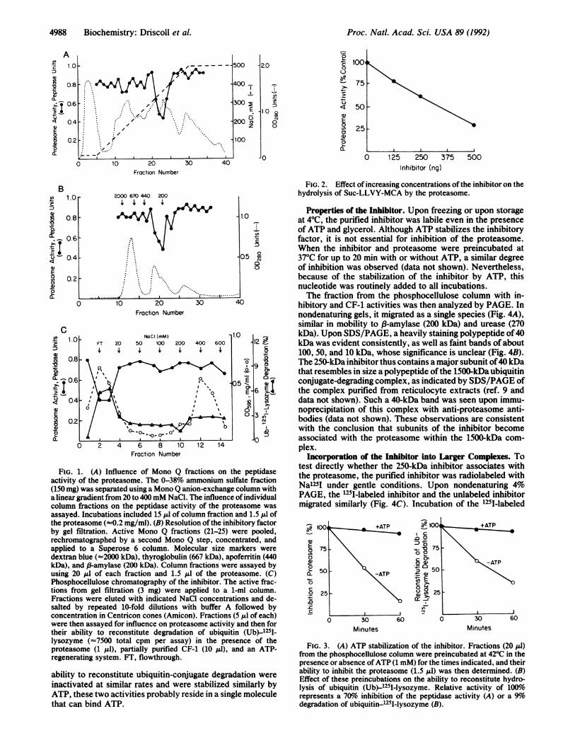
FIG. 1. (A) Influence of Mono Q fractions on theactivity of the proteasome. The 0-38% ammonium sulfi(150 mg) was separated using a Mono Q anion-exchange c
a linear gradient from 20 to 400mM NaCl. The influence ocolumn fractions on the peptidase activity of the proteassayed. Incubations included 15 pl of column fraction and 1.5 A1 ofthe proteasome (-0.2 mg/ml). (B) Resolution ofthe inhibitory factorby gel filtration. Active Mono Q fractions (21-25) were pooled,rechromatographed by a second Mono Q step, concentrated, andapplied to a Superose 6 column. Molecular size markers weredextran blue (-2000 kDa), thyroglobulin (667 kDa), apoferritin (440kDa), and p-amylase (200 kDa). Column fractions were assayed byusing 20 Al of each fraction and 1.5 Aul of the proteasome. (C)Phosphocellulose chromatography of the inhibitor. The active frac-tions from gel filtration (3 mg) were applied to a 1-ml column.Fractions were eluted with indicated NaCl concentrations and de-salted by repeated 10-fold dilutions with buffer A followed byconcentration in Centricon cones (Amicon). Fractions (5 A.l of each)were then assayed for influence on proteasome activity and then fortheir ability to reconstitute degradation of ubiquitin (Ub)-1251-lysozyme (-7500 total cpm per assay) in the presence of theproteasome (1 Al), partially purified CF-1 (10 pl), and an ATP-regenerating system. FT, flowthrough.
ability to reconstitute ubiquitin-conjugate degradation were
inactivated at similar rates and were stabilized similarly byATP, these two activities probably reside in a single moleculethat can bind ATP.
Fraction Number
E
0z
2.0
_enc
1.0 o0aN
O
4-
4-
C
aL)E0(n
oa).0
a.
75
50
25
JO 125 250 375Inhibitor (ng)
500
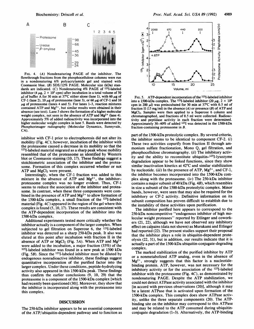
FIG. 2. Effect of increasing concentrations of the inhibitor on thehydrolysis of Suc-LLVY-MCA by the proteasome.
Properties of the Inhibitor. Upon freezing or upon storage.0 _at 40C, the purified inhibitor was labile even in the presence
e~ of ATP and glycerol. Although ATP stabilizes the inhibitoryfactor, it is not essential for inhibition of the proteasome.When the inhibitor and proteasome were preincubated at
0.5 C 37TC for up to 20 min with or without ATP, a similar degree8 of inhibition was observed (data not shown). Nevertheless,
because of the stabilization of the inhibitor by ATP, thisnucleotide was routinely added to all incubations.The fraction from the phosphocellulose column with in-
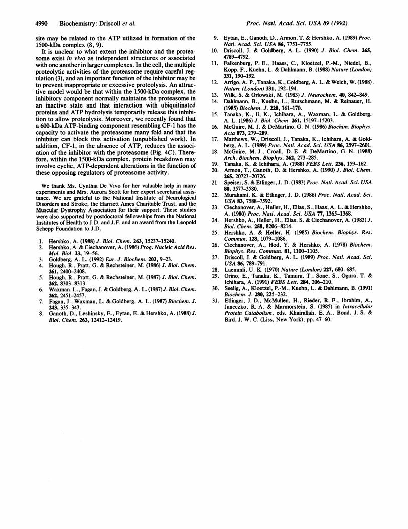
0 hibitory and CF-1 activities was then analyzed by PAGE. Innondenaturing gels, it migrated as a single species (Fig. 4A),similar in mobility to P-amylase (200 kDa) and urease (270
12 kDa). Upon SDS/PAGE, a heavily staining polypeptide of4012_0 kDa was evident consistently, as well as faint bands of about
100, 50, and 10 kDa, whose significance is unclear (Fig. 4B).90n The 250-kDa inhibitor thus contains a major subunit of40 kDa
-Q a, that resembles in size a polypeptide ofthe 1500-kDa ubiquitinconjugate-degrading complex, as indicated by SDS/PAGE of
E 6 the complex purified from reticulocyte extracts (ref. 9 anddata not shown). Such a 40-kDa band was seen upon immu-
c,'-~ noprecipitation of this complex with anti-proteasome anti-o 3 bodies (data not shown). These observations are consistentwith the conclusion that subunits of the inhibitor become
0o associated with the proteasome within the 1500-kDa com-plex.
Incorporation of the Inhibitor into Larger Complexes. Topeptidase test directly whether the 250-kDa inhibitor associates with
ate fraction the proteasome, the purified inhibitor was radiolabeled witholumn with Na125I under gentle conditions. Upon nondenaturing 4%;findividual PAGE, the 125I-labeled inhibitor and the unlabeled inhibitorasome was migrated similarly (Fig. 4C). Incubation of the 1251-labeled
0750
a)0E 500-
0
C
25
c
. +ATP
30Minutes Minutes
FIG. 3. (A) ATP stabilization of the inhibitor. Fractions (20 1l)from the phosphocellulose column were preincubated at 42TC in thepresence or absence ofATP (1 mM) for the times indicated, and theirability to inhibit the proteasome (1.5 ILI) was then determined. (B)Effect of these preincubations on the ability to reconstitute hydro-lysis of ubiquitin (Ub)-1251-lysozyme. Relative activity of 100%yorepresents a 70o inhibition of the peptidase activity (A) or a 9%odegradation of ubiquitin-1251-lysozyme (B).
C
4988 Biochemistry: Driscoll et al.
Proc. Natl. Acad. Sci. USA 89 (1992) 4989
A B C
1 2 3 4 5
- Proteosome98
-Urease -66
-A-amyIse*-45
-36
:2924
FIG. 4. (A) Nondenaturing PAGE of the inhibitor. Theflowthrough fractions from the phosphocellulose columns were run
in a nondenaturing 6% polyacrylamide gel and stained withCoomassie blue. (B) SDS/12% PAGE. Molecular size (kDa) stan-dards are indicated. (C) Nondenaturing 4% PAGE of 125I-labeledinhibitor (4 ,ug, 2 x 10⁵ cpm) after incubation in a total volume of 50jul of buffer A for 30 min at 370C either alone (lane 1), with 60 ,ug ofCF-1 (lane 2), 10 ,ug of proteasome (lane 3), or 60 ,ug of CF-1 and 10,ug of proteasome (lanes 4 and 5). For lanes 1-3, reaction mixturescontained ATP and Mg2+, but similar results were obtained in theirabsence (see text). Lane 5 shows the formation of a higher molecularweight complex, not seen in the absence of ATP and Mg2+ (lane 4).Approximately 5% of added radioactivity was incorporated into thehigher molecular weight complex in lane 5. Bands were detected byPhosphorimager radiography (Molecular Dynamics, Sunnyvale,CA).
inhibitor with CF-1 prior to electrophoresis did not alter itsmobility (Fig. 4C); however, incubation of the inhibitor withthe proteasome caused a decrease in its mobility so that the125I-labeled material migrated as a sharp peak whose mobilityresembled that of the proteasome as identified by Westernblot or Coomassie staining (10, 17). These findings suggest astoichiometric association of the inhibitor and the protea-some. Formation of this complex occurred whether or notATP and MgCl2 were present.
Interestingly, when the CF-1 fraction was added to thismixture in the absence of ATP and Mg2+, the inhibitor-proteasome complex was no longer evident. Thus, CF-1seems to reduce the association of the inhibitor and protea-some. In contrast, when these three components were com-bined in the presence ofATP and Mg2+ to allow formation ofthe 1500-kDa complex, a small fraction of the 125I-labeledmaterial (Fig. 4C) appeared in the region of the gel where thiscomplex is found (5, 10, 17). These results are consistent withthe ATP-dependent incorporation of the inhibitor into the1500-kDa complex.
Additional experiments tested more critically whether theinhibitor actually is a component ofthe larger complex. Whensubjected to gel filtration on Superose 6, the 125I-labeledinhibitor was detected as a sharp 250-kDa peak. It also waseluted at this point after incubation with fraction II in theabsence of ATP or MgCl2 (Fig. 5A). When ATP and Mg2+were added to the incubation, a major fraction (35%) of the1251-labeled inhibitor was found in a new peak at 1500 kDa(Fig. 5B). Since the 125I-labeled inhibitor must be diluted byendogenous nonradioactive inhibitor, these findings suggestquantitative incorporation of the purified factor into thelarger complex. Under these conditions, peptide-hydrolyzingactivity also appeared in this 1500-kDa peak. These findingsthus confirm the earlier conclusions (9, 10, 29) that theproteasome is a component of the 1500-kDa complex [whichhad recently been questioned (30)]. Moreover, they show thatthe inhibitor is incorporated along with the proteasome intothis complex.
DISCUSSION
The 250-kDa inhibitor appears to be an essential componentof the ATP/ubiquitin-dependent pathway and to function as
4
* 3
C.)C.0
a
D 4.r_c 3_
I
3
2
-)c
a)
0~
'aOL
Q)>1
a)
0
()0a}0
Volume, ml ___
FIG. 5. ATP-dependent incorporation ofthe 1251-labeled inhibitorinto a 1500-kDa complex. The 1251-labeled inhibitor (20 ,ug, 2 x 10⁶cpm in 200 ,ul) was preincubated for 30 min at 37°C with 0.5 ml offraction II (15 mg/ml) in the absence (A) or presence (B) ofATP andMgCl2. Samples were then applied to a Superose 6 column andchromatographed, and fractions of 0.5 ml were collected. Radioac-tivity and peptidase activity in each fraction were determined.Approximately 30-40o of added 125I was detected in the 1500-kDafraction-containing proteasome in B.
part of the 1500-kDa proteolytic complex. By several criteria,the inhibitor seems to be identical to component CF-2. (i)These two activities copurify from fraction II through am-
monium sulfate fractionation, Mono Q, gel filtration, andphosphocellulose chromatography. (ii) The inhibitory activ-ity and the ability to reconstitute ubiquitin-1251-lysozymedegradation appear to be linked functions, since they showsimilar inactivation kinetics at 42°C and similar stabilizationby nucleotide. (iii) In the presence of ATP, Mg2+, and CF-1,the inhibitor becomes incorporated into the 1500-kDa com-
plex along with the proteasome. (iv) The 250-kDa inhibitorcontains a major subunit of40 kDa (Fig. 4B), which resemblesin size a subunit of the 1500-kDa proteolytic complex. Minorbands, however, were seen that may also be required for theinhibitory or CF-2 activity. Definitive information on itssubunit composition has proven difficult to establish due tothe instability of these activities upon purification.The inhibitor purified here appears to correspond to the
250-kDa noncompetitive "endogenous inhibitor of high mo-
lecular weight proteases" reported by Etlinger and cowork-ers (21, 22), although we have not observed any inhibitoryeffect on calpains (data not shown) as Murakami and Etlingerhad reported (22). The present studies support their proposalthat the inhibitor plays a role in ubiquitin-dependent prote-olysis (22, 31), but in addition, our results indicate that it isactually a part of the 1500-kDa ubiquitin-conjugate-degradingcomplex.The marked stabilization of the purified inhibitor by ATP
or a nonmetabolized ATP analog, even in the absence ofMg2+, strongly suggests that this factor is a nucleotide-binding protein. ATP, however, was not necessary for theinhibitory activity or for the association of the 125I-labeledinhibitor with the proteasome (Fig. 4C), as demonstrated bynondenaturing PAGE. Despite the ATP stabilization, we
could not detect ATPase activity associated with the inhibitor[in accord with previous observations (20)], although it maybe a latent ATPase that is activated upon formation of the1500-kDa complex. This complex does show ATPase activ-ity, unlike the three separate components (20). The ATP-binding site on the inhibitor may correspond to this ATPaseand may be related to the ATP consumed during ubiquitin-conjugate degradation (1-3). Alternatively, the ATP-binding
-2000 670 440 200Ii I I
on'
_*5-_%
0'
+ ATP
- p 1%sp,"\ 3
10 15O% 20 25
10 15 20 25
Biochemistry: Driscoll et al.
4990 Biochemistry: Driscoll et al.
site may be related to the ATP utilized in formation of the1500-kDa complex (8, 9).
It is unclear to what extent the inhibitor and the protea-some exist in vivo as independent structures or associatedwith one another in larger complexes. In the cell, the multipleproteolytic activities of the proteasome require careful reg-ulation (3), and an important function of the inhibitor may beto prevent inappropriate or excessive proteolysis. An attrac-tive model would be that within the 1500-kDa complex, theinhibitory component normally maintains the proteasome inan inactive state and that interaction with ubiquitinatedproteins and ATP hydrolysis temporarily release this inhibi-tion to allow proteolysis. Moreover, we recently found thata 600-kDa ATP-binding component resembling CF-1 has thecapacity to activate the proteasome many fold and that theinhibitor can block this activation (unpublished work). Inaddition, CF-1, in the absence of ATP, reduces the associ-ation of the inhibitor with the proteasome (Fig. 4C). There-fore, within the 1500-kDa complex, protein breakdown mayinvolve cyclic, ATP-dependent alterations in the function ofthese opposing regulators of proteasome activity.
We thank Ms. Cynthia De Vivo for her valuable help in manyexperiments and Mrs. Aurora Scott for her expert secretarial assis-tance. We are grateful to the National Institute of NeurologicalDisorders and Stroke, the Harriett Ames Charitable Trust, and theMuscular Dystrophy Association for their support. These studieswere also supported by postdoctoral fellowships from the NationalInstitutes of Health to J.D. and J.F. and an award from the LeopoldSchepp Foundation to J.D.
1. Hershko, A. (1988) J. Biol. Chem. 263, 15237-15240.2. Hershko, A. & Ciechanover, A. (1986) Prog. Nucleic Acid Res.
Mol. Biol. 33, 19-56.3. Goldberg, A. L. (1992) Eur. J. Biochem. 203, 9-23.4. Hough, R., Pratt, G. & Rechsteiner, M. (1986) J. Biol. Chem.
261, 2400-2408.5. Hough, R., Pratt, G. & Rechsteiner, M. (1987) J. Biol. Chem.
262, 8303-8313.6. Waxman, L., Fagan, J. & Goldberg, A. L. (1987) J. Biol. Chem.
262, 2451-2457.7. Fagan, J., Waxman, L. & Goldberg, A. L. (1987) Biochem. J.
243, 335-343.8. Ganoth, D., Leshinsky, E., Eytan, E. & Hershko, A. (1988) J.
Biol. Chem. 263, 12412-12419.
9. Eytan, E., Ganoth, D., Armon, T. & Hershko, A. (1989) Proc.Nat!. Acad. Sci. USA 86, 7751-7755.
10. Driscoll, J. & Goldberg, A. L. (1990) J. Biol. Chem. 265,4789-4792.
11. Falkenburg, P. E., Haass, C., Kloetzel, P.-M., Niedel, B.,Kopp, F., Kuehn, L. & Dahlmann, B. (1988) Nature (London)331, 190-192.
12. Arrigo, A. P., Tanaka, K., Goldberg, A. L. & Welch, W. (1988)Nature (London) 331, 192-194.
13. Wilk, S. & Orlowski, M. (1983) J. Neurochem. 40, 842-849.14. Dahlmann, B., Kuehn, L., Rutschmann, M. & Reinauer, H.
(1985) Biochem. J. 228, 161-170.15. Tanaka, K., Ii, K., Ichihara, A., Waxman, L. & Goldberg,
A. L. (1986) J. Biol. Chem. 261, 15197-15203.16. McGuire, M. J. & DeMartino, G. N. (1986) Biochim. Biophys.
Acta 873, 279-289.17. Matthews, W., Driscoll, J., Tanaka, K., Ichihara, A. & Gold-
berg, A. L. (1989) Proc. Natl. Acad. Sci. USA 86, 2597-2601.18. McGuire, M. J., Croall, D. E. & DeMartino, G. N. (1988)
Arch. Biochem. Biophys. 262, 273-285.19. Tanaka, K. & Ichihara, A. (1988) FEBS Lett. 236, 159-162.20. Armon, T., Ganoth, D. & Hershko, A. (1990) J. Biol. Chem.
265, 20723-20726.21. Speiser, S. & Etlinger, J. D. (1983) Proc. Natl. Acad. Sci. USA
80, 3577-3580.22. Murakami, K. & Etlinger, J. D. (1986) Proc. Nat!. Acad. Sci.
USA 83, 7588-7592.23. Ciechanover, A., Heller, H., Elias, S., Haas, A. L. & Hershko,
A. (1980) Proc. Nat!. Acad. Sci. USA 77, 1365-1368.24. Hershko, A., Heller, H., Elias, S. & Ciechanover, A. (1983) J.
Biol. Chem. 258, 8206-8214.25. Hershko, A. & Heller, H. (1985) Biochem. Biophys. Res.
Commun. 128, 1079-1086.26. Ciechanover, A., Hod, Y. & Hershko, A. (1978) Biochem.
Biophys. Res. Commun. 81, 1100-1105.27. Driscoll, J. & Goldberg, A. L. (1989) Proc. Natl. Acad. Sci.
USA 86, 789-791.28. Laemmli, U. K. (1970) Nature (London) 227, 680-685.29. Orino, E., Tanaka, K., Tamura, T., Sone, S., Ogura, T. &
Ichihara, A. (1991) FEBS Lett. 284, 206-210.30. Seelig, A., Kloetzel, P.-M., Kuehn, L. & Dahlmann, B. (1991)
Biochem. J. 280, 225-232.31. Etlinger, J. D., McMullen, H., Rieder, R. F., Ibrahim, A.,
Janeczko, R. A. & Marmorstein, S. (1985) in IntracellularProtein Catabolism, eds. Khairallah, E. A., Bond, J. S. &Bird, J. W. C. (Liss, New York), pp. 47-60.
Proc. Natl. Acad. Sci. USA 89 (1992)