Embed Size (px)
Citation preview

Research Article
Atrial-like cardiomyocytes from human pluripotentstem cells are a robust preclinical model forassessing atrial-selective pharmacologyHarsha D Devalla1,*, Verena Schwach1, John W Ford2, James T Milnes2, Said El-Haou2, Claire Jackson2,
Konstantinos Gkatzis1, David A Elliott3, Susana M Chuva de Sousa Lopes1,4, Christine L Mummery1,
Arie O Verkerk5 & Robert Passier1,**
Abstract
Drugs targeting atrial-specific ion channels, Kv1.5 or Kir3.1/3.4, arebeing developed as new therapeutic strategies for atrial fibrilla-tion. However, current preclinical studies carried out in non-cardiac cell lines or animal models may not accurately representthe physiology of a human cardiomyocyte (CM). In the currentstudy, we tested whether human embryonic stem cell (hESC)-derived atrial CMs could predict atrial selectivity of pharmaco-logical compounds. By modulating retinoic acid signaling duringhESC differentiation, we generated atrial-like (hESC-atrial) andventricular-like (hESC-ventricular) CMs. We found the expressionof atrial-specific ion channel genes, KCNA5 (encoding Kv1.5) andKCNJ3 (encoding Kir 3.1), in hESC-atrial CMs and further demon-strated that these ion channel genes are regulated by COUP-TFtranscription factors. Moreover, in response to multiple ion chan-nel blocker, vernakalant, and Kv1.5 blocker, XEN-D0101, hESC-atrialbut not hESC-ventricular CMs showed action potential (AP) prolon-gation due to a reduction in early repolarization. In hESC-atrialCMs, XEN-R0703, a novel Kir3.1/3.4 blocker restored the AP short-ening caused by CCh. Neither CCh nor XEN-R0703 had an effect onhESC-ventricular CMs. In summary, we demonstrate that hESC-atrial CMs are a robust model for pre-clinical testing to assessatrial selectivity of novel antiarrhythmic drugs.
Keywords arrhythmias; atrial cardiomyocytes; atrial fibrillation; COUP-TF; ion
channels
Subject Categories Cardiovascular System; Pharmacology & Drug Discovery;
Stem Cells
DOI 10.15252/emmm.201404757 | Received 17 October 2014 | Revised 18
January 2015 | Accepted 23 January 2015 | Published online 19 February 2015
EMBO Mol Med (2015) 7: 394–410
Introduction
Atrial fibrillation (AF) affects over 33 million people globally
(Chugh et al, 2014) and is characterized by irregular atrial rhythm
leading to a decline in atrial mechanical function. Untreated AF
increases the risk of life-threatening complications such as stroke or
heart failure (Wang et al, 2003; Marini et al, 2005). Current treat-
ment options for rhythm control in AF include interventional ther-
apy such as ablation or pharmacotherapy with antiarrhythmic
drugs. The latter is the preferred treatment of early AF in individuals
who prefer non-invasive treatment and as a follow-up therapy post-
electrical cardioversion, to prevent recurrence of AF (Wann et al,
2011). However, existing antiarrhythmic agents lack atrial selectiv-
ity and pose the risk of inducing undesirable cardiac events, such as
ventricular proarrhythmia (Dobrev & Nattel, 2010).
In order to overcome this limitation, pharmaceutical industry has
initiated design and development of compounds aimed at atrial-
specific targets (Li et al, 2009; Milnes et al, 2012). The notorious
difficulty in obtaining human cardiomyocytes (CMs) and propagat-
ing them in culture has precluded their use from many drug screen-
ing assays and instigated the use of alternative preclinical models.
However, many of the current preclinical screening assays used in
the identification of atrial-selective compounds are performed using
either non-cardiac recombinant cell lines expressing a non-native
ion channel or animal models. Both these models may not accu-
rately represent the ion channel composition as well as physiology
of a human CM and therefore have limitations in predicting drug
responses on the human heart.
Human pluripotent stem cell-derived CMs (hPSC-CMs) offer a
human-based, physiologically relevant model system for drug
discovery and development strategies. Despite the suitability of
these cells for cardiotoxicity testing and safety pharmacology (Braam
et al, 2010; Navarrete et al, 2013), their application in validating
1 Department of Anatomy & Embryology, Leiden University Medical Center, Leiden, The Netherlands2 Xention Ltd, Cambridge, UK3 Murdoch Childrens Research Institute, Royal Children’s Hospital, Melbourne, Vic., Australia4 Department for Reproductive Medicine, Ghent University Hospital, Ghent, Belgium5 Heart Failure Research Center, Academic Medical Center, University of Amsterdam, Amsterdam, The Netherlands
*Corresponding author. Tel: +31 71 5269528; Fax: +31 71 5268289; E-mail: [email protected]**Corresponding author. Tel: +31 71 5269359; Fax: +31 71 5268289; E-mail: [email protected]
EMBO Molecular Medicine Vol 7 | No 4 | 2015 ª 2015 The Authors. Published under the terms of the CC BY 4.0 license394

novel drug candidates for AF requires cultures enriched in atrial-
like CMs. Current protocols for cardiac differentiation of hPSCs
result in heterogeneous pools of CMs consisting predominantly of
ventricular-like cells with a small percentage of atrial-like and
nodal-like cells (Blazeski et al, 2012). Based on substantial evidence
from in vivo and in vitro studies (Niederreither et al, 2001; Hochgreb
et al, 2003; Gassanov et al, 2008; Zhang et al, 2011) indicating a
role for retinoic acid (RA) in atrial specification, we hypothesized
that RA would drive mesodermal progenitors from PSCs toward
an atrial fate.
In the current study, we show that transcriptional and electro-
physiological properties of human embryonic stem cell-derived
atrial CMs (hESC-atrial CMs), generated by modulating RA signal-
ing, closely resemble that of native human atrial CMs. We also
observed that transcription factors, COUP-TFI (NR2F1) and COUP-
TFII (NR2F2), are robustly upregulated in response to RA during
directed atrial differentiation. Short hairpin RNA (shRNA)-mediated
knockdown and chromatin immunoprecipitation (ChIP) of COUP-
TFs identified that they regulate atrial-specific ion channel genes
KCNA5 (encoding Kv1.5) and KCNJ3 (encoding Kir3.1). Furthermore,
hESC-atrial CMs express atrial-selective ion currents, IKur as well as
IK,ACh, and also respond to pharmacological compounds targeting
ion channels that conduct these currents (Kv1.5 and Kir3.1/3.4,
respectively).
Collectively, our data identify a key role for COUP-TF transcrip-
tion factors in RA-driven atrial differentiation and also demonstrate
that hESC-atrial CMs are a robust model for predicting atrial
selectivity of novel pharmacological compounds during preclinical
development.
Results
Treatment of differentiating hESCs with RA promotesatrial specification
A cocktail of cytokines (Fig 1A) was used to initiate cardiac differen-
tiation in NKX2-5-eGFP/w hESCs as described previously (Ng et al,
2008; Elliott et al, 2011). To direct differentiating hESCs toward an
atrial phenotype, timing and concentration of treatment with RA
were carefully optimized (Supplementary Fig S1A–C). We hypothe-
sized that specification of CM subtypes in vitro occurs post-
mesoderm formation and prior to the onset of cardiac progenitor
stage. Accordingly, embryoid bodies (EBs) were supplemented with
RA from day 4, just after the transient expression of early cardiac
mesoderm marker, MESP1, until day 7, a time point at which key
transcription factors such as NKX2.5, GATA4 and MEF2C important
for commitment and specification of cardiovascular lineages are
activated (Supplementary Fig S1A).
Adding low concentrations of RA (1–10 nmol/l) from day 4 to 7
enhanced cardiac differentiation as assessed by the percentage of
GFP+ cells at day 15 (Supplementary Fig S1B). On the other hand,
treatment with high concentrations of RA (1 lmol/l) in the same
time window resulted in GFP+ EBs with reduced expression of the
ventricular specific myosin gene, MLC2V (Supplementary Fig S1C).
Therefore, treatment of differentiating hESCs with 1 lmol/l of RA
from day 4 to 7 was considered to be most suitable for driving atrial
differentiation. As a control, every experiment included parallel
differentiating cultures treated with 0.002% DMSO (the final
concentration in RA-treated cultures) from day 4 to 7 (Fig 1A).
Morphologically, RA-treated EBs were similar compared with
control EBs (Supplementary Fig S1D), and contractile GFP+ areas
were observed in both groups at day 10 (Fig 1B). Flow cytometry
analysis of GFP expression at day 15 revealed a decrease in the
proportion of NKX2.5-expressing cells upon treatment with RA.
Sixty-five percent of all cells expressed GFP in control differentia-
tion, while only 50% cells in RA-treated differentiation were GFP+
(Fig 1C and Supplementary Fig S1E). These data are consistent with
earlier reports in zebrafish embryo which demonstrated that expo-
sure of anterior lateral plate mesoderm to RA signaling restricts the
size of the cardiac progenitor pool (Keegan et al, 2005). Also, EBs
treated with RA during differentiation displayed faster beating
frequencies upon differentiation (Supplementary Fig S1F). Finally,
immunofluorescence analysis of EBs from both control and
RA-treated differentiations confirmed that the contractile GFP+ areas
expressed both NKX2-5 and the myofilament marker, ACTN2
(Supplementary Fig S1G).
Transcriptional profiling of CMs from RA-treated differentiationsreveals an upregulation of atrial and downregulation ofventricular markers
To study gene expression in CMs resulting from control and RA-
treated conditions, cells were sorted on the basis of NKX2-5-eGFP.
GFP+ and GFP� fractions from control (CT+/CT�) and RA-treated
EBs (RA+/RA�) isolated at day 31 post-differentiation were assessed
by microarray and quantitative PCR (qPCR). Expression of contrac-
tility genes, TNNC1 and TNNT2, calcium handling genes, RYR2 and
ATP2A2, and cardiac transcription factors, MEF2C and NKX2.5 were
enriched in both CT+ and RA+ pools compared with CT� and
RA� populations, indicating efficient purification of CMs (Fig 1D).
A heat map of GFP+ and GFP� samples also demonstrates strong
correlation with each group (Supplementary Fig S2A). Microarray
analysis demonstrated upregulation of atrial markers such as SLN,
HEYL, PITX2 and NPPA (Fig 1E), while ventricular markers such as
MYL2, IRX4, HAND1 and HEY2 were downregulated in RA+ CMs
(Fig 1F). Measuring expression levels of selected targets by
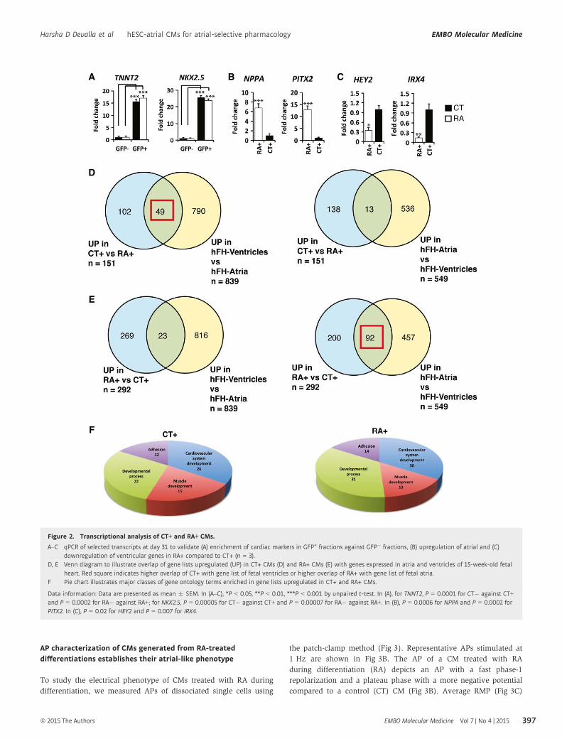
quantitative qPCR further validated the microarray data in which
expression of NKX2-5 and TNNT2 was significantly higher in GFP+
fractions (Fig 2A). qPCR also confirmed upregulation of atrial
and downregulation of ventricular transcripts in RA+ CMs (Fig 2B
and C).
In order to compare the expression profile of CT+ and RA+ CMs
at day 31 with that of human heart, we included atrial and ventricu-
lar tissue samples of a 15-week-old fetal heart for microarray analy-
sis. A total of 151 genes showed increased expression of more than
twofold in CT+ group compared to RA+. Thirty-two percent of these
genes (49 out of 151) were preferentially expressed in human ventri-
cles, whereas only 8% (13 out of 151) could be identified in the
group of genes that were enriched in the human atria (Fig 2D). On
the other hand, 292 genes showed increased expression of more
than twofold in RA+ group compared to CT+. Thirty-one percent of
the genes (92 out of 292) with enriched expression in RA+ group
were preferentially expressed in the human atria, while a mere 8%
(23 out of 292) of these genes were expressed in the human ventri-
cles (Fig 2E). Gene lists in Venn diagrams (Fig 2D and E) are
ª 2015 The Authors EMBO Molecular Medicine Vol 7 | No 4 | 2015
Harsha D Devalla et al hESC-atrial CMs for atrial-selective pharmacology EMBO Molecular Medicine
395

included in Supplementary Table S1. Pie charts illustrating the
cellular localization and molecular function of genes enriched in
CT+ and RA+ groups are shown in Supplementary Fig S2B and C.
Gene ontology (GO) analysis of microarray data was performed
with ConsensusPathDB-human, and terms satisfying a cutoff of
P < 0.01 were considered enriched. GO terms related to four
major classes, cardiovascular development, muscle development,
developmental process and adhesion, were overrepresented in both
groups (Fig 2F). In the category of cardiovascular development,
GO terms such as appendage development, cardiac atrium develop-
ment and cardiac septum development were enriched in RA+ CMs
while cardiac ventricle development, ventricular septum formation
and heart trabecular formation were enriched in CT+ CMs (Supple-
mentary Table S2). A detailed list of GO terms in each category
and the genes are included in Supplementary Table S3.
Therefore, the transcriptional profile of RA+ CMs suggested a
fetal atrial-like gene expression pattern compared with control CMs,
which expressed higher levels of ventricular transcripts.
A
B C
D E F
Figure 1. Treatment of differentiating hESCs with RA promotes atrial specification.
A Schematic of the cardiac differentiation protocol. Beating embryoid bodies (EBs) were observed at day 10. Differentiation efficiency in each experiment wasassessed by flow cytometry (FC) for GFP at day 15. Further characterization of EBs derived from control (CT) and RA-treated (RA) cultures was carried out bytranscriptional or functional analysis between days 27 and 31.
B GFP+ EBs derived from CT and RA cultures at day 10; scale bar: 100 lm.C Representative FC plots depicting percentage of GFP+ cells obtained at day 15, from CT and RA cultures in a typical experiment.D Heat map demonstrating enrichment of cardiac genes in GFP+ fractions (CT+, RA+) compared to GFP� fractions (CT�, RA�) at day 31.E, F Heat map of a select list of genes (E) upregulated and (F) downregulated in RA+ compared to CT+ at day 31. Fold change > 2.
EMBO Molecular Medicine Vol 7 | No 4 | 2015 ª 2015 The Authors
EMBO Molecular Medicine hESC-atrial CMs for atrial-selective pharmacology Harsha D Devalla et al
396
AP characterization of CMs generated from RA-treateddifferentiations establishes their atrial-like phenotype
To study the electrical phenotype of CMs treated with RA during
differentiation, we measured APs of dissociated single cells using
the patch-clamp method (Fig 3). Representative APs stimulated at
1 Hz are shown in Fig 3B. The AP of a CM treated with RA
during differentiation (RA) depicts an AP with a fast phase-1
repolarization and a plateau phase with a more negative potential
compared to a control (CT) CM (Fig 3B). Average RMP (Fig 3C)
A
D
E
F
B C
Figure 2. Transcriptional analysis of CT+ and RA+ CMs.
A–C qPCR of selected transcripts at day 31 to validate (A) enrichment of cardiac markers in GFP+ fractions against GFP� fractions, (B) upregulation of atrial and (C)downregulation of ventricular genes in RA+ compared to CT+ (n = 3).
D, E Venn diagram to illustrate overlap of gene lists upregulated (UP) in CT+ CMs (D) and RA+ CMs (E) with genes expressed in atria and ventricles of 15-week-old fetalheart. Red square indicates higher overlap of CT+ with gene list of fetal ventricles or higher overlap of RA+ with gene list of fetal atria.
F Pie chart illustrates major classes of gene ontology terms enriched in gene lists upregulated in CT+ and RA+ CMs.
Data information: Data are presented as mean � SEM. In (A–C), *P < 0.05, **P < 0.01, ***P < 0.001 by unpaired t-test. In (A), for TNNT2, P = 0.0001 for CT� against CT+and P = 0.0002 for RA� against RA+; for NKX2.5, P = 0.00005 for CT� against CT+ and P = 0.00007 for RA� against RA+. In (B), P = 0.0006 for NPPA and P = 0.0002 forPITX2. In (C), P = 0.02 for HEY2 and P = 0.007 for IRX4.
ª 2015 The Authors EMBO Molecular Medicine Vol 7 | No 4 | 2015
Harsha D Devalla et al hESC-atrial CMs for atrial-selective pharmacology EMBO Molecular Medicine
397

A B
C D
G H
E F
Figure 3. AP characterization of CMs generated from control and RA-treated differentiations.
A AP illustrating the analyzed parameters.B Representative APs of day 31 CMs from control (CT) and RA-treated (RA) groups at 1 Hz.C–E RMP, APAmax and APAplat (C), dV/dtmax (D) and APD20, APD50 and APD90 of CT and RA CMs (E).F Plot showing all measured APAplat values of CT and RA CMs.G Representative APs of CT and RA CMs at 0.5–4 Hz.H Average APAplat at 0.5–4 Hz. Please note that the AP differences in morphology are present at all measured frequencies.
Data information: Data are presented as mean � SEM. *P < 0.05 by unpaired t-test or Mann–Whitney rank-sum test for (C–E). In (C), P = 0.238 for RMP; P < 0.001 forAPAmax and APAplat. In (D), P = 0.598 for dV/dtmax. In (E), P ≤ 0.001 for APD20; P = 0.009 for APD50; P = 0.04 for APD90. Two-way repeated measures ANOVA followed bypairwise comparison using the Student–Newman–Keuls test for (H). *P = 0.002, 0.006, 0.003, 0.004 and 0.003, respectively, for comparison of APAplat between CT and RAgroups at frequencies of 0.5, 1.0, 2.0, 3.0 and 4.0 Hz. AP = action potential; APAmax = maximum AP amplitude; APAplat = AP plateau amplitude; APD20, APD50 andAPD90 = AP duration at 20, 50, and 90% repolarization, respectively; CMs = cardiomyocytes; dV/dtmax = maximum upstroke velocity; RMP = resting membrane potential.
EMBO Molecular Medicine Vol 7 | No 4 | 2015 ª 2015 The Authors
EMBO Molecular Medicine hESC-atrial CMs for atrial-selective pharmacology Harsha D Devalla et al
398

and dV/dtmax (Fig 3D) did not differ significantly between the two
groups.
In particular, APs of RA CMs had a significantly lower APAmax
(Fig 3C). They also repolarized faster resulting in significantly lower
APAplat (Fig 3C) and shorter APD20, APD50 and APD90 (Fig 3E). A
scatter-plot of individual APAplat values clearly shows that the
plateau amplitudes of individual cells in the RA group were typically
< 80 mV, whereas those in the CT group were > 80 mV (Fig 3F).
The AP differences between the two groups were also consistent
when the cells were paced at higher frequencies (Fig 3G and H). Of
25 cells measured from the control group, about 80% (of 25 cells)
displayed ventricular-like action potential properties while about
85% (of 26 cells) in the RA treatment group showed atrial-like
action potential properties. We observed a very small percentage
(< 1%) of nodal-like cells in both the groups.
The differences observed in AP duration and APAplat between RA
and CT CMs closely matched the AP differences observed between
atrial and ventricular CMs in vivo (Nerbonne & Kass, 2005). Taken
together, gene expression signature and electrophysiological proper-
ties demonstrated that CMs treated with RA during differentiation
displayed atrial-like phenotype (hereby referred to as hESC-atrial
CMs), while control CMs resembled ventricular-like cells (hereby
referred to as hESC-ventricular CMs).
COUP-TFI and COUP-TFII are upregulated in response to retinoicacid, and their expression persists in differentiatedhESC-atrial CMs
Transcriptional profiling experiments revealed that orphan nuclear
receptor transcription factors, COUP-TFI (NR2F1) and COUP-TFII
(NR2F2), are highly upregulated in hESC-atrial CMs. Based on previ-
ous reports by others indicating the involvement of COUP-TFs in RA
signaling (Jonk et al, 1994; van der Wees et al, 1996), we postulated
that these genes might play a central role downstream of RA during
atrial differentiation. This hypothesis was further supported by
atrial-specific expression of Coup-tfII in the mouse and severe atrial
abnormalities observed in the loss-of-function mouse mutant (Pereira
et al, 1999). A more recent study found that Coup-tfII regulates
atrial identity in the mouse heart (Wu et al, 2013).
In order to determine the dynamics of COUP-TF expression
following RA treatment, expression levels of both COUP-TFI and II
were analyzed by qPCR at different time points and compared to
control EBs. COUP-TFs were induced within 24 h of treatment with
RA followed by dramatic increase in expression thereafter. A line
graph plotting the relative mRNA levels of COUP-TFI and II between
control and RA-treated groups shows striking differences, indicating
that addition of RA induced the expression of these orphan nuclear
receptor transcription factors (Fig 4A and B). The expression of
COUP-TFs was maintained in differentiated CMs at day 31. COUP-TFI
was expressed 20-fold higher, and COUP-TFII was enriched over 30-
fold in hESC-atrial CMs compared to hESC-ventricular CMs (Fig 4A
and B). Antibodies selectively binding to COUP-TFI or COUP-TFII
were used to verify the expression of these proteins. While GFP+
areas in hESC-ventricular CMs at day 25 showed relatively low
expression of COUP-TFI and COUP-TFII, hESC-atrial CMs showed
robust expression of these transcription factors (Fig 4C and D).
To confirm our in vitro findings, which identified high levels of
COUP-TFs in hESC-atrial CMs, we sought to verify the expression of
COUP-TFI and COUP-TFII in the human heart. Previous studies by
others have reported preferential expression of Coup-tfII in the atrial
myocardium of the mouse heart (Pereira et al, 1999), but no data
are available for Coup-tfI. qPCR identified significantly higher mRNA
levels of COUP-TFI and COUP-TFII in the atria as opposed to ventri-
cles in both human fetal and adult heart (Supplementary Fig S3A
and B). In accordance with the mRNA expression levels, COUP-TFI
protein showed nuclear localization in the myocardium of the atrial
chambers stained with TNNI3 (Supplementary Fig S3D and E) while
no expression was detected in the myocardium of ventricles
(Supplementary Fig S3F and G) or elsewhere in two of the analyzed
hFHs at 12 weeks of gestation. Similarly, strong expression of
COUP-TFII was observed in the TNNI3-positive myocardium of the
atria (Supplementary Fig S3H and I), while no expression was found
in the ventricular myocardium (Supplementary Fig S3J and K) of the
hFH. Collectively, expression and histochemical analysis in human
fetal hearts demonstrate that COUP-TFI and II are indeed expressed
in the atrial myocardium of the human heart as observed in hESC-
atrial CMs in vitro.
COUP-TFs regulate expression of atrial-specific potassiumchannel genes, KCNA5 and KCNJ3
To investigate whether COUP-TFs have an essential role in differenti-
ated CMs, we used shRNAs to knockdown COUP-TFI or COUP-TFII in
hESC-atrial CMs and studied whether they regulate atrial-specific ion
channel genes. Lentiviral pLKO.1 constructs containing five different
shRNA sequences each (Supplementary Table S4), for COUP-TFI and
COUP-TFII (Supplementary Fig S4A–C), were tested in hESC-atrial
CMs. Two COUP-TFI-shRNAs (#2; #4) and two COUP-TFII-shRNAs
(#7; #10) gave efficient knockdown as assessed by qPCR (Supple-
mentary Fig S4D) and were selected for further experiments. Trans-
duction of COUP-TFI-shRNA or COUP-TFII-shRNA in hESC-atrial CMs
resulted in 70–75% reduction of the corresponding mRNA in
comparison with cells transduced with the scrambled-shRNA
(Fig 5A and B). Knockdown of COUP-TFI and COUP-TFII protein
following shRNA transduction was confirmed by Western blot
(Supplementary Fig S4E). hESC-atrial CMs transduced with scram-
bled-shRNA or COUP-TF-shRNAs maintained their cardiac pheno-
type. Knockdown of COUP-TFI or COUP-TFII in hESC-atrial CMs did
not affect GFP or cTNT expression (Supplementary Fig S5A and B).
Furthermore, shRNA-targeted knockdown of COUP-TFI did not affect
the expression of COUP-TFII and vice versa (Fig 5A and B).
However, knockdown of COUP-TFI or COUP-TFII in hESC-atrial
CMs led to significant decrease in the expression of KCNA5 (Fig 5C
and D). Similarly, knockdown of COUP-TFII decreased the expres-
sion of ion channel genes KCNJ3 and KCNJ5 (Fig 5D). Although
there was a small reduction in the expression of KCNJ3 and KCNJ5
in hESC-atrial CMs with decreased COUP-TFI expression (Fig 5C), it
did not reach significance.
To test whether the atrial-enriched ion channel genes KCNA5 and
KCNJ3 are direct targets of COUP-TFs, we performed ChIP-qPCR
assays using day 30 hESC-atrial CMs. COUP-TF genes regulate
transcription by interacting with direct repeats (DRs) of hormone
responsive elements with various spacings, but show highest affin-
ity to DR sequences separated by 1 nucleotide (DR1). Bioinformatic
analysis of promoter regions of human KCNA5 and KCNJ3 by
Genomatix-MatInspector revealed potential binding sites of the
ª 2015 The Authors EMBO Molecular Medicine Vol 7 | No 4 | 2015
Harsha D Devalla et al hESC-atrial CMs for atrial-selective pharmacology EMBO Molecular Medicine
399

Genomatix-defined NR2F matrix family (Supplementary Fig S5).
The promoter region of KCNA5 harbored two plausible NR2F
binding sites (Fig 5E), and analysis of immunoprecipitated DNA
with primers designed around site 1 confirmed binding of both
COUP-TFI and COUP-TFII (Fig 5G). Promoter analysis of KCNJ3
identified several putative NR2F binding sites (Fig 5F), and qPCR
for region encompassing site 1 confirmed interaction of both COUP-
TFs (Fig 5H).
Taken together, these data suggest that COUP-TFI and COUP-TFII
play a pivotal role in regulating ion channel genes responsible for
unique electrophysiological phenotype of human atrial cells.
Atrial-specific currents IKur and IK,ACh are functional inhESC-atrial CMs
The potassium ion channels Kv1.5 and the Kir3.1/3.4 are more abun-
dant in human atrial than in ventricular CMs (Wang et al, 1993;
Krapivinsky et al, 1995) and are responsible for functional differ-
ences between the two chambers. Kv1.5, encoded by the gene
KCNA5, conducts the ultrarapid delayed rectifier K+ current, IKur,which is a major repolarizing current in the human atrium. Hetero-
multimers of K+ channels Kir3.1/3.4 encoded by the genes KCNJ3
and KCNJ5, respectively, conduct the acetylcholine-activated current
IK,ACh in the human atria.
We observed significantly higher mRNA expression of both KCNA5
and KCNJ3 in hESC-atrial CMs compared with hESC-ventricular
CMs, in a manner similar to human atrial tissue (in comparison with
human adult ventricular tissue) (Fig 6A).
We next assessed the current densities of IKur and IK,ACh in hESC-
ventricular and hESC-atrial CMs. IKur, measured as the current sensi-
tive to 50 lmol/l 4-AP (Wang et al, 1993), was clearly present in
hESC-atrial CMs but absent in hESC-ventricular CMs (Fig 6B).
IK,ACh, measured as the current evoked by the muscarinic agonist,
CCh (10 lmol/l), was also present in hESC-atrial CMs but could not
A B
C D
Figure 4. Retinoic acid induces COUP-TFI and COUP-TFII during atrial differentiation.
A, B Line plot illustrating relative mRNA levels of (A) COUP-TFI and (B) COUP-TFII in VM and AM differentiations from day 5 through day 9 (left) and in GFP+ CMs at day31 (right); n = 3.
C, D COUP-TFI (C) and COUP-TFII (D) immunofluorescence at day 31 in AM (top) and VM (bottom). Scale bars: 40 lm.
Data information: Data are presented as mean � SEM. In (A, B), *P < 0.05, **P < 0.01, ***P < 0.001 by unpaired t-test. In (A), left panel, P = 0.03, 0.02, 0.03, 0.005 and0.0004 for comparison of COUP-TFI expression between AM and VM at days 5, 6, 7, 8 and 9 of differentiation. In (A), right panel, P = 0.0002 for comparison of COUP-TFIexpression at day 31 between AM and VM. In (B), left panel, P = 0.02, 0.03, 0.006, 0.004 and 0.003 for comparison of COUP-TFII expression between AM and VM at days5, 6, 7, 8 and 9 of differentiation. In (B), right panel, P = 0.0001 for comparison of COUP-TFII expression at day 31 between AM and VM. CT = control differentiation;hESC-atrial (AM); hESC-ventricular (VM).
EMBO Molecular Medicine Vol 7 | No 4 | 2015 ª 2015 The Authors
EMBO Molecular Medicine hESC-atrial CMs for atrial-selective pharmacology Harsha D Devalla et al
400

A B
C D
E F
G H
Figure 5. COUP-TFs regulate atrial-specific ion channel genes KCNA5 and KCNJ3.
A, B mRNA expression of COUP-TFI and COUP-TFII following shRNA-mediated knockdown of (A) COUP-TFI or (B) COUP-TFII in hESC-atrial cardiomyocytes (AM) at day 30.C, D mRNA expression of ion channel genes KCNA5, KCNJ3 and KCNJ5 after knockdown of (C) COUP-TFI or (D) COUP-TFII in AM at day 30.E, F Schematic of NR2F binding sites in (E) KCNA5 and (F) KCNJ3 promoters.G, H ChIP-qPCR analysis at day 30 shows enriched binding of COUP-TFI and COUP-TFII to the promoter region of (G) KCNA5 and (H) KCNJ3, compared to IgG in AM.
Data information: Data are presented as mean � SEM. *P < 0.05, **P < 0.01, ***P < 0.001 by unpaired t-test. In (A), P = 0.00004. In (B), P = 0.0001. In (C), P = 0.0001.In (D), P = 0.000004, 0.006 and 0.001, respectively. In (G), P = 0.0004 for COUP-TFI and P = 0.0002 for COUP-TFII. In (H), P = 0.0003 for COUP-TFI and P = 0.0001 forCOUP-TFII.
ª 2015 The Authors EMBO Molecular Medicine Vol 7 | No 4 | 2015
Harsha D Devalla et al hESC-atrial CMs for atrial-selective pharmacology EMBO Molecular Medicine
401

A
B
C
D
E
Figure 6.
EMBO Molecular Medicine Vol 7 | No 4 | 2015 ª 2015 The Authors
EMBO Molecular Medicine hESC-atrial CMs for atrial-selective pharmacology Harsha D Devalla et al
402

be detected in hESC-ventricular CMs (Fig 6C). Thus, hESC-atrial
CMs have substantially higher IKur and IK,ACh densities, consistent
with the greater mRNA expression of KCNA5 and KCNJ3.
Lastly, we evaluated the contribution of IKur and IK,ACh in the APs
of hESC-atrial and hESC-ventricular CMs. Blocking IKur by 4-AP
(50 lmol/l) reduced phase-1 repolarization resulting in AP prolon-
gation and an increase in APAplat in hESC-atrial but not hESC-
ventricular CMs (Supplementary Fig S6D and Supplementary
Table S5). These effects of IKur block observed in hESC-atrial CMs
confirmed its functional presence and are consistent with the effects
reported in freshly isolated human atrial CMs (Wang et al, 1993).
On the other hand, activation of IK,ACh by CCh resulted in hyperpo-
larization of the RMP in hESC-atrial CMs but not in hESC-ventricular
CMs (Fig 6E and Supplementary Table S5). The effects of IK,ACh acti-
vation on the AP of hESC-atrial CMs are consistent with findings in
isolated human atrial myocytes (Koumi et al, 1994).
These results suggest that hESC-atrial CMs derived from RA-
treated differentiations possess functional IKur and IK,ACh currents
and might therefore be a suitable model for testing drug responses
of pharmacological compounds selective for atrial cells.
Effects of vernakalant on APs of hESC-atrial andhESC-ventricular CMs
To validate hESC-atrial CMs as a preclinical model for screening the
selectivity of ion channel blockers, we tested the effects of antiar-
rhythmic agent, vernakalant (Wettwer et al, 2013). Intravenous
form of this drug has recently been approved by the European Medi-
cines Agency for cardioversion of recent-onset AF (Savelieva et al,
2014). In order to study the effects of this compound on the APs of
hESC-atrial and hESC-ventricular CMs, 30 lmol/l of the compound
(Wettwer et al, 2013) was administered while the cells were paced
at various frequencies (1–4 Hz) and compared with pre-drug
controls.
Figure 7A shows typical APs of hESC-atrial and hESC-ventricular
CMs at 1 Hz in the absence and presence of vernakalant. In hESC-
atrial CMs, at a frequency of 1 Hz, vernakalant significantly reduced
dV/dtmax (Fig 7A, inset) and increased APAmax (> 2.5 mV) and
APAplat (> 20%) resulting in prolongation of early as well as
late repolarization (APD20: > 7.5 ms; APD50: > 15 ms and APD90:
> 22 ms) in hESC-atrial CMs. These effects on APAplat and dV/dt
were also observed at higher stimulation frequencies (Fig 7B
and C). In hESC-ventricular CMs, vernakalant reduced dV/dtmax
but without affecting other AP parameters at 1 Hz (Fig 7A and
Supplementary Table S6). Interestingly, vernakalant depressed dV/
dtmax in a frequency-dependent manner in both hESC-atrial and
hESC-ventricular CMs by a similar amount (Fig 7C). The AP changes
in response to vernakalant were nearly reversible upon washout.
The effects of 30 lmol/l vernakalant on dV/dtmax, APA and
APD20 of hESC-atrial CMs were consistent with the results observed
in human atrial trabeculae in sinus rhythm (SR) (Wettwer et al,
2013).
Effects of XEN-D0101 on APs of hESC-atrial andhESC-ventricular CMs
Drugs developed to target Kv1.5 channels would ideally offer atrial
selectivity and have no proarrhythmic effect, since IKur conducted by
these channels is absent in the ventricles. To determine the response
of hESC-atrial CMs to blockers that act on repolarizing potassium
currents expressed preferentially in atrial CMs, we tested the effect
of a selective Kv1.5 blocker, XEN-D0101 (Ford et al, 2013).
Figure 8A shows typical APs of hESC-atrial and hESC-ventricular
CMs at 1 Hz in the absence and presence of 3 lmol/l XEN-D0101.
Treatment with XEN-D0101 caused robust elevation of APAplat
(> 26 mV) as well significant prolongation of APD20 (> 30 ms),
APD50 (> 35 ms) and APD90 (> 23 ms) in hESC-atrial cells, but the
compound did not significantly alter any AP parameter in hESC-
ventricular CMs (Fig 8A and Supplementary Table S7). AP changes
caused by XEN-D0101 were reversible upon washout. The effect of
XEN-D0101 on APD20 and APD50 of hESC-atrial CMs is consistent
with the effects observed in native human atrial trabeculae in SR
(Ford et al, 2013). On the contrary, XEN-D0101 significantly altered
APA (> 10 mV) and dV/dtmax (> 8 V/s) in hESC-atrial CMs
compared with atrial trabeculae in SR. Intriguingly, XEN-D0101
prolonged APD90 in hESC-atrial CMs as well as in human atrial
trabeculae (Ford et al, 2013) in AF while a reduction was observed
in SR.
Effects of XEN-R0703 on APs of hESC-atrial andhESC-ventricular CMs
Enhanced parasympathetic tone and constitutive activation of IK,AChare believed to be contributing factors to both paroxysmal AF
(clinically termed ‘vagal AF’) and chronic AF in man (Dobrev et al,
2005). Thus, antiarrhythmic drugs targeting the Kir3.1/3.4 channels
are a promising therapeutic option for AF termination and the main-
tenance of SR. To determine the presence of IK,ACh in hESC-atrial
and hESC-ventricular CMs, we tested XEN-R0703, a novel selective
IK,ACh-blocking antiarrhythmic drug. The ion channel pharmacology
of XEN-R0703 was investigated in HEK293 cells or CHO cells
expressing the channel of interest (Supplementary Table S8 and
Figure 6. Characterization of IKur and IK,ACh in hESC-ventricular and hESC-atrial CMs.
A Expression of KCNA5 (left) and KCNJ3 (right) in GFP+ pools of VM and AM CMs at day 31, as well as in ventricles and atria of human heart.B, C Typical examples (left) and current–voltage relationships (right) of (B) IKur and (C) IK,ACh in VM and AM CMs.D, E Representative APs of VM and AM at 1 Hz in response to (D) IKur block by 4-AP and (E) IK,ACh activation by CCh. AP parameters are shown in Supplementary
Table S5.
Data information: Data are presented as mean � SEM. In (A), *P < 0.05 by unpaired t-test. In (B, C), *P < 0.05 by two-way repeated measures ANOVA followed bypairwise comparison using the Student–Newman–Keuls test for (B) and Mann–Whitney rank-sum test for (C). In (B), P = 0.778, 0.350, 0.03, 0.02, 0.002, 0.001, < 0.001and < 0.001, respectively, for comparison between VM and AM within membrane potentials of �20, �10, 0, 10, 20, 30, 40 and 50 mV. In (C), P = 0.01, 0.01, 0.01, 0.01,0.03, 0.397, 0.397, 0.397, 0.671, 0.207, 0.207, 0.09, 0.01, 0.039 and 0.015, respectively, for comparison between VM and AM within membrane potentials of �120, �110,�100, �90, �80, �70, �60, �50, �40, �30, �20, �10, 0, 10 and 20 mV. CMs = cardiomyocytes; hESC-atrial (AM) and hESC-ventricular (VM) CMs; IK,ACh = acetylcholine-activated potassium current; IKur = potassium ultra-rapid delayed rectifier current. 4-AP = 4-aminopyridine; CCh = carbachol.
◀
ª 2015 The Authors EMBO Molecular Medicine Vol 7 | No 4 | 2015
Harsha D Devalla et al hESC-atrial CMs for atrial-selective pharmacology EMBO Molecular Medicine
403

Supplementary Fig S7). XEN-R0703 potently inhibited recombinant
Kir3.1/3.4 (IC50 = 57 nM, nH = 0.52 � 0.1) and had nominal effect
on other cardiac channels displaying 100-fold selectivity over hERG
(IC50 5.6 lM, nH = 0.99 � 0.2) and > 300-fold selectivity over
Nav1.5, Cav1.2 and Kir2.1 (IC50 � 10 lM for each) (Supplementary
Fig S7 and Supplementary Table S8). The effect of XEN-R0703 on
native human IK,ACh was confirmed using primary human atrial
CMs. Of 300 nM XEN-R0703 inhibited the CCh-activated native
human IK,ACh by 81 � 10% (Supplementary Fig S8).
To study the effect of 1 lmol/l XEN-R0703 on APs of hESC-atrial
and hESC-ventricular CMs, IK,ACh current was first activated with
10 lmol/l CCh as shown in Fig 6, followed by the addition of
XEN-R0703. Figure 8B shows typical APs of hESC-atrial and hESC-
ventricular CMs at 1 Hz in the absence and presence of XEN-R0703
as well as in the continuous presence of CCh. XEN-R0703 reversibly
depolarized the RMP and restored the AP shortening caused by
CCh in hESC-atrial CMs (Supplementary Table S9). In contrast to
hESC-atrial CMs, XEN-R0703 did not affect any AP parameter in
hESC-ventricular CMs, consistent with the absence of IK,ACh in these
cells. These results also demonstrate that XEN-R0703 does not affect
other membrane currents present in hESC-ventricular CMs, consistent
with the effects of the drug observed in HEK-293 cells expressing
various ion channels (Supplementary Table S8 and Supplementary
Fig S7).
In order to confirm the selectivity of XEN-R0703 predicted by the
effect on hESC-atrial CMs in vitro, the effect of XEN-R0703 was stud-
ied in an in vivo RAP dog model of persistent AF. XEN-R0703
increased right atrial effective refractory period (AERP) in dog by
10, 17 and 28% at 1, 3 and 10 mg/kg without affecting the Van de
Water’s QTc interval (Fig 8C). Furthermore, as depicted in Fig 8D,
XEN-R0703 reduced AF inducibility from 76% in vehicle to 43 and
11% following the administration of 3 and 10 mg/kg.
A
B C
Figure 7. Effects of vernakalant on APs of hESC-ventricular and hESC-atrial CMs.
A Representative APs at 1 Hz of VM and AM CMs in response to vernakalant. Inset shows dV/dtmax.B, C Average APAplat (B) and dV/dtmax (C)in the absence and presence of vernakalant at 1–4 Hz. AP parameters are shown in Supplementary Table S6. Abbreviations as in
Figs 3 and 6.
Data information: Data are presented as mean � SEM. *P < 0.05 by Mann–Whitney rank-sum test for (B). P = 0.01, 0.008, 0.008 and 0.007, respectively, for comparisonof APAplat between VM and AM groups at frequencies of 1.0, 2.0, 3.0 and 4.0 Hz. Two-way repeated measures ANOVA followed by pairwise comparison using theStudent–Newman–Keuls test for (C). P = 0.686 between VM and AM groups and hence not statistically significant. For VM, P = 0.06 for 1 versus 2 Hz; P < 0.001 for1 versus 3 Hz; P < 0.001 for 1 versus 4 Hz; P = 0.001 for 2 versus 3 Hz; P = 0.002 for 2 versus 4 Hz; and P = 0.857 for 3 versus 4 Hz. For AM, P = 0.03 for 1 versus 2 Hz;P < 0.001 for 1 versus 3 Hz; P < 0.001 for 1 versus 4 Hz; P = 0.02 for 2 versus 3 Hz; P = 0.02 for 2 versus 4 Hz; and P = 0.621 for 3 versus 4 Hz.
EMBO Molecular Medicine Vol 7 | No 4 | 2015 ª 2015 The Authors
EMBO Molecular Medicine hESC-atrial CMs for atrial-selective pharmacology Harsha D Devalla et al
404

Discussion
Despite the remarkable improvement in efficiency and robustness of
protocols for cardiac differentiation of hPSCs, the resulting CM
population is usually a heterogeneous pool of atrial-, ventricular-
and nodal-like cells (Mummery et al, 2012). Native atrial and
ventricular CMs exhibit distinct molecular and functional profiles
essential for their diverse physiological roles in the heart, and hPSC-
CM cultures enriched in these subtypes would have significant
added value in drug response assays.
In the study described here, we directed hESCs toward atrial-
like CMs by exogenous addition of RA during CM differentiation.
RA signaling is crucial for atrial chamber development in vivo
(Niederreither et al, 2001; Hochgreb et al, 2003), and its activation
has previously been shown to steer differentiation of mouse and
human ESCs toward atrial-like CMs (Gassanov et al, 2008; Zhang
et al, 2011). However, little is known about the molecular media-
tors that govern ion channel repertoire of RA-driven atrial-like
CMs and their suitability as a model system for preclinical drug
screenings.
Gene expression profiling of the resulting CMs, exposed to RA
during differentiation, indicated an upregulation of atrial transcripts
such as COUP-TFII, SLN, NPPA and PITX2 along with a downregula-
tion of ventricular transcripts such as HAND1, HEY2, IRX4 and
A
B
C D
Figure 8. Effects of XEN-D0101 and XEN-R0703 on APs of hESC-ventricular and hESC-atrial CMs.
A Representative APs of VM and AM CMs in the absence, presence and following washout of 3 lmol/l XEN-D0101. AP parameters are shown in SupplementaryTable S7.
B Representative APs (1 Hz) of VM and AM in the CCh, to activate IK,ACh and subsequent addition of XEN-R0703. AP parameters are shown in Supplementary Table S9.C, D Experiments performed in RAP conscious dogs in the presence of vehicle or following 1, 3 and 10 mg/kg XEN-R0703 show (C) mean right AERP values (left), mean
Van de Water’s QTc (right) and (D) AF inducibility plotted as a function of dose.
Data information: For RAP dog experiments, n = 5; statistical significance tested with paired t-test. Data are presented as mean � SEM. AERP = atrial effective refractoryperiod; AF = atrial fibrillation; RAP = rapid atrial pacing; N.S. = not significant. Other abbreviations as in Figs 3, 6 and 7.
ª 2015 The Authors EMBO Molecular Medicine Vol 7 | No 4 | 2015
Harsha D Devalla et al hESC-atrial CMs for atrial-selective pharmacology EMBO Molecular Medicine
405

MYL2. COUP-TFII is expressed in atrial chambers of the heart and
has been reported to determine atrial identity in mice (Wu et al,
2013). SLN is also atrial specific and is an integral part of the sarco-
plasmic reticulum calcium complex (Minamisawa et al, 2003).
NPPA is expressed in both atrial and ventricular chambers during
development and becomes progressively restricted to the atria
shortly after birth (Chuva de Sousa Lopes et al, 2006). Cardiac left–
right determinant PITX2 is expressed in the left atrium, and its insuf-
ficiency has been linked to atrial arrhythmogenesis (Kirchhof et al,
2011). Myosin light chain gene, MYL2, iroquois homeobox tran-
scription factor, IRX4, and basic helix-loop-helix transcription
factors, HAND1 and HEY2, are expressed in the developing ventri-
cles of the heart and have critical roles in ventricular chamber
development and function (Moorman & Christoffels, 2003).
Moreover, the global gene profile of hESC-atrial CMs showed a
higher overlap with that of human fetal atria, and hESC-ventricular
CMs showed an increased overlap with that of human fetal ventri-
cles. About 30% of the genes in GFP+ cells of hESC-atrial or
hESC-ventricular CMs overlapped with that of chamber-specific
genes in the fetal heart. Taking into account that we used atria and
ventricles from a 15-week-old human heart, consisting of cardio-
myocytes, endothelial cells, smooth muscle cells, fibroblasts and
other cardiac cells, a 30% overlap can be considered as a strong
correlation. An additional limiting factor is the stage of heart used
for microarray analysis. We used a second trimester human fetal
heart for comparison to hESC-derived CMs while a previous study
has noted that ESC-derived CMs resemble that of embryonic heart
tube (Fijnvandraat et al, 2003).
Multiple mechanisms such as transcriptional regulation or
programmed cell death leading to selective survival might contrib-
ute to subtype specification of atrial or ventricular CMs. Earlier
studies in amniotes had suggested a possibility that RA is required
for the formation of atrial cardiomyocytes and that in its absence,
cardiac precursors differentiate to ventricular cells (Hochgreb et al,
2003; Simoes-Costa et al, 2005). However, a recent study in zebra-
fish has shown that RA signaling acts via different mechanisms to
limit both atrial and ventricular cell numbers but not at the expense
of each other (Waxman et al, 2008). In the current study, we did
not observe any unusual apoptosis in RA-treated embryos compared
to controls during hPSC differentiation. Moreover, we observed
rapid and robust induction of COUP-TFs in response to RA and thus
proposed a central role for these transcription factors in RA-driven
atrial differentiation. COUP-TFI and COUP-TFII belong to the steroid
receptor super family of genes and display overlapping yet distinct
patterns of expression in all the three germ layers in mouse (Pereira
et al, 2000). Both these COUP-TF genes are induced by retinoids in
vivo in zebrafish brain (Jonk et al, 1994). In addition, determination
of the crystal structure of COUP-TFII led to its identification as a
RA-activated receptor (Kruse et al, 2008). Interestingly, deletion of
either RALDH2 or COUP-TFII in the mouse results in severe abnor-
malities of the atria and sinus venosus, implicating COUP-TFII as
a possible downstream effector of RA-driven posterior chamber
specification.
While a role for COUP-TFII in the heart and vasculature has been
identified, little is known about the function of COUP-TFI in the
heart. COUP-TFI was found to be expressed in whole heart protein
lysates obtained from embryonic and neonatal mouse hearts and
has been proposed to antagonize the activation of calreticulin
promoter by NKX2.5 (Guo et al, 2001). The localization of COUP-
TFI in the human heart and its role in cardiac lineage specification
have been unknown to date. We showed here that COUP-TFI is
induced by RA along with COUP-TFII during atrial differentiation in
vitro and was also expressed specifically in the atrial chambers of
the human heart. It is worth mentioning that COUP-TFI was
observed only in the atria of the human fetal heart while expression
of COUP-TFII spanned a broader region (endothelium, smooth
muscle cells—data not shown) than just the atria, suggesting non-
redundant functions of these genes in the human heart.
Based on our results as well as evidence from previous studies
pointing to an integral role for COUP-TFs in the retinoid network
and cell-fate determination, we investigated the functional signifi-
cance of robust expression of these genes in hESC-atrial CMs.
shRNA-mediated knockdown as well as ChIP experiments demon-
strated that COUP-TFs regulate the atrial-selective ion channel gene,
KCNA5. These experiments also established that KCNJ3 and KCNJ5
are regulated by COUP-TFII. Interestingly, although COUP-TFI
showed strong interaction with the KCNJ3 promoter, knockdown of
COUP-TFI itself did not result in a decrease in the expression of
KCNJ3. This suggests that COUP-TFI might be dispensable for the
expression of KCNJ3 and KCNJ5 in atrial CMs. Future studies aimed
at dissecting the roles of COUP-TFI and COUP-TFII during atrial
differentiation are required to understand whether these genes act
in synergy or possess functions independent and specific to one
another. It also remains to be tested whether loss of COUP-TFI can
be rescued by COUP-TFII and vice versa. However, since myocardial
ablation of COUP-TFII in the mouse results in a severe phenotype
(Wu et al, 2013), it seems unlikely that COUP-TFI can account for
loss of COUP-TFII. Nonetheless, it is likely that deletion of both
COUP-TFI and COUP-TFII might result in a more severe phenotype.
Ion channels Kv1.5, Kir3.1 and Kir3.4 encoded by KCNA5, KCNJ3
and KCNJ5, respectively, conduct the potassium currents IKur and
IK,ACh, which are major determinants of electrophysiological differ-
ences between atrial and ventricular CMs (Schram et al, 2002;
Ravens et al, 2013) in humans. Although mechanisms controlling
ion channel expression are far more complex than transcriptional
regulation alone, these findings identify a potential regulatory mech-
anism of KCNA5 and KCNJ3 in human atrial myocytes. Previous
work by others has shown that COUP-TFs regulate the expression of
Na+/H+exchanger (NHE) in differentiating P19 cells (Fernandez-
Rachubinski & Fliegel, 2001). The ability of these transcription
factors to regulate ion channels in diseased and non-diseased
human heart warrants further investigation to better understand
their role in pathophysiological states.
In addition to identifying a central role for COUP-TFs in the
transcriptional regulation of KCNA5 and KCNJ3, we also reported
the functional presence of potassium currents encoded by these
atrial-specific ion channel genes, in hESC-atrial but not hESC-
ventricular CMs. These findings prompted us to investigate
whether hESC-atrial CMs would be a suitable model for preclinical
testing of pharmacological compounds currently being developed
for AF. Drug responses of hESC-atrial CMs following treatment
with multiple ion channel blocker, vernakalant, and selective Kv1.5
blocker, XEN-D0101, recapitulated the effects observed on early
repolarization in human right atrial trabeculae in SR (Ford et al,
2013; Wettwer et al, 2013). Furthermore, we observed effects
of these compounds on other AP parameters such as APD90 in
EMBO Molecular Medicine Vol 7 | No 4 | 2015 ª 2015 The Authors
EMBO Molecular Medicine hESC-atrial CMs for atrial-selective pharmacology Harsha D Devalla et al
406

hESC-atrial CMs that differ from those previously reported in
human atrial trabeculae in SR. This may be due to the use of
dialyzed (whole-cell patch-clamp) single cells in this study,
whereas previous studies used multicellular preparations, in which
cells were non-dialyzed (sharp microelectrode) and electrically
coupled. Additionally, we tested a novel Kir3.1/3.4 blocker, XEN-
R0703, in an in vivo RAP dog model. Dog is regarded as the one of
the most predictive animal species of human cardiovascular toxic-
ity and is routinely used in the pharmaceutical industry to assess
cardiac safety and the risk of drug-induced ventricular arrhythmias
(Olson et al, 2000). In the dog, XEN-R0703 resulted in a dose-
dependent increase of AERP without affecting the QTc interval.
These results suggest lack of effect of XEN-R0703 on the ventricles
and confirm its atrial selectivity as predicted in hESC-atrial CMs.
hPSC-derived CMs are spontaneously active and exhibit depolar-
ized RMP. Evidence suggests that they are developmentally imma-
ture, resembling human fetal CMs rather than their adult
counterparts (Beqqali et al, 2006). In our study, RMP did not differ
between hESC-atrial CMs and hESC-ventricular CMs indicating simi-
lar level of maturity in both the groups. Nonetheless, hESC-atrial
CMs respond to atrial-selective ion channel blockers demonstrating
that an immature electrical phenotype does not preclude their use in
preclinical drug screening and pharmacology. Our findings have
important implications for integrating hPSC-derived atrial CMs into
high-throughput screenings for selection and validation of lead
compounds during early stages of drug discovery.
In conclusion, we addressed the void for a humanized preclinical
screening platform in the pharmaceutical industry for evaluating
selectivity of novel ion channel blockers for AF. We showed that
hESC-atrial CMs respond to atrial-selective compounds in a manner
similar to isolated human atrial CMs, thus demonstrating the poten-
tial of this tool as a robust model for preclinical atrial-selective
pharmacology.
Materials and Methods
For more detailed Materials and Methods, please see Supplementary
Information.
hESC culture and differentiation to CMs
A transgenic NKX2-5-eGFP/w hESC line that faithfully reports endog-
enous NKX2.5 expression by GFP was described previously (Elliott
et al, 2011). Undifferentiated hESCs were maintained on irradiated
mouse embryonic fibroblasts, and cardiac differentiation was
induced using a spin EB protocol. Briefly, hESCs were harvested
and resuspended on day 0 in BPEL medium (Ng et al, 2008)
containing 20–30 ng/ml hActivin-A (R&D Systems), 20–30 ng/ml
bone morphogenetic protein 4 (R&D Systems), 40 ng/ml stem cell
factor (Stem Cell Technologies), 30 ng/ml vascular endothelial
growth factor (R&D Systems) and 1.5 lmol/l CHIR 99021 (Axon
Medchem). EBs were refreshed on day 3 with BPEL and then trans-
ferred to gelatin-coated dishes on day 7.
To induce atrial specification in hESCs, cardiac differentiation
was initiated as described above and 1 lmol/l all-trans retinoic acid
(RA) (Sigma) was added on day 4 of differentiation. Cells were
refreshed with BPEL on day 7 of differentiation.
Cellular electrophysiology
Cell preparation, data acquisition and analysis
Spin EBs resulting from control and RA-treated differentiations were
dissociated at day 17 to single cells using TrypLETM Select (Life Tech-
nologies) and plated on gelatin-coated coverslips. Electrophysio-
logical measurements were performed 10–14 days after dissociation
from intrinsically quiescent single GFP+ CMs that were able to
contract upon field stimulation.
APs and membrane currents from hESC-CMs were recorded with
the amphotericin-B-perforated patch-clamp technique at 36 � 0.2°C
using an Axopatch 200B amplifier (Molecular Devices, Sunnyvale,
CA, USA). Cells were superfused with Tyrode’s solution containing
(in mmol/l) NaCl 140, KCl 5.4, CaCl2 1.8, MgCl2 1.0, glucose 5.5,
and HEPES 5.0; pH was adjusted to 7.4 with NaOH. Pipettes (boro-
silicate glass; resistance 2–3 MΩ) were heat polished and filled with
solution containing (in mmol/l) K-gluconate 125, KCl 20, NaCl 5,
amphotericin-B 0.22 and HEPES 10; pH was adjusted to 7.2 with
KOH. AP measurements were low-pass-filtered (cutoff frequency
10 kHz) and digitized at 40 kHz; membrane currents were
measured at 1 and 4 kHz, respectively. Capacitance and series resis-
tance were compensated by ≥ 80%, and APs were corrected for the
calculated liquid junction potential (Barry & Lynch, 1991). Voltage
control, data acquisition and analysis were accomplished using
custom software. Cell membrane capacitance (Cm) was estimated by
dividing the time constant of the decay of the capacitive transient in
response to 5 mV hyperpolarizing voltage clamp steps from �40 mV
by the series resistance.
Current clamp experiments
APs were elicited at 0.5 to 4 Hz by 3 ms, ~1.2× threshold current
pulses through the patch pipette. APs were characterized, as
depicted in Fig 2A, by resting membrane potential (RMP), maxi-
mum upstroke velocity (dV/dtmax), maximum AP amplitude
(APAmax), AP plateau amplitude (APAplat, defined as the potential
difference between RMP and potential at 20 ms after the upstroke),
and the duration at 20, 50 and 90% repolarization (APD20, APD50,
and APD90, respectively). Parameter values obtained from 10
consecutive APs were averaged.
Voltage clamp experiments
The ultrarapid delayed rectifier K+ current (IKur) and the acetylcho-
line-activated K+ current (IK,ACh) were measured as the current
sensitive to 50 lM 4-aminopyridine (4-AP) or 10 lM carbachol
(CCh), respectively, in the presence of 10 lM nifedipine to block the
L-type Ca2+ current. Voltage clamp protocols are shown in the
corresponding figures and have been described previously (Choisy
et al, 2012; Wang et al, 1993). Current density was calculated by
dividing current amplitude by Cm.
Statistics
qPCR, electrophysiology, pharmacology, ChIP and knockdown
experiments were performed on cardiomyocytes resulting from
three independent control and RA-treated differentiations.
Statistical analysis was carried out with SigmaStat 3.5 software.
Normality and equal variance assumptions were tested with the
Kolmogorov–Smirnov and the Levene median test, respectively.
ª 2015 The Authors EMBO Molecular Medicine Vol 7 | No 4 | 2015
Harsha D Devalla et al hESC-atrial CMs for atrial-selective pharmacology EMBO Molecular Medicine
407

Groups were compared with unpaired t-test or with Mann–Whitney
rank-sum test (in case of a failed normality and/or equal variance
test). Two-way repeated measures (RM) ANOVA followed by the
Student–Newman–Keuls post hoc test was used by comparing
groups in the frequency and I–V relationships. In case of a failed
normality and/or equal variance test, data were tested with the
Mann–Whitney rank-sum test per frequency or voltage. Paired
t-tests were used to compare drug effects within a group of cells.
Data obtained at a series of frequencies within a group were
compared with one-way RM ANOVA. Groups were compared using
paired/unpaired t-test or two-way repeated measures ANOVA
followed by pairwise comparison using the Student–Newman–Keuls
test. P < 0.05 defines statistical significance. Data are presented as
mean � SEM.
Ethics statement
Studies on hESCs were performed in the Netherlands, and their use
was approved by the medical ethical committee of Leiden University
Medical Center (LUMC). Collection and use of human fetal material
for research was also approved by the medical ethical committee of
LUMC (protocol 08.087). Specimens of human atrial appendage
were obtained from patients undergoing a range of cardiac surgical
procedures with written informed consent and conformed to the
principles outlined in the Declaration of Helsinki. Tissue was
obtained from consenting patients (from Papworth Hospital NHS
Trust, Cambridge, UK) following approval from the Local Research
Ethical Approval Committee (H03/035). Animal experiments were
carried out by CorDynamics, IL, USA, in compliance with the Guide
for the Care and Use of Laboratory Animals (U.S.A. NIH publication
No 85-23, revised 1985).
Supplementary information for this article is available online:
http://embomolmed.embopress.org
AcknowledgementsFunding for this study from the following sources is gratefully acknowl-
edged: Netherlands Organization for Health Research and Development
(ZonMw-TOP 40-00812-98-12086) to HDD and (ZonMw-MKMD-40-42600-
98-036) to RP; Leiden University Medical Center (BW-plus) doctoral grant to
VR; European Union (FP7-Health T2-2010-261057 ‘EUTRAF’) to Xention Ltd.
(JWF, JTM, CJ, SE-H); Netherlands Heart Foundation (NHS 2008B106) to KG;
Netherlands Organization for Scientific Research (NWO-ASPASIA 016.121.365)
and Interuniversity Attraction Poles Program (IUAP-07/07) to SMCdSL; and
European Research Council advanced grant (STEMCARDIOVASC-323182) to
CLM. The authors thank Dr. Milena Bellin for critical reading of the manu-
script and CASA (Leiden and Den Haag) for the collection of human fetal
material.
Author contributionsHDD designed the study, performed hESC differentiations, characterization of
cardiomyocytes, microarray data analysis, ChIP experiments and interpretation
of electrophysiology data. VS performed differentiations and COUP-TF shRNA
experiments. JWF and JTM performed protocol design, data analysis and inter-
pretation of experiments in native human myocytes and recombinant ion
channel screening. SEH and CJ performed data acquisition, analysis and inter-
pretation of experiments in human myocytes and recombinant ion channel
screening. KG performed gene ontology analysis. DAE provided the NKX2-5
(eGFP/w) hESC line and SMCdSL provided the fetal heart material. CLM
approved the final manuscript. AOV performed protocol design, data acquisi-
tion, analysis and interpretation of electrophysiology experiments. RP super-
vised the study and approved the final manuscript. HDD wrote the manuscript
with contributions from JTM and AOV.
Conflict of interestCLM and RP are co-founders and advisors of Pluriomics. JWF, JTM, SE-H and CJ
are employees of Xention Ltd and hold stock/stock options in Xention Ltd. The
remaining authors declare that they have no conflict of interest.
For more informationhttp://www.heart.org/atrialfibrillation
https://www.lumc.nl/org/anatomie-embriologie/research/902041006062533/
https://www.lumc.nl/org/anatomie-embriologie/medewerkers/
1112140234062531
www.xention.com
References
Barry PH, Lynch JW (1991) Liquid junction potentials and small cell effects in
patch-clamp analysis. J Membr Biol 121: 101 – 117
The paper explained
ProblemAtrial fibrillation (AF) is the most common sustained arrhythmia and itimposes a huge socioeconomic burden worldwide. Existing antiarrhy-thmic drugs for the treatment of AF carry the risk of causing ventricularproarrhythmia or negative ionotropy. This highlights the need for devel-oping atrial-selective drugs that promise safety and efficacy. However,current preclinical screening assays to identify atrial-specific drugs usenon-cardiac cell lines or animal models, both of which have limitationsin predicting the drug responses on the human heart.
ResultsWe generated and characterized human embryonic stem cell-derivedatrial-like cardiomyocytes (hESC-atrial CMs). We identified thatCOUP-TF transcription factors, induced in response to retinoic acidduring atrial differentiation, regulate atrial-specific ion channel genesin hESC-atrial CMs. Furthermore, we tested the effect of Vernakalant,a recently approved drug for the treatment of AF in Europe and notedthat hESC-atrial CMs elicited effects comparable to those observed innative human CMs in sinus rhythm. We also showed that hESC-atrialCMs predict atrial-selectivity of novel ion channel blockers, XEN-D0101 and XEN-R0703. Collectively, these results demonstrate thathESC-atrial CMs are a valuable model for preclinical drug screeningsto identify effective atrial-selective compounds.
ImpactWe uncovered a role for COUP-TFI and COUP-TFII in hESC-atrial CMs,which warrants further investigation into the role of these transcrip-tion factors in cardiac development and disease. Moreover, generationand characterization of cardiomyocyte subtypes such as atrial,ventricular and pacemaker cells in vitro is essential for their applica-tion in pre-clinical and clinical testing. In this study, we addressed theimpending need for a preclinical screening model resembling thephysiology of a human atrial cardiomyocyte. The finding that hESC-atrial CMs are a robust model for atrial-selective pharmacology hasmajor implications for drug discovery and development to combat AF.
EMBO Molecular Medicine Vol 7 | No 4 | 2015 ª 2015 The Authors
EMBO Molecular Medicine hESC-atrial CMs for atrial-selective pharmacology Harsha D Devalla et al
408

Beqqali A, Kloots J, Ward-van Oostwaard D, Mummery C, Passier R (2006)
Genome-wide transcriptional profiling of human embryonic stem cells
differentiating to cardiomyocytes. Stem Cells 24: 1956 – 1967
Blazeski A, Zhu R, Hunter DW, Weinberg SH, Boheler KR, Zambidis ET, Tung L
(2012) Electrophysiological and contractile function of cardiomyocytes
derived from human embryonic stem cells. Prog Biophys Mol Biol 110:
178 – 195
Braam SR, Tertoolen L, van de Stolpe A, Meyer T, Passier R, Mummery CL
(2010) Prediction of drug-induced cardiotoxicity using human
embryonic stem cell-derived cardiomyocytes. Stem Cell Res 4:
107 – 116
Choisy SPCM, James AF, Hancox JC (2012) Acute desensitization of
acetylcholine and endothelin-1 activated inward rectifier K+ current in
myocytes from the cardiac atrioventricular node. Biochem Biophys Res
Commun 423: 496 – 502
Chugh SS, Havmoeller R, Narayanan K, Singh D, Rienstra M, Benjamin EJ,
Gillum RF, Kim YH, McAnulty JH Jr, Zheng ZJ et al (2014) Worldwide
epidemiology of atrial fibrillation: a global burden of disease 2010 study.
Circulation 129: 837 – 847
Chuva de Sousa Lopes SM, Hassink RJ, Feijen A, van Rooijen MA, Doevendans
PA, Tertoolen L, Brutel de la Rivière A, Mummery CL (2006) Patterning the
heart, a template for human cardiomyocyte development. Dev Dynam 235:
1994 – 2002
Dobrev D, Friedrich A, Voigt N, Jost N, Wettwer E, Christ T, Knaut M, Ravens U
(2005) The G protein-gated potassium current IK, ACh is constitutively
active in patients with chronic atrial fibrillation. Circulation 112: 3697 – 3706
Dobrev D, Nattel S (2010) New antiarrhythmic drugs for treatment of atrial
fibrillation. The Lancet 375: 1212 – 1223
Elliott DA, Braam SR, Koutsis K, Ng ES, Jenny R, Lagerqvist EL, Biben C,
Hatzistavrou T, Hirst CE, Yu QC et al (2011) NKX2-5(eGFP/w) hESCs for
isolation of human cardiac progenitors and cardiomyocytes. Nat Methods
8: 1037 – 1040
Fernandez-Rachubinski F, Fliegel L (2001) COUP-TFI and COUP-TFII regulate
expression of the NHE through a nuclear hormone responsive element
with enhancer activity. Eur J Biochem 268: 620 – 634
Fijnvandraat AC, van Ginneken AC, de Boer PA, Ruijter JM, Christoffels VM,
Moorman AF, Lekanne Deprez RH (2003) Cardiomyocytes derived from
embryonic stem cells resemble cardiomyocytes of the embryonic heart
tube. Cardiovasc Res 58: 399 – 409
Ford J, Milnes J, Wettwer E, Christ T, Rogers M, Sutton K, Madge D, Virag L,
Jost N, Horvath Z et al (2013) Human electrophysiological and
pharmacological properties of XEN-D0101: a novel atrial selective Kv1. 5/I
Kur inhibitor. J Cardiovasc Pharm 61: 408 – 415
Gassanov N, Er F, Zagidullin N, Jankowski M, Gutkowska J, Hoppe UC
(2008) Retinoid acid induced effects on atrial and pacemaker cell
differentiation and expression of cardiac ion channels. Differentiation 76:
971 – 980
Guo L, Lynch J, Nakamura K, Fliegel L, Kasahara H, Izumo S, Komuro I,
Agellon LB, Michalak M (2001) COUP-TF1 antagonizes Nkx2.5-mediated
activation of the calreticulin gene during cardiac development. J Biol Chem
276: 2797 – 2801
Hochgreb T, Linhares VL, Menezes DC, Sampaio AC, Yan CY, Cardoso WV,
Rosenthal N, Xavier-Neto J (2003) A caudorostral wave of RALDH2 conveys
anteroposterior information to the cardiac field. Development 130:
5363 – 5374
Jonk LJC, de Jonge MEJ, Vervaart JMA, Wissink S, Kruijer W (1994) Isolation
and developmental expression of retinoic-acid-induced genes. Devl Biol
161: 604 – 614
Keegan BR, Feldman JL, Begemann G, Ingham PW, Yelon D (2005) Retinoic
acid signaling restricts the cardiac progenitor pool. Science 307:
247 – 249
Kirchhof P, Kahr PC, Kaese S, Piccini I, Vokshi I, Scheld HH, Rotering H,
Fortmueller L, Laakmann S, Verheule S et al (2011) PITX2c is expressed in
the adult left atrium, and reducing Pitx2c expression promotes atrial
fibrillation inducibility and complex changes in gene expression. Circ
Cardiovasc Genet 4: 123 – 133
Koumi S, Arentzen CE, Backer CL, Wasserstrom JA (1994) Alterations in
muscarinic K+ channel response to acetylcholine and to G protein-
mediated activation in atrial myocytes isolated from failing human hearts.
Circulation 90: 2213 – 2224
Krapivinsky G, Gordon EA, Wickman K, Velimirovic B, Krapivinsky L, Clapham
DE (1995) The G-protein-gated atrial K+ channel IKACh is a
heteromultimer of two inwardly rectifying K(+)-channel proteins. Nature
374: 135 – 141
Kruse SW, Suino-Powell K, Zhou XE, Kretschman JE, Reynolds R, Vonrhein C,
Xu Y, Wang L, Tsai SY, Tsai MJ et al (2008) Identification of COUP-TFII
orphan nuclear receptor as a retinoic acid-activated receptor. PLoS Biol 6:
e227
Li D, Sun H, Levesque P (2009) Antiarrhythmic drug therapy for atrial
fibrillation: focus on atrial selectivity and safety. Cardiovasc Hematol
Agents Med Chem 7: 64 – 75
Marini C, De Santis F, Sacco S, Russo T, Olivieri L, Totaro R, Carolei A (2005)
Contribution of atrial fibrillation to incidence and outcome of ischemic
stroke: results from a population-based study. Stroke 36: 1115 – 1119
Milnes JT, Madge DJ, Ford JW (2012) New pharmacological approaches to
atrial fibrillation. Drug Discov Today 17: 654 – 659
Minamisawa S, Wang Y, Chen J, Ishikawa Y, Chien KR, Matsuoka R (2003)
Atrial chamber-specific expression of sarcolipin is regulated during
development and hypertrophic remodeling. J Biol Chem 278: 9570 – 9575
Moorman AFM, Christoffels VM (2003) Cardiac chamber formation:
development, genes, and evolution. Physiol Rev 83: 1223 – 1267
Mummery CL, Zhang J, Ng ES, Elliott DA, Elefanty AG, Kamp TJ (2012)
Differentiation of human embryonic stem cells and induced pluripotent
stem cells to cardiomyocytes: a methods overview. Circ Res 111:
344 – 358
Navarrete EG, Liang P, Lan F, Sanchez-Freire V, Simmons C, Gong T, Sharma
A, Burridge PW, Patlolla B, Lee AS et al (2013) Screening drug-induced
arrhythmia events using human induced pluripotent stem cell-derived
cardiomyocytes and low-impedance microelectrode arrays. Circulation 128:
S3 – S13
Nerbonne JM, Kass RS (2005) Molecular physiology of cardiac repolarization.
Physiol Rev 85: 1205 – 1253
Ng ES, Davis R, Stanley EG, Elefanty AG (2008) A protocol describing the use
of a recombinant protein-based, animal product-free medium (APEL) for
human embryonic stem cell differentiation as spin embryoid bodies. Nat
Protoc 3: 768 – 776
Niederreither K, Vermot J, Messaddeq N, Schuhbaur B, Chambon P, Dollé P
(2001) Embryonic retinoic acid synthesis is essential for heart
morphogenesis in the mouse. Development 128: 1019 – 1031
Olson H, Betton G, Robinson D, Thomas K, Monro A, Kolaja G, Lilly P, Sanders
J, Sipes G, Bracken W et al (2000) Concordance of the toxicity of
pharmaceuticals in humans and in animals. Regul Toxicol Pharmacol 32:
56 – 67
Pereira FA, Qiu Y, Zhou G, Tsai MJ, Tsai SY (1999) The orphan nuclear
receptor COUP-TFII is required for angiogenesis and heart development.
Genes Dev 13: 1037 – 1049
ª 2015 The Authors EMBO Molecular Medicine Vol 7 | No 4 | 2015
Harsha D Devalla et al hESC-atrial CMs for atrial-selective pharmacology EMBO Molecular Medicine
409
Pereira FA, Tsai MJ, Tsai SY (2000) COUP-TF orphan nuclear receptors in
development and differentiation. Cell Mol Life Sci 57: 1388 – 1398
Ravens U, Poulet C, Wettwer E, Knaut M (2013) Atrial selectivity of
antiarrhythmic drugs. J Physiol 591: 4087 – 4097
Savelieva I, Graydon R, Camm AJ (2014) Pharmacological cardioversion of
atrial fibrillation with vernakalant: evidence in support of the ESC
Guidelines. Europace 16: 162 – 173
Schram G, Pourrier M, Melnyk P, Nattel S (2002) Differential distribution of
cardiac ion channel expression as a basis for regional specialization in
electrical function. Circ Res 90: 939 – 950
Simoes-Costa MS, Vasconcelos M, Sampaio AC, Cravo RM, Linhares VL,
Hochgreb T, Yan CY, Davidson B, Xavier-Neto J (2005) The evolutionary
origins of cardiac chambers. Devl Biol 277: 1 – 15
Wang Z, Fermini B, Nattel S (1993) Sustained depolarization-induced outward
current in human atrial myocytes. Evidence for a novel delayed rectifier K+
current similar to Kv1.5 cloned channel currents. Circ Res 73: 1061 – 1076
Wang TJ, Larson MG, Levy G, Vasan RS, Leip EP, Wolf PA, D’Agostino RB,
Murabito JM, Kannel WB, Bemjamin EJ (2003) Temporal relations of atrial
fibrillation and congestive heart failure and their joint influence on
mortality: the Framingham Heart Study. Circulation 107: 2920 – 2925
Wann LS, Curtis AB, January CT, Ellenbogen KA, Lowe JE, Estes NA 3rd, Page
RL, Ezekowitz MD, Slotwiner DJ, Jackman WM et al (2011) 2011 ACCF/
AHA/HRS focused update on the management of patients with atrial
fibrillation (Updating the 2006 guideline). J Am Coll Cardiol 57: 223 – 242
Waxman JS, Keegan BR, Roberts RW, Poss KD, Yelon D (2008) Hox5b acts
downstream of retinoic acid signaling in the forelimb field to restrict
heart field potential in zebrafish. Dev Cell 15: 923 – 934
van der Wees J, Matharu PJ, de Roos K, Destrée OH, Godsave SF, Durston AJ,
Sweeney GE (1996) Developmental expression and differential regulation
by retinoic acid of Xenopus COUP-TF-A and COUP-TF-B. Mech Dev 54:
173 – 184
Wettwer E, Christ T, Endig S, Rozmaritsa N, Matschke K, Lynch JJ, Pourrier M,
Gibson JK, Fedida D, Knaut M et al (2013) The new antiarrhythmic drug
vernakalant: ex vivo study of human atrial tissue from sinus rhythm and
chronic atrial fibrillation. Cardiovasc Res 98: 145 – 154
Wu S-P, Cheng C-M, Lanz RB, Wang T, Respress JL, Ather S, Chen W, Tsai SJ,
Wehrens XH, Tsai MJ et al (2013) Atrial identity is determined by a
COUP-TFII regulatory network. Dev Cell 25: 417 – 426
Zhang Q, Jiang J, Han P, Yuan Q, Zhang J, Zhang X, Xu Y, Cao H, Meng Q,
Chen L et al (2011) Direct differentiation of atrial and ventricular
myocytes from human embryonic stem cells by alternating retinoid
signals. Cell Res 21: 579 – 587
License: This is an open access article under the
terms of the Creative Commons Attribution 4.0
License, which permits use, distribution and reproduc-
tion in any medium, provided the original work is
properly cited.
EMBO Molecular Medicine Vol 7 | No 4 | 2015 ª 2015 The Authors
EMBO Molecular Medicine hESC-atrial CMs for atrial-selective pharmacology Harsha D Devalla et al
410