Embed Size (px)
Citation preview

FERTILITY PRESERVATION
Attempted application of bioengineered/biosyntheticsupporting matrices with phosphatidylinositol-trisphosphate-enhancing substances to organ culture of human primordialfollicles
Galit Lerer-Serfaty & Nivin Samara & Benjamin Fisch &
Michal Shachar & Olga Kossover & Dror Seliktar &
Avi Ben-Haroush & Ronit Abir
Received: 25 February 2013 /Accepted: 8 July 2013 /Published online: 11 August 2013# Springer Science+Business Media New York 2013
AbstractPurpose To improve human primordial follicle culture.Methods Thin or thick ovarian slices were cultured on alginatescaffolds or in PEG-fibrinogen hydrogels with or without bpV(pic), which prevents the conversion of phosphatidylinositol-trisphosphate (PIP3) to phosphatidylinositol-bisphosphate(PIP2) or 740Y-P which converts PIP2 to PIP3. Folliculargrowth was evaluated by follicular counts, Ki67 immunohis-tochemistry, and 17β-estradiol (E2) levels.
Results BpV (pic) had a destructive effect on cultured folli-cles. Thawed-uncultured samples had more primordial folli-cles than samples cultured in basic medium and fewer de-veloping follicles than samples cultured in PEG-fibrinogenhydrogels with 740Y-P. There were more atretic follicles insamples cultured on alginate scaffolds than in PEG-fibrinogen hydrogels, and in samples cultured in PEG-fibrinogen hydrogels with 740Y-P than in PEG-fibrinogenhydrogels with basic medium. Ki67 staining was higher inPEG-fibrinogen hydrogels than on alginate scaffolds. E2
levels were higher in thick than in thin slices.Conclusions PEG-fibrinogen hydrogels appear to have anadvantage over alginate scaffolds for culturing human pri-mordial follicles. Folliculogenesis is not increased in thepresence of substances that enhance PIP3 production or withthin rather than thick sectioning.
Keywords Alginate scaffolds . 17β-estradiol (E2) . Humanprimordial follicles . Ki67 . Polyethylene glycol (PEG)-fibrinogen hydrogels . PIP3
Introduction
With the increase in cancer survival rates among youngwomen of reproductive age, problems of radiation- andchemotherapy-related ovarian failure have come to the fore-front [1]. The options for fertility preservation in this settingare currently limited. Cryopreservation of ovarian corticaltissue containing immature primordial follicles followed byautotransplantation [1, 2] has so far resulted in livebirths [2,3], but it carries a risk of reintroducing the malignancy [1, 4,5]. This risk could be potentially eliminated by embryo
Galit Lerer-Serfaty and Dr. Nivin Samara contributed equally to thisstudy as first authors.
Capsule PEG-fibrinogen hydrogels seem to have an advantage overalginate scaffolds for culturing human primordial follicles.Folliculogenesis is not enhanced with the addition of PIP3-inducingsubstances or use of thin-slice sectioning.
G. Lerer-Serfaty :N. Samara :B. Fisch :A. Ben-Haroush :R. Abir (*)Infertility and IVF Unit, Beilinson Hospital for Women, RabinMedical Center, Petach Tikva 49100, Israele-mail: [email protected]
G. Lerer-Serfaty :N. Samara : B. Fisch :A. Ben-Haroush : R. AbirSackler Faculty of Medicine, Tel Aviv University, Tel Aviv, Israel
M. ShacharDepartment of Chemical Engineering, Sami Shamoon College ofEngineering, Beer-Sheva, Israel
O. Kossover :D. SeliktarFaculty of Biomedical Engineering, Technion–Israel Institute ofTechnology, Haifa, Israel
Present Address:N. SamaraDepartment of Obstetrics and Gynecology, Meir Medical Center,Kfar Saba, Israel
J Assist Reprod Genet (2013) 30:1279–1288DOI 10.1007/s10815-013-0052-8

transfer after in vitro fertilization of oocytes from primordialfollicles matured in culture [4, 6–9]. Culturing primordialfollicles in whole ovarian tissue slices (organ culture) wouldretain the interactions of the follicles with the surroundingstroma cells. Indeed, studies using this method promotedmammalian (including human) activation of primordial fol-licles to secondary stages [4, 7–9]. Its main draw-back,however, is that the follicles cannot be monitored duringgrowth. Moreover, thin, flat slices (as opposed to thick ones)were shown to accelerate follicular maturation [6], and thesignals that prompt the initial activation of the primordialfollicles are still uncertain [4].
The PTEN gene, encodes a negative regulator-lipid phospha-tase enzyme that converts phosphatidylinositol-triphosphate(PIP3) to phosphatidylinositol-biphosphate (PIP2) [10, 11].PTEN regulates the primary oocyte effector, forkhead transcrip-tion factor (Foxo) 3 [12]. The balance between PTEN andphosphatidylinositide 3 (PI3) kinase influences PIP3 levels andthereby apparently affects oocyte growth and maintenance [12].Therefore, in attempts to develop a better culture system, re-searches have incubated mouse and human primordial follicleswith the PTEN inhibitors bpV (pic), bpV (HOpic), and 740Y-P,the Akt-activating phosphopeptide, in order to increase PIP3levels. The results in terms of primordial follicular activation arepromising [10, 13]. At the same time advances in biomaterialshave made it possible to develop culturing systems that mimicthe physiological environment and generate an extracellularmatrix-like milieu adjacent to the tissue [14, 15]. Soluble fibrin-ogen can be combined with polyethylene glycol (PEG), a highlyhydrophilic, easily functionalized, non-toxic, biologically inertcompound, useful for modifying proteins/glycoproteins [16].The PEG-fibrinogen hydrogel network provides successfulstructural control to various cultured cell types, and preventsmaterial biodegradation [17].
The alginate scaffold is also biocompatible, mechanicallystable, and hydrophilic, and has been applied successfullyfor efficient culture transport [9]. We previously showed thatorgan culture for human ovarian primordial follicles yieldedbetter results on alginate scaffolds than on Matrigel [9].
The aim of the present study was to compare theeffectivenss of PEG-fibrinogen hydrogel [17] with alginatescaffold [9] using thin-flat [6] and thick [8, 9] ovarian slicesfor the culture of human primordial follicles. In addition, wesought to determine if adding substances that enhance PIP3production in the ovarian slices increases follicular activa-tion and development [10, 13]. As the availability of humantissue for research is scarce, we conducted a multifactorialanalysis on the same samples. To improve the assessment offollicular development in organ culture, we applied bothmorphological and endocrinological methods.
Materials and methods
Sample sources and retrieval
The study was performed on samples of frozen-thawed ovar-ian tissue obtained from 15 girls and women aged 5–27 years(mean±SD=17±7 years) who underwent gynecological lap-aroscopy for ovarian cryopreservation before chemotherapy[9] or for removal of ovarian cysts (Table 1). The EthicsCommittee of Rabin Medical Center approved the studyprotocol, and informed consent was received from everyadult patient or parents of minors.
All samples were handled in our laboratory within 1 h ofsurgery [9]. One slice measuring 1–2 mm from each patientwas fixed in Bouin’s solution (components purchased fromBDH Chemicals Ltd., Poole, England, and Sigma, St. Louis,MO, USA) immediately after ovarian dissection (fresh-uncultured sample), and the remaining ovarian tissue wasfrozen.
Cryopreservation and thawing of ovarian tissue
Before freezing, the samples were kept on ice withdimethylsulfoxide (DMSO) solution (Sigma). Tissue sliceswere frozen slowly and gradually with a 1.5 M DMSO in aprogrammable freezer (Kryo 10; series 10/20, PlanerBiomed, Sunbury on Thames, UK), and immediately placedin liquid nitrogen [7–9].
The slices were thawed by washouts with decreasingconcentration gradients of DMSO (1.0 M, 0.5 M) and phos-phate buffered saline (Biological Industries, Beit Ha’emek,Israel) followed by incubation at 37 °C. One slice of everythawed ovarian sample, similar in size to the fresh-uncultured slices, was fixed in Bouin's solution immediatelyafter thawing (thawed-uncultured sample).
Preparation of PEG-fibrinogen solutions and hydrogels
Concentrated PEG solutions (at least 10mg/ml) were preparedaccording to routine procedures and frozen [17]. The solutionswere thawed at room temperature, followed by centrifugationfor 5 min at 2500 rpm. They were then diluted to 9.9 mg/mlwith 0.1 % W/v Irgacure™2959 (Ciba Specialty ChemicalsInc., Basel, Switzerland) and phosphate buffered saline (Bio-logical Industries) to a volume of 1 ml. It is noteworthy thatthe photo-initiator, Irgacure™2959, is non-toxic to variouscell types [18], and at this low concentration creates sufficientphoto-polymerization.
Molds for hydrogel formation were prepared from plastictubes 0.5 cm in diameter (Avni and Sons, Haifa, Israel) and
1280 J Assist Reprod Genet (2013) 30:1279–1288

cut at lengths of 0.5 cm. The tubes were placed on micro-scope slides (StarFrost, Braunschweig, Germany) smearedwith high- vacuum grease (Dow Corning, Wiesbaden, USA)(Fig. 1a) and sterilized under ultraviolet (UV) light in abiological hood for 1 h. The sterile molds were filled withdiluted PEG-fibrinogen solution. One or two ovarian sliceswere placed in each PEG-fibrinogen-containing-mold andthe solution was stirred gently with a sterile 21G needle(Shanghai Kindly Enterprise Development Group, Shang-hai, China). The mold-containing solutions with the ovarianslices were then photo-polymerized by exposure to UVAlight (365 nm, 5 mW/cm2) for 5 min in order to form crosslinks [17]. Studies have shown that cell survival and viabilityis not affected by exposure to UVA light for periods of up to10 min at wavelengths required for hydrogel photo-polymerization [19, 20]. The PEG-fibrinogen hydrogels withthe encapsulated ovarian slices (Fig. 1b) were released fromthe molds by gently pipetting culture medium and transferredinto 24-well plates (CELLSTAR, Greiner Bio-One, GmbH,Freickenhausen, Germany) containing 1 ml of culture medi-um (see Culturing methods).
Alginate scaffold preparation
Alginate scaffolds (Fig. 1c) were prepared from alginate witha high gluconic acid concentration (NovaMatrix FMC Bio-polymers, Drammen, Norway) using a freeze-dry technique[9]. The scaffolds were sterilized by exposure to UV light in abiological hood for 1 h.
Culturing methods
Only frozen-thawed samples were used for incubation [7–9],so follicular density could be studied histologically from thefresh-uncultured sample (see “Histological preparation”). Thispractice eliminated the risk of utilizing poorly populated ovar-ian tissue. It was supported by earlier studies showing nodifferences in morphology or in in vitro development betweenfresh and frozen-thawed human follicles [7–9, 21]. Becausethe ultimate fertility preservation and restoration technique forcancer patients will require the use of frozen-thawed follicles[4], studies using frozen-thawed ovaries are crucial.
Samples from the same 7 patients were used to obtain boththin slices (width <1 mm) and thick slices (width 1–2 mm,twice the size of the thin slices) (Table 1). In addition, samplesfrom four of the remaining patients were only used for thinslicing, and from the other 4, only for thick slicing (total, 15samples from 15 patients) (Table 1). Half the samples wereplaced in PEG-fibrinogen hydrogels in 24-well culture plates(CELLSTAR, Greiner Bio-One) (Fig. 1b) and half wereplaced on alginate scaffolds (Fig. 1c) in 48-well culture plates(CELLSTAR, Greiner Bio-One). The wells were then filledwith one of three culture-medium combinations:
(1) Basic culture medium: alpha minimal essential medium(αMEM) (Biological Industries), human recombinantfollicle stimulating hormone (1 IU/ml) (Gonal-F,Serono, Aubonne, Switzerland), 10 % human serumalbumin (Irvine Scientific, Santa Ana, CA, USA), in-sulin, transferrin, and selenium (Sigma) [8].
Table 1 Description of the pa-tients and the allocation to thickand thin slices
Patient Age Disease Thin/thick slices
1 5 Neuroblastoma Thin
2 6 Brain tumor Thick
3 10 Brain tumor Thick
4 13 Brain tumor Thick
5 13 Ewing sarcoma Thin + thick
6 13 Ewing sarcoma Thin
7 16 Hodgkin lymphoma Thin
8 17 Hodgkin lymphoma Thick
9 19 Ewing sarcoma Thin + thick
10 20 Ewing sarcoma Thin + thick
11 22 Acute lymphoblastic leukemia Thin + thick
12 22 Osteosarcoma Thin + thick
13 23 Ovarian cyst Thin
14 24 Hodgkin lymphoma Thin + thick
15 28 Ovarian cyst Thin + thick
J Assist Reprod Genet (2013) 30:1279–1288 1281

(2) Basic culture medium supplemented with bpV (pic)(100 μM) [10] (Santa Cruz Biotechnology, Santa Cruz,CA, USA, catalogue number sc-221379).
(3) Basic culture medium supplemented with 740Y-P(500 μg/ml) [10] (Tocris Bioscience, Ellsville, Mo,USA, catalogue number 1983) [10].
Because a normal cellular PIP3/PIP2 balance is essentialfor many other cellular signals [22], culture medium combi-nations 2 and 3 were rinsed after 24 h [10, 13] and 48 h,respectively [10], and the samples were further incubatedwith the basic culture medium.
In the initial studies, samples from seven patients wereincubated for 6 and 12 days. More atretic follicles were iden-tified in the secondweek than the first week (data not included),as noted also in previous studies [6, 9]. Samples from the 8remaining patients were cultured for only 6 days [6].
Culturing was performed in a standard incubator (95 %air, 5 % CO2) [8]. The culture media were changed everysecond day, starting (in groups 2 and 3) after the initial bpV(pic) or 740Y-P rinse. Once in 6 days, one slice was removedfrom culture and fixed immediately in Bouin’s solution(BDH Chemicals, East Yorkshirt, UK, and Sigma), and thespent medium samples were collected for 17β-estradiol (E2)measurement [8, 9].
Histological preparation
All fixed specimens were prepared for paraffin embedding,sectioning, and staining with hematoxylin and eosin [7, 8].The number of follicles in the uncultured and cultured sam-ples was counted in two different section-levels per sample(50 μm between sections, to avoid counting the same follicletwice), The follicles were classified according to Gougeon[23]: primordial (with a single flat layer of granulosa cellssurrounding the oocyte), primary (with a single cuboidalgranulosa cell layer surrounding the oocyte) or secondary(with at least two granulosa cell layers and a theca layersurrounding the oocyte). Developing follicles were definedas primary or secondary [7, 8]; atretic follicles were charac-terized by pyknotic cells, eosinophilia of the ooplasm, andclumping of the chromatin material [23]. Three independentinvestigators assessed the histological sections (G.L.-S, N.S.,R.A.). Unstained sections were placed on OptiPlus positivecharged microscope slides (BioGenex Laboratories, SanRamon, CA, USA) for immunohistochemistry assay.
Immunohistochemistry for the proliferating marker Ki67
Ki67 is a cell-cycle-associated nucleoprotein antigen that servesas a proliferationmarker during the active cell-cycle phases (G1,S, G2 and mitosis) [24–29]. An increase in Ki67 staining hasbeen reported to correlate directly with activation to cuboidalgranulosa cells as well as with follicular growth [26–28].
Ki67 immunohistochemical staining was conducted essen-tially as described previously for proliferating cell nuclear anti-gen [7–9]. The sections were incubated for 1 h with a rabbitpolyclonal anti-Ki67 antibody (1: 10 and 1: 30, Santa CruzBiotechnology, Santa Cruz, CA,USA, sc-15402). Sections wereincubated with a negative control solution that replaced theprimary antibody: a normal rabbit IgG antibody (Santa CruzBiotechnology, sc-2027) diluted to the same concentration as theprimary antibody. Thereafter, the samples were incubated withhorseradish peroxidase polymer conjugate against mouse andrabbit primary antibodies (SuperPicture HRP, Zymed Laborato-ries Inc., San Francisco, CA, USA; catalogue number: 879363).Red-brown 3-amino-9-ethylcarbazole staining (Zymed Labora-tories) indicated Ki67 expression. We defined a follicle as beingpositively stained if at least one of its granulosa cells expressedKi67 [7–9].
Fig. 1 Photographs of the culture systems. aMolds for PEG-fibrinogenhydrogel formation. b Ovarian slice encapsulated in a PEG-fibrinogenhydrogel. c Human ovarian slice on alginate scaffold
1282 J Assist Reprod Genet (2013) 30:1279–1288

E2 accumulation in the culture medium
E2 concentrations were measured by a double antibody ra-dioimmunoassay kit (Diagnostic Products Corp., LosAngeles, CA, USA) [7–9].
Statistical analysis
Data were analyzed by analysis of variance, chi-square testand Fisher’s exact test, as required. P values less than 0.05were considered statistically significant.
Results
Follicular counts and classification
The distribution of follicles (after 6 days in culture) by typeof treatment is presented in Table 2. As our previous expe-rience showed no difference in the growing potential offollicles from adults and girls [8, 9, Abir et al., unpublisheddata], the results were not differentiated by patient age. For
the present analysis, we combined the results for thin andthick samples because a relatively low number of follicleswere identified in the thin slices. Figure 2 presents a graphicillustration of the main findings in Table 1. Figure 3 showshistological sections containing follicles at various stages ofthe experiment.
Several statistically significant findings were noted:
& Thawed uncultured samples had more primordial folli-cles than samples cultured with bpV (pic) or with basicmedium alone regardless of the culture matrix (alginatescaffold or PEG-fibrinogen hydrogel) (P<0.05). In ad-dition, Thawed uncultured samples had more developingfollicles than samples cultured with bpV (pic) regardlessof the culture matrix (P<0.04, respectively).
& Samples cultured with basic medium alone or with 740Y-P had higher E2 levels than samples cultured withbpV(pic) (P<0.03 and P<0.05, respectively), regardlessof the matrix.
& Samples cultured on alginate scaffold had a higher atreticfollicle count than samples cultured in PEG-fibrinogenhydrogel (P<0.04), regardless of the treatment.
Table 2 Follicular counts in samples and E2 secretion after 1 week
Study condition Treatment Primordial follicles Developing follicles Atretic follicles Total E2 (pg/ml) (mean±SD)
Thawed – 161a (74 %)* 48b (22 %) 8 (4 %) 217 –
12±3** 4±1 1±0.2 17±5
Matrix
Basic medium 94 (51 %) 55 (30 %) 35 (19 %) 184 251±89c
5±1 3±1 2±0.4 9±2
Alginate Scaffoldd +bpV (pic) 0 3 (15 %) 17e (85 %) 20 0.5±0.41±0.3 3±2 4±2
+740Y-P 147 (38 %) 218 (57 %) 19 (5 %) 384 318±134c
12±4 18±5 2±0.5 32±9
Basic medium 92 (33 %) 197 (68 %) 2 (1 %) 291 174±70c
7±2 14±4 0.14±0.04 21±6
PEG-Fibrinogen +bpV (pic) 1 (33 %) 0 2 (67 %) 3 36±350.3±0.2 1±0.4 1±1
+740Y-P 83 (55 %) 56f (37 %) 11g (7 %) 150 149±38c
14±6 9±4 2±1 25±10
*Sum of follicles per treatment group. Percents were calculated from the total number of follicles in the treatment group
**Mean±SD per samplea Significantly higher primordial follicle count in thawed-untreated samples than after culture with bpV (pic) or basic medium alone, regardless of thematrix (P<0.05)b Significantly higher developing follicle count in thawed-untreated samples than after culture with bpV (pic), regardless of the matrix (P<0.04)c Significantly higher E2 level after culture with basic medium alone (P<0.03) or with 740Y-P (P<0.05) than with bpV (pic), regardless of the matrixd Significantly higher atretic follicle count on alginate scaffolds than atretic follicles in PEG-fibrinogen hydrogels, regardless of treatment (P<0.04)e Significantly higher atretic follicle count on alginate scaffolds with bpV (pic) than atretic follicles in thawed samples (P<0.05) and in PEG-fibrinogen hydrogels, regardless of treatment (P<0.04)f Significantly higher developing follicles count in PEG-fibrinogen with 740Y-P than developing follicles in PEG-fibrinogen hydrogels with bpV(pic) (P<0.05) and in thawed samples (P<0.03)g Significantly higher atretic follicle count in PEG-fibrinogen with 740Y-P than atretic follicles in PEG-fibrinogen hydrogels with basic medium(P<0.02)
J Assist Reprod Genet (2013) 30:1279–1288 1283

& Samples cultured on alginate scaffolds with bpV (pic) hadmore atretic follicles than thawed uncultured samples(P<0.05) and samples cultured in PEG-fibrinogen hydro-gel (P<0.04), regardless of the treatment.
& Samples cultured in PEG-fibrinogen hydrogel with 740Y-P had higher developing follicle counts than samplescultured in PEG-fibrinogen hydrogel with bpV (pic)(P<0.05) and thawed uncultured samples (P<0.03).
a b
c d
e f
g h
i j
1284 J Assist Reprod Genet (2013) 30:1279–1288

& Samples cultured in PEG-fibrinogen hydrogel with740Y-P had a significantly higher atretic follicle countthan samples cultured in PEG-fibrinogen hydrogel withbasic medium (P<0.02).
Ki67 staining
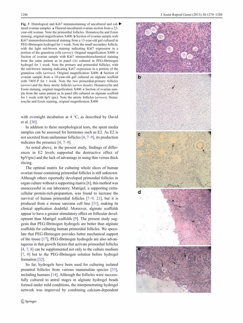
The results for Ki67 staining are shown in Figs. 2e and 3b, c.In most cases, Ki67 staining was identified in a portion of thegranulosa cells from primordial-primary and secondary fol-licles, and it was generally weak. More stained follicles werefound in samples cultured on alginate scaffold and in PEG-fibrinogen hydrogel with basic medium and with 740Y-Pthan in thawed uncultured samples (P<0.0001); in samplescultured in PEG-fibrinogen hydrogel with basic mediumalone than on alginate scaffolds with basic medium alone(P<0.0001); and in samples cultured on alginate scaffoldwith 740Y-P than on alginate scaffold with basic mediumalone (P<0.0001).
E2 secretion
E2 production was calculated per slice [7–9]. Secretion fromthe thick and thin slices was adjusted by dividing the valuesfor every thick slice by 2 (Table 2). E2 production wassignificantly higher with basic medium alone (P<0.03) andwith 740Y-P (P<0.05) than with bpV (pic), regardless of the
matrix. There was significantly more E2 production in thickslices than thin slices, regardless of the treatment or thematrix (250±59 pg/ml and 49±14 pg/ml, respectively,P<0.0004) and on alginate scaffold than in PEG-fibrinogenhydrogel, regardless of the treatment (267±87 pg/ml and52±18 pg/ml, respectively, P<0.04).
Discussion
The morphology-based results of the present study suggestthat PEG-fibrinogen hydrogels have an advantage over algi-nate scaffolds for growing human primordial follicles inorgan culture. More developing follicles were identified inPEG-fibrinogen hydrogel (with 740Y-P or with basic medi-um alone); more Ki67-stained follicles seemed to be detectedin PEG-fibrinogen hydrogels; and there were more atreticfollicles on alginate scaffolds. The addition of PIP3-inducingagents did not improve follicular development; Indeed bpV(pic) caused follicular deterioration. Use of thin-flat slicesdid not improve follicular development over thick slices, asindicted by the higher E2 levels in the thin slices.
As different tissue slices were evaluated during organculture and we could not monitor the same follicle [7–9],we applied several methods of evaluation to ensure accuracy.Presumably because each method was designed to examine adifferent tissue level, rather than obtaining a direct correla-tion of the results among the methods, we found that eachmethod provided information on a different aspect of follic-ular development. The stained sections were used to deter-mine the number and classification (primordial, primary andsecondary) of the follicles, reflecting the growth and survivalof follicles in culture [4], and the cultured vs. unculturedcontrol samples were used to count the follicles periodicallythroughout culture and compare the findings among thefollicular classes. Thus, a decrease in the number of primor-dial follicles and a corresponding increase in the number ofgrowing follicles, from primary stages onwards, would sig-nify follicular growth. We used uniform-size samples, andthe follicles were counted throughout the field (magnifica-tion X100) in 2 to 3 levels per specimen (with at least 50 μmbetween levels to avoid counting the same follicle twice).Increase in follicular atresia were due to the destructive effectof bpV (pic). There were also more atretic follicles on algi-nate scaffolds than in PEG-fibrinogen hydrogels.
Various immunocytochemical methods have been devel-oped to evaluate DNA division or granulosa cell prolifera-tion [4, 7–9]. In the present study, Ki67 staining was used toidentify granulosa cell activation [24–29]. We detected onlyweak Ki67 immunostaining, similar to previous studies [8,9]. It is possible that staining intensity could be increased bysubstituting the 1 h incubation with the primary antibody
Fig. 2 Graphs representing main and significant results from Table 1. I.Activation in culture by follicular counts. a Results are in percent ofprimordial follicles. Primordial follicle count is significantly higher inthawed-untreated samples than after culture with basic medium alone,regardless of the matrix (P<0.05). b Results are in percent of develop-ing follicles. Developing follicle count is higher in samples culturedwith basic medium alone than in thawed-untreated samples, regardlessof the matrix (NS). II. PEG-fibrinogen hydrogels versus alginate scaf-folds. c Results are in percent of atretic follicles. Atretic follicle count issignificantly higher on alginate scaffolds than atretic follicles in PEG-fibrinogen hydrogels, regardless of treatment (P<0.04). d Results are inpercents of developing follicles. Developing follicle count is higher insamples cultured in PEG-fibrinogen hydrogels than in thawed-untreatedsamples (NS). e Results are in percent of Ki67 positively stainedfollicles. The proportion of stained follicles is significantly higher onalginate scaffolds or in PEG-fibrinogen hydrogels with basic mediumthan in thawed-uncultured samples (P<0.0001). III. Destructive effectsof bpV (pic). Results are in percent of primordial follicles. f Primordialfollicle count is significantly higher in thawed-untreated samples thanin samples cultured with bpV (pic), regardless of the matrix (P<0.05).(G) Developing follicles count is significantly higher in samples cul-tured in PEG-fibrinogen hydrogel with 740Y-P than in PEG-fibrinogenhydrogel with bpV (pic) (P<0.05). h E2 level is significantly higher insamples cultured with 740Y-P (P<0.05) or with basic medium alone(P<0.03) than with bpV (pic), regardless of the matrix. IV. Thick versusthin samples. Results are in pg/ml. Values represent mean±standarddeviation. i E2 level is significantly higher after culture of thick slicesthan thin slices (P<0.0004), regardless of the matrix. j E2 level issignificantly higher after culture of thick slices than thin slices onalginate scaffolds (P<0.04)
�
J Assist Reprod Genet (2013) 30:1279–1288 1285
with overnight incubation at 4 °C, as described by Davidet al. [30].
In addition to these morphological tests, the spent mediasamples can be assessed for hormones such as E2. As E2 isnot secreted from unilaminar follicles [4, 7–9], its productionindicates the presence [4, 7–9].
As noted above, in the present study, findings of differ-ences in E2 levels supported the destructive effect ofbpV(pic) and the lack of advantage in using thin versus thickslicing.
The optimal matrix for culturing whole slices of humanovarian tissue containing primordial follicles is still unknown.Although others reportedly developed primordial follicles inorgan culture without a supportingmatrix [6], this methodwasunsuccessful in our laboratory. Matrigel, a supporting extra-cellular protein-rich-preparation, was found to increase thesurvival of human primordial follicles [7–9, 21], but it isproduced from a mouse sarcoma cell line [31], making itsclinical application doubtful. Moreover, alginate scaffoldsappear to have a greater stimulatory effect on follicular devel-opment than Matrigel scaffolds [9]. The present study sug-gests that PEG-fibrinogen hydrogels are better than alginatescaffolds for culturing human primordial follicles. We specu-late that PEG-fibrinogen provides better mechanical supportof the tissue [17], PEG-fibrinogen hydrogels are also advan-tageous in that growth factors that activate primordial follicles[4, 7, 8] can be supplemented not only to the culture medium[7, 8] but to the PEG-fibrinogen solution before hydrogelformation [32].
So far, hydrogels have been used for culturing isolatedpreantral follicles from various mammalian species [33],including humans [14]. Although the follicles were success-fully cultured to antral stages in alginate hydrogel beadsformed under mild conditions, the interpenetrating hydrogelnetwork was improved by combining calcium-dependent
Fig. 3 Histological and Ki67 immunostaining of uncultured and cul-tured ovarian samples. a Thawed-uncultured ovarian section from a 22-year-old woman. Note the primordial follicles. Hematoxylin and Eosinstaining, original magnification X400. b Section of ovarian sample withKi67 immunohistochemical staining from a 13-year-old girl cultured inPEG-fibrinogen hydrogel for 1 week. Note the small secondary follicle,with the light red-brown staining indicating Ki67 expression in aportion of the granulosa cells (arrow). Original magnification X400. cSection of ovarian sample with Ki67 immunohistochemical stainingfrom the same patient as in panel (A) cultured in PEG-fibrinogenhydrogel for 1 week. Note the primary and primordial follicles, withthe red-brown staining indicating Ki67 expression in a portion of thegranulosa cells (arrows). Original magnification X400. d Section ofovarian sample from a 10-year-old girl cultured on alginate scaffoldwith 740Y-P for 1 week. Note the two primordial-primary follicles(arrow) and the three atretic follicles (arrow heads). Hematoxylin andEosin staining, original magnification X400. e Section of ovarian sam-ple from the same patient as in panel (B) cultured on alginate scaffoldfor 1 week with bpV (pic). Note the atretic follicles (arrows). Hema-toxylin and Eosin staining, original magnification X400
�
1286 J Assist Reprod Genet (2013) 30:1279–1288

alginate with calcium-dependent fibrin [34]. Alginate in-creases hydrogel-bead density, provides additional? mechan-ical support and becomes more rigid with follicular expan-sion [9]. Fibrin is a natural fibrinogen biopolymer; it entrapsgrowth factors and furnishes the hydrogel with mechanicaldynamic properties. Its cell-secreted-protease degradation isbalanced by the non-degradable alginate. Indeed, fibrin-alginate hydrogel beads were found to enhance the growthof encapsulated mouse follicles and to improve oocyte mei-otic competence [33, 34]. Accordingly, it is possible thatvarious compounds may be combined to enhance the encap-sulating matrix for ovarian slices.
Our evaluation of the possible role of PIP3-enhancingsubstances in in vitro follicular development was promptedby previous studies [10, 13] wherein bpV (pic) (mouse andhuman), bpV (HOpic) (mouse), or 740Y-P (mouse) supple-mentation of the culture medium promoted primordial folli-cle activation. Subsequent implantation of these ovaries intomurine hosts resulted in follicular growth to the preovulatorystages (mice and human), production of mature oocytes(mice), successful oocyte fertilization, and birth of healthypups [10, 13]. Moreover, mice with oocyte-specific deletionsin PTEN or Foxo3 showed enhanced primordial follicleactivation [10, 12, 35].
However, the present results contradict these findings, de-spite our use of both bpV (pic) and 740Y-P at the same lowconcentrations and for the same short-term culture periods asLi et al. [10]. Although higher concentrations of these sub-stances might induce more follicular activation, they couldalso damage other intracellular pathways [22, 35]. Indeed, inour study, bpV (pic) apparently heightened follicular destruc-tion. The discrepancy between our study and that of Li et al.[10] might be due to our purchasing the compound bpV (pic)from another manufacturer, as Calbiochem no longer pro-duces it. In a follow-up study, Adhikari et al. [13] addedanother PTEN inhibitor, bpV(HOpic), to the culture mediumof murine primordial follicles, without testing PIP3 level.Therefore, it seemed unnecessary to test PIP3 levels in theculture medium in the present study. Our results might beimproved by cultivating ovarian slices with bpV (HOpic) atthe same low concentration (1 μm) used by Adhikari et al.[13], (100-fold lower than the bpV (pic) concentration usedpreviously [10]). Regarding 740Y-P, the positive results re-ported by Li et al. [10] pertained to murine primordial folli-cles, whereas we tested human primordial follicles. Althoughwe carefully rinsed off bpV (pic) and 740Y-P, by increasingPIP3 levels in vitro we may have interfered with other impor-tant cellular processes in which the PI3K-Akt intracellularsignaling pathway is normally involved [22]. At the sametime it is conceivable that in the process of ovarian tissuetransplantation after the short-term culture period used in theprevious studies [10, 13], the in vivo environment corrected orbypassed damage to the PI3K-Akt pathway. This was
impossible in our in vitro system. Nonetheless, it is highlyunlikely that mature oocytes obtained in mice engrafted withovarian tissue will be used for clinical fertility restoration inwomen.
By contrast to Telfer et al. [6] we found that thick ovarianslices promoted better follicular development than thinslices. This difference can be explained by our cutting slicesfrom frozen-thawed rather than fresh specimens. In addition,it is possible that despite numerous attempts, we failed toproduce samples that were thin and flat enough or that sometissue-damage occurred during slicing. Results with thinslices might be improved by the use of special tissue-chopper devices [3, 36] rather than manual cutting. It isnoteworthy that to the best of our knowledge, no furtherevidence of benefits of culturing thin slices [6] has beenreported to date.
In summary, this study suggests an advantage for PEG-fibrinogen hydrogel for the short-term culture of humanprimordial follicles. A two-step culturing system might bethe optimal strategy [6–9]; i.e., short-term culture of ovarianslices encapsulated in PEG-fibrinogen hydrogels to obtainlarge secondary follicles, which are then isolated and cul-tured [6, 37, 38]. So far, however, in vitro follicular growthfrom primordial stages to mature oocytes has been achievedonly in mice [37, 38] with the birth of live pups [38]. Furtherstudies are needed to investigate additional culture matricesand improvements in the culture medium for the growth ofhuman primordial follicles [6, 8, 9].
Acknowledgments The authors are grateful to Prof. Smadar Cohenfrom The Avram and Stella Goldstein-Goren Department of Biotech-nology Engineering, Ben-Gurion University of the Negev, Beer-Sheva,Israel for the production of alginate scaffolds in her laboratory. Theauthors are greatly indebted to Ms. Gloria Ganzach from the EditorialBoard of Rabin Medical Center, Beilinson Hospital for the Englishediting. We are also indebted to our laboratory technician, Ms. CarmelaFelz for the histological sectioning. The study was partially funded by aresearch grant from Tel Aviv University, LeoMinz Fund (to R. A and B.F) and the Israel Cancer Association (to R. A and B. F).
Disclosure summary RA and BF received grant support from LeoMinz Fund, Tel Aviv University and from the Israel Cancer Association,GL-S, NS, MS, OK, DS, SC and AB-H have nothing to declare.
References
1. Feigin E, Freud E, Fisch B, Orvieto R, Kravarusic D, Avrahami G,et al. Fertility preservation in female adolescents with malignan-cies. In: Moorland MT, editor. Cancer in female adolescents.Hauppauge: Nova Science Publishers; 2008. p. 38–103.
2. Silber SJ. Ovary cryopreservation and transplantation for fertilitypreservation. Mol Hum Reprod. 2012;18:59–67.
3. Revel A, Laufer N, Ben Meir A, Lebovich M, Mitrani E. Mirco-organ ovarian transplantation enables pregnancy: a case report.Hum Reprod. 2011;26:1033–97.
J Assist Reprod Genet (2013) 30:1279–1288 1287

4. Abir R, Nitke S, Ben-Haroush A, Fisch B. In vitro maturation ofhuman primordial ovarian follicles: clinical significance, progressin mammals, and methods for growth evaluation. HistolHistopathol. 2006;21:887–98.
5. Abir R, Feinmesser M, Yaniv I, Fisch B, Cohen IJ, Ben-Haroush A,et al. Occasional involvement of the ovary in Ewing sarcoma. HumReprod. 2010;25:1708–12.
6. Telfer EE, McLaughlin M, Ding C, Thong KJ. A two-step serum-free culture system supports development of human oocytes fromprimordial follicles in the presence of activin. Hum Reprod.2008;23:1151–8.
7. Garor R, Abir R, Erman A, Felz C, Nitke S, Fisch B. Effects ofbasic fibroblast growth factor on in vitro development of humanovarian primordial follicles. Fertil Steril. 2009;91:1967–75.
8. Kedem A, Fisch B, Garor R, Ben-Zaken A, Gizunterman T. Growthdifferentiating factor 9 (GDF9) and bone morphogenetic protein 15both activate development of human primordial follicles in vitro,with seemingly more beneficial effects of GDF9. J Clin EndocrinolMetab. 2011;96:1246–54.
9. Kedem A, Hourvitz A, Fisch B, Shachar M, Cohen S. Alginatescaffold for organ culture of cryopresereved-thawed human ovariancortical follicles. J Assist Reprod Genet. 2011;28:761–9.
10. Li J, Kawarmura K, Cheng Y, Liu S, Klein C, Liu S, et al. Activa-tion of dormant follicle to generate mature eggs. PNAS.2010;107:10280–4.
11. Kim SR, Lee YC. PTEN as a unique promising therapeutic targetfor occupational asthma. Immunotoxicology. 2008;30:793–814.
12. John GB, Gallardo TD, Shirley LJ, Castrillon DH. Foxo3 is a PI3K-dependent molecular switch controlling the initiation of oocytegrowth. Dev Biol. 2008;321:197–204.
13. Adhikari D, Gorre N, Risal S, Zhao Z, Zhang H, Shen Y, et al. Thesafe use of a PTEN inhibitor for the activation of dormant mouseprimordial follicles and generation of fertilizable eggs. PLoS One.2012;7:e39034.
14. Xu M, West-Farrell ER, Stouffer RL, Shea LD, Woodruff TK,Zelinski MB. Encapsulated three-dimensional culture supports de-velopment of nonhuman primate secondary follicles. Biol Reprod.2009;81:587–94.
15. Seliktar D. Designing cell-compatible hydrogels for biomedicalapplications. Science. 2012;336:1124–8.
16. Dikovskey D, Bianco-Peled H, Seliktar D. Investigating the mo-lecular structure and physical properties of PEG-Fibrinogenhydrogels. Adv Eng Mater. 2010;12:B200–9.
17. Peled E, Boss J, Bejar J, Zinman C, Seliktar D. A novel poly(eth-ylene glycol)-fibrinogen hydrogel for tibial segmental defect repairin a rat model. J Biomed Mater Res A. 2007;80:874–84.
18. Federovich NE, Oudshoorn MH, Van Geemen D, Hennink WE,Alblas J. The effect of photopolymerization on stem cells embed-ded in hydrogels. Biomaterials. 2009;30:344–53.
19. Bryant SJ, Nuttelman CR, Anseth KS. Cytocompatibility of UVand visible light photoinitiating systems on cultured NIH/3T3 fi-broblasts in vitro. J Biomater Sci Polym Ed. 2000;11:439–57.
20. Elisseeff J, McIntosh W, Anseth K, Riley S, Ragan P, Langer R.Photoencapsulation of chondrocytes in poly(ethylene oxide)-basedsemi-interpentrating networks. J Biomed Mater Res. 2000;51:164–71.
21. Hovatta O, Silye R, Abir R, Krausz T, Winston RM. Extracellularmatrix improves survival of both stored and fresh human primordialand primary ovarian follicles in long-term culture. Hum Reprod.1997;12:1032–6.
22. Blanco-Aparicio C, Renner O, Leal JF, Carnero A. PTEN, morethan the AKT pathway. Carcinogenesis. 2007;28:1379–86.
23. Gougeon A. Regulation of ovarian follicular development in pri-mates: facts and hypotheses. Endocr Rev. 1996;17:55–121.
24. Gerdes J, Lemke H, Baisch J. Cell cycle analysis of cell prolifera-tion associated human nuclear antigen defined by the monoclonalantibody Ki67. J Immunol. 1984;133:1710–5.
25. Scholzen T, Geredes J. The Ki67 protein: from the known and theunknown. J Cell Physiol. 2000;182:311–22.
26. Rivera OE, Varayoud J, Rodrigues HA, Munoz-de-Toro M, LuqueEH. Neonatal exposure to bisphenol A or diethylstilbestrol altersthe ovarian follicular dynamics in the lamb. Reprod Toxicol.2011;32:304–12.
27. Vendola KA, Zhou J, Adesanya OO, Weil SJ, Bondy CA. Andro-gens stimulate early stages of follicular growth in the primate ovary.J Clin Invest. 1998;101:2622–9.
28. Fabbri R, Venturoli S, D'Errico A, Iannascoli C, Gabusi E, Valeri B,et al. Ovarian tissue banking and fertility preservation in cancerpatients: histological and immunohistochemical evaluation.Gynecol Oncol. 2003;89:259–66.
29. Rahmanzadeh R, Hüttmann G, Gerdes J, Scholzen T.Chromophore-assisted light inactivation of pKi67 leads to in-hibition of ribosomal RNA synthesis. Cell Prolif. 2007;40:422–30.
30. David A, Van Langendonckt A, Gilliaux S, Dolmans MM, DonnezJ, Amorim CA. Effect of cryopreservation and transplantation onthe expression of kit ligand and anti-Mullerian hormone in humanovarian tissue. Hum Reprod. 2012;27:1088–95.
31. Kleinman HK. Preparation of basement membrane componentsfrom EHS tumors. Curr Protoc Cell Biol. 2001;10:10.2.1–10.
32. Seliktar D, Zisch AH, Lutolf MP, Wrana IJL, Hubbell JA. MMP-2sensitive, VEGF bearing bioactive hydrogels for promotion ofvascular healing. Biomed Mater Res A. 2004;68:704–16.
33. Xu M, Kreeger PK, Shea LD, Woodruff TK. Tissue-engineeredfollicles produce live, fertile offspring. Tissue Eng. 2006;12:2739–46.
34. Shikanov A, Xu M, Woodruff TK, Shea LD. Interpenetratingfibrin-alginate matrices for in vitro ovarian follicle development.Biomaterials. 2009;30:5476–85.
35. Castrillon DH, Miao L, Kollipara R, Horner JW, DePinho RA.Suppression of ovarian follicle activation in mice by the transcrip-tion factor Foxo3a. Science. 2003;301:215–8.
36. Kagawa N, Sliber S, Kuwayama M. Successful vitrification ofbovine and human ovarian tissue. Reprod Biomed Online.2009;18:568–77.
37. Jin SY, Lei L, Shikanov A, Shea LD, Woodruff TK. A novel two-step strategy for in vitro culture of early-stage ovarian follicles inthe mouse. Fertil Steril. 2010;93:2633–9.
38. O'Brien MJ, Pendola JK, Eppig JJ. A revised protocol for in vitrodevelopment of mouse oocytes from primordial follicles dramati-cally improves their developmental competence. Biol Reprod.2003;68:1682–6.
1288 J Assist Reprod Genet (2013) 30:1279–1288


![Buparlisib, another step in the quest to cure ......PIP3 – Phosphatidylinositol 3,4,5 trisphosphate [PI(3,4,5)P3] PTEN – Phosphatase and tensin homologue RAEB – Refractory anemia](https://img.pdfslide.net/doc/110x75/5f3382fe83ae4a328035e0d8/buparlisib-another-step-in-the-quest-to-cure-pip3-a-phosphatidylinositol.jpg)