Embed Size (px)
Citation preview
Diabetic FootAttlee B. Benally DPM
University of New Mexico Department of Orthopedic
Neither the University of New Mexico Medical school, Hospital, nor the Author of this presentation assume any liability or responsibility for the accuracy, completeness, or usefulness of any information, product, or endorse or seek any financial gain from the products or process discussed in this presentation.
Disclaimer:
To understand the process of evaluating and treating the Diabetic foot and streamlining the treatment of the infected Diabetic foot.
Objective:
2012: 29.1 M Americans with Diabetes, 1.25 M Am. Children & Adults have type 1 diabetes
Undiagnosed: Of the 29.1M, 21.0 were Diagnosed, 8.1 were undiagnosed.
Seniors: Age >65 (25.9% or 11.8M). New Cases: 2012, 1.7 M. Deaths: 7th leading cause of Death in America in 2010. 69K death cert., listed has underlining
cause of death. 234K death cert., contributing cause of death.
Statistic’s of Diabetes & Prediabetes
Prediabetes: 2012, 86M Americans age 20 and older had prediabetes. Up from 79M in 2010.
Continuation of Statistic’s of Diabetes & Prediabetes
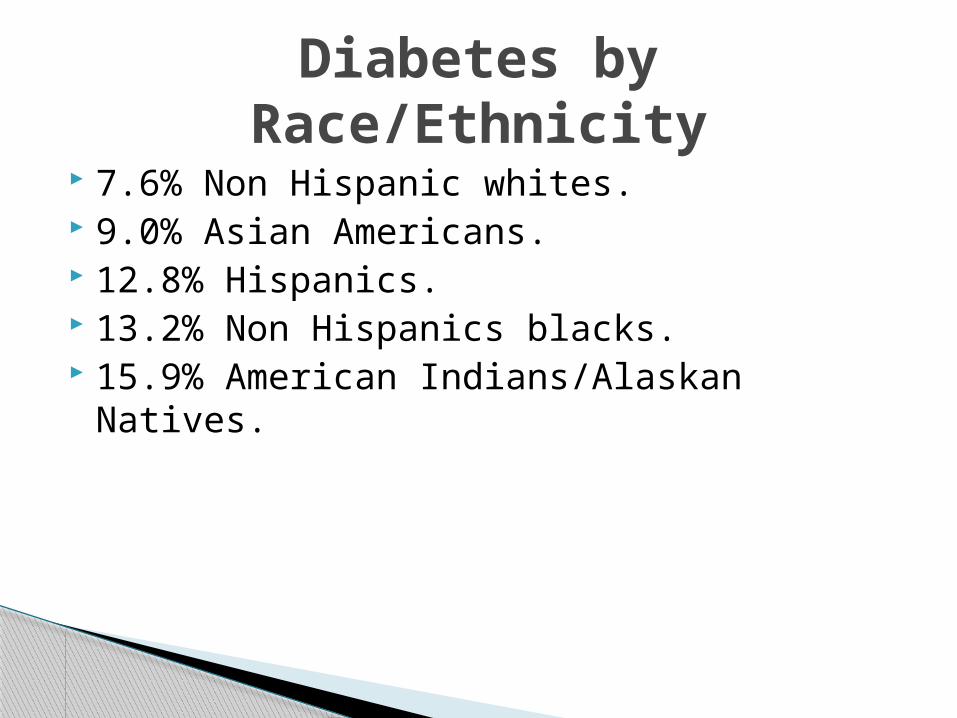
7.6% Non Hispanic whites. 9.0% Asian Americans. 12.8% Hispanics. 13.2% Non Hispanics blacks. 15.9% American Indians/Alaskan Natives.
Diabetes by Race/Ethnicity
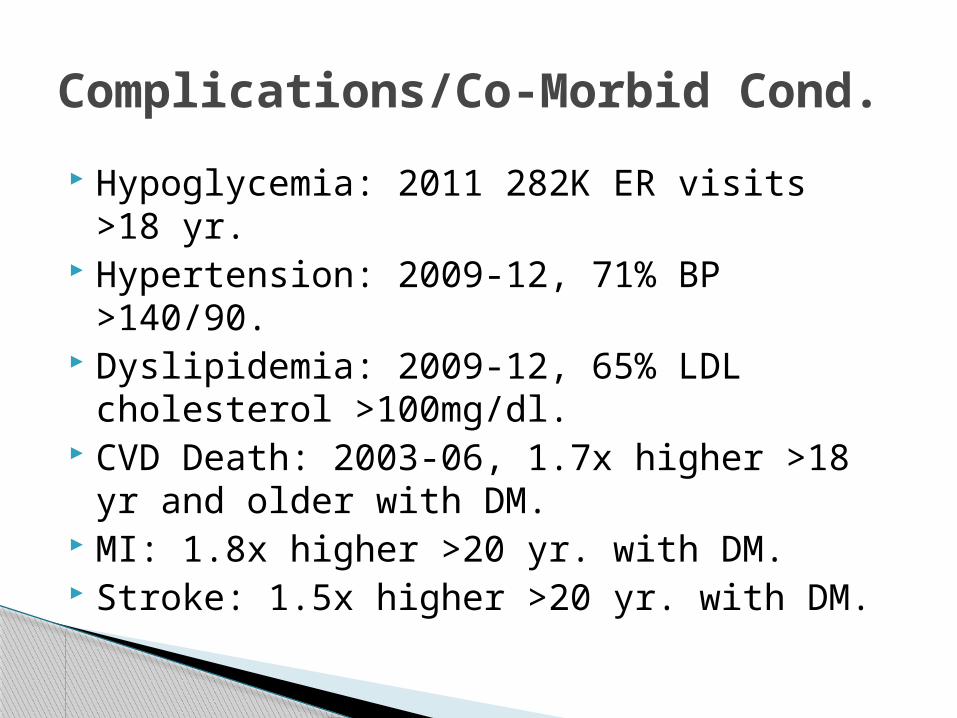
Hypoglycemia: 2011 282K ER visits >18 yr. Hypertension: 2009-12, 71% BP >140/90. Dyslipidemia: 2009-12, 65% LDL cholesterol
>100mg/dl. CVD Death: 2003-06, 1.7x higher >18 yr
and older with DM. MI: 1.8x higher >20 yr. with DM. Stroke: 1.5x higher >20 yr. with DM.
Complications/Co-Morbid Cond.
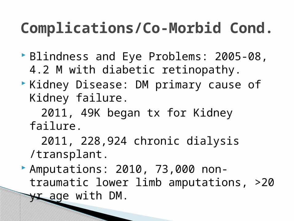
Blindness and Eye Problems: 2005-08, 4.2 M with diabetic retinopathy.
Kidney Disease: DM primary cause of Kidney failure.
2011, 49K began tx for Kidney failure. 2011, 228,924 chronic dialysis /transplant. Amputations: 2010, 73,000 non-traumatic
lower limb amputations, >20 yr age with DM.
Complications/Co-Morbid Cond.
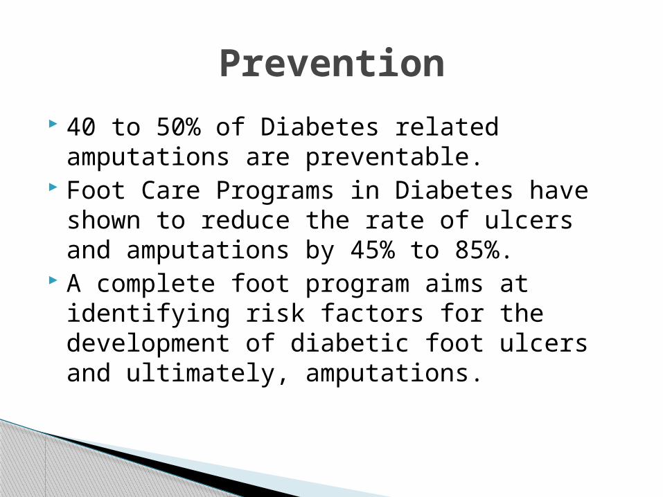
40 to 50% of Diabetes related amputations are preventable.
Foot Care Programs in Diabetes have shown to reduce the rate of ulcers and amputations by 45% to 85%.
A complete foot program aims at identifying risk factors for the development of diabetic foot ulcers and ultimately, amputations.
Prevention
Peripheral neuropathy. Absent pedal pulses. Trophic skin changes, decreased hair
growth, abnormal toenails, or discoloration of skin.
Calf or leg pain indicative of claudication. History of previous foot ulcer or lower
extremity amputation. Previous hospitalization for any diabetic foot
infection. Obesity.
Prevention: Risk Factors forDevelopment of Ulcers
Foot deformity and callus formation resulting in focal areas of high pressure.
Autonomic neuropathy, causing decreased sweating and dry, fissured skin.
Impaired vision. Poor glucose control-Impaired wound
healing. Poor footwear that causes skin breakdown,
or inadequately protects the skin from high pressure and shear forces.
Prevention: Risk Factors forDevelopment of Ulcers
ADA recommends Diabetic’s to have a through foot examination about once a year.
foot examination would include: 1) DERMATOLOGY: Pedal hair, skin integrity, The Temperature of skin, nails-normal, or mycotic. 2) VASCULAR: Pedal Pulses DP and PT intact or absent, Cap refill +3 seconds, Edema.
Prevention: Foot Examination
foot examination would include: 3) NEUROLOGY: Protective threshold intact
with Semmes Weinstein 5.07 monofilament. 4) MUSCULOSKELETAL: Strength, check
all four quadrants, look for foot deformities, hammer toe’s, bunions, rocker bottom foot, boney protuberance’s, Pain with
range of motion.
Prevention: Foot Examination
Poor glucose control increases risk of developing peripheral neuropathy in the lower extremities.
Leads to insensitive foot and ulcer formation. Diabetic Complications and Control Trial (DCCT): 1) Type 1, achieved near normal glucose, experience reduction in clinical neuropathy by 57% to 69%. 2) Type 2, reduces micro vascular and neuropathic complications.
Prevention: Glycemic Control

Cigarette smoking is a major risk factor for PVD and amputation in people without diabetes.
Relationship between tobacco and diabetic foot ulcers has not been clearly established.
Evidence of an association between smoking and micro vascular disease as seen in retinopathy and nephropathy.
Prevention: Smoking Cessation
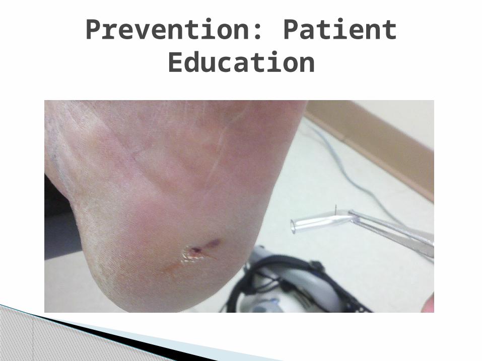
Studies have shown that patient education about foot care results in a significant reduction of diabetic foot ulcerations.
Diabetic patients need to be taught that their feet need daily surveillance and that the primary care physician needs to be notified of any abnormality.
Diabetic patients need to be educated on the importance of diet and nutrient.
Prevention: Patient Education
Prevention: Patient Education
Medicare coverage of therapeutic shoes for diabetics became available in 1993.
Diabetic shoes are seamless and come with a molded insert.
Properly fitted footwear can reduce the formation of calluses and ulcers.
Purchase new shoes in the afternoon when feet are the largest.
Select shoes that have rounded toes, high toe box and soft leather uppers.
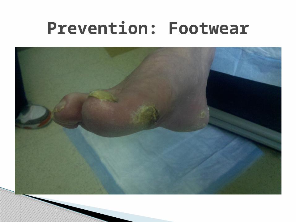
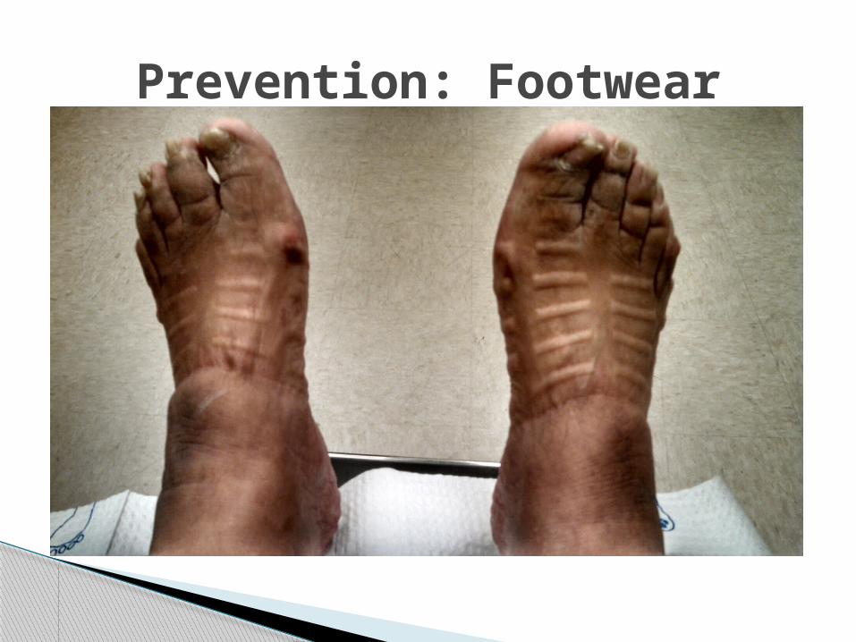
Prevention: Footwear
The ball of the foot should rest in the widest part of the shoe.
Wear shoes that are of adequate length, width and depth. Shoes should not compress the toes or foot. The uppers should be soft leather.
Wear new shoes around the house on carpeting for one hour and check for any hot spots or problem areas.
Prevention: Footwear
Prevention: Footwear
Prevention: Footwear
Replace your shoes regularly. Have at least two pairs of well-fitting shoes to alternate so that shoes can dry between wearing.
Have your feet measured every time you purchase a pair of shoes.
People prone to diabetes-related foot problems should not wear a new shoe more than 2 hours without checking there feet for pressure areas. Five hours should be the maximum daily length of wear.
Prevention: Footwear
Prevention: Footwear
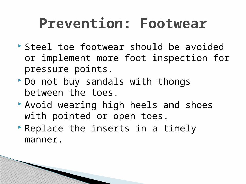
Steel toe footwear should be avoided or implement more foot inspection for pressure points.
Do not buy sandals with thongs between the toes.
Avoid wearing high heels and shoes with pointed or open toes.
Replace the inserts in a timely manner.
Prevention: Footwear
Prevention: Footwear
Exercise is very important in the management of diabetes.
Exercise must be modified in patients with high-risk factors, such as peripheral neuropathy or PVD.
Subjecting insensitive feet to repetitive weight bearing exercises such as jogging, prolong walking, treadmill, and step exercises my lead to ulceration and fractures.
Prevention: Exercise
Swimming. Bicycling. Rowing. Chair exercises. Arm exercises. Other non-weight bearing exercises.
Prevention: Exercises for Diabetic Patients with neuropathy
Treadmill. Prolong walking. Jogging. Step exercises.
Prevention: Exercises Contraindicated for Diabetic Patients with neuropathy
Diabetic foot infection is usually polymicrobial.
Organisms isolated from an infected diabetic foot ranges from one and five organisms.
Average number of bacterial isolates per culture-positive specimen was 2.8 (Lipsky et al).
The most common isolated pathogen was S aureus(44%). 80% methicillin sensitive.
Microbiology

Other common isolates were: Peptostreptococcus spp
Prevotella-Porphyromonas group Enterobacteriaceae
Enterococcus spp Streptococcus agalactiae Bacteroides fragilis Pseudomonas
Microbiology
There are more hospital admissions for Diabetics with related foot pathology.
Neuropathy, peripheral vascular disease, neuro-osteoarthropathy (Charcot’s joint) contribute to foot ulcers, cellulitis, soft tissue abscesses, gangrene, and osteomyelitis.
Differentiating these conditions and the infection often require imaging studies.
Plain radiography, radionuclide studies, CT, and MRI help with these assessment.
Imaging of the Diabetic foot
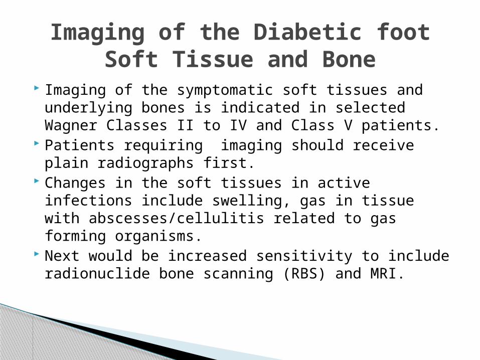
Imaging of the symptomatic soft tissues and underlying bones is indicated in selected Wagner Classes II to IV and Class V patients.
Patients requiring imaging should receive plain radiographs first.
Changes in the soft tissues in active infections include swelling, gas in tissue with abscesses/cellulitis related to gas forming organisms.
Next would be increased sensitivity to include radionuclide bone scanning (RBS) and MRI.
Imaging of the Diabetic footSoft Tissue and Bone
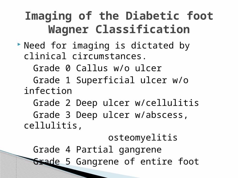
Need for imaging is dictated by clinical circumstances.
Grade 0 Callus w/o ulcer Grade 1 Superficial ulcer w/o infection Grade 2 Deep ulcer w/cellulitis Grade 3 Deep ulcer w/abscess, cellulitis,
osteomyelitis Grade 4 Partial gangrene Grade 5 Gangrene of entire foot
Imaging of the Diabetic footWagner Classification
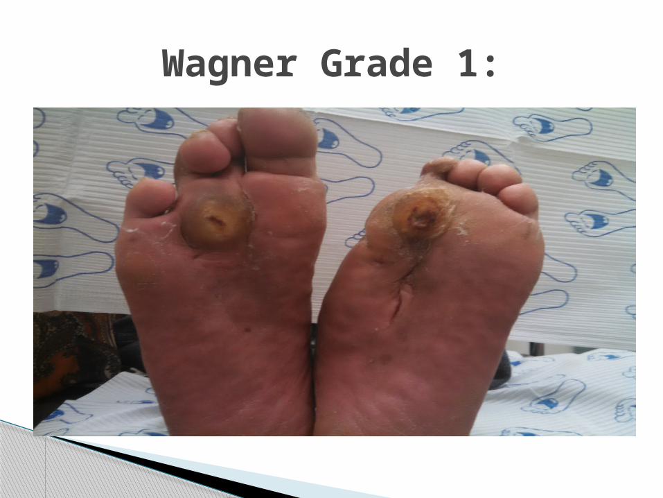
Wagner Grade 1:
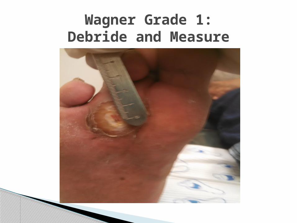
Wagner Grade 1:Debride and Measure

Wagner Grade 2:Deep ulcer/Cellulitis
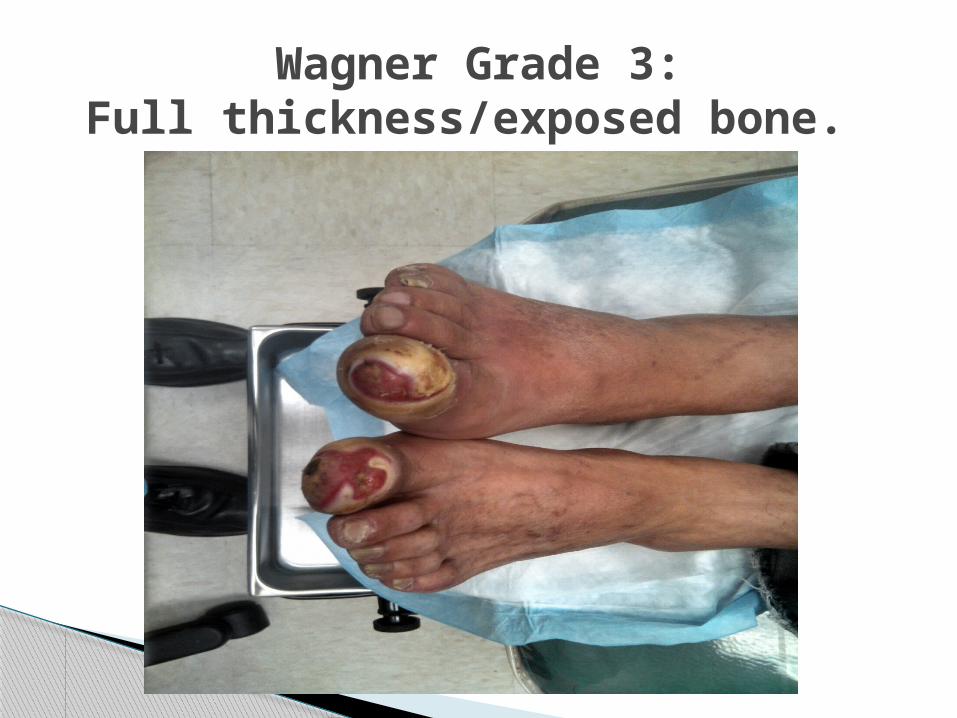
Wagner Grade 3:Full thickness/exposed bone.
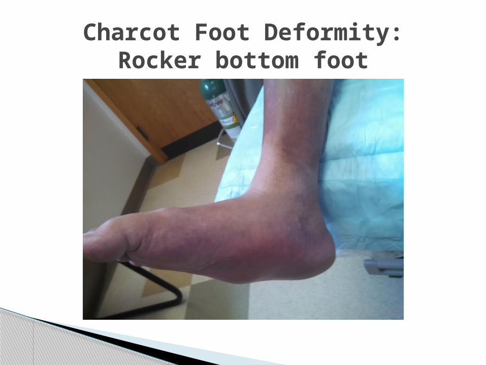
Charcot Foot Deformity:Rocker bottom foot
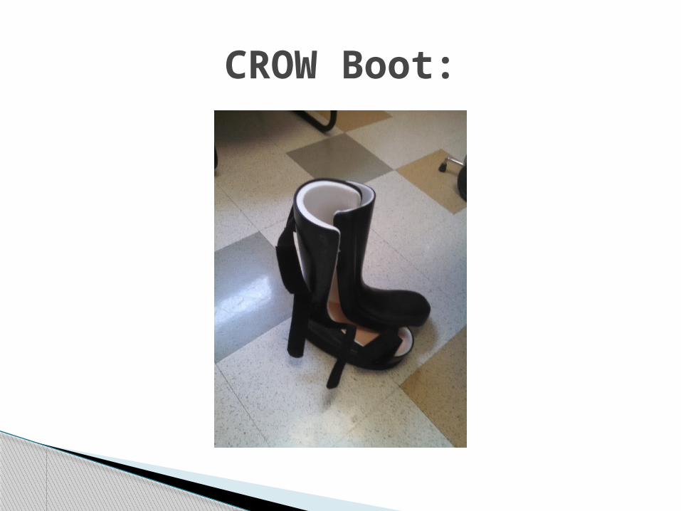
CROW Boot:
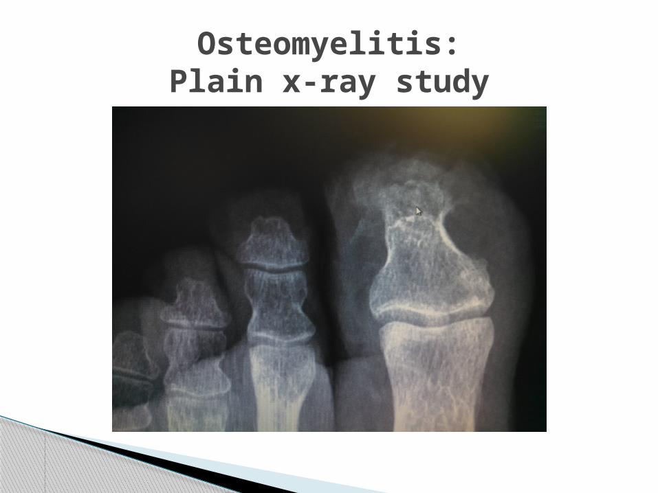
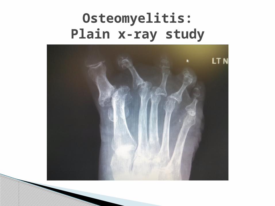
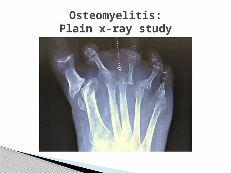
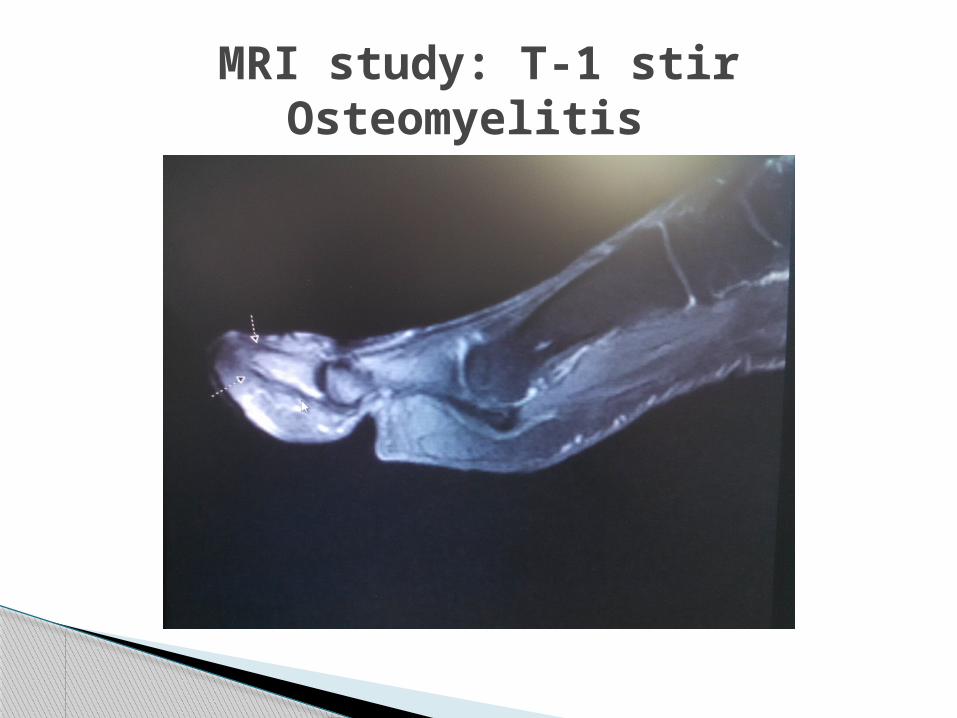
Osteomyelitis is one of the most feared complications of the diabetic foot. It is almost always related to the spread of infection from a diabetic ulcer to the bone (secondary osteomyelitis) rather than from hematogenous spread.
Plain film radiography is specific but not particularly sensitive in osteomyelitis diagnosis.
MRI superior to other methods for suspected soft tissue and or osteomyelitis.
Imaging of the Diabetic footPlain x-ray’s & MRI study
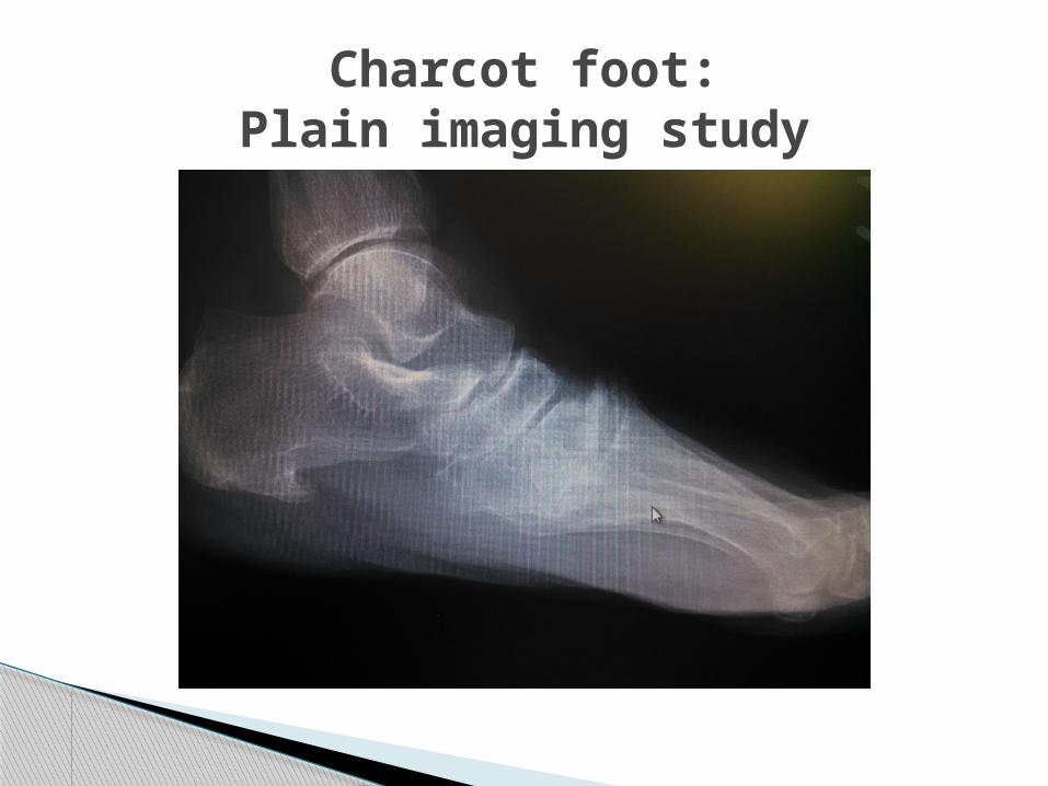
Charcot foot:Plain imaging study
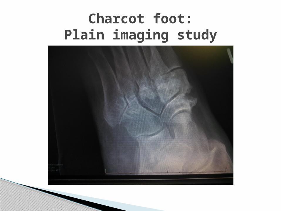
Charcot foot:Plain imaging study
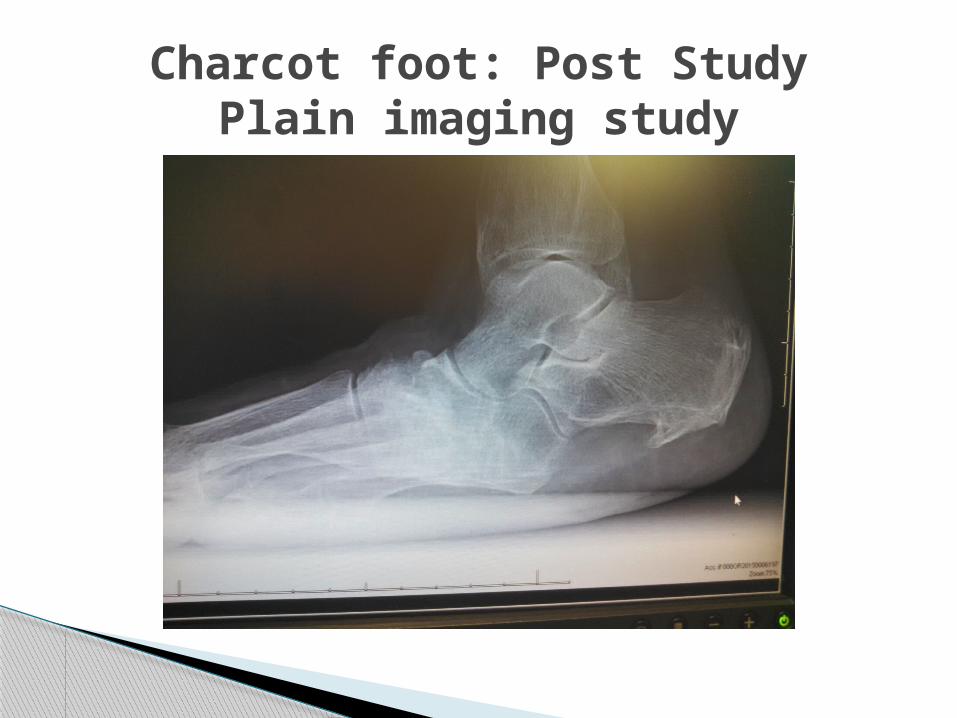
Charcot foot: Post StudyPlain imaging study
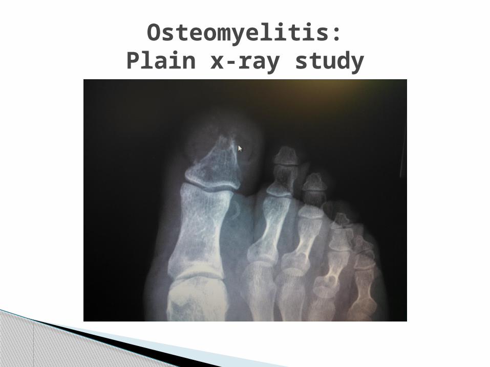
Osteomyelitis:Plain x-ray study
Osteomyelitis:Plain x-ray study
Osteomyelitis:Plain x-ray study
Osteomyelitis:Plain x-ray study
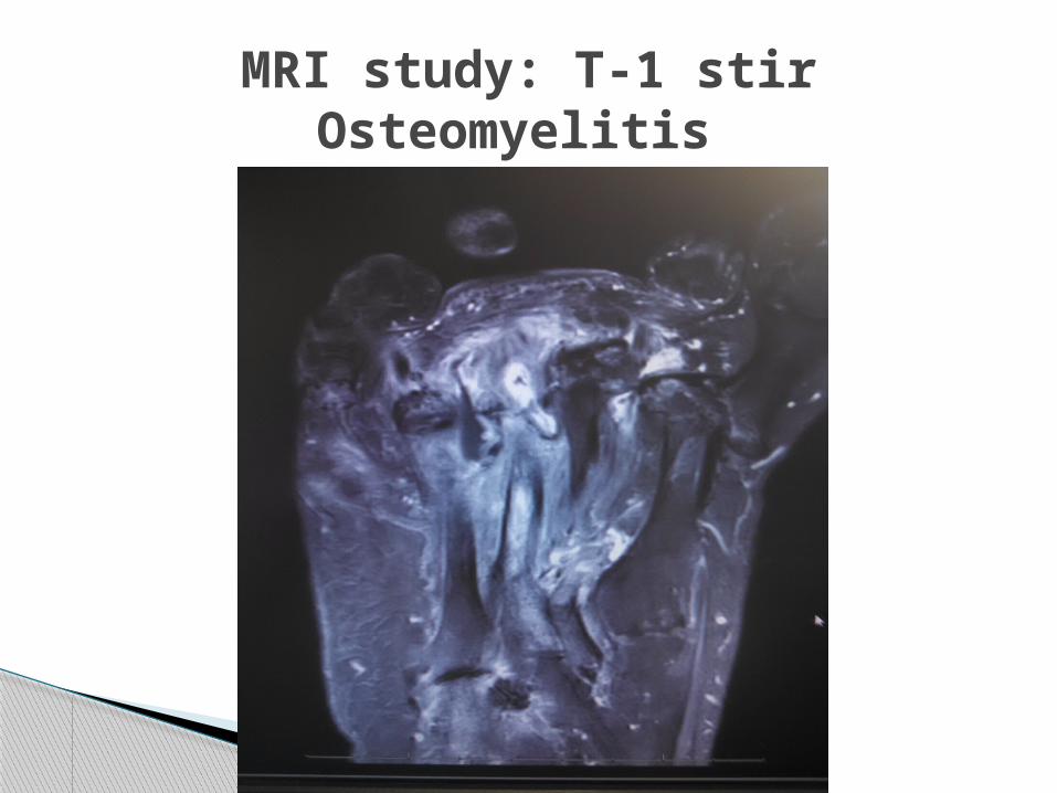
MRI study: T-1 stirOsteomyelitis
MRI study: T-1 stirOsteomyelitis
Evaluate the Diabetic Patient with the foot wound infection.
Diagnose infection on symptoms and signs of inflammation (erythema, warmth, tenderness, pain, induration, or purulent secretions.
Identify factors that increase the risk for DFI, such as: Probe to bone, Tobacco usage, PVD, loss of Protective sensation, History of foot ulcers, Trauma, Presence of renal insufficiency, ABI, TCPO2.
Medical treatmentAssessment

Podiatrist: Outline’s treatment plan, reads MRI, x-rays, C&S test, resects infected bone.
Vascular Surgeon: TCPO2, Angiogram, Angioplasty, Stent, resects infected bone.
Infectious Disease: Appropriate antibiotic regimen, IV or oral treatment.
Wound specialist: Periodic debridement of wound and application of biologics or other.
Medical treatmentConsultation:
All patients with severe infection, selected patients with complicating features (eg. Diabetics, Severe peripheral arterial disease, lack of home support).
Any patient unable to comply with required outpatient treatment regimen (psychological or social).
Infectious Disease monitoring of IV antibiotic.
Medical treatmentHospitalization:
Do not collect a specimen for clinically uninfected wounds.
Infected wounds, send appropriately obtained specimens for culture prior to starting empirical antibiotic therapy.
Specimens for culture should be from deep tissue, biopsy or curettage after the wound has been debrided.
Infectious Disease application of appropriate antibiotics based on C&S, gram stain etc.
Medical treatmentMicrobiology:
Patients presenting with new DFI should have plain radiographs of the affected foot, look for bone abnormalities, soft tissue gas, and foreign objects. Space study by two weeks.
MRI is study of choice for more specific imaging, soft tissue abscess, osteomyelitis.
MRI unavailable or contraindicated, consider radionuclide bone scan and labeled white blood cell scan.
Medical treatmentImaging Studies:

Prescribe antibiotics for all infected wounds. Select an empirical antibiotic regimen based
on the severity of the infection. Base definitive therapy on both the results
of obtained culture and sensitivity and patient’s clinical response to the regimen.
Base therapy on infection severity, parenteral for severe, oral when patient improves.
Continue therapy until resolution of infection.
Medical treatmentAntibiotic Regimen:
Nonsurgical clinicians consider assessment by a surgeon for moderate or severe DFI.
Urgent surgical intervention required for most foot infections with gas in tissue, abscess or necrotizing fasciitis, or extensive bone or joint involvement.
Involve vascular surgeon early for revascularization when ischemia complicates a DFI.
Medical treatmentSurgical Intervention:
Diabetic patients with foot wounds should receive the following:
Debridement of wound. Off-loading wound. Bioengineered skin equivalents Growth factors Granulocyte colony stimulating factors Hyperbaric oxygen therapy Negative pressure wound therapy
Medical treatmentWound Care:
Lipsky BA, Berendt AR, Deery HG, et al: Diagnosis and treatment of Diabetic Foot Infections. Clin Infect Dis 2004; 39:885-910.
Lee C. Rodgers, DPM, and Robert G. Frykberg, DPM. Emerging Evidence on Treatment of the Diabetic Charcot Foot. Podiatry Today, March 2012.
Statistics about Diabetes. American Diabetes Association, February 19, 2015.
Sartoris DJ. Cross-sectional imaging of the diabetic foot. J Foot Ankle Surg 1994;33:531-545.
Valerie L. Schade, DPM, and Nathan S. Higa, DPM. Keys to the Diagnostic Workup for Patients with Diabetic Foot Infections. Podiatry Today, March 2012.
References:

Questions: