Embed Size (px)
Citation preview
378 15 August 1970 Vagotomy and Gastric Ulcer-Burge et al. MBDICTALOUNLFiqure 4
CU? NC
| , A B
Fiqure 5
INADEQUATE ADEOUATEif maliqnant if maliqnantUNNECESSARY A SERIOUS ERRORif beniqn if beniqn
GU
D ~~~~~~~~E
FIG. 4.-Total gastric resection for "doubtful" high lesser curve ulcer. FIG.5.-Sites of gastric resection in "doubtful" mid-lesser curve ulcer.
gastric resection (the Kelling-Madlener operation) or prefer-ably by vagotomy and simple drainage.When the "doubtful" ulcer is on the mid-lesser curve the
surgeon, not wanting to perform a very high gastric resection,may choose to divide the stomach a little distance above theulcer (Fig. 5). If in this case the ulcer proves to be malignantthen the operation was inadequate. If it proves to be benignthen the gastric resection again was unnecessary. If in thissame case the surgeon decides on a high gastric resection
because he has doubt about the nature of the ulcer thenagain a serious error has been made, for the patient is leftwith very little stomach and all the disabilities and sequelaewhich may follow such a high resection.How, then, is the problem to be solved? There would seem
to be three ways of dealing with it. The first is to treat thepatient on a medical regimen for a matter of weeks and tofollow the course of healing. All benign gastric ulcers,especially large ones, are best allowed to heal before selectivevagotomy is undertaken. Selective vagotomny is easier whenthe ulcer is completely healed. The second method is toexcise the ulcer completely at the time of vagotomy and touse frozen section or await the "paraffin" report. The thirdmethod is to open the stomach and palpate the ulcer edgewith the finger, and to take one or more biopsy specimensfrom the edge, using frozen section, or, failing this, to per-form the lesser operation of vagotomy and simple drainage,then await the paraffin report. This is the method advised byDragstedt and the one which we have chosen, so far withoutregret. The more carefully this method of biopsy is practisedthe fewer mistakes will arise. In fact there should be none.As with most mistakes in surgery the fault lies not with themethod but with how well it is carried out. Probably, veryrarely a mistake will arise and a malignant ulcer will beoverlooked. The late results of gastric cancer are not good,and the harm done by a very occasional error must beweighed against that arising from routine gastric resectionsometimes radically and unnecessarily done for benign ulcer.
REFERNCESBurge, H. (1964). Vagotomy, p. 110. London, Arnold.Burge, H. (1966). Annals of the Royal College of Surgeons of England, 38, 349.Burge, H. (1970). Proceedings of the Royal Society of Medicine, 63, 766.Burge, H., Gill, A. M., and Lewis, R. H. (1963). Lancet, 1, 73.Burge, H., MacLean, G., Stedeford, R., Pinn, G., and Holanders, D. (1969).
British Medical Journal, 2, 690.Carman, R. D. (1917). American3Journal of Roentgenology, Radinm Therapy,
and Nuclear Medicine, 4, 552.De Miguel, J. (1968). Revista EspaTnola de las Enfermedades del Aparato
Digestivo y de la Nutrici6n, 27, 24.Grassi, G. (1967). Chirurgia Gastroenterologica, 1, 431.Hansky, J., and Cain, M. D. (1969). Lancet, 2, 1388.Hendry, W. G. (1961). Postgraduate MedicalJournal, 37, 137.Hendry, W. G., and Al Bahrani, Z. (1965). British Journal of Surgery, 52,
588.Kennedy, T., and George, J. D. (1967). Gut, 8, 632.McNeill, A. D., McAdam, W. A. F., and Hutchison, J. S. F. (1969). Surgery,
Gynecology and Obstetrics, 128, 91.
Tropical Splenomegaly Syndrome: Long-term Proguami TherapyCorrelated with Spleen Size, Serum IgM, and Lymphocyte Transformation*
ABA-SEGUA SAGOEt M.B., B.S.
British Medical Journal, 1970, 3, 378-382
Suimmary: Forty-three patients with an initialdigosof tropical splenomegaly syndrome were placed
on long-term proguanil therapy. AU patients who failed torespond to proguanil and who were adequately followedup developed identifiable disease, usually malignantlymphoma or chronic lymphatic leukaemia. In patientswho responded to proguanil IgM values were always veryhigh and phytohaemagglutinin (P.H.A.)-lymphocyte-trans-formation scores were always normal before treatmentwas started. In patients who failed to respond IgM valueswere within the normal range or below, whileP.H.A.-lymphocyte-transformation scores were abnormallylow. During proguanil treatment IgM values fellgradually, dosely paralleling the decrease in spleen size.
* Work supported by grant from the Rockefeller Foundation.t Research ellow l)epartment of Haematology, University College Hos-
pital, Ibadan, Nigeria.
Introduction
A syndrome of chronic splenomegaly without definableaetiology is found in some people living in tropical areas andhas been reported from the Congo (Charmot and Vargues,1963), Uganda (Marsden et al., 1965), Sudan (Mustafa, 1965),Zambia (Lowenthal et al., 1966), New Guinea (Pryor, 1967)and Nigeria (Edington, 1967; Watson-Williams and Allan,1968) and reviewed by Pitney (1968). Increased values forserum IgM in this syndrome have been reported from theCongo, Algeria, and the Ivory Coast (Charmot and Andre,1964) and reviewed by (Trincflo et al., 1966) and from NewGuinea (Wells, 1968). Hepatic sinusoidal lymphocytosis wasfound in many of the liver biopsy specimens from Ugandaand New Guinea (Marsden et al., 1965, 1967). In Ibadan ithas been found that the lymphocytosis is not limited to theliver but also involves the peripheral blood and bone marrow
of patients with this syndrome. In a previous paper from thisdepartment Watson-Williams and Allan (1968) reported that alarge majority of patients responded with complete remissionto the prolonged administration of the antimalarial drugproguanil (Paludrine).The diagnosis of the syndrome is by exclusion of all other
causes of splenomegaly encountered in tropical areas. More-over, the differential diagnosis from a malignant lymphoma,and in Ibadan chronic lymphatic leukaemia in particular,can be a difficult problem when there is no palpablelymphadenopathy. This study is designed to give, if possible,positive criteria for the diagnosis of the syndrome. Clinicaland biochemical findings of patients with an initial diagnosisof tropical splenomegaly syndrome (T.S.S.) are given. Beforestarting antimalarial therapy, observations were made of theresponse of each patient's peripheral blood lymphocytes tostimulation by phytohaemagglutinin (P.H.A.) and their serum
IgM levels, and during therapy serial estimations of serumimmunoglobulin levels were made. The fate of those whofailed to respond to antimalarial-therapy is recorded.
Patients and Methods
Adult patients were admitted to this study only if they ful-filled the following criteria:
(a) Splenomegaly of more than 10 cm. measured from thecostal margin (at the anterior axillary line) to the apex of thespleen. This corresponds to grade 4 or more of the Hackettclassification (World Health Organization, 1963).
(b) Exclusion of other causes of gross splenomegaly as
follows: (i) Bacterial infections by examination of blood andstool; x-ray, culture of sputum and in some cases culture ofbone marrow and splenic aspirates for Mycobacteriumtuberculosis. (ii) Parasite infections by repeated examinationof thick films for malaria parasites; stool and urine examina-tion for ova of schistosomes; marrow culture and in a fewcases splenic cultures for Leishmania. (iii) Primary blooddyscrasia by full haematological examination of blood andbone marrow. (iv) Liver function tests, liver biopsy (32 cases),and splenoportograms (12 cases) were done to excludeprimary liver diseases. Hepatic sinusoidal lymphocytosis(H.S.L.) in liver biopsies was graded according to Marsden etal. (1967).
Determination of Immunoglobulins
Commercially prepared immunoplates (Hyland Labora-tories, California, U.S.A.) were used for the estimationof IgG, IgA, and IgM according to the method of Fahey andMcKelvey (1965). Three standard sera of known concentra-tion (supplied by Hyland) were included in each plate. When-ever the precipitin rings obtained with IgM tests were largerthan those obtained with the standards the original sera were
diluted 5-20 times with phosphate-buffered saline pH 7-2, sothat the ring diameters would fall within the range foundwith the standards used.
In order to confirm the high values of IgM in many of thesera, the same method was employed but with speciallyprepared immunoplates, using lyophilized anti-IgMt so thatthe concentration of antibody incorporated per ml. of agar
was increased threefold.
P.H.A. Transformation
Phytohaemagglutinin (P.H.A.) "Wellcome" was used to initi-ate blastic transformation of peripheral blood lymphocytes inculture with the method suggested by Wellcome ResearchLaboratories, Beckenham, with modifications. The cells were
harvested after 72 hours and slide preparations were stained
t Supplied by Professor Houba of the W.H.O. Immunology and ResearchTraining Centre, Ibadan.
MEDICAL JOURNAL 379
with May-Grunwald-Giemsa. From each culture 500 cells werecounted, being scored according to the method suggested byPentycross (1968).
Results
Forty-three patients fulfilled initially the criteria for thediagnosis of tropical splenomegaly syndromet (as set outunder Methods). All patients were treated with proguanil, 100mg./day, for at least six months. At the end of this period amarked regression in spleen size was recorded in 32 patients,hereinafter referred to as "responders." No reduction, butsometimes an increase, in spleen size was noted in theremaining 11 patients, hereinafter referred to as "non-responders."
Clinical and Haematological Findings.-All patients com-plained of abdominal swelling with discomfort of a fewmonths to several years' duration. Mild symptoms of anaemiawere present in the majority. The anaemia was normocyticand normochromic. The total white blood cell count variedwidely, but the typical feature, especially in responders, was alymphocytosis in the peripheral blood and bone marrowwhich also showed normoblastic hyperplasia. The plateletcount was often below normal, but there was no evidence ofbleeding diatheses. Repeated thick blood films for malariaparasites were negative.Immunoglobulin Values (Table I).-IgG and IgA: No sig-
nificant difference was found in the levels of IgG and IgA in
TABLE I.-Serum Levels of Immunoglobulins in 43 Patients with an InitialDiagnosis of T.S.S., Shown in Those Who Responded and Those Who DidNot Respond to Proguanil Therapy. Spleen Sizes Range from 10-30 cm. inBoth Groups. In Responders the Spleen Sizes During Therapy are Expressed
as a Percentage of the Initial Value
SpleenSize as a IgG IgA IgM
Group Percentage (mg./100 ml.) (mg./100 ml.) (mg./100 ml.)of InitialMeasured
lu
Range Mean Range Mean Range Mean
Responders r 100 750-5,100 2,175 150-300 185 860-3,000 1,799(32) 50 1,050-4,400 2,443 78-210 130 310-860 553
L 0 1,050-4,000 1,972 45-260 152 20-420 308
Non-responders(1 1) 100 750-3,800 1,810 48-400 135 20-325 81
Normal adultNigerians 1,250-4,100 2,500 60-360 183 50400 189
*40 blood donors without splenomegaly.
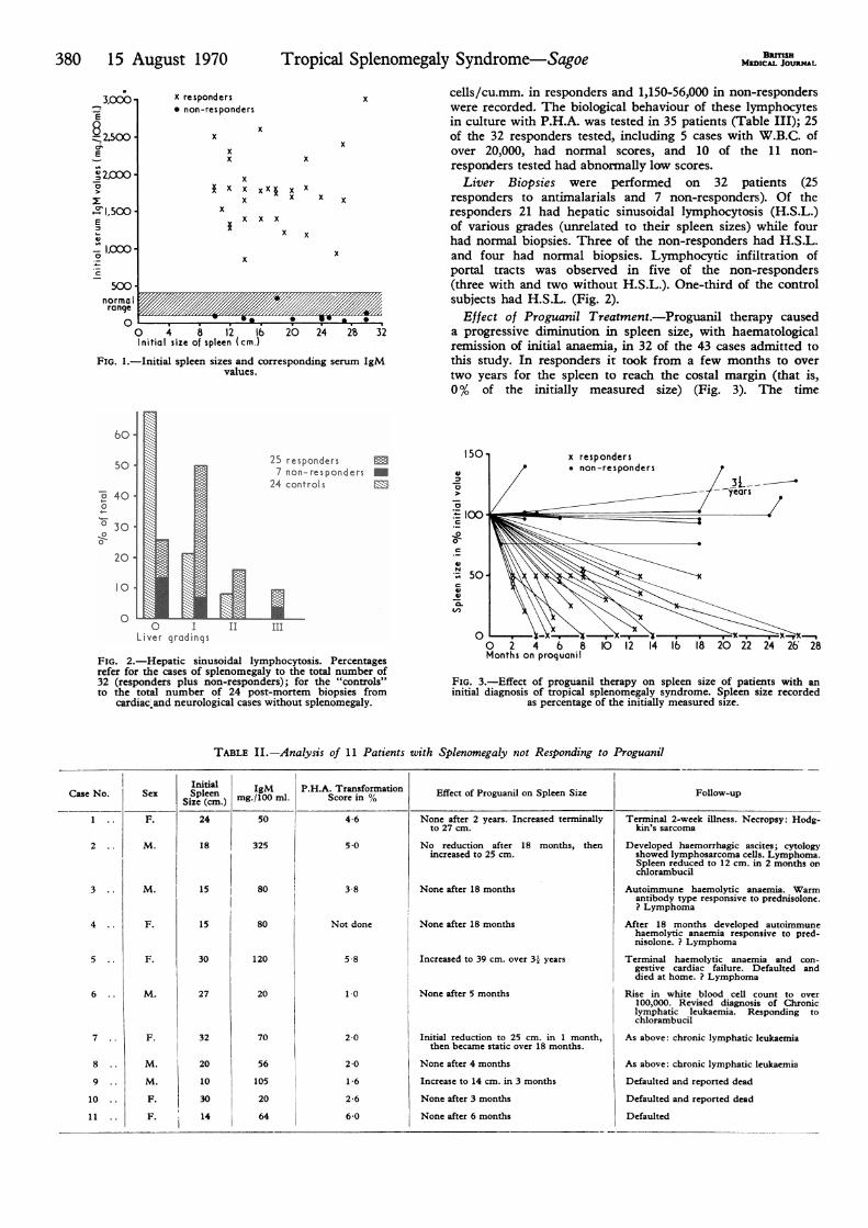
our patients and the controls. IgM: Before treatment theresponders had 3-10 times the values for normal adultNigerians, while all the non-responders had serum IgM levelseither within or below the normal range. A scattergram of theinitial IgM values against the initial values of the spleen sizeis shown in Fig. 1. It can be seen that the Individual IgMvalues bear no correlation to the size of the spleen in eithergroup, and that responders have much higher IgM valuesthan the non-responders. The non-overlap of IgM valuesfound in this study between the group of T.S.S. patients andthe control group is in contrast to the findings of Wells,(1968) in New Guinea. This can be explained by the selectivenature of the patients in this study: all had splenomegaly ofgrade 4 or more; all were investigated to exclude other causesof splenomegaly, and all made significant response toproguanil therapy.Lymphocytes and P-H.A.-induced Lymphocyte Transforma-
tion.-Absolute lymphocyte counts varied widely, dependingon the total white cell count (W.B.C.). Values of 1,380-35,000
t None of these was induded in the series published by Watson-Williamsand Allan (1968).
15 August 1970 Tropical Splenomegaly Syndrome-Sagoe
380 15 August 1970 Tropical Splenomegaly Syndrome-Sagoe
x responders* non-responders
xx
xx x
x* x x XX* x x
x x xx
x x x
x x
x
x
x
x
normalI Vranger *. .
0 4 8 12 Ib 20 24 28 32Initial size of spleen (cm.)
FIG. 1.-Initial spleen sizes and corresponding serum IgMvalues.
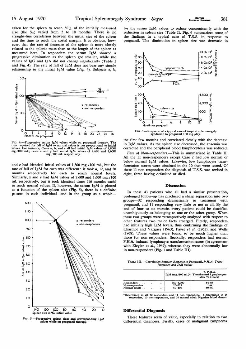
cells/cu.mm. in responders and 1,150-56,000 in non-responderswere recorded. The biological behaviour of these lymphocytesin culture with P.H.A. was tested in 35 patients (Table III); 25of the 32 responders tested, including 5 cases with W.B.C. ofover 20,000, had normal scores, and 10 of the 11 non-responders tested had abnormally low scores.Liver Biopsies were performed on 32 patients (25
responders to antimalarials and 7 non-responders). Of theresponders 21 had hepatic sinusoidal lymphocytosis (H.S.L.)of various grades (unrelated to their spleen sizes) while fourhad normal biopsies. Three of the non-responders had H.S.L.and four had normal biopsies. Lymphocytic infiltration ofportal tracts was observed in five of the non-responders(three with and two without H.S.L.). One-third of the controlsubjects had H.S.L. (Fig. 2).
Effect of Proguanil Treatment.-Proguanil therapy causeda progressive diminution in spleen size, with haematologicalremission of initial anaemia, in 32 of the 43 cases admitted tothis study. In responders it took from a few months to overtwo years for the spleen to reach the costal margin (that is,0% of the initially measured size) (Fig. 3). The time
60
50 -
0
4530 'wo
20 -
10.
0
25 responders -7 non-responders -
24 controls
0 I II mLiver grodings
FIG. 2.-Hepatic sinusoidal lymphocytosis. Percentagesrefer for the cases of splenomegaly to the total number of32 (responders plus non-responders); for the "controls"to the total number of 24 post-mortem biopsies from
cardiac and neurological cases without splenomegaly.
TABLE II.-Analysis of 11 Patients with
150Iv4,
a
a0c:t
-0-
.I,
._
c
0 2 4 6 8 10 12 14Months on proquanil
FIG. 3.-Effect of proguanil therapy on spleen size of patients with aninitial diagnosis of tropical splenomegaly syndrome. Spleen size recorded
as percentage of the initially measured size.
Splenomegaly not Responding to Proguanil
Effect of Proguanil on Spleen Size
None after 2 years. Increased terminallyto 27 cm.
No reduction after 18 months, thenincreased to 25 cm.
None after 18 months
None after 18 months
Increased to 39 cm. over 3- years
None after 5 months
Initial reduction to 25 cm. in 1 month,then became static over 18 months.
None after 4 months
Increase to 14 cm. in 3 months
None after 3 months
None after 6 months
Follow-up
Terminal 2-week illness. Necropsy: Hodg-kin's sarcoma
Developed haemorrhagic ascites; cytologyshowed lymphosarcoma cells. Lymphoma.Spleen reduced to 12 cm. in 2 months onchlorambucil
Autoimmune haemolytic anaemia. Warmantibody type responsive to prednisolone.? Lymphoma
After 18 months developed autoimmunehaemolytic anaemia responsive to pred-nisolone. ? Lymphoma
Terminal haemolytic anaemia and con-gestive cardiac failure. Defaulted anddied at home. ? Lymphoma
Rise in white blood cell count to over100,000. Revised diagnosis of Chroniclymphatic leukaemia. Responding tochlorambucil
As above: chronic lymphatic leukaemia
As above: chronic lymphatic leukaemia
Defaulted and reported dead
Defaulted and reported dead
Defaulted
3,000E
_2IXC0'
°>
Z', 1.500E
I2.M
0
BamSsMEDICAL JOURNAL
24
18
15
15
30
27
32
20
10
30
14
3
4
5
6
7
8
9
10
11
M.
F.
F.
M.
F.
M.
M.
F.
F.
80
80
120
20
70
56
105
20
64
3-8
Not done
5-8
1-0
2-0
2-0
1-6
26
6-0
- - Cl,CXX)
5,00A
15 August 1970 Tropical Splenomegaly Syndrome-Sagoe B Qm1MwICu. JOURnL 381
taken for the spleen to reach 50% of the initially measuredsize (the S5o) varied from 2 to 18 months. There is nostraight-line correlation between the initial size of the spleenand the time to reach the costal margin. It is obvious, how-ever, that the rate of decrease of the spleen is more closelyrelated to the splenic mass than to the length of the spleen as
measured here. In responders the serum IgM showed a
progressive diminution as the spleen got smaller, while thevalues of IgG and IgA did not change significantly (Table Iand Fig. 4). The rate of fall of IgM does not bear any simplerelationship to the initial IgM value (Fig. 4). Subjects a, b,
0 2 4 8 12 14 1 18 20Months on proquanil
FIG. 4.-Progressive serum IgM values while on proguanil therapy. Thetime required for fall of IgM to normal values is not proportional to initialvalues. For instance, Cases a, b, and c all had initial IgM values of 1,800/mg./100 ml.; cases x and y had initial IgM values of 2,600 and 1,600/
mg./100 ml. respectively.
and c had identical initial values of 1,800 mg./100 ml., but therate of fall of IgM for each was different: it took 4, 12, and 20months respectively for each to reach normal levels.Similarly, x and y had IgM values of 2,600 and 1,600 mg./100ml. respectively, but it took identical times (16 months each)to reach normal values. If, however, the serum IgM is plottedas a function of the spleen size (Fig. 5), there is a definitepattern in each individual-and in the group as a whole-
120-
110 X
100 -x responders
non-responders
90
IV 80
70
6b0 -X X
so50
E X
40-
30 -
x
20 -~
10
0
140 120 100 80 60 40 20 0Spleen size in t/o initial value
FIG. 5.-Progressive spleen sizes and corresponding IgMvalues while on proguanil therapy.
for the serum IgM values to reduce concomitantly with thereduction in spleen size (Table I). Fig. 6 summarizes some ofthe findings in a typical case of T.S.S. in response toproguanil. The diminution in spleen size was dramatic in
laoOx 103
u0 8; x 68-OxlO13 -
8b0 .1 [0 l0°40 - X."^ lymphocytes%~/ 4-OslO' n
E 20 abXsolt = 2 0x 103
30
30 -1500E C
v20- .l.000's20- sp"t^^ spleen size 510 X___ x 500
A3
0 Ig M0 30 2 4 b 8 10 12 -
Months on proquani
FIG. 6.-Response of a typical case of tropical splenomegalysyndrome to proguanil 100 mg./day.
the first few months and correlated closely with the decreasein IgM values. As the spleen size decreased, the anaemia wascorrected and the peripheral blood lymphocytosis was reduced.
Fate of Non-responders.-This is summarized in Table II.All the 11 non-responders except Case 2 had low normal orbelow normal IgM values. Likewise, low lymphocyte trans-formation scores were obtained in the 10 that were tested. Ofthese 11 non-responders the diagnosis of T.S.S. was revised ineight, three having defaulted or died.
Discussion
In these 43 patients who all had a similar presentation,prolonged follow-up has produced a sharp separation into twogroups-32 responding dramatically to treatment withproguanil, and 11 responding very little or not at all. By theend of four to six months every patient could be classifiedunambiguously as belonging to one or the other group. Whenthese two groups were retrospectively analysed with respect toother features two major facts emerged. Firstly, respondershad initially high IgM levels, thus confirming the findings ofCharmot and Vargues (1963), Payet et al. (1963), and Wells(1968). These values were found to be much higher thanthose for non-responders. Secondly, responders had normalP.H.A.-induced lymphocyte transformation scores (in agreementwith Ziegler et al., 1969), whereas they were abnormally lowin non-responders (Fig. 1 and Table III).
TABLE III.---Correlation Between Response to Proguanil, P.H.A. Trans-formation and IgM values
% P.H.A.IgM (mg./100 ml.)* Transformed Lymphocytes
after 72 Hourst
Responders . . 860-3,000 64-88Non-responders 20-325 1-6Normal adults.. .. 50-400 60-80
*Determined in all 32 responders and 11 non-responders. tDetermined in 25responders, 10 non-responders, and 20 normal adult Nigerian blood donors.
Differential DiagnosisThese features seem of value, especially in relation to two
differential diagnoses. Firstly, cases of malignant lymphoma
BIUTISH382 15 August 1970 Tropical Splenomegaly Syndrome-Sagoe MEDICAL JOURAL
presenting with gross splenomegaly but withoutlymphadenopathy could be, and have been, misclassified asT.S.S. In such cases lack of a rise in IgM and abnormal P.H.A.transformation will militate against T.S.S. Secondly, cases ofT.S.S. with a lymphocytic leukemoid reaction might be andhave been misclassified as chronic lymphocytic leukaemia(Watson-Williams and Allan, 1968). In such cases high IgMand normal P.H.A. transformation will not favour such a diag-nosis. Since these conditions differ widely in treatment ofchoice and in prognosis, it is important to differentiate betweenthem as soon as possible-that is, before a time-consumingtherapeutic trial.The development of a neoplastic lymphoproliferative
disorder could be established definitely in only five of thenon-responders. Three others, however, had haemolyticanaemia of the type usually associated with malignantlymphomas; and three had a suggestive history and a fataloutcome (Table II). Nevertheless the possibility cannot beentirely ruled out that with continued reticuloendothelialhyperplasia in T.S.S. under certain host conditions or whentimely therapy is not instituted, the cellular proliferation mayproceed to a frank malignant proliferation.
Hepatic sinusoidal lymphocytosis (H.S.L.) found in mostcases of T.S.S. was typical but not pathognomonic (Fig. 2).Some responders did not show this feature and a few normalcontrols had H.S.L., which could not be correlated with thedegree of lymphocytosis in the peripheral blood as seen inIbadan.
Proguanil Therapy in T.S.S.Proguanil has been used for many years in T.S.S. in this
hospital. It is safe and the mode of administration simple-one 100-mg. tablet daily. The response to proguanil consistsnot only in a decrease of spleen size (Fig. 3, and Watson-Williams and Allan, 1968), but also in a return to normal ofhaematological and IgM values (Figs. 4 and 6). Successfultreatment with proguanil cannot be considered sufficient toincriminate malaria as the aetiological agent of the syndrome,for Houba and Adam (1964) have shown that the antimalarialdrug chloroquine has additional-for example, antibacterial-actions. Whether proguanil has this combined effect is notcertain, and neither is it known what effect, if any, proguanilmay have in man on some proliferating cells or on cellsactively producing immunoglobulins.
Source and Significance of Raised IgMThe spleen in T.S.S. constitutes the largest collection of
lymphoreticular tissue in the body. In animals the spleenundergoes morphological changes characteristic of cellssynthesizing proteins after antigenic stimulation (Hanna et al.,1966); and van Furth et al. (1966) showed that human spleencells can synthesize immunoglobulins in vitro. Notwithstand-ing the biosynthesis of IgM in other organs, the progressivediminution of serum levels of IgM accompanying theprogressive diminution in splenic mass suggests that theabnormally large amounts of macroglobulins in T.S.S. maybe formed in the spleen.As to the possible causes involved in stimulating cell prolif-
eration and immunoglobulin production in the spleen, twosets of factors must be considered.
Environmental FactorsOwing to the geographical distribution of T.S.S. and the
finding of Plasmodium malariae in patients with the syndromein Uganda (Marsden et al., 1965) malaria has been implicatedin the aetiology of the syndrome (Pitney, 1968). We foundthat the incidence of malaria parasitaemia was not different
from that in the general population in Ibadan, and malariapigment was not seen in liver biopsies from our patients withT.S.S. IgM is the antibody of the primary immune response,and the macroglobulinaemia of trypanosomiasis is attributedto the frequent antigenic variation in that protozoon. Intropical areas the whole population is exposed to innumerableantigens-viral, bacterial, protozoal, and metazoan. Possiblyrepeated antigenic stimulation by different organisms orantigenic variation of a particular agent giving repeatedprimary immune responses may play a part in thesplenomegaly and macroglobulinaemia of T.S.S.Though antigenic variation may explain a continued
primary immune response it is not clear how this processcould protract itself over many decades in a particular per-son. Also, probably by adulthood an individual would havemet most antigens in his environment. Taking into considera-tion the cross-reactivity of many antigens, one would expectmost immune responses at that age to be not of the primarytype (with IgM production), but rather of the secondary tyre(with IgG production). In T.S.S. the IgG level is not signifi-cantly different from normal: the abnormality is clearly in theIgM fraction.
Host FactorsIt is not yet known whether the macroglobulins of T.S.S.
have any protective antibody function, but probably the hostis responding in an unusual manner to extraneous antigens,especially as only a few persons in tropical areas develop thissyndrome. This finding is increasingly pertinent since asyndrome not unlike T.S.S. has been reported in Britain(Dacie et al., 1969). These host factors, possibly genetic, mayinitiate or perpetuate the condition, with proliferation of cellsin the spleen. The same cells may be producing IgM, andthus account for the serum macroglobulinaemia typical ofT.S.S.
r am grateful for the helpful supervision from Professor G. M.Edington, department of pathology, and Professor V. Houba, ofW.H.O. Immunology and Research Training Centre. I thankProfessor Lucio Luzzatto, department of haematology, for hisencouragement throughout the study.
REFERENCESCharmot, G., and Andr6, L. J. (1964). Semaine des Hopitaux de Paris, 106,
2779.Charmot, G., and Vargues, R. (1963). Semaine des H6pitaux de Paris, 39,
1421.Dacie, J. V., Brain, M. C., Lewis, S. M., and Woriledge, S. M. (1969).
British Journal of Haematology, 17, 317.Edington, G. M. (1967). British Medical3Journal, 1, 715.Fahey, J. L., and McKelvey, E. M. (1965). 7ournal of Immunology, 94, 84.Hanna, M. G., jun., Swartzendruber, D. C., and Congdon, C. C. (1966).
Experimental and Molecular Pathology, 5, No. 3, p. 75.Houba, B., and Adam, M. (1964). Zentralblatt fur Bakteriologie, Parasiten-
kunde, Infektionskrankheiten und Hygiene. I. Abt. Originale, 192, 526.Lowenthal, M. N., Hamilton, P. J. S., Hutt, M. S. R., and Wilks, N. E.
(1966). Central African3Journal of Medicine, 12, 99.Marsden, P. D., et al. (1965). British MedicalJ7ournal, 1, 89.Marsden, P. D., et al. (1967). Bulletin of the World Health Organization, 36,
901.Mustafa, D. (1965). Journal of Tropical Medicine and Hygiene, 68, 183.Payet, M., Sankak, M., Mattern, P., Chambon, L., and Pesse, P. (1963).
Nouvelle Revue Fran9aise d'Hematologie, 3, 630.Pentycross, C. R. (1968). Journal of Clinical Pathology, 21, 175.Pitney, W. R. (1968). Transactions of the Royal Society of Tropical Medicine,
62, 717.Pryor, D. S. (1967). Quarterly Journal of Medicine, 36, 321.Trincao, C., dos Santos Nascimento, J., and Cordeiro Ferreira, N. (1966).
Acta Haematologia, 36, 26.van Furth, R., Schuit, H. R. E., and Hijmans, W. (1966). Immunology, 11,
19.Watson-Williams, E. J., and Allan, N. C. (1968). British Medical Journal,
4, 793.Wells, J. V. (1968). Clinical and Experimental Immunology, 3, 943.World Health Organization (1963). Terminology of Malaria and of Malarial
Eradication, p. 40. Geneva, W.H.O.Ziegler, J. L., Cohen, M. H., and Hutt, M. S. R. (1969). British Medical
.Journal, 4, 15.


![TUBERCULOUS SPLENOMEGALY - SUNY … · TUBERCULOUS SPLENOMEGALY [1] ... A sono or CT-guided percutaneous drainage of associated collections greater than 5 cm may be added to the ATT](https://img.pdfslide.net/doc/110x75/5b9bdc0009d3f272468b9596/tuberculous-splenomegaly-suny-tuberculous-splenomegaly-1-a-sono-or-ct-guided.jpg)