Embed Size (px)
DESCRIPTION
Auscultation
Citation preview
CDHB Clinical Skills Unit
Skills Development Pack (07/2013)
Auscultation of heart and lungs
1
CDHB CLINICAL SKILLS UNIT
HEART AND LUNG AUSCULTATION
CDHB Clinical Skills Unit
Skills Development Pack (07/2013)
Auscultation of heart and lungs
2
Contents: Acknowledgements
Learning objectives Pre-requisites to learning this skill Tutorial outline (preparation, procedure, finishing) Appendix References Self / peer assessment form
User feedback sheet
ACKNOWLEDGEMENTS This pack has been produced in consultation with
Lutz Beckert (consultant physician),
Angela Kraiger (Respiratory Nurse specialist)
AUSCULTATION Learning objectives
CDHB Clinical Skills Unit
Skills Development Pack (07/2013)
Auscultation of heart and lungs
3
Completion of this package will encourage learners to:
Communicate with patients in a way, which reduces anxiety, provides necessary information, earns their trust and ensures safe practice.
1. Describe the relevant anatomy of the heart and lungs and related structures in relation to auscultation.
2. Competently perform a thorough and appropriate examination on a teaching model or peer and correctly identify sounds on the Smartscope model.
3. Document findings accurately in a structured systematic way, which communicates effectively and meets quality standards.
Before learning this skill, it is expected that learners will;
Have up to date knowledge of related anatomy and physiology
Be aware of cultural sensitivities relating to this procedure (see associated “Maori Healthcare, Clinical Skills Information” document)
Have read through the whole package before starting
Identify own learning needs relating to this procedure This pack can be used for: Practical group teaching session using simulation models and / or training video Individual self-directed learning session, with / without peer support using
simulation models and / or training video Using this pack is intended to help learners to:
Meet stated objectives
Meet some / all own learning needs
Feel prepared for any formative / summative assessment It is recommended that learners:
Complete self evaluation form (in this pack) and amend on-going professional development action plan – useful for professional portfolio
Complete user feedback sheet (in this pack) to contribute to the on-going improvement of Clinical Skills Unit facilities.
CDHB Clinical Skills Unit
Skills Development Pack (07/2013)
Auscultation of heart and lungs
4
TUTORIAL OUTLINE
The following guidance is offered in an attempt to improve your technique when required to examine a patient’s heart and/or lungs. If you are new to this skill, you are encouraged to study the written guidance and practice the skill in the safety of the unit, as frequently as you feel necessary before being assessed and ultimately taking responsibility for performing this procedure with patients. Alternatively, even if you have experience, the opportunity to revise your knowledge and practice the skill in a safe environment will improve your technique, thus increasing your confidence and competence.
Your patients will be thankful that you spent time with this activity.
This procedure is performed to assess the Respiratory and Cardiac sounds of the patient and to note any deviations from normal, so that appropriate referrals and recommendations can be made for the client.
To perform this task in a sensitive and well-organised way, you need to apply your of anatomy and physiology (Appendix 1 ) good communication including common courtesy appropriate cultural considerations Having considered the issues raised in the associated document “Maori healthcare, Clinical Skills Information Pack”, think about how you may:
Involve the family/whanau in care of the patient
Reinforce the holistic care perspectives, including the 4 components of well being described in the associated document, in your practice and make this explicit to the patient
Show through words and actions that you understand Maori concepts of health and wellbeing.
CDHB Clinical Skills Unit
Skills Development Pack (07/2013)
Auscultation of heart and lungs
5
AUSCULTATION PREPARE a) ENVIRONMENT
Ensure the room is warm and quiet, that privacy can be maintained during examination and that you will not be interrupted.
b) SELF Ensure that you incorporate appropriate greeting processes, such as getting up from your seat and interacting personally and warmly with the person coming to see you, and their whanau/supporters who may be present, looking for cues about whom the whanau identify as their spokesperson. A Mäori person may not immediately reveal their name or their situation, without the preliminary formalities having been appropriately completed. Time needs to be allowed for issues to be set out and explained, talked through sufficiently for a clear decision pathway to emerge. Be aware that silence does not equal assent – and may be more likely to indicate that further debate is required. Think through the whole procedure and consider the potential problems you might encounter (Appendix 2) Wash your hands carefully in warm water.
c) PATIENT Introduce yourself and confirm the client’s identity. Explain and discuss the procedure, to both reduce patient anxiety and embarrassment, and to ensure understanding, so that consent which is given, is well informed. Ask if the client wishes for a chaperone / whanau / support person to be present All upper clothing should be lifted clear of the area to be examined. Be alert to the possibility of “whakamä” being exhibited ie. When Mäori are embarrassed, shy, feeling powerless, frustrated, under scrutiny or at a disadvantage, they may express unhappiness, and this will require time and sensitivity to discover what is creating the unhappiness. Note the potential influence of “cultural inhibitions on modesty and what is or isn’t proper exposure is ingrained into most Maori girls at an early age.” (Cartwright 1988:115). This may be equally difficult for non-Maori women during examination of the chest.
CDHB Clinical Skills Unit
Skills Development Pack (07/2013)
Auscultation of heart and lungs
6
AUSCULTATION PROCEED
Step Action (Rationale in italics)
PHOTO
1. Observe for general signs of heart or respiratory disease (see Appendix 2).
2. Ask the patient to lower the gown. Indicating patient in control of exposure
3. INSPECT the chest for asymmetry, deformity, injury, scars, skin colour, lifts/ heaves or pulsations, and increased or decreased antero-posterior chest diameter, or use of accessory muscles.
4. Observe rate, rhythm, depth and effort of breathing, noting if expiratory phase is prolonged or any bulges or retractions present. Record findings.
5. PALPATE the ribs and sternum noting any tenderness, muscle spasm, surgical emphysema. Helps to distinguish traumatic chest pain from lung or cardiac pain.
6. Confirm that the trachea is near the midline. Unilateral change in pressure in the chest may result in displacement of the trachea.
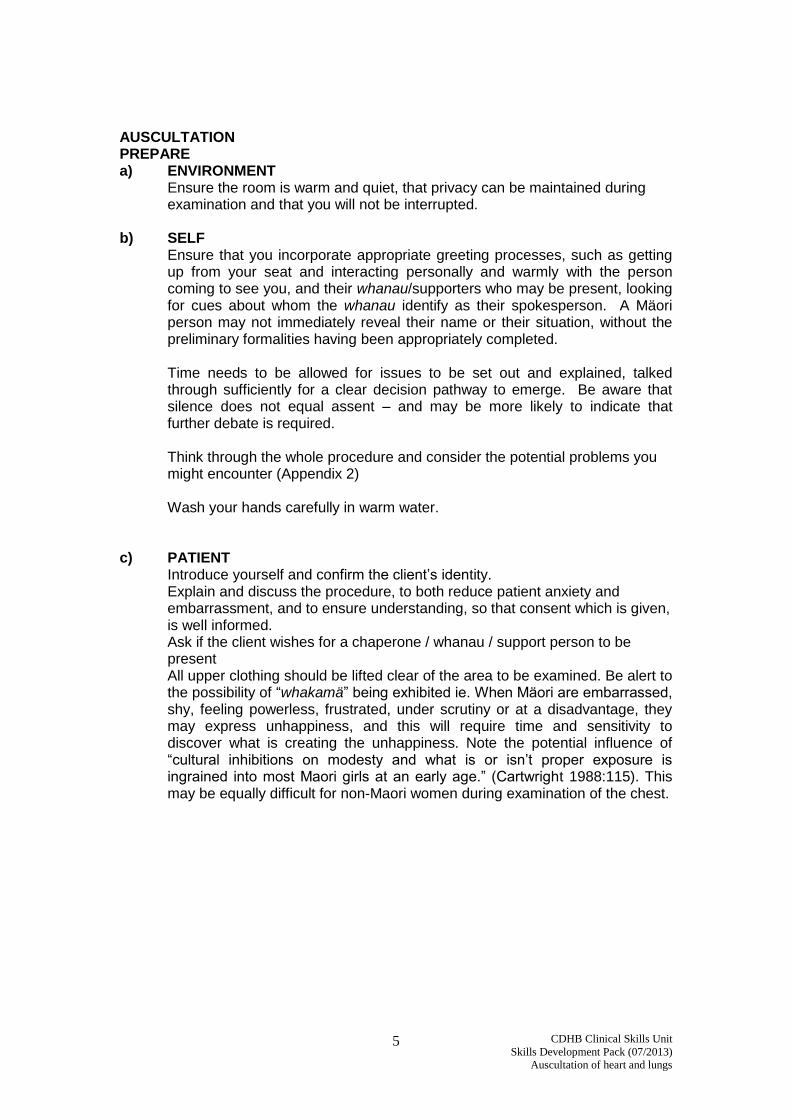
7. Assess chest expansion and symmetry by placing hands on patient’s back at the level of and parallel to 10th ribs, with thumbs sliding together at the midline to raise loose skin folds and ask client to breathe deeply. Note any lag / asymmetry/ pain /amount of movement and elasticity of chest wall.
8. Check for tactile fremitus by asking patient to say “99” with either the “ball” or ulnar surface of one hand against the posterior chest in the pattern shown
CDHB Clinical Skills Unit
Skills Development Pack (07/2013)
Auscultation of heart and lungs
7
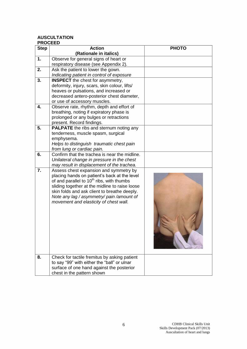
9. Asking the patient to cross arms across front
of chest, PERCUSS the intercostal spaces on posterior chest in the pattern shown. Note the symmetry, location and quality of percussion note and locate diaphragmatic dullness on both sides.
10. Estimate diaphragmatic excursion by comparing the level of dullness on full expiration and full inspiration – normally a distance of about 5-6cm.
11. AUSCULTATE the posterior chest, with the stethoscope diaphragm, asking the patient to breathe deeply through open mouth to identify breath sounds (Appendix 3). Follow the same pattern as for percussion, listening through at least one full breath at each location. Note quality of sounds and location and timing in cycle of breathing or any adventitious (additional) sounds
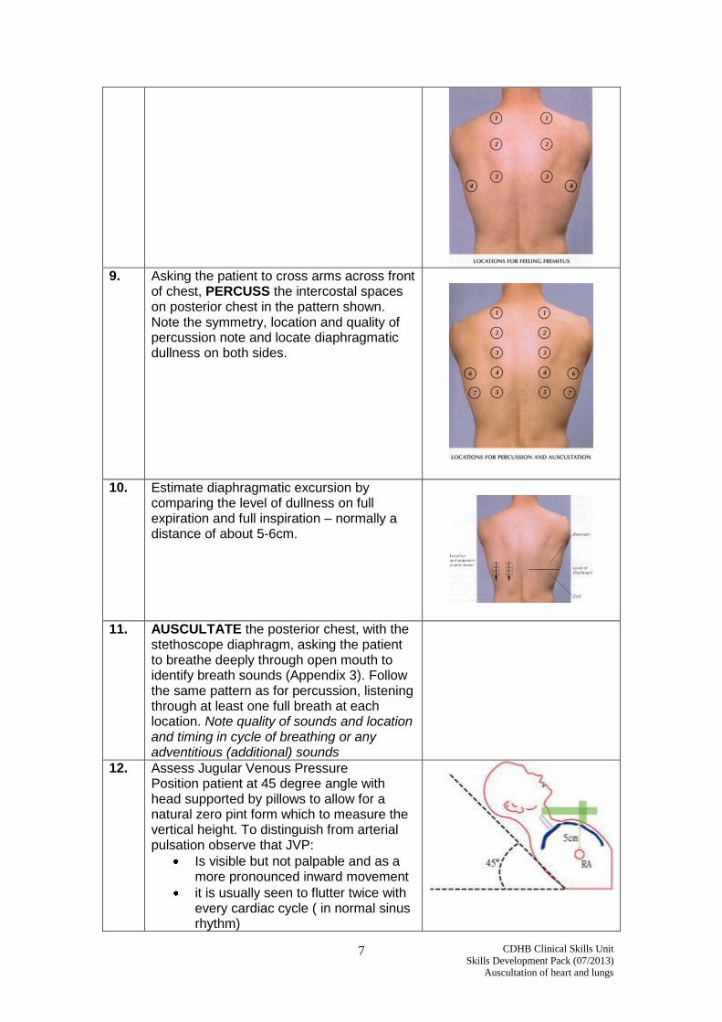
12. Assess Jugular Venous Pressure Position patient at 45 degree angle with head supported by pillows to allow for a natural zero pint form which to measure the vertical height. To distinguish from arterial pulsation observe that JVP:
Is visible but not palpable and as a more pronounced inward movement
it is usually seen to flutter twice with every cardiac cycle ( in normal sinus rhythm)
CDHB Clinical Skills Unit
Skills Development Pack (07/2013)
Auscultation of heart and lungs
8
when applying light pressure to the base of the neck it will disappear and return from the top
(Appendix 4)
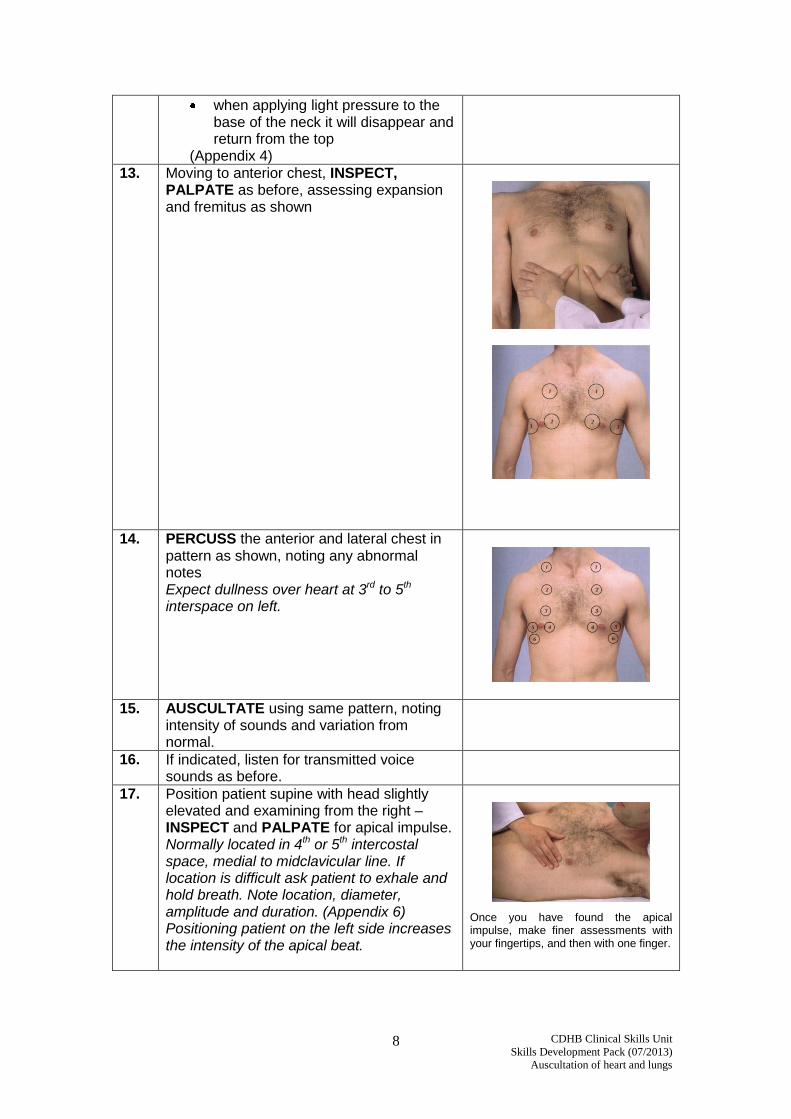
13. Moving to anterior chest, INSPECT, PALPATE as before, assessing expansion and fremitus as shown
14. PERCUSS the anterior and lateral chest in pattern as shown, noting any abnormal notes Expect dullness over heart at 3rd to 5th interspace on left.
15. AUSCULTATE using same pattern, noting intensity of sounds and variation from normal.
16. If indicated, listen for transmitted voice sounds as before.
17. Position patient supine with head slightly elevated and examining from the right – INSPECT and PALPATE for apical impulse. Normally located in 4th or 5th intercostal space, medial to midclavicular line. If location is difficult ask patient to exhale and hold breath. Note location, diameter, amplitude and duration. (Appendix 6) Positioning patient on the left side increases the intensity of the apical beat.
Once you have found the apical impulse, make finer assessments with your fingertips, and then with one finger.
CDHB Clinical Skills Unit
Skills Development Pack (07/2013)
Auscultation of heart and lungs
9
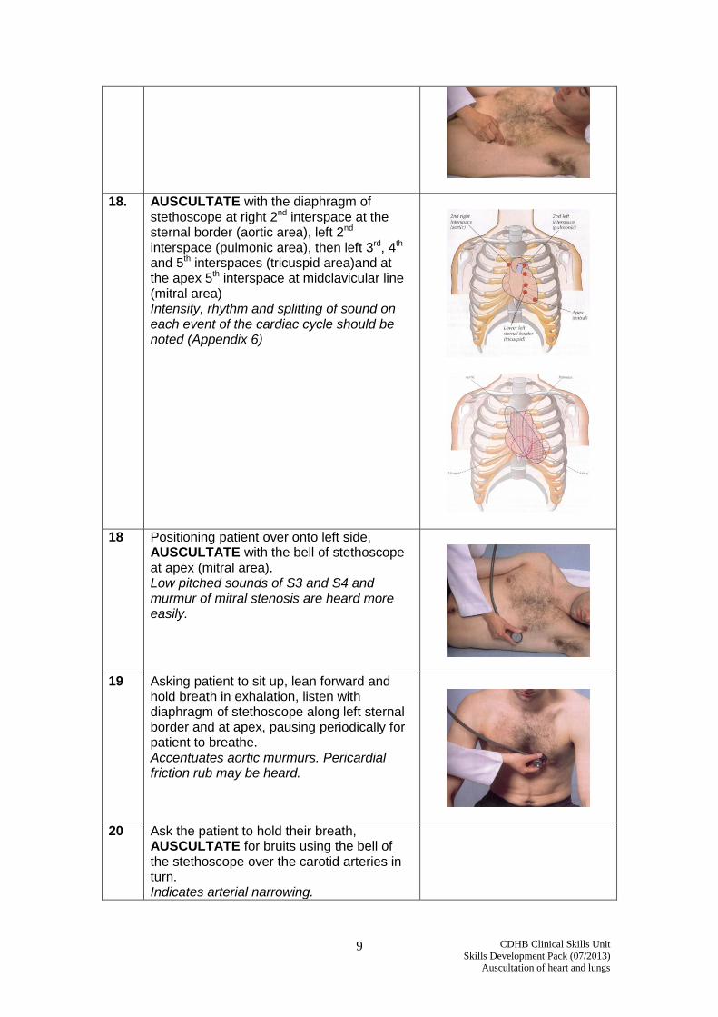
18. AUSCULTATE with the diaphragm of stethoscope at right 2nd interspace at the sternal border (aortic area), left 2nd interspace (pulmonic area), then left 3rd, 4th and 5th interspaces (tricuspid area)and at the apex 5th interspace at midclavicular line (mitral area) Intensity, rhythm and splitting of sound on each event of the cardiac cycle should be noted (Appendix 6)
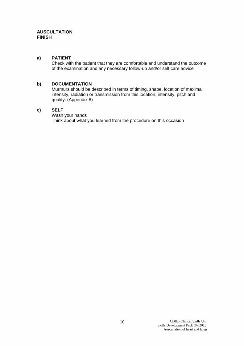
18 Positioning patient over onto left side, AUSCULTATE with the bell of stethoscope at apex (mitral area). Low pitched sounds of S3 and S4 and murmur of mitral stenosis are heard more easily.
19 Asking patient to sit up, lean forward and hold breath in exhalation, listen with diaphragm of stethoscope along left sternal border and at apex, pausing periodically for patient to breathe. Accentuates aortic murmurs. Pericardial friction rub may be heard.
20 Ask the patient to hold their breath, AUSCULTATE for bruits using the bell of the stethoscope over the carotid arteries in turn. Indicates arterial narrowing.
CDHB Clinical Skills Unit
Skills Development Pack (07/2013)
Auscultation of heart and lungs
10
AUSCULTATION FINISH
a) PATIENT
Check with the patient that they are comfortable and understand the outcome of the examination and any necessary follow-up and/or self care advice
b) DOCUMENTATION
Murmurs should be described in terms of timing, shape, location of maximal intensity, radiation or transmission from this location, intensity, pitch and quality. (Appendix 8)
c) SELF
Wash your hands Think about what you learned from the procedure on this occasion

CDHB Clinical Skills Unit
Skills Development Pack (07/2013)
Auscultation of heart and lungs
11
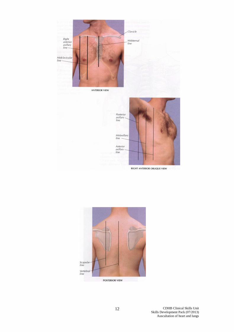
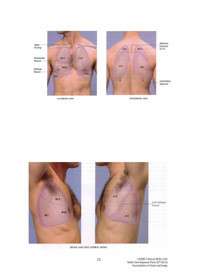
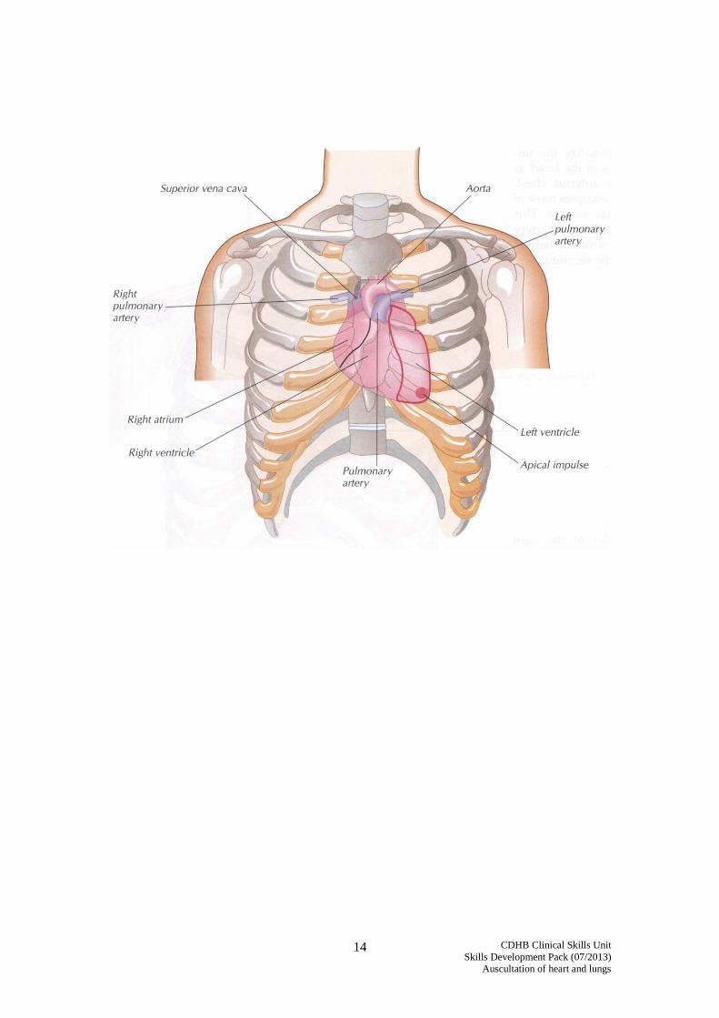
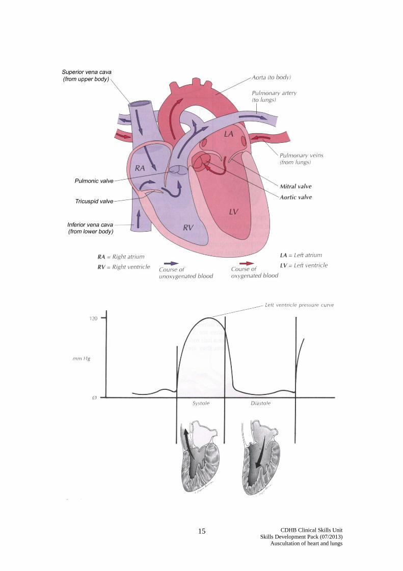
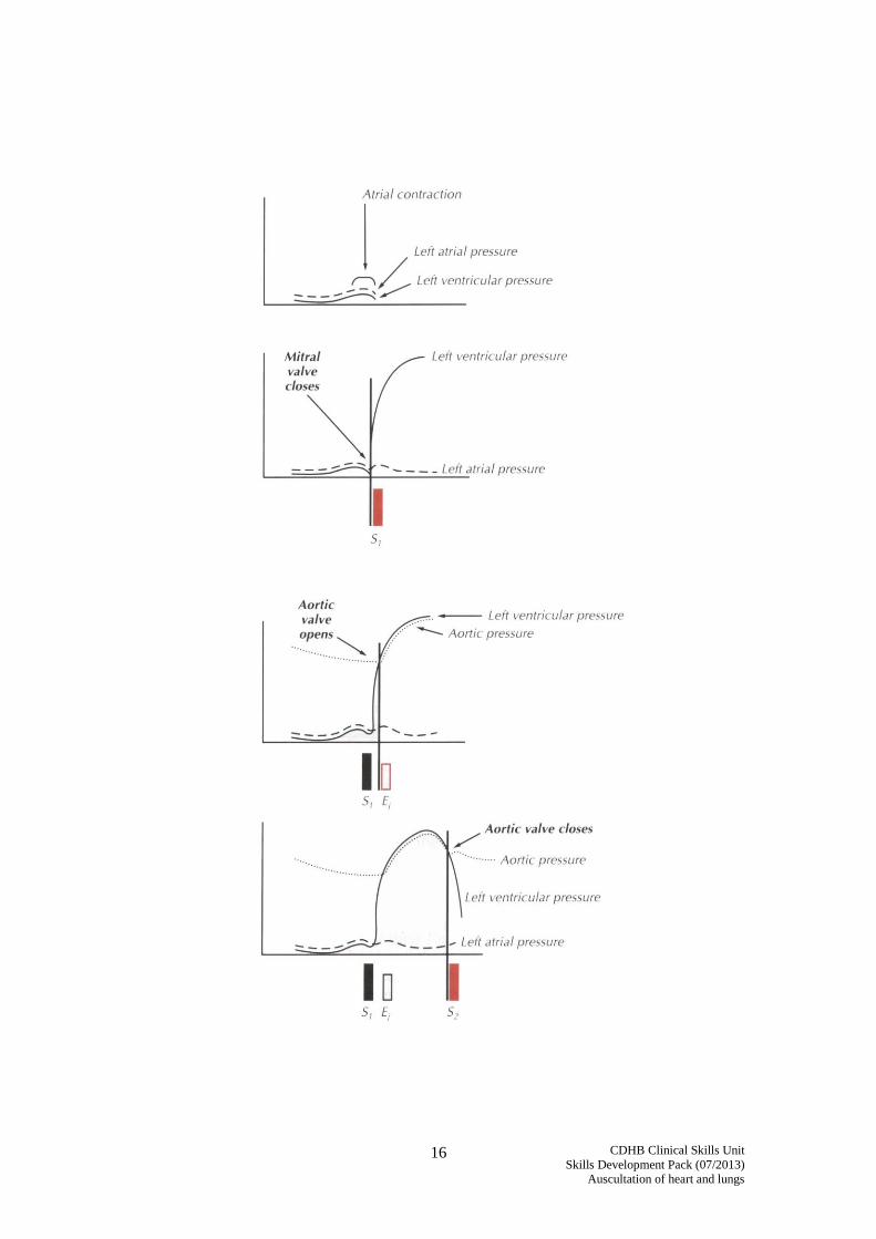
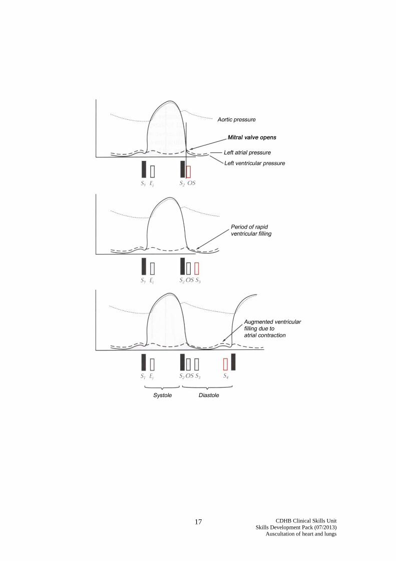
APPENDIX 1 ANATOMY AND PHYSIOLOGY
CDHB Clinical Skills Unit
Skills Development Pack (07/2013)
Auscultation of heart and lungs
12
CDHB Clinical Skills Unit
Skills Development Pack (07/2013)
Auscultation of heart and lungs
13
CDHB Clinical Skills Unit
Skills Development Pack (07/2013)
Auscultation of heart and lungs
14
CDHB Clinical Skills Unit
Skills Development Pack (07/2013)
Auscultation of heart and lungs
15
CDHB Clinical Skills Unit
Skills Development Pack (07/2013)
Auscultation of heart and lungs
16
CDHB Clinical Skills Unit
Skills Development Pack (07/2013)
Auscultation of heart and lungs
17

CDHB Clinical Skills Unit
Skills Development Pack (07/2013)
Auscultation of heart and lungs
18
APPENDIX 2
GENERAL SIGNS OF RESPIRATORY OR CARDIAC DISEASE – Well developed sternomastoid muscles Finger clubbing Cyanosis Air hunger Poor exercise tolerance Poor capillary refill Pursed lip breathing “Tripod” resting body position Head bobbing
CDHB Clinical Skills Unit
Skills Development Pack (07/2013)
Auscultation of heart and lungs
19
APPENDIX 3
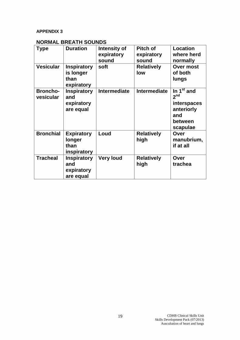
NORMAL BREATH SOUNDS
Type Duration Intensity of expiratory sound
Pitch of expiratory sound
Location where herd normally
Vesicular Inspiratory is longer than expiratory
soft Relatively low
Over most of both lungs
Broncho-vesicular
Inspiratory and expiratory are equal
Intermediate Intermediate In 1st and 2nd interspaces anteriorly and between scapulae
Bronchial Expiratory longer than inspiratory
Loud Relatively high
Over manubrium, if at all
Tracheal Inspiratory and expiratory are equal
Very loud Relatively high
Over trachea
CDHB Clinical Skills Unit
Skills Development Pack (07/2013)
Auscultation of heart and lungs
20
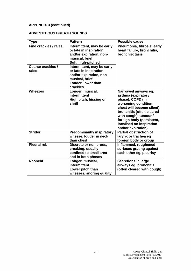
APPENDIX 3 (continued) ADVENTITIOUS BREATH SOUNDS
Type Pattern Possible cause
Fine crackles / rales
Intermittent, may be early or late in inspiration and/or expiration, non-musical, brief Soft, high-pitched
Pneumonia, fibrosis, early heart failure, bronchitis, bronchiectasis
Coarse crackles / rales
Intermittent, may be early or late in inspiration and/or expiration, non-musical, brief Louder, lower than crackles
Wheezes Longer, musical, intermittent High pitch, hissing or shrill
Narrowed airways eg. asthma (expiratory phase), COPD (in worsening condition chest will become silent), bronchitis (often cleared with cough), tumour / foreign body (persistent, localised on inspiration and/or expiration)
Stridor Predominantly inspiratory wheeze, louder in neck than chest
Partial obstruction of larynx or trachea eg foreign body or croup
Pleural rub Discrete or numerous, creaking, usually confined to small area and in both phases
Inflammed, roughened surfaces grating against each other eg. pleurisy
Rhonchi Longer, musical, intermittent Lower pitch than wheezes, snoring quality
Secretions in large airways eg. bronchitis (often cleared with cough)
CDHB Clinical Skills Unit
Skills Development Pack (07/2013)
Auscultation of heart and lungs
21
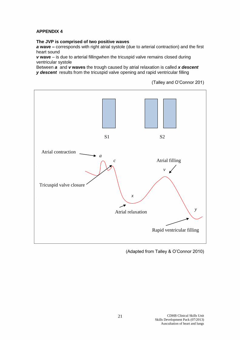
APPENDIX 4 The JVP is comprised of two positive waves a wave – corresponds with right atrial systole (due to arterial contraction) and the first heart sound v wave – is due to arterial fillingwhen the tricuspid valve remains closed during ventricular systole Between a and v waves the trough caused by atrial relaxation is called x descent y descent results from the tricuspid valve opening and rapid ventricular filling
(Talley and O’Connor 201)
a
a
(Adapted from Talley & O’Connor 2010)
c
v
y
a x
a a
a Atrial relaxation
a Rapid ventricular filling
Atrial filling
Tricuspid valve closure
a Atrial contraction
a S1 a S2
CDHB Clinical Skills Unit
Skills Development Pack (07/2013)
Auscultation of heart and lungs
22
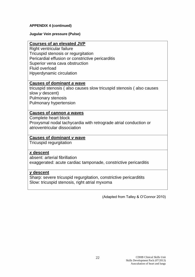
APPENDIX 4 (continued) Jugular Vein pressure (Pulse)
Courses of an elevated JVP Right ventricular failure Tricuspid stenosis or regurgitation Pericardial effusion or constrictive pericarditis Superior vena cava obstruction Fluid overload Hpyerdynamic circulation
Causes of dominant a wave tricuspid stenosis ( also causes slow tricuspid stenosis ( also causes slow y descent) Pulmonary stenosis Pulmonary hypertension
Causes of cannon a waves Complete heart block Proxysmal nodal tachycardia with retrograde atrial conduction or atrioventricular dissociation
Causes of dominant v wave Tricuspid regurgitation
x descent absent: arterial fibrillation exaggerated: acute cardiac tamponade, constrictive pericarditis
y descent Sharp: severe tricuspid regurgitation, constrictive pericarditits Slow: tricuspid stenosis, right atrial myxoma
(Adapted from Talley & O’Connor 2010)
CDHB Clinical Skills Unit
Skills Development Pack (07/2013)
Auscultation of heart and lungs
23
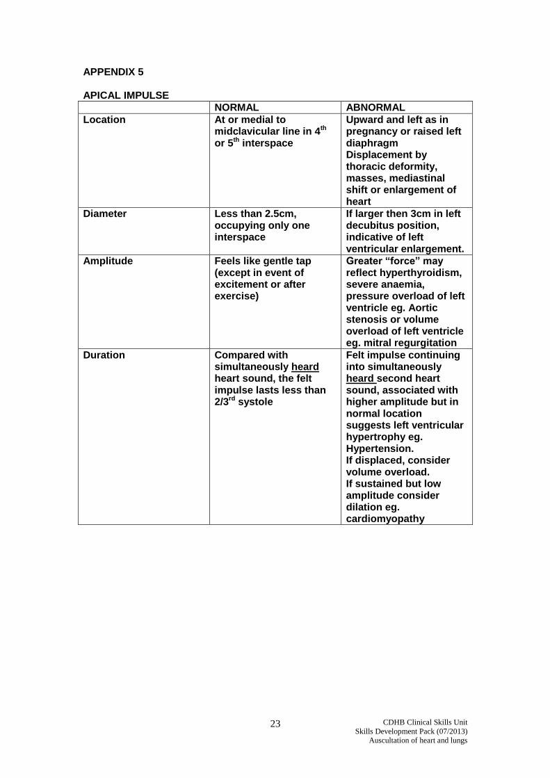
APPENDIX 5 APICAL IMPULSE
NORMAL ABNORMAL
Location At or medial to midclavicular line in 4th or 5th interspace
Upward and left as in pregnancy or raised left diaphragm Displacement by thoracic deformity, masses, mediastinal shift or enlargement of heart
Diameter Less than 2.5cm, occupying only one interspace
If larger then 3cm in left decubitus position, indicative of left ventricular enlargement.
Amplitude Feels like gentle tap (except in event of excitement or after exercise)
Greater “force” may reflect hyperthyroidism, severe anaemia, pressure overload of left ventricle eg. Aortic stenosis or volume overload of left ventricle eg. mitral regurgitation
Duration Compared with simultaneously heard heart sound, the felt impulse lasts less than 2/3rd systole
Felt impulse continuing into simultaneously heard second heart sound, associated with higher amplitude but in normal location suggests left ventricular hypertrophy eg. Hypertension. If displaced, consider volume overload. If sustained but low amplitude consider dilation eg. cardiomyopathy
CDHB Clinical Skills Unit
Skills Development Pack (07/2013)
Auscultation of heart and lungs
24
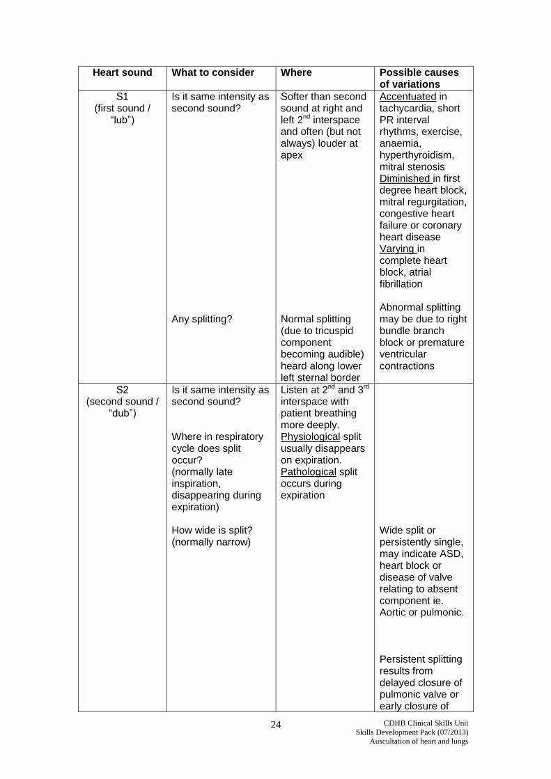
Heart sound
What to consider
Where
Possible causes of variations
S1 (first sound /
“lub”)
Is it same intensity as second sound? Any splitting?
Softer than second sound at right and left 2nd interspace and often (but not always) louder at apex Normal splitting (due to tricuspid component becoming audible) heard along lower left sternal border
Accentuated in tachycardia, short PR interval rhythms, exercise, anaemia, hyperthyroidism, mitral stenosis Diminished in first degree heart block, mitral regurgitation, congestive heart failure or coronary heart disease Varying in complete heart block, atrial fibrillation Abnormal splitting may be due to right bundle branch block or premature ventricular contractions
S2 (second sound /
“dub”)
Is it same intensity as second sound? Where in respiratory cycle does split occur? (normally late inspiration, disappearing during expiration) How wide is split? (normally narrow)
Listen at 2nd and 3rd interspace with patient breathing more deeply. Physiological split usually disappears on expiration. Pathological split occurs during expiration
Wide split or persistently single, may indicate ASD, heart block or disease of valve relating to absent component ie. Aortic or pulmonic. Persistent splitting results from delayed closure of pulmonic valve or early closure of
CDHB Clinical Skills Unit
Skills Development Pack (07/2013)
Auscultation of heart and lungs
25
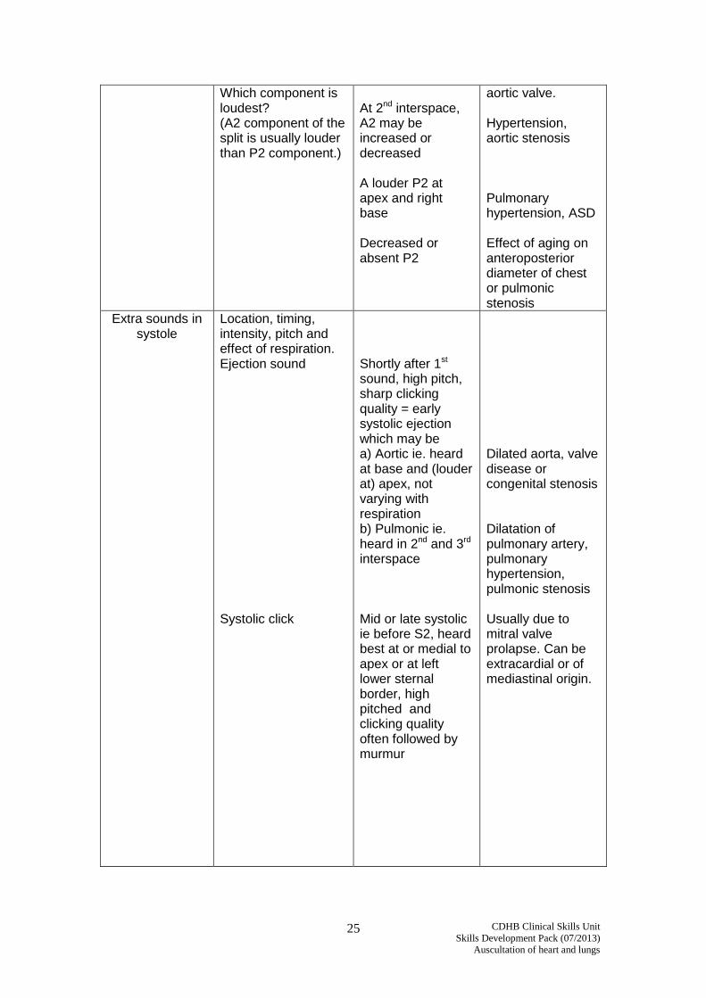
Which component is loudest? (A2 component of the split is usually louder than P2 component.)
At 2nd interspace, A2 may be increased or decreased A louder P2 at apex and right base Decreased or absent P2
aortic valve. Hypertension, aortic stenosis Pulmonary hypertension, ASD Effect of aging on anteroposterior diameter of chest or pulmonic stenosis
Extra sounds in systole
Location, timing, intensity, pitch and effect of respiration. Ejection sound Systolic click
Shortly after 1st sound, high pitch, sharp clicking quality = early systolic ejection which may be a) Aortic ie. heard at base and (louder at) apex, not varying with respiration b) Pulmonic ie. heard in 2nd and 3rd interspace Mid or late systolic ie before S2, heard best at or medial to apex or at left lower sternal border, high pitched and clicking quality often followed by murmur
Dilated aorta, valve disease or congenital stenosis Dilatation of pulmonary artery, pulmonary hypertension, pulmonic stenosis Usually due to mitral valve prolapse. Can be extracardial or of mediastinal origin.
CDHB Clinical Skills Unit
Skills Development Pack (07/2013)
Auscultation of heart and lungs
26
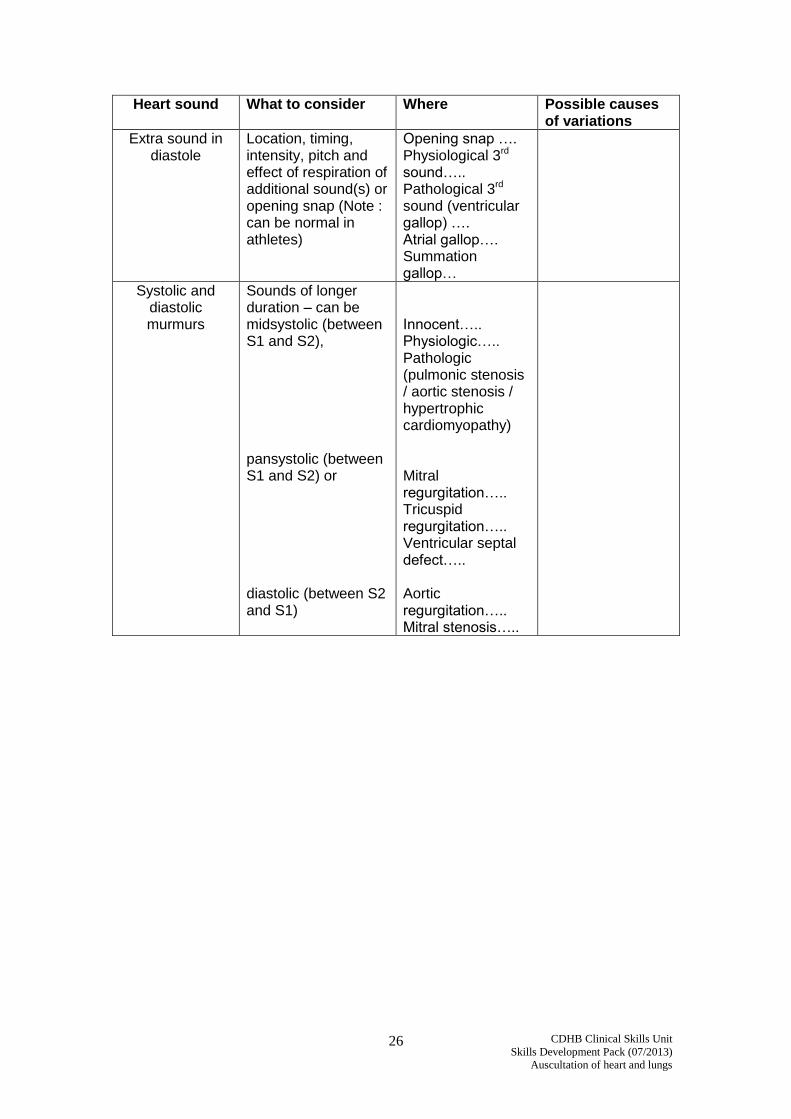
Heart sound
What to consider
Where
Possible causes of variations
Extra sound in diastole
Location, timing, intensity, pitch and effect of respiration of additional sound(s) or opening snap (Note : can be normal in athletes)
Opening snap …. Physiological 3rd sound….. Pathological 3rd sound (ventricular gallop) …. Atrial gallop…. Summation gallop…
Systolic and diastolic murmurs
Sounds of longer duration – can be midsystolic (between S1 and S2), pansystolic (between S1 and S2) or diastolic (between S2 and S1)
Innocent….. Physiologic….. Pathologic (pulmonic stenosis / aortic stenosis / hypertrophic cardiomyopathy) Mitral regurgitation….. Tricuspid regurgitation….. Ventricular septal defect….. Aortic regurgitation….. Mitral stenosis…..
CDHB Clinical Skills Unit
Skills Development Pack (07/2013)
Auscultation of heart and lungs
27
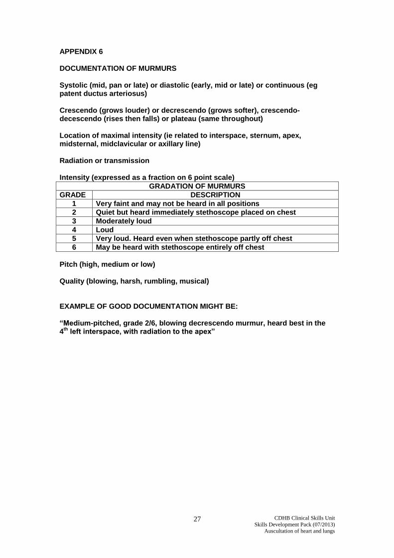
APPENDIX 6 DOCUMENTATION OF MURMURS Systolic (mid, pan or late) or diastolic (early, mid or late) or continuous (eg patent ductus arteriosus) Crescendo (grows louder) or decrescendo (grows softer), crescendo-decescendo (rises then falls) or plateau (same throughout) Location of maximal intensity (ie related to interspace, sternum, apex, midsternal, midclavicular or axillary line) Radiation or transmission Intensity (expressed as a fraction on 6 point scale)
GRADATION OF MURMURS
GRADE DESCRIPTION
1 Very faint and may not be heard in all positions
2 Quiet but heard immediately stethoscope placed on chest
3 Moderately loud
4 Loud
5 Very loud. Heard even when stethoscope partly off chest
6 May be heard with stethoscope entirely off chest
Pitch (high, medium or low) Quality (blowing, harsh, rumbling, musical) EXAMPLE OF GOOD DOCUMENTATION MIGHT BE: “Medium-pitched, grade 2/6, blowing decrescendo murmur, heard best in the 4th left interspace, with radiation to the apex”
CDHB Clinical Skills Unit
Skills Development Pack (07/2013)
Auscultation of heart and lungs
28
REFERENCES
Bickley, L. S., Hoekelman, R. A.1999. Bates Guide to Physical Examination and History Taking. 7th Edition. Lippincott.
Talley, N. J., & O’Connor, S. ((2010). Clinical Examination A systematic guide to physical diagnosis. 6th ed. NSW: Elsevier.
CDHB Clinical Skills Unit
Skills Development Pack (07/2013)
Auscultation of heart and lungs
29
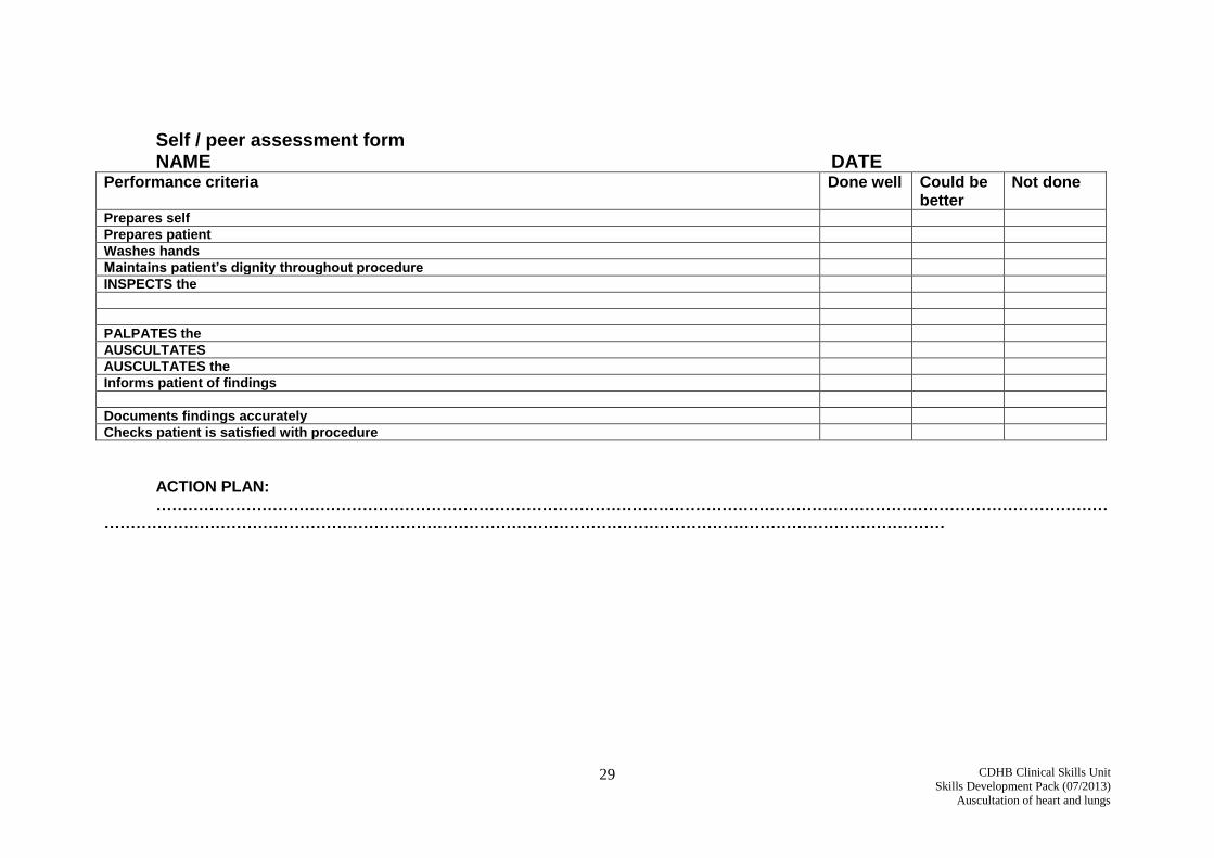
Self / peer assessment form NAME DATE
Performance criteria Done well Could be better
Not done
Prepares self
Prepares patient
Washes hands
Maintains patient’s dignity throughout procedure
INSPECTS the
PALPATES the
AUSCULTATES
AUSCULTATES the
Informs patient of findings
Documents findings accurately
Checks patient is satisfied with procedure
ACTION PLAN: ………………………………………………………………………………………………………………………………………………………………
……………………………………………………………………………………………………………………………………………
CDHB Clinical Skills Unit
Skills Development Pack (07/2013)
Auscultation of heart and lungs
30
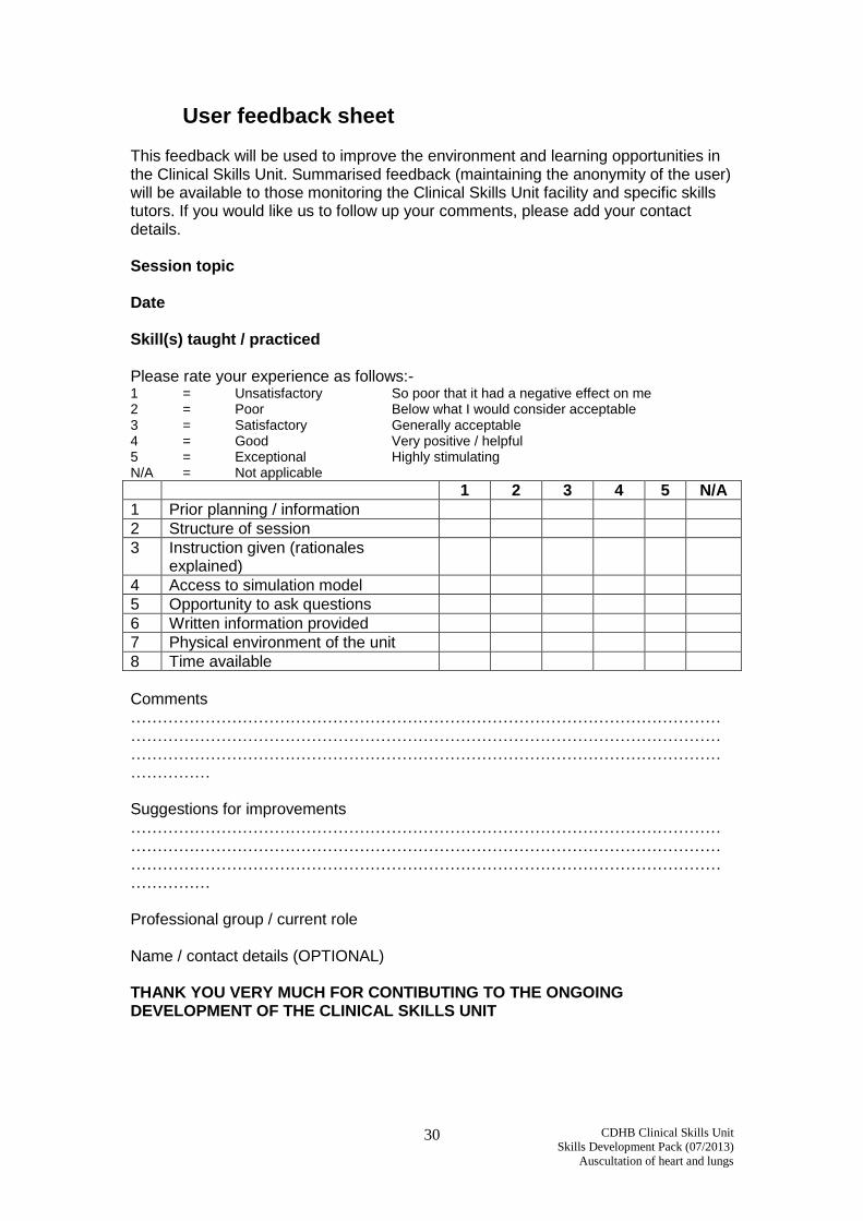
User feedback sheet This feedback will be used to improve the environment and learning opportunities in the Clinical Skills Unit. Summarised feedback (maintaining the anonymity of the user) will be available to those monitoring the Clinical Skills Unit facility and specific skills tutors. If you would like us to follow up your comments, please add your contact details. Session topic Date Skill(s) taught / practiced Please rate your experience as follows:- 1 = Unsatisfactory So poor that it had a negative effect on me 2 = Poor Below what I would consider acceptable 3 = Satisfactory Generally acceptable 4 = Good Very positive / helpful 5 = Exceptional Highly stimulating N/A = Not applicable
1 2 3 4 5 N/A
1 Prior planning / information
2 Structure of session
3 Instruction given (rationales explained)
4 Access to simulation model
5 Opportunity to ask questions
6 Written information provided
7 Physical environment of the unit
8 Time available
Comments …………………………………………………………………………………………………………………………………………………………………………………………………………………………………………………………………………………………………………………… Suggestions for improvements …………………………………………………………………………………………………………………………………………………………………………………………………………………………………………………………………………………………………………………… Professional group / current role Name / contact details (OPTIONAL) THANK YOU VERY MUCH FOR CONTIBUTING TO THE ONGOING DEVELOPMENT OF THE CLINICAL SKILLS UNIT