Embed Size (px)
Citation preview
Wnt signaling arrests effector T cell differentiation and generatesCD8+ memory stem cells
Luca Gattinoni1,*, Xiao-Song Zhong1,*, Douglas C Palmer1, Yun Ji1, Christian S Hinrichs1,Zhiya Yu1, Claudia Wrzesinski1, Andrea Boni1, Lydie Cassard1, Lindsay Church1, ChrystalM Paulos1, Pawel Muranski1, and Nicholas P Restifo11 Center for Cancer Research, National Cancer Institute, National Institutes of Health, Bethesda,Maryland 20892
AbstractSelf-renewing cell populations such as hematopoietic stem cells and memory B and T lymphocytesmight be regulated by shared signaling pathways1. Wnt/β-catenin is an evolutionarily conservedpathway that promotes hematopoietic stem cell self-renewal and multipotency by limiting stem cellproliferation and differentiation2,3, but its role in the generation and maintenance of memory T cellsis unknown. We found that the induction of Wnt/β-catenin signaling using inhibitors of glycogen-sythase-kinase-3β or the Wnt protein family member, Wnt3a, arrested CD8+ T cell development intoeffector cells. By blocking T-cell differentiation, Wnt signaling enabled the generation ofCD44low, CD62Lhigh, Sca-1high, CD122high, Bcl-2high self-renewing, multipotent CD8+ memorystem cells with proliferative and anti-tumor capacities exceeding those of central and effector memoryT cell subsets. These findings reveal a key role for Wnt signaling in the maintenance of stemness inmature memory CD8+ T cells and have important implications for the design of novel vaccinationstrategies and adoptive immunotherapies.
T cell factor (Tcf) 1 and lymphoid enhancer-binding factor (Lef) 1 are downstreamtranscription factors of the Wnt/β-catenin signaling pathway. Tcf1 and Lef1 are required fornormal thymic T cell development, but less is known about Wnt function in mature T cells2,4. Although experiments using multimerized TCF/LEF binding site reporter system haverevealed that Wnt signaling is active in mature CD8+ T cells, the impact of this pathway to thiscell population has yet to be fully elucidated5. At least three lines of evidence indicate that Wntsignaling might regulate the maturation of post-thymic T lymphocytes: i.) CD8+ T cells fromTcf7−/− mice, which are missing the gene that encodes for TCF1, display a more differentiatedphenotype (CD44high and CD62Llow) than WT T cells6; ii.) expression of Lef1 and Tcf7,decreases with progressive differentiation of CD8+ T cells from naive (TN) → central memory(TCM) → effector memory (TEM) inhumans7 and mouse (Supplementary Fig. 1); and iii.) highlevels of Ctnnb1 (which encodes β-catenin), Lef1, Tcf7 have been detected in T cells withincreased potential to form memory in vivo8,9. Thus, triggering the activities of the Wntsignaling transcription factors TCF1 and LEF1 could promote stem-like self-renewal capacityin mature T cells.
To test the impact of Wnt/β-catenin signaling on mature CD8+ T cells, we primed TN in thepresence of the 4,6-disubstituted pyrrolopyrimidine TWS119, a potent inhibitor of the serine/threonine kinase glycogen-synthase-kinase-3β (Gsk-3β)10. Gsk-3β blockade mimics Wnt
Address correspondence to Luca Gattinoni or Nicholas P. Restifo, National Cancer Institute, National Institutes of Health, ClinicalResearch Center, Room 3-5762, 10 Center Drive, Bethesda, MD 20892-1502. E-mail: (LG): [email protected], (NPR):[email protected].*These authors contributed equally to the present work.
NIH Public AccessAuthor ManuscriptNat Med. Author manuscript; available in PMC 2010 January 1.
Published in final edited form as:Nat Med. 2009 July ; 15(7): 808–813. doi:10.1038/nm.1982.
NIH
-PA Author Manuscript
NIH
-PA Author Manuscript
NIH
-PA Author Manuscript
signaling by promoting the accumulation of β-catenin, the molecule that tethers the TCF andLEF transcription factors to targeted DNA2. TWS119 triggered a rapid accumulation of β-catenin (mean 6.8 +/− SD 1.7-fold increase by densitometry; p < 0.05) (Fig. 1a), augmentednuclear protein-TCF/LEF oligonucleotide interaction (Fig. 1b) and sharply up-regulatedTcf711, Lef112 and other Wnt target genes including Jun13, Frizzled7 (Fzd7)14, Nemo-like-kinase (Nlk)15 (Fig. 1c). By contrast, T cell activation in the absence of the Gsk-3β inhibitorresulted in the down-regulation of the Wnt/β-catenin pathway at these steps of the signalingcascade (Fig. 1a–c). Thus, TWS119 activated the Wnt/β-catenin pathway in naive T cells andreversed the physiological down-regulation of Tcf7 and Lef1 induced by T cell activation7.
We sought to assess the effect of Wnt signaling on CD8+ T cell differentiation and proliferation.We stimulated CFSE-labeled CD8+ T cells from pmel-1 TCR transgenic mice16 with thecognate antigen, gp100, in the presence of titrated doses of TWS119 and analyzed them forthe expression of the differentiation markers CD44 and CD62L. CD44 expression is known toincrease with T cell differentiation while CD62L is progressively lost17. TWS119 increasedthe frequency of T cells that retained CD62L expression in a dose-dependent manner, indicatingthat it inhibited CD8+ T cell differentiation (Fig. 2a). Interestingly, 46% of CD8+ T cellscultured in the presence of the highest concentration of Gsk-3β inhibitor failed to up-regulateCD44, maintaining a “naive” CD44lowCD62Lhigh phenotype (Fig. 2a). Low doses of TWS119(≤ 1 μM) preserved CD62L expression without affecting T cell proliferation, while higher drugconcentrations promoted a dose-dependent inhibition of cell cycling (Fig. 2b). Arresteddifferentiation and proliferation of CD8+ T cells by TWS119 was not secondary to the impactof the drug on dendritic cells (DC), because we observed similar results stimulating purifiedCD8+ T cells in a DC-free system (Supplementary Fig. 2a,b). Similar to TWS119, we foundthat the structurally unrelated Gsk-3β inhibitor, 6-bromo-substituted indirubin, BIO18,19,inhibited T cell differentiation (Supplementary Fig. 3a) and induced the expression of the Wnttranscription factors Tcf7 and Lef1 (Supplementary Fig. 3b). The use of an analog, BIO-acetoxime19, with a greater Gsk-3β kinase inhibitory specificity, retained the observed activitywhile the use of N-methylated analog (Methyl-BIO)19, a kinase inactive control, had no effect(Supplementary Fig. 3a,b). These results are in contrast with those obtained using lithiumchloride20 as a Gsk-3β inhibitor, which is less active and specific than the inhibitors used inthe present study19. Because Gsk-3β regulates several signaling pathways other than Wnt, wesought to more directly test whether the impact of the pharmacological blockade of Gsk-3βwas dependent on mimicking the downstream signals of the Wnt/β-catenin pathway. Weprimed CD8+ T cells in the presence of Wnt3A, a Wnt protein that has been shown to promotestem cell self-renewal and pluripotency via β-catenin accumulation in the cell nucleus21. LikeTWS119, we found that Wnt3A itself inhibited T cell differentiation and proliferation(Supplementary Fig. 4). Thus, T cell proliferation and differentiation could be restrainedthrough the activation of the Wnt/β-catenin pathway by the naturally-occuring ligand, Wnt3A,and by the pharamcologic inhibition of Gsk-3β downstream. Neverthelss, our data did not ruleout the possibility that Gsk-3β inhibitors were regulating T cell differentiation by affectingother pathways in addition to Wnt.
We sought to evaluate whether the phenotypic differences of pmel-1 CD8+ T cells induced byTWS119 were associated with functional changes. It has been previously shown thatdifferentiating CD8+ T cells lose the capacity to produce IL-2 as they acquire the ability to killtarget cells and release high levels of IFN-γ17,22. We found that TWS119 induced a dose-dependent decrease in T cell-specific killing and IFN-γ release associated with a preservationin the ability to produce IL-2 (Fig. 2c). These functional data confirmed the phenotypic findingsthat TWS119 was a negative regulator of TEFF differentiation. We next assessed if theinhibition of TEFF development was associated with the suppression of eomesodermin(Eomes), a master regulator of CD8+ T-cell effector function23. We found that the exposureof cells to TWS119 abrogated the induced expression of Eomes that occurred within 8h after
Gattinoni et al. Page 2
Nat Med. Author manuscript; available in PMC 2010 January 1.
NIH
-PA Author Manuscript
NIH
-PA Author Manuscript
NIH
-PA Author Manuscript
T cell priming, indicating that TWS119-mediated suppression of the effector program was anearly event (Fig. 2d). Altogether, phenotypic, functional and molecular data indicated thatinduction of Wnt signaling during T cell priming resulted in an inhibition of CD8+ T celldifferentiation into TEFF cells. Given our in vitro findings, we tested whether TWS119 couldinfluence the qualities of adoptively transferred pmel-1 CD8+ T cells in response to fowlpox-based gp100 immunization and prevent the induction of highly differentiated, senescent T cells,a major pitfall of current T cell-based vaccines24. We found that there were no differences infrequency or numbers of pmel-1 T cells in spleens six days after vaccination, but thedevelopment of TEFF was inhibited (Fig. 2e). More importantly, TWS119 augmented thenumbers of CD44high, CD62Lhigh TCM in responding CD8+ T cells in the post-vaccinationsetting(Fig. 2e). Although the present approach does not enable the dissection of the cellularmechanism of action in vivo, TWS119 effectively altered CD8+ T cell differentiation afterimmunization.
We observed a cell population that expressed low levels of CD44 and high levels ofCD62Lwhen TWS119 was administered in vitro and in vivo (Fig. 2a,e) but it was unclearwhether these cells remained naive after antigen encounter or had entered into a primordialmemory state that retained some phenotypic traits of TN. Using a murine model of graft-versus-host disease, Zhang et al. have described population of CD44low CD62Lhigh memory CD8+ Tcells that expressed high levels of stem cell antigen-1 (Sca-1), B-cell lymphoma protein 2(Bcl-2), and common IL-2/IL-15 receptor β chain (CD122)25. Because these cells displayedrobust self-renewal and the multipotent capacity to derive TCM, TEM, and TEFF they weredesignated “T memory stem cells” (TSCM)25. We sought to explore whether the CD44low
CD62Lhigh pmel-1 T cells generated in vitro with TWS119 were TSCM. We found thatTWS119-induced CD44low CD62Lhigh T cells had undergone up to four divisions asmanifested by CFSE dilution, and uniformly expressed high levels of the core phenotypic traitsof TSCM, namely Sca-1, CD122 and Bcl-2 (Fig. 3a). By contrast, TN expressed these markersat low levels (Fig. 3a). Similarly, Sca-1, CD122 and Bcl-2 were up-regulated in CD44low,CD62Lhigh pmel-1 T cells generated in vitro in the presence of BIO-acetoxime (SupplementaryFig. 5) or Wnt3A (Supplementary Fig. 6) and in vivo after specific-vaccination and TWS119administration (Supplementary Fig. 7). These activation/memory T cell markers were notexclusively expressed by TSCM but instead only defined TSCM in the context of cells the werealso CD44low CD62Lhigh (not shown). Antigen-experienced memory T cells can bedistinguished from TN not only by phenotype but also by a number of functional properties:i.) rapid acquisition of effector functions upon antigen re-challenge26–28; ii.) pronounced cellcycling capacity29; iii.) robust homeostatic proliferation30; and iv.) independence of MHC classI for persistence31 and anti-tumor activity32. We found that, unlike TN, TWS119-inducedCD44low, CD62Lhigh T cells rapidly released cytokines (IFN-γ and IL-2) upon antigenencounter (Fig. 3b), had undergone more cell division after adoptive transfer into alymphoreplete host (Fig. 3c) or into sublethally irradiated or genetically lymphodepletedTcra−/− (T cell-deficient) or Rag1−/− (T and B cell-deficient) hosts (Fig. 3d), and persisted andmediated tumor destruction (P < 0.05) in B2m−/− mice (which are MHC class I-deficient) (Fig.3e,f). Altogether, these findings indicated that Wnt signaling induced the generation of aTSCM-like population that possessed the rapid recall ability, proliferative capacity and MHCclass I independency that are distinct from naive T cells and characteristic of memory T cells.
The concept of “stemness” encompasses the capability to both self-renew and generate moredifferentiated, specialized cells. We sought to determine the “stemness” of TWS119-generatedCD44low, CD62Lhigh T cells by evaluating their fate after adoptive transfer cells intosublethally irradiated mice. Four weeks later, we found that high percentages of TWS119-generated cells preserved their original CD44low, CD62Lhigh phenotype even after multiplecell divisions, while TN rapidly acquired CD44 as a function of CFSE dilution (P < 0.02) (Fig.3g,h). The differentiation of TWS119-induced CD44low, CD62Lhigh T cells was not arrested
Gattinoni et al. Page 3
Nat Med. Author manuscript; available in PMC 2010 January 1.
NIH
-PA Author Manuscript
NIH
-PA Author Manuscript
NIH
-PA Author Manuscript
however, because they acquired CD44high expression, albeit at a slower pace. Unlike TN,TWS119-derived CD44low, CD62Lhigh, Sca-1high Tcells robustly proliferated (by 28 days,95% had undergone cell division) (Fig. 3h), enabling their re-isolation and transfer to asecondary recipient (Supplementary Fig. 8). After four weeks, we found that the secondarilytransferred CD44low, CD62Lhigh, Sca-1high Tcells had again regenerated all T cell subsets.Importantly, 43% of the cells retained the TSCM phenotype in spite of a second round of robustproliferation in vivo (Supplementary Figure 8). These findings indicated that Wnt signalingpromoted the generation of self-renewing, multipotent TSCM.
Having established that the CD44low, CD62Lhigh T cells generated in the presence of TWS119were bona fide TSCM and not merely naive, we sought to compare them with the well definedmemory T cell subsets, TCM and TEM, in secondary challenge experiments. Highly-purifiedpmel-1 thy1.1+ memory T cell subsets were adoptively transferred into sublethally-irradiatedmicein combination with recombinant vaccinia-based gp100 vaccine and IL-2. As previouslyreported by our group and others33,34, the in vivo recall responsesof TCM were greater thanTEM (Fig. 4a). TSCM, however, robustly expanded by approximately 200-fold in the spleenalone. These levels were ≈ 10-times higher than TCM and ap; 30-times higher than TEM (Fig.4a). Furthermore, TSCM exhibited an enhanced survival capacity as revealed by the frequenciesof pmel-1 cells in the spleens of vaccinated mice one month after transfer (Fig. 4b). T cellproliferative and survival capacities have been correlated with tumor responses in mice andhumans receiving adoptive T cell-based therapies35,36. To assess whether the enhancedreplicative capability of TSCM would result in superior anti-tumor activity, we adoptivelytransferred limiting numbers of pmel-1 memory T cell subsets into sublethally-irradiated hostsbearing B16 tumors, in combination with gp100 vaccine and IL-2. As we previouslyreported33, TCM conferred superior anti-tumor immunity compared with TEM (tumorregression P < 0.01; overall survival P = 0.0644) (Fig. 4c). It was striking that minusculenumbers (4 × 10⁴) of TSCM were able to trigger the destruction of bulky tumors (1 cm3
containing ap; 109 cells) and to improve survival (TSCM vs.TCM, P < 0.005; TSCM vs. TEMP < 0.005) (Fig. 4c). Thus, adoptive transfer of TSCM in combination with tumor-antigenvaccination and exogenous IL-2 produced a far more robust and therapeutically significantsecondary response compared with the other memory T cell subsets.
Arrest of lymphocyte differentiation in order to maintain long-lived, self-renewing antigen-experienced T cells with the stem cell-like properties has been postulated as the basis of thecontinual generation of effector T cells37, but the transcription factors that regulate this processhave not been fully elucidated. Although the physiologic role of Wnt signaling in post-thymicT cell development remains unknown, our data indicate that Wnt can regulate the “stemness”of CD8+ T cells by suppressing their differentiation into TEFF. In addition, other circumstantialevidence supports our findings. We recently reported that high levels of the Wnt transcriptionfactors Tcf7 and Lef1 were expressed by T cells whose development into TEFF had been arrestedusing IL-218. Conversely, Tcf7−/− mice reportedly have an increased frequency of T cellsdisplaying a CD44high, CD62Llow TEFF/TEM phenotype6. Paralleling our own observations inCD8+ T cells, expression of a stabilized form of β-catenin inhibited the proliferation andeffector function of CD4+ T cells in a model of inflammatory bowel disease38. Furthermore,productive CD4+ T cell memory precursors overexpressed Ctnnb1 and Tcf7 as well as Bcl2and Il2rb9. These factors were all implicated in the formation and maintenance of TSCM in thepresent study.
Our findings also have parallels in stem cell biology where Wnt signaling plays a pivotal rolein promoting self-renewal while limiting proliferation and differentiation2,3. Using miceoverexpressing the Wnt signaling inhibitor Dickkopf1 in the hematopoietic stem cell niche,Fleming et al., have found that hematopoietic stem cells exposed to a Wnt-inhibitoryenvironment were less quiescent and had profound defects in their ability to reconstitute the
Gattinoni et al. Page 4
Nat Med. Author manuscript; available in PMC 2010 January 1.
NIH
-PA Author Manuscript
NIH
-PA Author Manuscript
NIH
-PA Author Manuscript
host after serial transplants3. T cell lineage commitment to effector versus memory subsets hasbeen recently linked to the asymmetric segregation and inheritance of proteins that control cellfate specification39. Although we do not know the role of Wnt in the asymmetric division ofCD8+ T cells, unequal localization and activities of Wnt signaling component including β-catenin have been implicated in cell fate specification in Caenorhabditis elegans40.
The ability to pharmacologically induce TSCM has significant implications for the field ofadoptive immunotherapy. The findings that small numbers of TSCM in conjunction withspecific vaccination and IL-2 can cause the regression of large established vascularized tumorscould reduce the cost and complexity of highly effective therapies based on the adoptivetransfer of anti-tumor T cells35. Using existing technology36, it may be possible to geneticallyengineer the human counterpart of CD8+ TSCM to express T cell receptors or chimeric antigenreceptors while pharamacologically mimicking Wnt signaling. This might ultimately allow forthe widespread application of adoptive immunotherapy based on multipotent, highlyproliferative, tumor-specific TSCM. Finally, the modulation of Wnt signaling to induce long-term T cell memory might be important for T cell-based vaccines designed to target intracellularpathogens. Increasing data indicate that protection against intracellular pathogens correlateswith the induction and maintenance of TCM responses, but these responses have not beenreproducibly obtained using current vaccination strategies24,28,34. In vivo administration ofsmall molecule agonists of Wnt might be useful to consistently achieve this goal.
MethodsMice and tumor lines
Pmel-1 TCR transgenic (Tg) mice (Jackson Laboratories) and pmel-1 ly5.1 double Tg miceand pmel-1 thy1.1 double Tg mice were previously described16,17. B16 (H-2b), a gp100+
murine melanoma, and the gp100− MCA-205 (National Cancer Institute Tumor Repository)were maintained in culture media.
Antibodies, flow cytometry and cell sortingAll antibodies were purchased from BD Biosciences. Flow cytometry acquisition wasperformed on a BD FACSCanto™, or BD FACSCanto™ II Flow cytometer. Samples wereanalyzed using FlowJo software (Tree Star). Naive and T cell memory subsets were sortedusing the BD FACSAria™ cell sorter.
Pharmacologic inhibitors of Gsk-3β and Wnt3ATWS119, BIO, BIO-acetoxime, Methyl-BIO (EMD biosciences) and Wnt3a (R&D systems)were reconstituted according to the manufacturer’s instructions and used in experiments invitro or in vivo at indicated doses.
In vitro activation of CD8+ T cellsPmel-1 splenocytes were stimulated with 1 μM hgp10025–33 in a culture media containing 10ng ml−1 of IL-2 (Chiron Corporation). Enriched CD8+ T cells from WT were activated onplate-bound anti-CD3 and 1 μg ml−1 soluble anti-CD28 antibody in a culture media containing10 ng ml−1 of IL-2. Enriched CD8+ T cells were depleted of non-CD8+ T cells using a MACSnegative selection column (Miltenyi Biotech, Inc).
Cytokine Release and Cytolytic AssaysMCA-205 (2.5 × 10⁴) cells were pulsed with 1 μM hgp10025–33 or irrelevant influenzanucleoprotein peptide (np) and incubated overnight with T cells at 1:1 ratio at 37°C.Supernatants were analyzed by murine IFN-γ and murine IL-2 ELISA kits (Pierce-Endogen).
Gattinoni et al. Page 5
Nat Med. Author manuscript; available in PMC 2010 January 1.
NIH
-PA Author Manuscript
NIH
-PA Author Manuscript
NIH
-PA Author Manuscript
For 51Cr release assays, MCA-205 target cells were pulsed with 1 μM hgp10025–33 or np and100 μCi of 51Cr (Amersham Biosciences) for 2 h at 37°C. Labeled cells (103) were incubatedwith T cells at indicated ratios for 4 h at 37°C. Maximal and spontaneous 51Cr releases weredetermined by incubating 103 labeled targets in either 2% SDS or medium.
Adoptive cell transferAdoptive cell transfer, immunization with recombinant poxvirus-based vaccines and tumorexperiments were performed as described16,17,32. C57BL/6, Tcra−/− and Rag1−/− (JacksonLaboratories), B2m−/− (Taconic) were used as recipients. All animal experiments wereconducted with theapproval of the NCI Animal Use and Care Committee.
Enumeration of Adoptively Transferred CellsAt days indicated, mice were sacrificed. Spleens were harvested and homogenized into a singlecell suspension using the rubber end of a 3cc syringe and a 40 μM filter cup. Samples wereenumerated using trypan blue exclusion and were analyzed by flow cytometry for CD8 andthy.1 expression by cells. The absolute number of pmel-1 cells was calculated by multiplyingthe splenocyte count by the ratio CD8+ thy1.1+/lymphocyte.
CFSE proliferation assaysCD8+ T cells labeled with 1 μM CFSE (Molecular Probes) were used in adoptive experimentsor stimulated in vitro as described above.
Real-time reverse transcription-polymerase chain reactionRNA was isolated using RNeasy mini kit (QIAGEN). cDNA was generated by reversetranscription (Applied Biosystems). Real time-polymerase chain reaction was performed usingcommercially available probes and primers for the indicated genes (Applied Biosystems) anda Prism 7900HT (Applied Biosystems). The levels of gene expression were calculated relativeto the housekeeping genes β-actin (Actb).
Detection of β-catenin by Western Blot analysisCells were lysed in RIPA buffer (Cell Signaling Technology) with protease inhibitor. Proteinconcentration was quantified with Bio-Rad protein assay. 20 μg of total protein were separatedon 4–12% SDS-PAGE gel followed by standard immunoblot with anti-β-catenin (BDBioscience), anti-Gapdh (Chemicon international) and horseradish peroxidase-conjugated goatanti-mouse antibodies (Santa Cruz Biotechnology).
Electrophoretic mobility shift assayNuclear extracts were isolated using the nuclear extract kit (Active Motif) and then incubated(5 μg protein) with poly-dI-dC (1 μg μl−1 in TE), 5X Binding Buffer (Promega) and γ32P-ATPlabeled oligonucleotide probes (Sense: CACAGAGAAAACAAAGCGCGCTATT;Antisense: AATAGCGCGCTTTGTTTTCTCTGTG), containing the TCF/LEF binding motifAACAAAG. Unlabeled oligonucleotide probes were used as competitors. Samples wereincubated 30 min at RT and loaded onto the gel (NuPAGE 4–12% or Bis-Tris Gel Invitrogen).Gel was blotted on a nitrocellulose membrane and exposed to the X-ray film.
Statistical analysisTumor slopes were compared using Wilcoxon rank sum test. Survival analyses andgraphs wereperformed using Kaplan–Meier methods.
Gattinoni et al. Page 6
Nat Med. Author manuscript; available in PMC 2010 January 1.
NIH
-PA Author Manuscript
NIH
-PA Author Manuscript
NIH
-PA Author Manuscript
Supplementary MaterialRefer to Web version on PubMed Central for supplementary material.
AcknowledgmentsThis research was supported by the Intramural Research Program of the NIH, National Cancer Institute, Center forCancer Research. The authors have no conflicting financial interests. The authors would like to thank S.A. Rosenberg,C.A. Klebanoff and S. Kerkar for critical review of the manuscript, and A. Mixon and S. Farid of the Flow CytometryUnit for Flow Cytometry analyses and sort.
Reference List1. Luckey CJ, et al. Memory T and memory B cells share a transcriptional program of self-renewal with
long-term hematopoietic stem cells. Proc Natl Acad Sci U S A 2006;103:3304–3309. [PubMed:16492737]
2. Staal FJ, Luis TC, Tiemessen MM. WNT signalling in the immune system: WNT is spreading its wings.Nature Rev Immunol. 2008
3. Fleming HE, et al. Wnt signaling in the niche enforces hematopoietic stem cell quiescence and isnecessary to preserve self-renewal in vivo. Cell Stem Cell 2008;2:274–283. [PubMed: 18371452]
4. Verbeek S, et al. An HMG-box-containing T-cell factor required for thymocyte differentiation. Nature1995;374:70–74. [PubMed: 7870176]
5. Jeannet G, et al. Long-term, multilineage hematopoiesis occurs in the combined absence of beta-cateninand gamma-catenin. Blood 2008;111:142–149. [PubMed: 17906078]
6. Schilham MW, et al. Critical involvement of Tcf-1 in expansion of thymocytes. J Immunol1998;161:3984–3991. [PubMed: 9780167]
7. Willinger T, et al. Human naive CD8 T cells down-regulate expression of the WNT pathwaytranscription factors lymphoid enhancer binding factor 1 and transcription factor 7 (T cell factor-1)following antigen encounter in vitro and in vivo. J Immunol 2006;176:1439–1446. [PubMed:16424171]
8. Hinrichs CS, et al. IL-2 and IL-21 confer opposing differentiation programs to CD8+ T cells foradoptive immunotherapy. Blood 2008;111:5326–5333. [PubMed: 18276844]
9. Williams MA, Ravkov EV, Bevan MJ. Rapid culling of the CD4+ T cell repertoire in the transitionfrom effector to memory. Immunity 2008;28:533–545. [PubMed: 18356084]
10. Ding S, et al. Synthetic small molecules that control stem cell fate. Proc Natl Acad Sci U S A2003;100:7632–7637. [PubMed: 12794184]
11. Roose J, et al. Synergy between tumor suppressor APC and the beta-catenin-Tcf4 target Tcf1. Science1999;285:1923–1926. [PubMed: 10489374]
12. Hovanes K, et al. Beta-catenin-sensitive isoforms of lymphoid enhancer factor-1 are selectivelyexpressed in colon cancer. Nature Genet 2001;28:53–57. [PubMed: 11326276]
13. Mann B, et al. Target genes of beta-catenin-T cell-factor/lymphoid-enhancer-factor signaling inhuman colorectal carcinomas. Proc Natl Acad Sci U S A 1999;96:1603–1608. [PubMed: 9990071]
14. Katoh M, Katoh M. Comparative integromics on FZD7 orthologs: conserved binding sites for PU.1,SP1, CCAAT-box and TCF/LEF/SOX transcription factors within 5′-promoter region of mammalianFZD7 orthologs. Int J Mol Med 2007;19:529–533. [PubMed: 17273804]
15. Zeng YA, Verheyen EM. Nemo is an inducible antagonist of Wingless signaling during Drosophilawing development. Development 2004;131:2911–2920. [PubMed: 15169756]
16. Overwijk WW, et al. Tumor regression and autoimmunity after reversal of a functionally tolerantstate of self-reactive CD8+ T cells. J Exp Med 2003;198:569–580. [PubMed: 12925674]
17. Gattinoni L, et al. Acquisition of full effector function in vitro paradoxically impairs the in vivoantitumor efficacy of adoptively transferred CD8+ T cells. J Clin Invest 2005;115:1616–1626.[PubMed: 15931392]
Gattinoni et al. Page 7
Nat Med. Author manuscript; available in PMC 2010 January 1.
NIH
-PA Author Manuscript
NIH
-PA Author Manuscript
NIH
-PA Author Manuscript
18. Sato N, Meijer L, Skaltsounis L, Greengard P, Brivanlou AH. Maintenance of pluripotency in humanand mouse embryonic stem cells through activation of Wnt signaling by a pharmacological GSK-3-specific inhibitor. Nature Med 2004;10:55–63. [PubMed: 14702635]
19. Meijer L, Flajolet M, Greengard P. Pharmacological inhibitors of glycogen synthase kinase 3. TrendsPharmacol Sci 2004;25:471–480. [PubMed: 15559249]
20. Ohteki T, et al. Negative regulation of T cell proliferation and interleukin 2 production by the serinethreonine kinase GSK-3. J Exp Med 2000;192:99–104. [PubMed: 10880530]
21. Willert K, et al. Wnt proteins are lipid-modified and can act as stem cell growth factors. Nature2003;423:448–452. [PubMed: 12717451]
22. Pantaleo G, Harari A. Functional signatures in antiviral T-cell immunity for monitoring virus-associated diseases. Nature Rev Immunol 2006;6:417–423. [PubMed: 16622477]
23. Pearce EL, et al. Control of effector CD8+ T cell function by the transcription factor Eomesodermin.Science 2003;302:1041–1043. [PubMed: 14605368]
24. Appay V, Douek DC, Price DA. CD8+ T cell efficacy in vaccination and disease. Nature Med2008;14:623–628. [PubMed: 18535580]
25. Zhang Y, Joe G, Hexner E, Zhu J, Emerson SG. Host-reactive CD8+ memory stem cells in graft-versus-host disease. Nature Med 2005;11:1299–1305. [PubMed: 16288282]
26. Sallusto F, Geginat J, Lanzavecchia A. Central memory and effector memory T cell subsets: function,generation, and maintenance. Annu Rev Immunol 2004;22:745–763. [PubMed: 15032595]
27. Kaech SM, Wherry EJ. Heterogeneity and cell-fate decisions in effector and memory CD8+ T celldifferentiation during viral infection. Immunity 2007;27:393–405. [PubMed: 17892848]
28. Klebanoff CA, Gattinoni L, Restifo NP. CD8+ T-cell memory in tumor immunology andimmunotherapy. Immunol Rev 2006;211:214–224. [PubMed: 16824130]
29. Tough DF, Sprent J. Turnover of naive- and memory-phenotype T cells. J Exp Med 1994;179:1127–1135. [PubMed: 8145034]
30. Surh CD, Sprent J. Homeostatic T cell proliferation: how far can T cells be activated to self-ligands?J Exp Med 2000;192:F9–F14. [PubMed: 10952731]
31. Murali-Krishna K, et al. Persistence of memory CD8 T cells in MHC class I-deficient mice. Science1999;286:1377–1381. [PubMed: 10558996]
32. Wrzesinski C, et al. Hematopoietic stem cells promote the expansion and function of adoptivelytransferred antitumor CD8 T cells. J Clin Invest 2007;117:492–501. [PubMed: 17273561]
33. Klebanoff CA, et al. Central memory self/tumor-reactive CD8+ T cells confer superior antitumorimmunity compared with effector memory T cells. Proc Natl Acad Sci U S A 2005;102:9571–9576.[PubMed: 15980149]
34. Wherry EJ, et al. Lineage relationship and protective immunity of memory CD8 T cell subsets. NatureImmunol 2003;4:225–234. [PubMed: 12563257]
35. Gattinoni L, Powell DJ Jr, Rosenberg SA, Restifo NP. Adoptive immunotherapy for cancer: buildingon success. Nat Rev Immunol 2006;6:383–393. [PubMed: 16622476]
36. Morgan RA, et al. Cancer regression in patients after transfer of genetically engineered lymphocytes.Science 2006;314:126–129. [PubMed: 16946036]
37. Fearon DT, Manders P, Wagner SD. Arrested differentiation, the self-renewing memory lymphocyte,and vaccination. Science 2001;293:248–250. [PubMed: 11452114]
38. Ding Y, Shen S, Lino AC, Curotto de Lafaille MA, Lafaille JJ. Beta-catenin stabilization extendsregulatory T cell survival and induces anergy in nonregulatory T cells. Nature Med 2008;14:162–169. [PubMed: 18246080]
39. Chang JT, et al. Asymmetric T lymphocyte division in the initiation of adaptive immune responses.Science 2007;315:1687–1691. [PubMed: 17332376]
40. Mizumoto K, Sawa H. Two betas or not two betas: regulation of asymmetric division by beta-catenin.Trends Cell Biol 2007;17:465–473. [PubMed: 17919911]
Gattinoni et al. Page 8
Nat Med. Author manuscript; available in PMC 2010 January 1.
NIH
-PA Author Manuscript
NIH
-PA Author Manuscript
NIH
-PA Author Manuscript
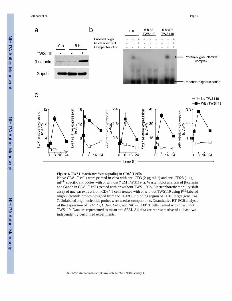
Figure 1. TWS119 activates Wnt signaling in CD8+ T cellsNaive CD8+ T cells were primed in vitro with anti-CD3 (2 μg ml−1) and anti-CD28 (1 μgml−1) specific antibodies with or without 7 μM TWS119. a, Western blot analysis of β-cateninand Gapdh in CD8+ T cells treated with or without TWS119. b, Electrophoretic mobility shiftassay of nuclear extract from CD8+ T cells treated with or without TWS119 using P32-labeledoligonucleotide probes designed from the TCF/LEF binding region of TCF1 target gene Fzd7. Unlabeled oligonucleotide probes were used as competitor. c, Quantitative RT-PCR analysisof the expression of Tcf7, Lef1, Jun, Fzd7, and Nlk in CD8+ T cells treated with or withoutTWS119. Data are represented as mean +/− SEM. All data are representative of at least twoindependently performed experiments.
Gattinoni et al. Page 9
Nat Med. Author manuscript; available in PMC 2010 January 1.
NIH
-PA Author Manuscript
NIH
-PA Author Manuscript
NIH
-PA Author Manuscript
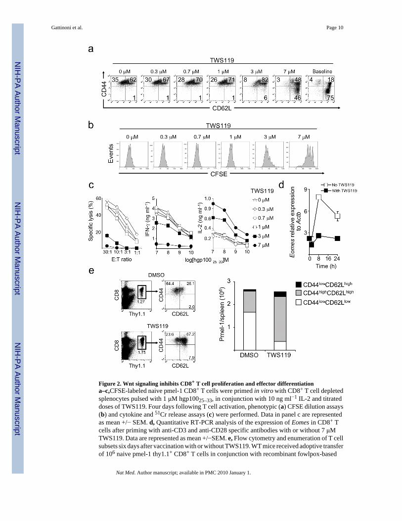
Figure 2. Wnt signaling inhibits CD8+ T cell proliferation and effector differentiationa–c,CFSE-labeled naive pmel-1 CD8+ T cells were primed in vitro with CD8+ T cell depletedsplenocytes pulsed with 1 μM hgp10025–33, in conjunction with 10 ng ml−1 IL-2 and titrateddoses of TWS119. Four days following T cell activation, phenotypic (a) CFSE dilution assays(b) and cytokine and 51Cr release assays (c) were performed. Data in panel c are representedas mean +/− SEM. d, Quantitative RT-PCR analysis of the expression of Eomes in CD8+ Tcells after priming with anti-CD3 and anti-CD28 specific antibodies with or without 7 μMTWS119. Data are represented as mean +/−SEM. e, Flow cytometry and enumeration of T cellsubsets six days after vaccination with or without TWS119. WT mice received adoptive transferof 106 naive pmel-1 thy1.1+ CD8+ T cells in conjunction with recombinant fowlpox-based
Gattinoni et al. Page 10
Nat Med. Author manuscript; available in PMC 2010 January 1.
NIH
-PA Author Manuscript
NIH
-PA Author Manuscript
NIH
-PA Author Manuscript
hgp100 vaccine. Mice received four daily doses of TWS119 (at 30 mg kg−1) from day 0 to day3 or DMSO as control. All data are representative of at least two independently performedexperiments.
Gattinoni et al. Page 11
Nat Med. Author manuscript; available in PMC 2010 January 1.
NIH
-PA Author Manuscript
NIH
-PA Author Manuscript
NIH
-PA Author Manuscript
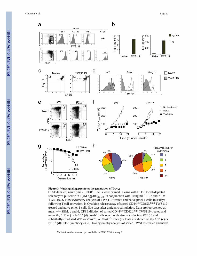
Figure 3. Wnt signaling promotes the generation of TSCMCFSE-labeled, naive pmel-1 CD8+ T cells were primed in vitro with CD8+ T cell-depletedsplenocytes pulsed with 1 μM hgp10025–33, in conjunction with 10 ng ml−1 IL-2 and 7 μMTWS119. a, Flow cytometry analysis of TWS119-treated and naive pmel-1 cells four daysfollowing T cell activation. b, Cytokine release assay of sorted CD44lowCD62Lhigh TWS119-treated and naive pmel-1 cells five days after antigenic stimulation. Data are represented asmean +/− SEM. c and d, CFSE dilution of sorted CD44lowCD62Lhigh TWS119-treated andnaive thy 1.1+ (c) or ly5.1+ (d) pmel-1 cells one month after transfer into WT (c) andsublethally-irradiated WT, or Tcra−/−, or Rag1−/− mice (d). Data are shown on thy 1.1+ (c) orly5.1+ (d) CD8+ lymphocytes. e, Flow cytometry analysis of sorted TWS119-treated and naive
Gattinoni et al. Page 12
Nat Med. Author manuscript; available in PMC 2010 January 1.
NIH
-PA Author Manuscript
NIH
-PA Author Manuscript
NIH
-PA Author Manuscript
ly5.1+ pmel-1 cells one month after transfer into WT or B2m−/− mice. Data are shown only5.1+, CD8+ lymphocytes. f, Tumor treatment of myeloablated WT or B2m−/− mice bearingB16 tumors established for 7 days. Mice received age-matched, lineage-depleted bone marrowcells. On the following day, 106 CD44lowCD62Lhigh TWS119-treated or naive pmel-1 cellswere transferred in conjunction with exogenous IL-2.g and h, Flow cytometry analysis ofCFSE-labeled, sorted CD44lowCD62Lhigh TWS119-treated and naive ly5.1+ pmel-1 cells onemonth after transfer into sublethally-irradiated WT mice. Data are represented as the percentageof CD44lowCD62Lhigh cells as a function of CFSE dilution of two independent experiments(g) and as fraction of cells with any given number of divisions (h). All data are representativeof at least two independently performed experiments.
Gattinoni et al. Page 13
Nat Med. Author manuscript; available in PMC 2010 January 1.
NIH
-PA Author Manuscript
NIH
-PA Author Manuscript
NIH
-PA Author Manuscript
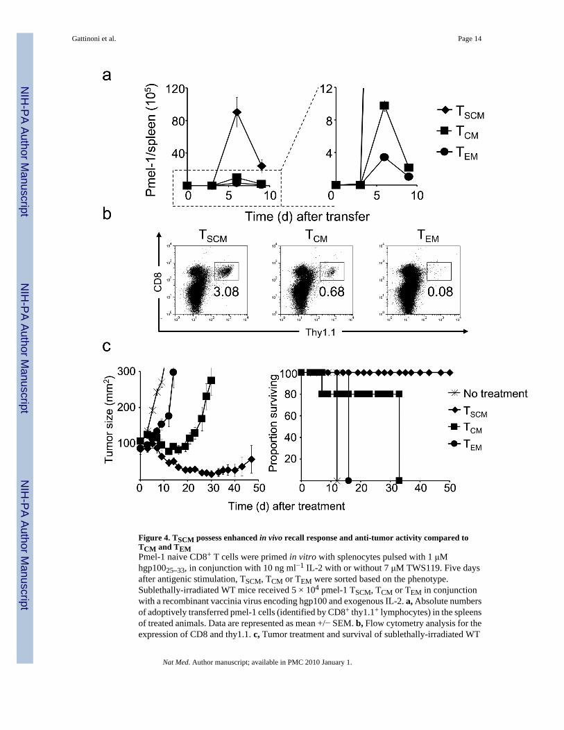
Figure 4. TSCM possess enhanced in vivo recall response and anti-tumor activity compared toTCM and TEMPmel-1 naive CD8+ T cells were primed in vitro with splenocytes pulsed with 1 μMhgp10025–33, in conjunction with 10 ng ml−1 IL-2 with or without 7 μM TWS119. Five daysafter antigenic stimulation, TSCM, TCM or TEM were sorted based on the phenotype.Sublethally-irradiated WT mice received 5 × 10⁴ pmel-1 TSCM, TCM or TEM in conjunctionwith a recombinant vaccinia virus encoding hgp100 and exogenous IL-2. a, Absolute numbersof adoptively transferred pmel-1 cells (identified by CD8+ thy1.1+ lymphocytes) in the spleensof treated animals. Data are represented as mean +/− SEM. b, Flow cytometry analysis for theexpression of CD8 and thy1.1. c, Tumor treatment and survival of sublethally-irradiated WT
Gattinoni et al. Page 14
Nat Med. Author manuscript; available in PMC 2010 January 1.
NIH
-PA Author Manuscript
NIH
-PA Author Manuscript
NIH
-PA Author Manuscript
mice bearing B16 tumors established for 10 days (n = 5 for all groups) receiving 4 × 10⁴ pmel-1TSCM, TCM or TEM in conjunction with a recombinant vaccinia virus encoding hgp100 andexogenous IL-2. Data are represented as mean +/− SEM. All data shown are representative ofat least two independently performed experiments.
Gattinoni et al. Page 15
Nat Med. Author manuscript; available in PMC 2010 January 1.
NIH
-PA Author Manuscript
NIH
-PA Author Manuscript
NIH
-PA Author Manuscript