Embed Size (px)
Citation preview

This article appeared in a journal published by Elsevier. The attachedcopy is furnished to the author for internal non-commercial researchand education use, including for instruction at the authors institution
and sharing with colleagues.
Other uses, including reproduction and distribution, or selling orlicensing copies, or posting to personal, institutional or third party
websites are prohibited.
In most cases authors are permitted to post their version of thearticle (e.g. in Word or Tex form) to their personal website orinstitutional repository. Authors requiring further information
regarding Elsevier’s archiving and manuscript policies areencouraged to visit:
http://www.elsevier.com/copyright

Author's personal copy
Trans- and cis-stilbene polyphenols induced rapid perinuclear mitochondrialclustering and p53-independent apoptosis in cancer cells but not normal cells
Alexander Gosslau, Srihari Pabbaraja, Spencer Knapp, Kuang Yu Chen ⁎Department of Chemistry and Chemical Biology, Rutgers, The State University of New Jersey, Piscataway, New Jersey 08854-8087, United StatesNew Jersey Cancer Institute, New Brunswick, New Jersey, 08901, United States
A B S T R A C TA R T I C L E I N F O
Article history:Received 7 September 2007Received in revised form 26 February 2008Accepted 10 March 2008Available online 1 April 2008
Keywords:ResveratrolTrans- and cis-stilbenesApoptosisMitochondriap53
We previously reported that 3,4,5,4′-tetramethoxy-trans-stilbene (MR-4) induces p53 and perinuclearmitochondrial clustering in cancer cells [Gosslau, A., Chen, M., Ho, C.-T., Chen, K.Y., 2005, A methoxyderivative of resveratrol analogue selectively induced activation of the mitochondrial apoptotic pathway intransformed fibroblasts. Br. J. Cancer 92, 513-521.]. Here we extended the study to over 20 trans-stilbenederivatives and their cis-isomers to explore structure activity relationship. Among them, 3,4,5,4′-tetramethoxy-cis-stilbene (MC-4), the cis-isomer of MR-4, was most potent, with IC50 of 20 nM for growthinhibition. MC-4 induced a rapid perinuclear mitochondrial clustering, membrane permeability transition,cytochrome c release and DNA fragmentation. To determine whether trans- and cis-stilbene polyphenolsmay share a common mechanism, we compared the effects of MC-4 and MR-4 in three isogenic cell linesderived from the colorectal carcinoma HCT116 cells: p53+/+ (p53-wt), p53−/− (p53-null) and p21−/− (p21-null). Deletion of either p53 or p21 neither blocked the effects of MC-4 or MR-4 on mitochondrial clusteringnor inhibited apoptosis, indicating that the actions of both stilbenes are independent of p53 and p21.Although microtubule disruption has been proposed to account for the action of some cis-stilbenepolyphenols, we did not observe differences in microtubule dynamics between cells treated with MC-4 andMR-4. These findings suggest that MC-4 and MR-4 may share a common mechanism whereby the perinucearmitochondrial clustering, rather than p53, p21, or microtubule depolymerization, is critical for their pro-apoptotic action.
© 2008 Elsevier B.V. All rights reserved.
1. Introduction
Resveratrol, a trans-stilbene polyphenol isolated from grapes,inhibits cancer cell growth in vitro and tumor progression in mousemodels (reviewed in Savouret and Quesne 2002; Aziz et al., 2003; Bhatand Pezzuto, 2002). Recently, resveratrol has also been shown toenhance the life-span in several organisms (Baur and Sinclair, 2006).Combretastatin A-4 is a cis-stilbene polyphenol isolated from Com-bretum caffrum, an African bush willow tree, and is known for itspotent antitumor and antiangiogenic activity (Pettit et al., 1987). Aprodrug derived from combretastatin A-4 is under clinical trials as anantitumor vascular-targeting agent (Pettit et al., 2005). The wide-range anti-disease activities and potential health benefits of resver-atrol and combretastatin have generated a great interest in developing
trans- and cis-stilbene polyphenols into useful chemopreventive andchemotherapeutic agents.
Both resveratrol and combretastatin A-4 share a stilbene backbone.However, their respective cellular targets have not been unequivocallyestablished despite their simple chemical structure. Several targetshave been proposed for resveratrol and other trans-stilbene poly-phenols, including p53/Bax pathway (Lu et al., 2001; Gosslau et al.,2005), CD95-CD95 ligand pathway (Dorrie et al., 2001), or the putativeresveratrol-binding proteins (Hsieh et al., 2005). On the other hand,the cellular target of combretastatin A-4 has been thought to bemicrotubules (Lin et al., 1988; McGown and Fox, 1989). Since the invitro tubulin binding activity of combretastatin A-4 and its analoguesis usually two or three orders of magnitude less than their growthinhibitory activity, it has been questioned whether tubulin bindingdirectly account for the cytotoxic effect of combretastatins (Cushmanet al., 1992). Indeed, some combretastatin analogues lack tubulinbinding activity but still retain potent cytotoxic activity (Borrel et al.,2005).
We have previously reported that 3,4,5,4′-tetrahydroxy-trans-stil-bene (R-4) and 3,4,5,4′-tetramethoxy-trans-stilbene (MR-4) induce p53,Bax and cancer cell apoptosis (Lu et al., 2001; Gosslau et al., 2005). Theseresveratrol analogues also induce rapid perinuclear mitochondrial
European Journal of Pharmacology 587 (2008) 25–34
⁎ Corresponding author. Department of Chemistry and Chemical Biology, RutgersUniversity, 610 Taylor Road, Piscataway, NJ 08854-8087. Tel.: +1 732 445 3739; fax: +1732 445 5312.
E-mail address: [email protected] (K.Y. Chen).
0014-2999/$ – see front matter © 2008 Elsevier B.V. All rights reserved.doi:10.1016/j.ejphar.2008.03.027
Contents lists available at ScienceDirect
European Journal of Pharmacology
j ourna l homepage: www.e lsev ie r.com/ locate /e jphar

Author's personal copy
clustering in transformed WI38VA cells but not in their normalcounterparts (Gosslau et al., 2005). We have now extended this studyby synthesizingover twenty trans- andcis-stilbeneanalogues inorder toexplore structure-activity relationship. Among the stilbene derivativeswe tested, 3,4,5,4′-tetramethoxy-cis-stilbene (MC-4), the cis-isomer ofMR-4, inhibited cancer cell growthwith an IC50of 20nM.MC-4 andMR-4were used here respectively as themodel compound for cis- and trans-stilbene polyphenols to determine whether they may share commonmechanism in inducing apoptosis. We compared the dose–responsecurve of MC-4 and MR-4 on apoptosis and growth inhibition, andexamined the time course of mitochondrial clustering, membranepermeability transition, cytochrome c release, and DNA fragmentation.Since mitochondria play a critical role in p53-dependent apoptoticpathway (Polyak et al., 1997) and p53 may be directly involved in themitochondria-mediated apoptosis (Moll et al., 2005), we investigatedpossible relationship between p53 activation and mitochondrialclustering by investigating the effects of MC-4 and MR-4 in the p53-null and p21-null colorectal carcinoma cell lines. Our results show thatperinuclear mitochondrial clustering represents one of the earliestbiological events elicited by both MC-4 and MR-4, and suggest that thiseventmay be responsible for initiating apoptotic cascade in cancer cells.Despite the importance of p53 and p21 in mitochondria-mediatedapoptosis, they were not required in the mitochondrial clustering andapoptotic action of MR-4 and MC-4.
2. Materials and methods
2.1. Materials and chemicals
Dulbecco's modified Eagle's medium (DMEM) and fetal bovineserum (FBS) were obtained from Gibco BRL (Gaithersburg, MD). Anti-p53 antibody conjugated to horseradish peroxidase (polyclonal BMG-1B1) was purchased from Roche (Indianapolis, IN), Anti-α tubulinantibody (mouse monoclonal, A-11126) was from Invitrogen (Carls-bad, CA) and anti-cytochrome c antibody (mouse monoclonal,7H8.2C12) from Calbiochem (San Diego, CA). Other biologicalchemicals were purchased from Sigma (St. Louis, MO).
2.2. Preparation of trans- and cis-stilbenes
The synthesis of resveratrol using 4-methoxybenzyl alcohol and 3,5-dimethoxy-benzaldehyde as the starting materials has been described(Bachelor et al., 1970; Drewes and Fletcher, 1974). Similar strategy wasemployed to prepare trans- and cis-stilbene polyphenols. Variousderivatized benzyl triphenylphosphonium bromides were prepared fromcorresponding benzyl bromide. Wittig reactionwas then performed usingvarious benzaldehyde and appropriate benyl triphenylphosphoniumbromide. The identity and purity of each of these compounds has beenconfirmed by thin layer chromatography, NMR andGC-mass spectroscopy.
Fig. 1. Structure and activity of cis- and trans-stilbene polyphenol analogues. Resveratrol (2.1) was used as the prototype compound to synthesize trans- and cis-stilbene analogues.The compounds are grouped according to their cytotoxicity. The IC50 for compounds in each group is: Group 1, N100 µM; Group 2, 20–80 µM; Group 3, 2–10 µM; Group 4, 0.1–1 µM;Group 5, 20–50 nM. The compounds are referred by a numbering systemwith the first digit indicating the group that the compound belongs to and the second digit identifying thecompound within the group. Compound 1.1, however, is not a bona fide stilbene due to the presence of a triple bond instead of a double bond that connects the two benzene rings.
26 A. Gosslau et al. / European Journal of Pharmacology 587 (2008) 25–34

Author's personal copy
2.3. Cell culture and treatment
All cell lines used in this studywere cultured at 37 °C in a humidified,10%CO2 atmosphere inDMEMsupplementedwith10% FBS. Normal skinfibroblastWI38andSV40virally transformedWI38VAcellswereusedasan isogeneticmodel for normal and transformedhumancells (Lawrence,2002; Gosslau et al., 2005; Scaglia et al., 2005). HCT116 wild type, p53-null and p21-null cells were kindly provided by Dr. Bert Vogelstein(Johns Hopkins School of Medicine). Before experiments, cells wereseededeither in culturedishesor inmulti-well plates as indicated for thedifferent assays. Stock solutions of all stilbene analogues were made inDMSO. For the control cultures the solvent vehicle DMSO was added tothe medium instead of the stock solution.
2.4. Cellular proliferation assays
Cell proliferation was measured by the MTT (3 (4,5-dimethylthia-zol-2-yl)-2,5-diphenyl-tetrazolium-bromide) method, crystal violetstaining, or by cell counting as previously described (Gosslau et al.,2005). Because of the concern that MTT assay may yield false-positiveresults for certain cell types when treated with flavonoids orpolyphenols (Bernhard et al., 2003), we also included the crystalviolet dye staining assay in this study (Lu et al., 2001). All threemethods yielded comparable results.
2.5. DNA fragmentation assay
The assay was carried out as previously described (Gosslau et al.,2005). Briefly, cells were suspended in buffer containing sodium laurylsarkosinate (0.5%), proteinase K and RNase A. DNAwas extracted witha phenol/chloroform/isopropanol mixture (25:24:1, pH 8.0), analyzedon a 2% agarose gel, stained by ethidium bromide and visualized underUV illumination.
2.6. Caspase activation assay
Caspase activation was analyzed by CaspACE™FITC-VAD-FMK insitu marker (Promega, Madison, WI), a fluoroisothiocyanate (FITC)conjugate of the cell permeable caspase inhibitor VAD-FMK. Cellswere seeded on coverslips two days before experiment. At the end ofdrug treatment, the coverslips were transferred to a 35 mm dishcontaining 1 ml of medium containing CaspACE-solution. After anincubation of 20 min, cells were analyzed by fluorescent microscopyusing an excitation wavelength of 480 nm.
2.7. Rhodamine 123 fluorescence assay
Morphology of mitochondria was monitored using the cationicfluorophore rhodamine 123. Briefly, cells were grown on glasscoverslips in 35 mm culture dishes for 2 days. At the end of drugtreatment, rhodamine 123 (1.5 mM in medium containing 10% DMSO)was added to the dish to a final concentration of 1.5 μM. After anincubation of 20 min, cells were washed twice with medium andanalyzed by fluorescence microscopy with excitation and emissionwavelength set at 480 or 525 nm, respectively.
2.8. Mitochondrial potential
Changes in the mitochondrial membrane potential (ΔΨm) wasmonitored by the Mitochondrial Potential Assay Kit (ATCC, Manassas,VA). Delta-Psi reagent is a lipophilic cation (5, 5′, 6, 6′, tetrachloro-1,1′,
Table 1The IC50 of stilbene analogues measured with human WI38VA transformed cells
ID# Compound name IC50 (μM) Acronym
Group 11.1 3,4,5 trimethoxy 4′-diazirine ethyne NE1.2 3,4,5,3′4′-pentamethoxy trans-stilbene NE1.3 3,4,5,2′-tetramethoxy trans-stilbene NE1.4 3,5-dimethoxy trans-stilbene NE1.5 4,4′dimethoxy, 3-nitro trans-stilbene NE
Group 22.1 3,5,4′-trihydroxy trans-stilbene 50 R-3, resveratrol2.2 3,5,4′-trimethoxy trans-stilbene 25–502.3 3,4,5-trimethoxy 4-bromo trans-stilbene 25–502.4 3,4,5-trimethoxy trans-stilbene 802.5 3,5-dimethoxy trans-stilbene 80
Group 33.1 3,4,5,4′-tetramethoxy trans-stilbene 2 MR-43.2 3,5,4′-trimethoxy 3′-amino trans-stilbene 53.3 3,4,5,4′-tetrahydoxy trans-stilbene 5 R-43.4 3,4,5,4′-tetramethoxy 3′-nitro trans-stilbene 103.5 4,4′-dimethoxy 3′-nitro cis-stilbene 10
Group 44.1 3,5,4′-trimethoxy 3′-nitro cis-stilbene 0.54.2 3,5,4′-trimethoxy 3′-azido cis-stilbene 0.254.3 3,5,4′-trimethoxy 4′-bromo cis-stilbene 0.14.4 3,4,5,4′-tetramethoxy 3′-nitro cis-stilbene 0.1
Group 55.1 3,4,5,4′-tetramethoxy cis-stilbene 0.02 MC-45.2 3,5,4′-trimethoxy 3′-amino cis-stilbene 0.055.3 3,5,4′-trimethoxy cis-stilbene 0.03
IC50 (concentration that gives 50% of growth inhibition relative to the control) wasdetermined by proliferation assay using WI38VA cells. The number represents anaverage of three measurements with standard errors less than 10%.NE: no inhibitory effect at 100 µM or higher.
Fig. 2. Antiproliferative effects of trans- and cis-stilbene polyphenol analogues. HeLa cells (A) and WI38VA fibroblasts (B) were subcultured at 1:20 dilution and then treated withstilbene analogues 5.2, 5.1, 3.1 and 2.1 at indicated concentrations. The cell growth was monitored by the MTT assay on day 5 as described in Material and methods section.
27A. Gosslau et al. / European Journal of Pharmacology 587 (2008) 25–34

Author's personal copy
3, 3′-tetrathylbenzimidazolyl carbocyanin iodide) that aggregatesupon membrane polarization and forms an orange fluorescentcompound in intact mitochondria. If the mitochondrial potential isdisrupted, the dye cannot access the transmembrane space andremains as greenmonomers (Cossarizza et al., 1993). Cells were grownon coverslips in 35 mm culture dish for 2 days. At the end of drugtreatment, cells on coverslips were incubated with 1 ml of Delta-Psireagent solution for 20 min, washed and analyzed by phase contrastmicroscopy and fluorescence microscopy.
2.9. Cytochrome c release and western blot analysis
Cells were treated with MC-4 for various times and then harvestedfor subcellular fractionation using Dounce homogenizer. The mito-chondrial fraction was isolated by a centrifugation at 14,000 ×g for10min, and the supernatant was designated as cytosolc fraction. Equalamounts of protein were loaded on SDS-polyacrylamide gel forelectrophoresis. The gel was transblotted to a nitrocellulose mem-brane for western blot analysis using mouse anti-cytochrome cantibody and anti-mouse antibody conjugated to horseradish perox-
idase as secondary antibody (Amersham Pharmacia). Immuno-complexes were detected with the ECL Plus Western blot detectionkit (Amersham Pharmacia).
3. Results
3.1. Trans- and cis-stilbene polyphenol analogues and their antiproliferativeactivity
Using resveratrol (3,5,4′-trihydroxy trans-stilbene) as a prototypewe have synthesized over 20 trans- and cis-stilbene analogues (Fig. 1).We have grouped these analogues according to their IC50 values(Table 1). For example, resveratrol (3,5,4′-trihydroxy trans-stilbene) inTable 1 is encoded as 2.1, indicating that it belongs to Group 2 whichhas IC50 in the range of 20–80 µM. Group 5 analogues are the mostpotent ones with IC50 values comparable to or better than manyclassical anti-cancer chemotherapeutic agents such as daunorubicinand etoposide. Fig. 2 shows a comparison of antiproliferative activityof a trans- and two cis-stilbene derivatives with that of resveratrol inhuman cancer cells. The cis-stilbenes, MC-4 (5.1) and 3,5,4′-
Fig. 3. Differential apoptotic effects of stilbene analogues on normal and transformed fibroblasts. (A) Caspase activation-assay. MC-4 (5.1) was added to the culture of normal WI38and transformed WI38VA cells for 48 h. Caspase activation was analyzed by incubating the culture with the fluorphore CaspACE™ (FITC-VAD-FMK) for 20 min, cells were examinedunder a fluorescent microscope. Phase contrast (left panels) and fluorescent micrographs (right panels) were presented for each cell type. (B) DNA laddering analysis. ConfluentWI38or WI38VA cultures were treated with MC-4, MR-4 and resveratrol (R-3) at indicated concentrations for 48 h and processed for DNA isolation and agarose electrophoresis. DNAwasvisulaized by ethidium bromide staining.
28 A. Gosslau et al. / European Journal of Pharmacology 587 (2008) 25–34

Author's personal copy
trimethoxy-3′-amino cis-stilbene (5.2) are more than 1,000-foldeffective than resveratrol (2.1) in inhibiting the growth of cancercells. MR-4 (3.1), the most potent trans-stilbene derivative in ourseries, is 200-fold less potent than MC-4 (5.1). In view of their similarstructures, it is striking that the antiproliferative activity of theseanalogues can vary by as much as 3 to 4 orders of magnitude. Theseresults suggest that structure-activity relationship of the stilbenebackbone can be further exploited to develop even more potentderivatives. Several features of structure-activity relationship can bederived already: (i) among the six pairs of trans- and cis-isomer, thecis- isomers are always 50–200 times more potent than their trans-counterparts. For example, the IC50 values for isomers 3.1 and 5.1 are,respectively, 5 µM and 20 nM. (ii) An addition of amino group atposition C-3′ increases the activity of 3,5,4′ trimethoxy cis-stilbene by2-fold (5.3 vs. 5.2). However, an addition of nitro group at C-3′ reducesthe activity of 3,5,4′ trimethoxy cis-stilbene by 10-fold (5.3 vs. 4.1).Similarly, an addition of a nitro group at C-3′ position reduces theactivity of 3,4,5,4′ tetramethoxy cis-stilbene by 4-fold (5.1 vs. 4.4). It islikely that an electron-withdrawing group such as nitro-group at C-3′of a cis-stilbene diminishes its antiproliferative activity. However, anaddition of a nitro group at C-3′ position of a trans-stilbene does notaffect its activity (e.g. 3.4 vs. 3.1). (iii) An addition of methoxy group atC-3′ position drastically reduces its antiproliferative activity of a trans-stilbene (3.1 vs. 1.2). (iv) The addition of a methoxy group at C-4
enhances the activity for both cis- and trans-stilbenes by at least 4-fold (2.2 vs. 3.1 and 5.3 vs. 5.1) (v) the addition of a methoxy group atC-4′ of a trans-stilbene enhances its antiproliferative activity by atleast 10-fold (1.4 vs. 2.2 and 2.4 vs. 3.1). These features, althoughpreliminary, are in line with the notion that the antiproliferativeactivity of stilbene analogues is due to specific interaction with theircellular targets.
3.2. Apoptosis in transformed human cancer cells
To determinewhether trans- and cis-stilbene analogues may sharesimilar mechanism in their anti-proliferative action, we used thestereoisomeric pair, MC-4 and MR-4, as the representative compoundfor cis- and trans-stilbene polyphenols. We first examined the effect ofMC-4 on caspase activation. Fig. 3A shows that the MC-4 inducedcaspase 3/7 activation in WI38VA cells but not in normal WI38 cells,suggesting that, similar to MR-4, this cis-stilbene specifically inducedapoptosis in transformed cells. We then compared the dose–responseof MC-4, MR-4 and resveratrol on apoptosis as monitored by DNAfragmentation. Fig. 3B shows that at their respective IC50 concentra-tions these stilbene analogues induced extensive DNA fragmentationin WI38VA cancer cells, but not in normal WI38 cells. The closecorrelation between apoptosis and growth inhibition suggests thatapoptosis is the key mechanism underlying the anti-proliferative
Fig. 4.Mitochondrial distribution in normal and transformed fibroblasts. (A)WI38 andWI38VA cells were treated with MR-4 (50 µM) or MC-4 (50 nM) for 3 h, cells were then stainedwith rhodamine 123 as described in Materials and methods section and then monitored under a fluorescent microscope. For each cell type, the corresponding phase contrast (leftpanel) and fluorescence micrographs (right panel) were presented. (B) Pernuclear clustering of mitochondria in WI38VA cells. Cells were treated with MR-4 and MC-4 for 3 h andthen examined under phase contrast microscopy (upper panels), and fluorescent microscopy (middle panels). Bottom panels represent merged micrographs.
29A. Gosslau et al. / European Journal of Pharmacology 587 (2008) 25–34

Author's personal copy
action of both trans- and cis-stilbene polyphenols. Additional normaland cancer cells, including Caco-2, HeLa, IMR-90SV, IMR-90 and BJ-Twere tested and similar results were obtained (data not shown),suggesting that the differential apoptotic effect of stilbene analoguesis a general one.
3.3. Effects on mitochondrial morphology and membrane potential
Since MR-4 can cause perinuclear aggregation of mitochondria incancer cells, we have proposed that mitochondria may be the earlytarget of MR-4 (Gosslau et al., 2005). To determine whether cis-stilbene polyphenols also induce similar mitochondrial redistribution,we compared the effect of MC-4 on the mitochondrial morphology inboth normal and cancer cells. Fig. 4A shows that MC-4 elicited amarked perinuclear clustering in WI38VA cells, but had almost noeffect in their normal counterparts. Thus, bothMC-4 and MR-4 causedperinuclear mitochondrial clustering and apoptosis only in cancercells but not in normal cells (Fig. 4A vs. Fig. 3B). However, furtherexamination of the mitochondrial morphology in WI38VA cellsrevealed that the mitochondrial pattern in cells treated with MC-4was more diffuse than that treated with MR-4. We suspected thatdepolarization of mitochondria may have already occurred in the MC-4-treated cells. We therefore examined this possibility using amitochondrial membrane potential probe. Fig. 5 shows that MC-4caused a collapse of mitochondrial membrane potential, termedmembrane permeability transition, within 1 h of treatment. Incontrast, MR-4 elicited perinuclear mitochondrial clustering but didnot initiate membrane permeability transition. Instead, a longerexposure (N3 h) was required for MR-4 to initiate the onset ofmembrane permeability transition (data not shown). Neither MC-4nor MR-4 caused any membrane permeability transition in normalcells, even after prolonged treatment for up to 9 h (data not shown).Thus, although both MC-4 and MR-4 induced rapid perinuclearmitochondrial clustering, the subsequent onset of membrane perme-ability transition occurred earlier in cells treated with MC-4 thanwithMR-4.
3.4. Release of mitochondrial cytochrome c
The release of mitochondrial cytochrome c, a key event in initiatingthe mitochondria-mediated apoptosis signaling pathway (Liu et al.,1996), has been shown to be tightly coupled to membrane perme-
ability transition and mitochondrial depolarization (Heiskanen et al.,1999). To determine whether the early action of MC-4 on perinuclearmitochondrial clustering and membrane permeability transition didinitiate the apoptotic cascade, we examined the time course ofmitochondrial cytochrome c release in the MC-4-treated cancer cells.Fig. 6 shows that cytochrome c was detectable in the cytosol after 1 hof the treatment with MC-4, consistent with the notion thatcytochrome c release is coupled to membrane permeability transition.Given that cytochrome c release is a hallmark of mitochondria-dependent apoptosis, the early occurrence of mitochondrial redis-tribution, membrane potential collapse and cytochrome c releasesuggests that mitochondria may serve as the cellular target of MC-4.
Fig. 6. Effect of MC-4 on the release of mitochondrial cytochrome c. WI38VA cells at 80%confluence was treated with MC-4 (50 nM). At indicated time points, cells wereharvested for cytosolic and mitochondrial fractionation as described in Materials andmethods section. (A) Western blot analysis. Mitochondrial and cytosolic fractions wereanalyzed by SDS-PAGE. Cytochrome c in each fraction was detected by western blottingusing mouse anti-cytochrome c antibody and peroxidase-conjugated anti-mouseantibody (1:3000). (B) Time course of cytochrome c release. Cytochrome c in cytosolicfractions was detected by western blotting and quantified. Each data point representedan average of three separate experiments with standard errors less than 10%.
Fig. 5. Effects of MR-4 and MC-4 on Mitochondrial membrane potential in normal and transformed cells. WI38 and WI38VA cells were grown on glass coverslips in a 35 mm culturedish for 2 days. After treatment with MC-4 (50 nM) and MR-4 (50 µM) for 1 h, mitochondrial membrane potential was monitored with Delta-Psi reagent by fluorescence microscopy(excitation wavelength 480 nm) as described in Materials and methods section. For each cell type, the corresponding phase contrast (left panels) and fluorescent micrographs (rightpanels) were presented.
30 A. Gosslau et al. / European Journal of Pharmacology 587 (2008) 25–34
Author's personal copy
3.5. Effects on microtubules dynamics
It is generally thought that the biological effects cis-stilbenepolyphenols, but not their trans-analogues, are due to tubulin bindingandmicrotubule depolymerization (e.g. Gaukroger et al., 2003; Tron etal., 2006). Given that both MC-4 and MR-4 are capable of inducing arapid perinuclear mitochondrial clustering, we wonder what is therole of microtubules in this process. To address this question, wecompared the immunofluorescent staining pattern of microtubules inboth normal and cancer cells after the treatment with MC-4 or MR-4.Fig. 7 shows that MC-4, MR-4 or R-3 did not cause appreciable changesof microtubule distribution in normal cells. In cancer cells, treatmentwith either MC-4 or MR-4 gave similar pattern, which was slightlydifferent from that treated with R-3 or the control. These resultsindicate that mitochondrial clustering could occur without anysignificant change in microtubule dynamics, suggesting that micro-tubule depolymerization may not be directly responsible for causingperinuclear mitochondrial clustering.
3.6. Possible role of p53 pathway in the action of stilbene analogues
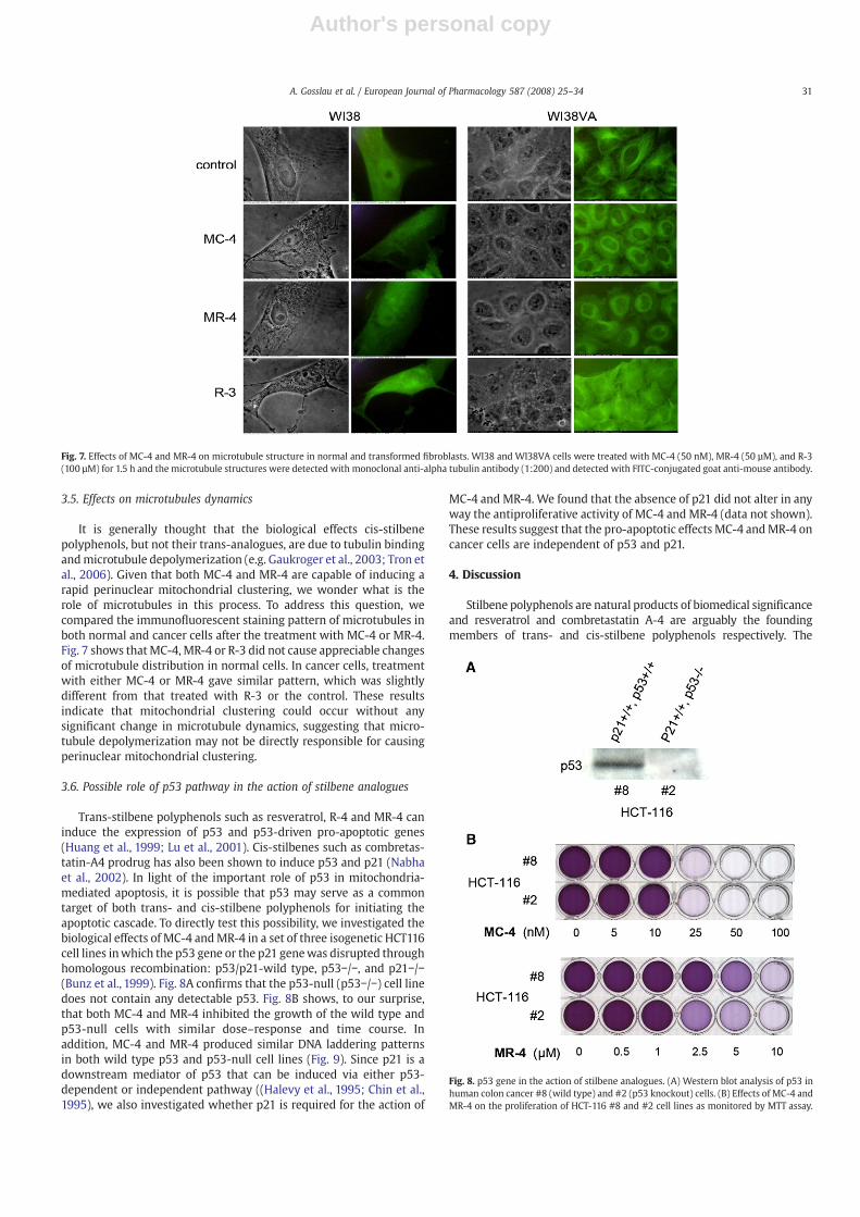
Trans-stilbene polyphenols such as resveratrol, R-4 and MR-4 caninduce the expression of p53 and p53-driven pro-apoptotic genes(Huang et al., 1999; Lu et al., 2001). Cis-stilbenes such as combretas-tatin-A4 prodrug has also been shown to induce p53 and p21 (Nabhaet al., 2002). In light of the important role of p53 in mitochondria-mediated apoptosis, it is possible that p53 may serve as a commontarget of both trans- and cis-stilbene polyphenols for initiating theapoptotic cascade. To directly test this possibility, we investigated thebiological effects of MC-4 andMR-4 in a set of three isogenetic HCT116cell lines inwhich the p53 gene or the p21 genewas disrupted throughhomologous recombination: p53/p21-wild type, p53−/−, and p21−/−(Bunz et al., 1999). Fig. 8A confirms that the p53-null (p53−/−) cell linedoes not contain any detectable p53. Fig. 8B shows, to our surprise,that both MC-4 and MR-4 inhibited the growth of the wild type andp53-null cells with similar dose–response and time course. Inaddition, MC-4 and MR-4 produced similar DNA laddering patternsin both wild type p53 and p53-null cell lines (Fig. 9). Since p21 is adownstream mediator of p53 that can be induced via either p53-dependent or independent pathway ((Halevy et al., 1995; Chin et al.,1995), we also investigated whether p21 is required for the action of
MC-4 and MR-4. We found that the absence of p21 did not alter in anyway the antiproliferative activity of MC-4 and MR-4 (data not shown).These results suggest that the pro-apoptotic effects MC-4 andMR-4 oncancer cells are independent of p53 and p21.
4. Discussion
Stilbene polyphenols are natural products of biomedical significanceand resveratrol and combretastatin A-4 are arguably the foundingmembers of trans- and cis-stilbene polyphenols respectively. The
Fig. 8. p53 gene in the action of stilbene analogues. (A) Western blot analysis of p53 inhuman colon cancer #8 (wild type) and #2 (p53 knockout) cells. (B) Effects of MC-4 andMR-4 on the proliferation of HCT-116 #8 and #2 cell lines as monitored by MTT assay.
Fig. 7. Effects of MC-4 and MR-4 on microtubule structure in normal and transformed fibroblasts. WI38 and WI38VA cells were treated with MC-4 (50 nM), MR-4 (50 µM), and R-3(100 µM) for 1.5 h and the microtubule structures were detected with monoclonal anti-alpha tubulin antibody (1:200) and detected with FITC-conjugated goat anti-mouse antibody.
31A. Gosslau et al. / European Journal of Pharmacology 587 (2008) 25–34

Author's personal copy
structure-activity analysis of stilbene analogues (e.g., Pettit et al., 2005;Tron et al., 2006; Hadfield et al., 2005; Fig. 1 and Table 1) has revealedseveral key features: (i) the cis-stilbene compounds are always morepotent than their trans-isomers (Fig. 2); (ii) the 3,4′,5-trihydroxy or3,4′,5-trimethoxy group appears to be the minimal structural require-ment for either trans- or cis-stilbenes to be biologically active; and (iii)the nature and positioning of functional groups on B rings candramatically affect the biological activity. These features suggest thepresence of a specific receptor for stilbene analogues and the highpotency of MC-4 may be due to its preferential interaction with thisreceptor. In this regard it is interesting to note that Wang et al. (2004)have reported the binding of resveratrol to mitochondrial quinonereductase. In addition, in view of the fact that both trans- and cis-stilbene analogues are small molecules containing similar functionalgroups and that resveratrol uptake is mediated mainly by passivediffusion (Lancon et al., 2004), it is unlikely that the differential effect ofstilbene analogues is due to their difference in uptake.
Among the stilbene analogues tested in this study, MR-4 and MC-4are the most potent trans- and cis-stilbene polyphenol, respectively.(Table 1). MR-4 and MC-4 inhibit cell growth (Fig. 2) and induce DNAfragmentation (Fig. 3). The pro-apoptotic effects of both compounds arelimited only to cancer cells, but not to their normal counterparts (Fig. 3).Both stilbenes also cause a rapid perinuclearmitochondrial clustering incancer cells (Fig. 4), making it one of the earliest detectable eventsinduced by either trans- or cis-stilbene polyphenols. Mitochondrialclustering has been shown to precede cell death in TNF-treatedfibrosarcoma cells (De Vos et al., 1998; Desagher and Martinou, 2000).Membrane permeability transition or mitochondrial membrane per-meabilization is the decisive event that delimits at cellular level theboundary between life and death (Kroemer et al., 2007). The temporal
relationship between loss of mitochondrial membrane potential andcytochrome c release has also been established (Heiskanen et al., 1999).In our study, both membrane permeability transition and cytochrome crelease occur in cancer cells within 60min after the treatmentwithMC-4 (Figs. 5 and 6). Thefindings thatMR-4 andMC-4 inducemitochondrialclustering, followed by mitochondrial membrane depolarization andmitochondrial cytochrome c release suggest that eithermitochondria orcellular sites upstream to mitochondrial clustering may serve as acommon target for both trans- and cis- stilbene analogues.
As noted by Tron et al. (2006), not only many combretastatinanalogues display discrepancies between cytotoxicity and anti-tubulinactivities, but the pharmacodynamic and clinical profile of combretas-tatins differs from that of other tubulin ibhibitors. Thus, a legitimatequestion can be raised onwhether the cytotoxic effect of combretastatinanalogues is dependent on their action on tubulin (Tron et al., 2006). Inthis regard, our finding that both MC-4 and MR-4 induced rapidperinuclear clustering seems relevant. It is well known that mitochon-dria move along microtubules as cargoes with the help of molecularmotors such as kinesin and dynein and, as such, the cellular distributionof mitochondria is closely linked to microtubule dynamics (Hirokawa,1998; Yaffe, 1999). Since microtubules have been considered to be thetarget of cis-stilbene polyphenols, one possible scenario is that MC-4first induces microtubule depolymerization, which in turn leads toperinuclear mitochondrial clustering. However, this scenario is notapplicable to MR-4, since trans-stilbene polyphenols have little or noinhibitory effect on tubulin polymerization. The microtubule distribu-tion monitored by immunostaining does not show significant differ-ences between cell cultures treated with MC-4 or MR-4 (Fig. 7),suggesting thatmitochondrial clustering induced by either trans- or cis-stilbene analogues may not be directly related to microtubule
Fig. 9. Apoptotic effect of MC-4 andMR-4 on p53-null HCT116 cells. Confluent HCT-116 p53-wildtype and p53-null cells were treatedwithMC-4 (lanes 2–4), MR-4 (lanes 5–7) and R-3(lanes 8–10) at indicated concentrations for 48 h and processed for DNA isolation and agarose electrophoresis. DNA was visualized by ethidium bromide staining.
32 A. Gosslau et al. / European Journal of Pharmacology 587 (2008) 25–34

Author's personal copy
depolymerization. Indeed, it has been reported that microtubuledepolymerization actually blocks mitochondrial clustering (De Vos etal., 1998; Kim et al., 2007). However, we do not know at this stagewhether kinesin and kinesin-related proteins are involved. Mitochon-dria are dynamic structures constantly undergoing fusion and fissionthat are controlled by shaping proteins such asmitofusins 1 and 2, OPA1,drp1 and Fis1 (Okamoto and Shaw, 2005; Chen and Chan, 2005; Chan,2006). The stilbene analogues may also target at some of thesemitochondrial shaping proteins, since disruption of fission can resultin perinuclear clustering of mitochondria (Stojanovski et al., 2004). Weare currently investigating this possibility. Alternatively, since apoptoticcells tend to shrink, perinuclear clustering ofmitochondria could also bea result of simple physical action due to cell shrinkage.
p53 can act as a gatekeeper by controlling cell cycle checkpoints oras a tumor suppressor by modulating the Bax to Bcl-2 ratio andmediating apoptosis (el-Deiry, 1998; Pietenpol and Stewart, 2002).p53 can also directly interact with mitochondria and promote theintrinsic mitochondria-mediated apoptosis (Moll et al., 2005). Abun-dant literature evidence indicates a close correlation between the up-regulation of p53 and apoptosis in cancer cells treated with trans-stilbene poyphenols (Huang et al., 1999; Gosslau et al., 2005). It istherefore a surprise to find that neither p53 nor p21 is required for theantiproliferative and pro-apoptotic action of not only cis-stilbene MC-4 but also trans-stilbeneMR-4 (Figs. 8 and 9). However, it can be notedthat resveratrol has been shown to induce apoptosis independent ofp53 (Mahyar-Roemer et al., 2001). Taken together, we conclude thatp53 or p21 is not the primary target for the pro-apoptotic action ofeither MC-4 or MR-4. On the other hand, our data suggest that themitochondrial clustering or the upstream events that lead tomitochondrial clustering could be the common underlying cause forapoptosis induced by either trans- or cis-stilbene polyphenols.Although the mechanism that triggers mitochondrial clustering isstill unclear, the time course of its occurrence, as short as 60 min,provides a convenient window for further defining the upstreamevents and elucidating the mechanism. Finally, the fact that the pro-apoptotic action of both trans- and cis-stilbene polyphenols such asMR-4 and MC-4 is independent of p53 and p21 further emphasizesthat stilbene analogues are potentially useful to be developed intochemotherapeutic agents against p53-resistant cancer cells.
Acknowledgments
This work was supported by the Commission on Science andTechnology, State of New Jersey as a component of the PioneeringNutraceutical Research Program. We acknowledge Dr. B. Vogelstein,Johns Hopkins School of Medicine, for the p53-null and p21-nullHCT116 cell lines.
References
Aziz, M., Kumar, R., Ahmad, N., 2003. Cancer chemoprevention by resveratrol: in vitroand in vivo studies and the underlying mechanisms (review). Int. J. Oncol. 23,17–28.
Bachelor, F., Loman, A., Snowdon, L., 1970. Synthesis of pinosylvin and relatedheartwood stilbenes. Can. J. Chem. 48, 1554–1557.
Baur, J.A., Sinclair, D.A., 2006. Therapeutic potential of resveratrol: the in vivo evidence.Nat. Rev. Drug Dis. 5, 493–506.
Bernhard, D., Schwaiger, W., Crazzolara, R., Tinhofer, I., Kofler, R., Csordas, A., 2003.Enhanced MTT-reducing activity under growth inhibition by resveratrol in CEM-C7H2 lymphocytic leukemia cells. Cancer Lett. 195, 193–199.
Bhat, K., Pezzuto, J., 2002. Cancer chemopreventive activity of resveratrol. Ann. N. Y.Acad. Sci. 957, 210–229.
Borrel, C., Thoret, S., Cachet, X., Guénard, D., Tillequin, F., Koch, M., Michel, S., 2005. Newantitubulin derivatives in the combretastatin A4 series: synthesis and biologicalevaluation. Bioorg. Med. Chem. 13, 3853–3864.
Bunz, F., Hwang, P.M., Torrance, C., Waldman, T., Zhang, Y., Dillehay, L., Williams, J.,Lengauer, C., Kinzler, K.W., Vogelstein, B., 1999. Disruption of p53 in human cancercells alters the responses to therapeutic agents. J. Clin. Invest. 104, 263–269.
Chan, D.C., 2006. Mitochondrial fusion and fission in mammals. Annu. Rev. Cell Dev.Biol. 22, 79–99.
Chen, H., Chan, D.C., 2005. Emerging functions of mammalian mitochondrial fusion andfission. Hum. Mol. Genet. 14, R283–R289.
Chin, Y.E., Kitagawa,M., Su,W.C., You, Z.H., Iwamoto, Y., Fu, X.Y.,1995. Science 272, 719–722.Cossarizza, A., Baccarani-Contri, M., Kalashnikova, G., Franceschi, C., 1993. A new
method for the cytofluorimetric analysis of mitochondrial membrane potentialusing the J-aggregate forming lipophilic cation 5,5′,6,6′-tetrachloro-1,1′,3,3′-tetraethylbenzimidazolcarbocyanine iodide (JC-1). Biochem. Biophys. Res. Com-mun. 197, 40–45.
Cushman, M., Nagarathnam, D., Gopal, D., He, H.M., Lin, C.M., Hamel, E., 1992. Synthesisand evaluation of analogues of (Z)-1-(4-methoxyphenyl)-2-(3,4 trimethoxyphenyl)ethene as potential cytotoxic and antimitotic agents. J. Med. Chem. 35, 2293–2306.
Desagher, S., Martinou, J.C., 2000.Mitochondria as the central control point of apoptosis.Trends Cell Biol. 10, 369–377.
De Vos, K., Goossens, V., Boone, E., Vercammen, D., Vancompernolle, K., Vandenabeele,P., Haegeman, G., Fiers, W., Grooten, J., 1998. The 55-kDa tumor necrosis factorreceptor induces clustering of mitochondria through its membrane-proximalregion. J. Biol. Chem. 273, 9673–9680.
Dorrie, J., Gerauer, H., Wachter, Y., Zunino, S.J., 2001. Resveratrol induces extensiveapoptosis by depolarizing mitochondrial membranes and activating caspase-9 inacute lymphoblastic leukemia cells. Cancer Res. 61, 4731–4739.
Drewes, S., Fletcher, L., 1974. Polyhydroxystilbenes from the heartwood of Schotiabrachypetala. J. Chem. Soc. Perkin Trans. 1, 961–962.
el-Deiry, W.S.,1998. Regulation of p53 downstream genes. Semin. Cancer Biol. 8, 345–357.Gaukroger, K., Hadfield, J.A., Lawrence, N.J., Nolan, S., McGown, A.T., 2003. Structural
requirements for the interaction of combretastatins with tubulin: how important isthe trimethoxy unit? Org. Biomol. Chem. 1, 3033–3037.
Gosslau, A., Chen, M., Ho, C.-T., Chen, K.Y., 2005. A methoxy derivative of resveratrolanalogue selectively induced activation of the mitochondrial apoptotic pathway intransformed fibroblasts. Br. J. Cancer 92, 513–521.
Hadfield, J.A., Gaukroger, K., Hirst, N., Weston, A.P., Lawrence, N.J., McGown, A.T., 2005.Synthesis and evaluation of double bond substituted combretastatins. Eur. J. Med.Chem. 40, 529–541.
Halevy, O., Novitch, B.G., Spicer, D.B., Skapek, S.X., Rhee, J., Hannon, G.J., Beach, D., Lassar,A.B., 1995. Correlation of terminal cell cycle arrest of skeletal muscle with inductionof p21 by MyoD. Science 267, 1018–1021.
Heiskanen, K.M., Bhat, M.B., Wang, H.W., Ma, J., Nieminen, A.L., 1999. Mitochondrialdepolarization accompanies cytochrome c release during apoptosis in PC6 cells.J. Biol. Chem. 274, 5654–5658.
Hirokawa, N., 1998. Kinesin and dynein superfamily proteins and the mechanism oforganelle transport. Science 279, 519–526.
Hsieh, H.P., Liou, J.P., Mahindroo, N., 2005. Pharceutical design of antimitotic agentsbased on combretastatins. Curr. Pharm. Des. 11, 1655–1677.
Huang, C., Ma, W., Goranson, A., Dong, Z., 1999. Resveratrol suppresses cell transformationand induces apoptosis through a p53-dependent pathway. Carcinogenesis 20, 237–242.
Kim, S., Kim, H.Y., Lee, S., Kim, S.W., Sohn, S., Kim, K., Cho, H., 2007. Hepatitis B virus xprotein induces perinuclear mitochondrial clustering in microtubule- and Dynein-dependent manners. J. Virol. 81, 1714–1726.
Kroemer, G., Galluzzi, L., Brenner, C., 2007. Mitochondrial membrane permeabilizationin cell death. Physiol. Rev. 87, 99–163.
Lancon, A., Delmas, D., Osman, H., Thnot, J.-P., Jannin, B., Latruffe, N., 2004. Humanhepatic cell uptake of resveratrol: involvement of both passive diffusion andcarrier-mediated process. Biochem. Biophys. Res. Commun. 316, 1132–1137.
Lawrence, D.A., 2002. Deficient R-smad/smad4 complex formation in fibroblastsgrowth-stimulated by TGF-beta 1. Int. J. Oncol. 20, 803–806.
Lin, C.M., Singh, S.B., Chu, P.S., Dempcy, R.O., Schmidt, J.M., Pettit, G.R., Hamel, E., 1988.Interactions of tubulin with potent natural and synthetic analogues of theantimitotic agent combretastatin: a structure-activity study. Mol. Pharmacol. 34,200–208.
Liu, X., Kim, C.N., Yang, J., Jemmerson, R., Wang, X., 1996. Induction of apoptotic programin cell-free extracts: requirement of dATP and cytochrome c. Cell 86, 147–157.
Lu, J., Ho, C., Ghai, G., Chen, K.Y., 2001. Resveratrol analog, 3,4,5,4′-tetrahydroxystilbene,differentially induces pro-apoptotic p53/Bax gene expression and inhibits thegrowth of transformed cells but not their normal counterparts. Carcinogenesis 22,321–328.
Mahyar-Roemer, M., Katsen, A., Mestres, P., Roemer, K., 2001. Resveratrol induces colontumor cell apoptosis independently of p53 and preceded by epithelial differentia-tion, mitochondrial proliferation andmembrane potential collapse. Int. J. Cancer 94,615–622.
McGown, A.T., Fox, B.W., 1989. Structural and biochemical comparison of the anti-mitotic agents colchicine, combretastatin A4 and amphethinile. Anticancer DrugDes. 3, 249–254.
Moll, U.M., Wolff, S., Speidel, D., Deppert, W., 2005. Transcription-independent pro-apoptotic functions of p53. Transcription-independent pro-apoptotic functions ofp53. Curr. Opin. Cell Biol. 17, 631–636.
Nabha, S.M., Mohammad, R.M., Dandashi, M.H., Coupaye-Gerard, B., Aboukameel, A.,Pettit, G.R., Al-Katib, A.M., 2002. Combretastatin-A4 prodrug induces mitoticcatastrophe in chronic lymphocytic leukemia cell line independent of caspaseactivation and poly(ADP-ribose) polymerase cleavage. Clin. Cancer Res. 8,2735–2741.
Okamoto, K., Shaw, J.M., 2005. Mitochondrial morphology and dynamics in yeast andmulticellular eukaryotes. Annu. Rev. Genet. 39, 503–536.
Pettit, G.R., Cragg, G.M., Singh, S.B., 1987. Antineoplastic agents, 122. Constituents ofCombretum caffrum. J. Nat. Prod. 50, 386–391.
Pettit, G.R., Minardi, M.D., Rosenberg, H.J., Hamel, E., Bibby, M.C., Martin, S.W., Jung, M.K.,Pettite, R.K., 2005. Antineoplastic agents. 509. Synthesis of fluorocombretastatinphosphate and related 3-halostilbenes. J. Nat. Prod. 68, 1450–1458.
33A. Gosslau et al. / European Journal of Pharmacology 587 (2008) 25–34

Author's personal copy
Pietenpol, J., Stewart, Z., 2002. Cell cycle checkpoint signaling: cell cycle arrest versusapoptosis. Toxicology 181-182, 475–481.
Polyak, K., Xia, Y., Zweier, J.L., Kinzler, K.W., Vogelstein, B., 1997. A model for p53-inducedapoptosis. Nature 389, 300–305.
Savouret, J., Quesne, M., 2002. Resveratrol and cancer: a review. Biomed. Pharmacother.56, 84–87.
Scaglia, N., Caviglia, J.M., Igal, R.A., 2005. High stearoyl-CoA desaturase protein andactivity levels in simian virus 40 transformed-human lung fibroblasts. Biochim.Biophys. Acta 1687, 141–151.
Stojanovski, D., Koutsopoulos, O.S., Okamoto, K., Ryan, M.T., 2004. Levels of human Fis1at the mitochondrial outer membrane regulate mitochondrial morphology. J. CellSci. 117, 1201–1210.
Tron, G.C., Pirali, T., Sorba, G., Pagliai, F., Busacca, S., Genazzani, A.A., 2006. Medicinalchemistry of combretastatin A4: present and future directions. J. Med. Chem. 49,3033–3044.
Wang, Z., Hsieh, T., Zhang, Z., Ma, Y., Wu, J.M., 2004. Identification and purification ofresveratrol targeting proteins using immobilized resveratrol affinity chromatogra-phy. Biochem. Biophys. Res. Commun. 323, 743–749.
Yaffe, M.P., 1999. Dynamic mitochondria. Nat. Cell Biol. 1, E149–E150.
34 A. Gosslau et al. / European Journal of Pharmacology 587 (2008) 25–34