Embed Size (px)
Citation preview

Auto-reverse nuclear migration in bipolar vertebrate cells on micropatterned surfaces
B. Szabó, Zs. Környei,A. Czirók, G. Csúcs, J. Zách, D. Selmeczi, T. Vicsek
Motivation Methods - micropatterning - video-microscopy - inhibitors, immuno- cytochemistry - digital image processing
Results Model

Nuclear positioning/migration versus bipolarity
Bipolar cells are abundant in nature Fission yeast (J.Cell.Sci., 1998, 701)
Where ?
Yeasts (meiotic prophase, fast oscillations)
In roots hairs (oscillations)
Epithelium (positioning)
Nucleokinesis (locomotion)
Ventricular zone (developing
vertebrate brain)
Arabidopsis thaliana root hairs (Mol. Biol. Of the Cell)
roothairs.mov

Nuclear positioning/migration versus
bipolarity
Positioning of nuclei in the epithelium (wing, fruitfly)
Ventricular zone (developing vertebrate brain)G1 MG2S G1
Lumen of neural tube
Newly formed chick neuronal tube
(Nature, 4212003, 83)

METHODS: Nuclear motility assayMicropatterning:Alternating adhesive/nonadhesive strips are prepared by microcontact printing. “Stamps” are made of PDMS obtained from a silicon “negative” produced by photolitogaphy (design, photoresist layer on metal film, chemical etching using the patterned metal film)
Stamping:
PDMS stamp
Inking with the protein solution
Short dryingBackfill with passivating PLL-PEG
Stamping – transfer of the protein structures

METHODS: Long-term videomicroscopy

Inhibitors: Drugs targeting cytoskeletal proteins were
used (vinblastine, taxol --> MT dynamics; vanadate, AMP-PNP,
ML-7 --> mol. motors; cytochalasin --> F-actin)
Immunocytochemistry: combining selective staining with
videomicroscopy (comparing spatial information with velocity data)
Digital image processing: recording data from video sequences,
calculating velocities of nuclei, and determining such features of
the oscillatory motion as distribution of periods, effect of drugs
on average velocity, etc
METHODS (continued)

Results
A typical experiment:
Adhesive: fibronectin
Strip width: 20 micron
Cell: C6 glioma cell line
Duration: 36 hours
A single cell:

Auto-reverse motion in bipolar cells
C6 – yes, average period 5 h
Primary mouse fibroblast:
- yes, average period 1.8 h
Primary mouse astroglia:
migration, no oscillations
3T3 nuclei migrate, no reverse
U87 no nuclear motility C6 on plain on stripe fibroblast on stripe

Time dependence of the position of a nucleus and the two corresponding edges of the cell
Time dependence of the velocity of the above nucleus

Distribution of the maximum velocities in an ensemble of C6 cells
Peaks are at
+/- 50 micron/hour
Distribution of the duration of the periods in an ensemble of the above cells. Peak is at
5 hours

Effect of drugs
Inhibition of microtubule dynamics: e.g., adding after one day 20 nM Vinblastine blocks auto-reverse migration
Drugs targeting actin and molecular motors did not have effect

Results from immunocytochemistry
The microtubules run parallel to the main axis (even over the nucleus, where their number density is significantly smaller)
Staining for microtubules

Centrosome position is correlated with the direction of nuclear motion
Staining for tubulin reveals that the centrosome is never located in the front part of the nucleus
no migration
centrosome
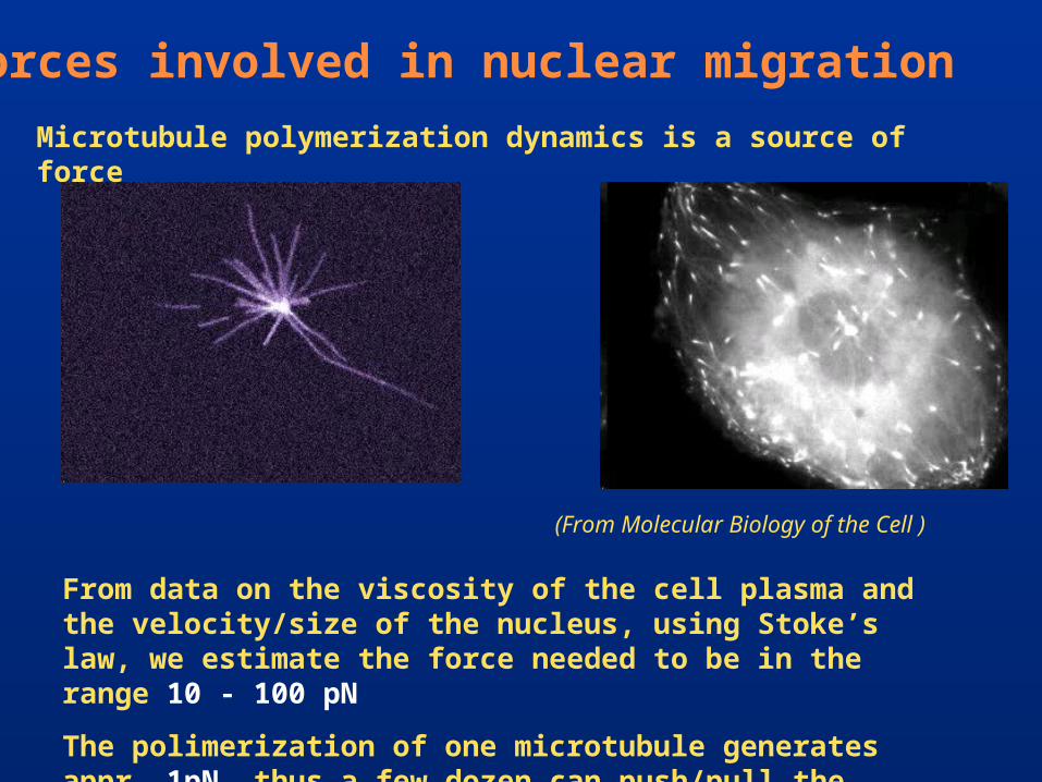
Forces involved in nuclear migration
From data on the viscosity of the cell plasma and the velocity/size of the nucleus, using Stoke’s law, we estimate the force needed to be in the range 10 - 100 pN
The polimerization of one microtubule generates appr. 1pN, thus a few dozen can push/pull the nucleus
Microtubule polymerization dynamics is a source of force
(From Molecular Biology of the Cell )

Experimental evidence for microtubules pushing an aster in a microfabricated chamber
(PNAS, 94,6228 1997)

MODEL
Based on our following observations: a) Array of microtubules is highly organized b) Centrosome is on the trailing side c) Estimated force is high enough
(dynein can play some role close to the cortex)
Reversal of direction is due to the repositioning of the centrosome (pushed by microtubules) at the edge of the cell
Only microtubules playing the most important role in the migration of the nucleus are shown (there are many more)

CONCLUSIONS
- First direct observation of oscillatory nuclear migration in vertebrate cells
- Proposition/demonstration of a nuclear motility assay for in vitro study
- Determination of the main characteristics of auto-reverse nuclear migration
- Evidence for the major role of MT dynamics and the position of the centrosome
- Presentation of a corresponding modelAcknowledgements:Hung. Natl. Sci Funds: OTKA and NKFP