Embed Size (px)
Citation preview
Submit Manuscript | http://medcraveonline.com
Abbreviations: AIHA, auto-immune hemolytic anemia; HL, Hodgkin’s lymphoma; RS, reed sternberg; RA, rheumatoid arthritis; SS, sjögren syndrome; SLE, systemic lupus erythematous; DLBCL, diffuse large b-cell lymphoma
Introduction AIHA is a recognized complication of lympho proliferative
disorders.1 AIHA is rarely seen in HL patients with a reported incidence of 0.2-4.2%.2 Sporadic case reports and reviews have shown that when AIHA occurs in HL patients it happens mostly at stage III and IV of nodular sclerosis HL or mixed cellularity HL.2 HL is usually manifested as lymphadenopathy typically in the cervical (70%), axillary (25%,), mediastinal areas (60%), and in 16-34 % of the cases presents as nodal disease below the diaphragm. HL presenting as AIHA has been reported in very few case reports in literature. We hereby report a case HL who presented as AIHA.1
Case report An 82 year old male patient with history of borderline diabetes
since 12years presented with on an off fever, malaise, generalized weakness, decreased appetite , weight loss, dizziness and itching all over the body since 1 month. There was history of hemiparesis, incontinence of urine and stool. On examination, his vital parameters were stable. Systemic examination revealed mild hepatomegaly and splenomegaly and bilateral basal crepitations on auscultation of the chest. He was found to have fever of 4°C and pallor and right axillary lymphadenopathy which was non tender and rubbery to feel. Laboratory investigations for liver function tests revealed liver enzymes within normal ranges, with unconjugated hyperbilirubinemia (with total bilirubin of 2.9 mg/dl and indirect bilirubin of 1.6mg/dl). LDH was 910U/L. Iron studies were within normal ranges.
The complete blood count revealed Hemoglobin of 5.6gm/dl, Hematocrit value 18.9%, MCV 113fL, and MCH 33.5pg and reticulocyte count of 30%. WBC count was 20,770cells/cumm with 1% Promyelocytes, 4% Myelocytes, 2% Metamyelocytes, 3% band cells, 66% Neutrophils, 4% Eosinophils,17% Lymphocytes, 3% Monocytes. The peripheral smear examination showed moderate anisocytosis and poikilocytosis with spherocytes, polychromatic cells, Howell-Jolly bodies, Cabot rings, basophilic stippling and
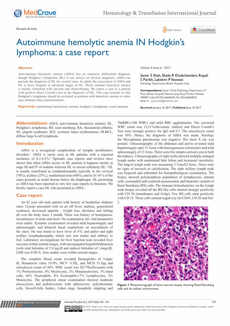
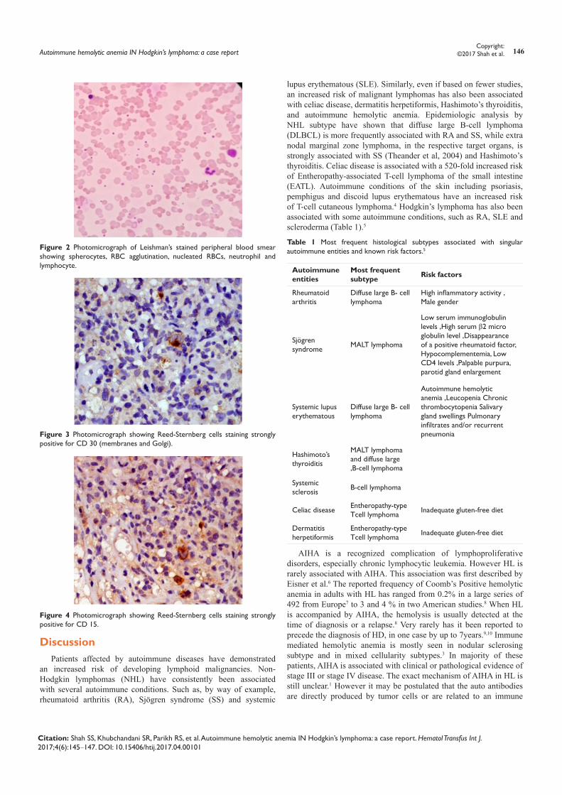
70nRBCs/100 WBCs and mild RBC agglutination. The corrected WBC count was 12,217cells/cumm. Indirect and Direct Coomb’s Test were strongly positive for IgG and C3. The reticulocyte count was 30%. Hence, the diagnosis of AIHA was made. Serology for Mycoplasma pneumoniae was negative. His chest X ray was normal. Ultrasonography of the abdomen and pelvis revealed mild hepatomegaly upto 15.3cms with heterogeneous echotexture and mild splenomegaly of 12.5cms. There were few simple cortical cysts in both the kidneys. Ultrasonography of right axilla showed multiple enlarged lymph nodes with maintained fatty hilum and increased vascularity. The largest lymph node was measuring 5.7cmsX2.4cms. There were no signs of necrosis or calcification. The right axillary lymph node was biopsied and submitted for histopathological examination. The biopsy showed polymorphous population of lymphocytes, plasma cells, eosinophils and scattered mononuclear and binuclear variants of Reed Sternberg (RS) cells. The immuno histochemistry on the lymph node biopsy revealed all the RS like cells stained strongly positively with CD 30 (membranes and Golgi). Few RS cells stain positively with CD 15. These cells stained negatively for CD45, CD 20 and Oct-2.
Figure 1 Photomicrograph of bone marrow biopsy showing Reed-Sternberg cells and its cellular environment.
Hematol Transfus Int J. 2017;4(6):145‒147. 145© 2017 Shah et al. This is an open access article distributed under the terms of the Creative Commons Attribution License, which permits unrestricted use, distribution, and build upon your work non-commercially.
Autoimmune hemolytic anemia IN Hodgkin’s lymphoma: a case report
Volume 4 Issue 6 - 2017
Samir S Shah, Shaila R Khubchandani, Rupali S Parikh, Lakshmi P Vaswani Pathology Department, Bhatia Hospital, India
Correspondence: Samir S Shah, Pathology Department, 2nd Floor, Bhatia Hospital, Tukaram Jivaji Road, Tardeo, Mumbai 400007, India, Tel 022-66660643, Fax 022-66660032, Email [email protected] Received: January 12, 2017 | Published: June 19, 2017
Abstract
Auto-immune hemolytic anemia (AIHA) has an extensive differential diagnosis though Hodgkin’s lymphoma (HL) is not always an obvious diagnosis. AIHA can precede the diagnosis of HL for several years. In adults the association of AIHA and HL is more frequent in advanced stages of HL. Warm immune hemolytic anemia is mainly controlled with steroids and chemotherapy. We report a case of a patient with positive direct Coomb’s test at the diagnosis of HL. This case reminds us that Hodgkin’s lymphoma should be excluded in patients with hemolytic anemia or other auto-immune (like) manifestations.
Keywords: autoimmune haemolytic anemia, hodgkin’s lymphoma, warm immune
Hematology & Transfusion International Journal
Review Article Open Access
Autoimmune hemolytic anemia IN Hodgkin’s lymphoma: a case report 146Copyright:
©2017 Shah et al.
Citation: Shah SS, Khubchandani SR, Parikh RS, et al. Autoimmune hemolytic anemia IN Hodgkin’s lymphoma: a case report. Hematol Transfus Int J. 2017;4(6):145‒147. DOI: 10.15406/htij.2017.04.00101
Figure 2 Photomicrograph of Leishman’s stained peripheral blood smear showing spherocytes, RBC agglutination, nucleated RBCs, neutrophil and lymphocyte.
Figure 3 Photomicrograph showing Reed-Sternberg cells staining strongly positive for CD 30 (membranes and Golgi).
Figure 4 Photomicrograph showing Reed-Sternberg cells staining strongly positive for CD 15.
Discussion Patients affected by autoimmune diseases have demonstrated
an increased risk of developing lymphoid malignancies. Non-Hodgkin lymphomas (NHL) have consistently been associated with several autoimmune conditions. Such as, by way of example, rheumatoid arthritis (RA), Sjögren syndrome (SS) and systemic
lupus erythematous (SLE). Similarly, even if based on fewer studies, an increased risk of malignant lymphomas has also been associated with celiac disease, dermatitis herpetiformis, Hashimoto’s thyroiditis, and autoimmune hemolytic anemia. Epidemiologic analysis by NHL subtype have shown that diffuse large B-cell lymphoma (DLBCL) is more frequently associated with RA and SS, while extra nodal marginal zone lymphoma, in the respective target organs, is strongly associated with SS (Theander et al, 2004) and Hashimoto’s thyroiditis. Celiac disease is associated with a 520-fold increased risk of Entheropathy-associated T-cell lymphoma of the small intestine (EATL). Autoimmune conditions of the skin including psoriasis, pemphigus and discoid lupus erythematous have an increased risk of T-cell cutaneous lymphoma.4 Hodgkin’s lymphoma has also been associated with some autoimmune conditions, such as RA, SLE and scleroderma (Table 1).5
Table 1 Most frequent histological subtypes associated with singular autoimmune entities and known risk factors.5
Autoimmune entities
Most frequent subtype Risk factors
Rheumatoid arthritis
Diffuse large B- cell lymphoma
High inflammatory activity , Male gender
Sjögren syndrome MALT lymphoma
Low serum immunoglobulin levels ,High serum β2 micro globulin level ,Disappearance of a positive rheumatoid factor, Hypocomplementemia, Low CD4 levels ,Palpable purpura, parotid gland enlargement
Systemic lupus erythematous
Diffuse large B- cell lymphoma
Autoimmune hemolytic anemia ,Leucopenia Chronic thrombocytopenia Salivary gland swellings Pulmonary infiltrates and/or recurrent pneumonia
Hashimoto’s thyroiditis
MALT lymphoma and diffuse large ,B-cell lymphoma
Systemic sclerosis B-cell lymphoma
Celiac diseaseEntheropathy-type Tcell lymphoma Inadequate gluten-free diet
Dermatitis herpetiformis
Entheropathy-type Tcell lymphoma Inadequate gluten-free diet
AIHA is a recognized complication of lymphoproliferative disorders, especially chronic lymphocytic leukemia. However HL is rarely associated with AIHA. This association was first described by Eisner et al.6 The reported frequency of Coomb’s Positive hemolytic anemia in adults with HL has ranged from 0.2% in a large series of 492 from Europe7 to 3 and 4 % in two American studies.8 When HL is accompanied by AIHA, the hemolysis is usually detected at the time of diagnosis or a relapse.8 Very rarely has it been reported to precede the diagnosis of HD, in one case by up to 7years.9,10 Immune mediated hemolytic anemia is mostly seen in nodular sclerosing subtype and in mixed cellularity subtypes.3 In majority of these patients, AIHA is associated with clinical or pathological evidence of stage III or stage IV disease. The exact mechanism of AIHA in HL is still unclear.1 However it may be postulated that the auto antibodies are directly produced by tumor cells or are related to an immune
Autoimmune hemolytic anemia IN Hodgkin’s lymphoma: a case report 147Copyright:
©2017 Shah et al.
Citation: Shah SS, Khubchandani SR, Parikh RS, et al. Autoimmune hemolytic anemia IN Hodgkin’s lymphoma: a case report. Hematol Transfus Int J. 2017;4(6):145‒147. DOI: 10.15406/htij.2017.04.00101
regulatory phenomenon. It is possible that there is an autoimmune process at the early stages of HL in which antibodies are produced against the tumor as well as the red blood cells as a Para- neoplastic phenomenon. The antibodies prevent the growth of the tumor at least initially, which later escapes the anti- tumor effect of the antibodies and finally manifests as HL.11 Patients with HL are known to have an impaired cell mediated immune response due to increase in number as well as function of T lymphocytes. Decreased number of cytotoxic T cells could lead to excessive autoantibody production. This appears to be partly due to hyper activation of B-cells.4 The other autoimmune manifestations may include the development of neurological disorders, including neuropathies, glomerulonephritis, vasculitis and rarely, arthritis. Taken together these manifestations highlight the prominent relationship between immune dysfunction and HD.12
ConclusionAIHA is a recognized complication of lymphoproliferative
disorders, especially chronic lymphocytic leukemia. HL however is rarely associated with AIHA but should be considered in the presence of warm agglutinin hemolytic anemia. Although the initial treatment of AIHA is steroids, immune haemolysis associated with Hodgkin’s disease requires definitive treatment with systemic chemotherapy. Our patient presented with severe AIHA for which no etiology was found at initial presentation. This report illustrates that a patient with AIHA should be carefully investigated particularly in treatment resistant cases for manifestations of HL.
AcknowledgementsThe authors have no conflicts of interests, including specific
financial interests, relationships and/or affiliations, relevant to the subject matter or materials included.
Conflict of interestThe author declares no conflict of interest.
References1. Shah MB, Nanjapp V, Devaraj HS, et al. AIHA in Hodgkin’s Lymphoma.
J Assoc Physicians India. 2013;61(7):492–494.
2. Feng Q, Zak D, Daya R. AIHA and classical Hodgkin’s lymphoma: a case report and literature review. Clinical advances in Hematology and Oncology. 2012;10(4):270–276.
3. Ozdemir F, Yilmaz M, Akdogan R, et al. Hodgkin’s disease and AIHA: a case report. Medical Principles and Practice. 2005;14(3):205–207.
4. Siddiqui N, Aleem A. AIHA preceding the diagnosis of Hodgkin’s disease: A Report of two cases and review of the literature. J Pak Med Assoc. 2009;59(5):316–318.
5. Dolcetti R, Ponzoni M, Mappa S, et al. Autoimmune disorders and lymphomas. Autoimmune Disorders-Pathogenic Aspects. 2011;18:339–386.
6. Eisner E, Ley AB, Mayer K. Coomb’s positive hemolytic anemia in Hodgkin’s disease. Ann Int Med. 1967;66(2):258–273.
7. Xiros N, Binder T, Anger B, et al. Idiopathic Thrombocytopenic pupura and Autoimmune Hemolytic Anemia in Hodgkin’s disease. Eur J Haematol. 1988;40(5):437–441.
8. Levine AM, Thornton P, Forman SJ, et al. Positive Coomb’s test in Hodgkin’s disease: Significance and Implications. Blood. 1980;55(4):607–611.
9. Bowdler AJ, Glick IW. Autoimmune hemolytic anemia as the herald state of Hodgkin’s disease. Ann Intern Med. 1966;65(4):761–767.
10. Cazenave JP, Gagnon JA, Girouard E, et al. Autoimmune hemolytic anemia terminating seven years later in Hodgkin’s disease. Can Med Assoc J. 1973;109(8):748–749.
11. Garratty G, Petz LD, Wallerstein RO, et al. Autoimmune haemolytic anaemia in Hodgkin’s disease associated with anti-It. Transfusion.1974;14(3):226–231.
12. Miller E. Review Hodgkin’s Lymphoma and Autoimmunity: A two-way street. Clinical advances in Hematology and Oncology. 2012;10(4):276–277.


![HISK 10 ANemia HEMOLITIK.ppt [Read-Only] - ocw.usu.ac.idocw.usu.ac.id/course/download/1110000096-hematology-and-immunology... · Autoimmune hemolytic anemia caused byAutoimmune hemolytic](https://img.pdfslide.net/doc/110x75/5c7c94c409d3f23a2a8b4fbf/hisk-10-anemia-read-only-ocwusuacidocwusuacidcoursedownload1110000096-hematology-and-immunology.jpg)