Embed Size (px)
Citation preview

Diagnostics 2021, 11, 300. https://doi.org/10.3390/diagnostics11020300 www.mdpi.com/journal/diagnostics
Article
Automatic Pharyngeal Phase Recognition in Untrimmed Videofluoroscopic Swallowing Study Using Transfer Learning with Deep Convolutional Neural Networks Ki-Sun Lee 1,*,†, Eunyoung Lee 2,3,†, Bareun Choi 2 and Sung-Bom Pyun 2,3,4,*
1 Medical Science Research Center, Ansan Hospital, Korea University College of Medicine, Gyeonggi do 15355, Korea
2 Department of Physical Medicine and Rehabilitation, Anam Hospital, Korea University College of Medicine, Seoul 02841, Korea; [email protected] (E.L.); [email protected] (B.C.)
3 Department of Biomedical Sciences, Korea University College of Medicine, Seoul 02841, Korea 4 Brain Convergence Research Center, Korea University College of Medicine, Seoul 02841, Korea * Correspondence: [email protected] (K.-S.L.); [email protected] (S.-B.P.) † These authors contribute equally to this work.
Abstract: Background: Video fluoroscopic swallowing study (VFSS) is considered as the gold stand-ard diagnostic tool for evaluating dysphagia. However, it is time consuming and labor intensive for the clinician to manually search the recorded long video image frame by frame to identify the in-stantaneous swallowing abnormality in VFSS images. Therefore, this study aims to present a deep leaning-based approach using transfer learning with a convolutional neural network (CNN) that automatically annotates pharyngeal phase frames in untrimmed VFSS videos such that frames need not be searched manually. Methods: To determine whether the image frame in the VFSS video is in the pharyngeal phase, a single-frame baseline architecture based the deep CNN framework is used and a transfer learning technique with fine-tuning is applied. Results: Compared with all experi-mental CNN models, that fine-tuned with two blocks of the VGG-16 (VGG16-FT5) model achieved the highest performance in terms of recognizing the frame of pharyngeal phase, that is, the accuracy of 93.20 (±1.25)%, sensitivity of 84.57 (±5.19)%, specificity of 94.36 (±1.21)%, AUC of 0.8947 (±0.0269) and Kappa of 0.7093 (±0.0488). Conclusions: Using appropriate and fine-tuning techniques and ex-plainable deep learning techniques such as grad CAM, this study shows that the proposed single-frame-baseline-architecture-based deep CNN framework can yield high performances in the full automation of VFSS video analysis.
Keywords: videofluoroscopic swallowing study; action recognition; deep learning; convolutional neural network; transfer learning
1. Introduction Dysphagia is defined as a clinical symptom of difficulty swallowing foods [1]. Neu-
rological, muscular, anatomical, and/or psychological factors may predispose a person to swallowing difficulty [2]. Swallowing for nutrition should include respiratory protective movements [3]. Hence, underlying health conditions may interact with dysphagia to pro-duce aspiration, pneumonia, and/or respiratory compromise [4]. Moreover, dysphagia may interfere with nutrition, delay clinical recovery and even results in death if not diag-nosed early and appropriately [5]. Therefore, earlier detection of dysphagia results in the earlier appropriate selection of a treatment method. This not only shortens the reestab-lishment of the overall health status but also reduces the overall rehabilitation efforts and costs [6,7].
Citation: Lee, K.-S.; Lee, E.; Choi, B.;
Pyun, S.-B. Automatic Pharyngeal
Phase Recognition in Untrimmed
Videofluoroscopic Swallowing
Study Using Transfer Learning with
Deep Convolutional Neural
Networks. Diagnostics 2021, 11, 300.
https://doi.org/10.3390/
diagnostics11020300
Academic editor: Alexandr Kalinin
Received: 3 December 2020
Accepted: 9 February 2021
Published: 13 February 2021
Publisher’s Note: MDPI stays neu-
tral with regard to jurisdictional
claims in published maps and institu-
tional affiliations.
Copyright: © 2021 by the authors. Li-
censee MDPI, Basel, Switzerland.
This article is an open access article
distributed under the terms and con-
ditions of the Creative Commons At-
tribution (CC BY) license (http://crea-
tivecommons.org/licenses/by/4.0/).

Diagnostics 2021, 11, 300 2 of 15
Videofluoroscopy swallowing study (VFSS) or a modified barium swallow study, is considered the gold standard tool for studying the oral and pharyngeal processes for eval-uating the swallowing process of dysphasia patients [8]. During the analysis of VFSS, pa-tients are asked to swallow solid and liquid food mixed with radiopaque materials. Sub-sequently, through fluoroscopy, the video data of the swallowing motion is collected. Cli-nicians repeatedly analyze the recorded video to evaluate abnormalities associated with the swallowing process [9].
The swallowing process is generally categorized into three phases—the oral, pharyn-geal and esophageal phases, as shown in Figure 1. During the oral phase, food is chewed and mixed with the saliva to form a bolus; next, the tongue pushes the bolus from the anterior to the posterior of the oral cavity. Subsequently, during the pharyngeal phase, the bolus is propelled from the oral cavity to the pharynx. At this moment, the hyoid bone and the larynx elevate and the epiglottis folds downward to protect the airway. This cru-cial point renders the pharyngeal phase a crucial phase of swallowing because it prevents the transport of the bolus to the airway system. After the airway is protected, the tail of the bolus exits through the opening of the upper esophageal sphincter. Finally, during the esophageal phase, the bolus passes down the esophagus to the stomach.
Figure 1. (A) Structural anatomy of normal swallowing of thick liquid bolus in (B) oral phase, (C–E) pharyngeal phase and (F) esophageal phase.
VFSS can illustrate the physiological process of the entire swallowing activity, in-cluding the motions of the jaws, tongue, palate, pharynx, larynx, esophagus and bolus of food [10]. Although VFSS is considered the standard for evaluating dysphasia and its video clips are collected as digital data, the evaluation of VFSS is a subjective interpreta-tion based on visual inspection. A previous study reported that VFSS analysis is time con-suming and laborious to a clinician [11]. Furthermore, another study reported that the consistency of the VFSS cannot be guaranteed owing to the subjectivity of the examiner when performing frame-by-frame analysis [12]. In particular, the recognition of the phar-yngeal phase frames in VFSS by clinicians is crucial for shortening the examination time

Diagnostics 2021, 11, 300 3 of 15
and revealing abnormalities in swallowing because aspiration or penetration occurs dur-ing the pharyngeal phase [13].
With recent efforts to obtain objective and consistent evaluations of VFSS image data, as well as with the rapid development of artificial intelligence (AI) research on medical imaging, several deep learning-based VFSS analysis methods have been suggested. In par-ticular, inspired by the recent success of temporal action detection technology on action classification and action recognition in videos, such as three-dimensional convolutional networks (3DConvNets) [14,15], medical researchers have attempted to adopt these tech-niques to detect the pharyngeal phase in the VFSS [16]. However, 3DConvNets incur a significant computational cost and video clips of at least 16 frames with no large intervals as input data for training and prediction. Moreover, this method only manages the tem-poral window spanning for 512 frames at the least (approximately 17 s) [17]. Because the pharyngeal phase in the entire long VFSS videos occurs during the short frame sequence, a recent study reported that this cutting-edge deep learning technology may present lim-itations in recognizing activity during short frames in long-sequence videos [17,18].
Therefore, to suggest a simple but practical computer-aided detection system using generic deep learning technology, this study proposes a single-frame-baseline-architec-ture-based [19] convolutional neural network (CNN) framework that recognizes the oc-currence of pharyngeal phase in every frame in VFSS videos.
2. Materials and Methods 2.1. Experimental Design
Figure 2 shows a conceptual diagram of the framework proposed herein.
Figure 2. Conceptual diagram of framework proposed herein.

Diagnostics 2021, 11, 300 4 of 15
To recognize the pharyngeal phase in a long frame of raw VFSS videos, this study proposes a framework composed of three stages: training, classifying and temporal action grouping stages. In the first training stage, a CNN model is trained by a dataset, where each frame is labeled whether it is the pharyngeal phase. In the second classification stage, each frame in the test dataset video is classified using a predicted score (0.0–1.0) regardless of whether each image corresponds to the pharyngeal phase using the trained CNN model. In the third stage, we integrate the classification results on each frame using the sliding window technique to recognize the pharyngeal phase in untrimmed VFSS videos.
2.2. Datasets The VFSS video data were taken from all 54 subjects who visited the Department of
Rehabilitation Medicine at Korea University Anam Hospital from 1 March to 30 June, 2020, who were experiencing subjective swallowing difficulties. The subjects were 19 to 94 years old (mean age 70.67 ± 14.73 years) and included 29 men and 25 women.
The collected VFSS dataset was recorded by rehabilitation medicine specialists who performed the VFSS based on the standard protocol [9]. During the VFSS, each subject was seated upright laterally in front of a fluoroscope and swallowed each of the following six substances that were mixed with diluted radio-opaque barium: 2 and 5 mL of liquid (orange juice), thick liquid (yogurt), semi-solid (boiled rice) and solid (rice). The radiolog-ical images of the lateral head and neck areas were sequentially recorded as a digital video file during the entire VFSS. The frame rate of the videos that had been collected was 30 frames per second. Because each subject swallowed six types of substances, 324 video clips were collected, including one pharyngeal phase. The length of video clips varied from 156 frames (5.2 s) to 2031 frames (67.7 s) with average 614.5 frames (20.5 s).
The entire collected video clips were randomly segmented into training and testing sets at a ratio of 80:20. In order to avoid over-estimation, the division was performed on a subject basis. Consequently, among 54 subjects (324 clips; 234,906 frames), 43 subjects (258 clips; 187,440 frames) were used for training and 11 subjects (66 clips; 47,466) were used for testing.
This study was conformity with the Declaration of Helsinki and Ethical Guidelines for Medical and Health Research Involving Human Subjects (https://www.wma.net/poli-cies-post/wma-declaration-of-helsinki-ethical-principles-for-medical-research-involving-human-subjects/, accessed on 14 September 2020). Because this study was designed as ret-rospective study, the requirement to obtain informed consent was waived. This study was approved by the Institutional Review Board of the Korea University Medical Center (IRB No. 2021AN0019) and carried out according to the guidelines of the committee.
2.3. CNN In deep learning, the CNN (or ConvNet) is a class of deep neural network that is the
most typically applied in analyzing visual images [20]. CNNs can extract the relevant fea-tures from images for classification tasks. CNNs are composed of convolutional layers that are groups of filters. One visualization is to obtain an input image that maximizes the activation of a particular filter. This provides insight into the learning of a particular filter within the CNN. This method can be extended to the final dense layer to visualize the features that are important for a particular output class.
This experiment was conducted using six different CNNs with different degrees of fine-tuning using VGG-16 [21] as the base CNN. VGG-16 is a pre-trained CNN developed from the Visual Geometry Group, Department of Engineering Science, University of Ox-ford. The VGG architecture has been widely applied and considered as a state-of-the-art architecture in both general and medical fields for various vision tasks, such as image feature extraction, image classification or object detection [22]. In VGG-16, 224 × 224 im-ages are passed through five blocks of convolutional layers, where each block is composed of increasing numbers of 3 × 3 filters. In the five blocks, the first two blocks comprise two

Diagnostics 2021, 11, 300 5 of 15
Conv layers, each followed by ReLU and MaxPool layers and the last three layers com-prise three Conv layers, each followed by ReLU and MaxPool layers. The five blocks of convolutional layers are followed by two fully connected layers. The final layer is a soft-max layer that outputs class probabilities. Figure 3 shows the six experimental deep CNN groups, the schematic diagrams of the layer composition and the fine-tuning degree of VGG-16.
Figure 3. Schematic diagram of six experimental groups based on fine-tuning degree in VGG-16 backbone convolutional neural network (CNN).

Diagnostics 2021, 11, 300 6 of 15
When the training dataset is relatively small, transferring a CNN pretrained by a large annotated dataset and fine-tuning it for a specific task can be an efficient method for achieving acceptable goals and lower training costs [23]. Although the classification of each frame image from VFSS videos differs from object classification and natural images, they can share similar learned features [24]. During transfer learning with a deep CNN via fine-tuning, weights in the CNN models were initialized based on pretraining on a general image dataset. However, some of the last blocks or layers in the CNN were unfro-zen and learnable; therefore, their weights were updated in each training step. In this study, the VGG-16 used in this study as a backbone neural network comprised five blocks. Therefore, fine-tuning was performed in six approaches that were unfrozen sequentially from 0 to 5 blocks starting from the last block, depending on the number of unfrozen blocks. Consequently, VGG-16 was segmented into six subgroups according to the fine-tuning degree.
2.4. Training The 258 video clips selected as the training dataset were randomly segmented into
five folds to perform five-fold cross validation to evaluate the model training while avoid-ing overfitting or bias [25]. During each iteration, the dataset was independently parti-tioned into training and validation sets with a 80:20 ratio. The selected fold as validation set was a completely independent from the other folds as training and was used to evalu-ate the training performance during the training. After one iteration was completed, the other independent fold was used as a validation and the previous validation fold was reused as part of the training fold to evaluate the training performance. An overview of the five-fold cross validation conducted in this study is presented in Figure 4.
The training process above was repeated for all 12 experimental groups (Figure 3). All deep CNN models were trained, validated and evaluated on an NVIDIA DGX Sta-tionTM (NVIDIA Corporation Santa Clara, CA, USA) with an Ubuntu 18 operating system, 256 GB of system memory and four NVIDIA Telsa V100 GPU. All the experiments were performed using the Keras [26] library and TensorFlow [27] backend engine. The initial training rate of each model was 0.00001. A ReduceLROn-Plateau method was employed because it reduces the learning rate when it stops improving the training performance. The RMSprop algorithm was used as the solver. After training all the five-fold deep CNN models, the best model was identified by testing using the test dataset.
Figure 4. Overview of five-fold cross validation applied in this study.

Diagnostics 2021, 11, 300 7 of 15
2.5. Performance Evaluation Three specialists in rehabilitation medicine annotated and validated the pharyngeal
phase occurrence. They annotated the start and end frames of all occurrences of the phar-yngeal phase in all experimental VFSS video clips. According to medical criteria [28,29], the beginning of the pharyngeal phase is defined as the moment when the head of the bolus is propelled to the pharynx, when the soft palate elevates and presses against the posterior wall of the pharynx. The end of the pharyngeal phase is defined as the point when the tail of the bolus exits through the opening of the upper esophageal sphincter.
To comprehensively evaluate the recognition performance of the pharyngeal phase on the test dataset, the accuracy, sensitivity, specificity, false positive rate (FPR), false neg-ative rate (FNR), positive prediction value (PPV), Negative Prediction Value (NPV), diag-nostic odds ratio (DOR), area under the receiver operating characteristic curve (AUC), Matthews correlation coefficient (MCC) and kappa were calculated as follows: Accuracy (ACC) = 𝑇𝑃 + 𝑇𝑁𝑇𝑃 + 𝑇𝑁 + 𝐹𝑁 + 𝐹𝑃
Sensitivity (True Positive Rate, TPR) = 𝑇𝑃𝑇𝑃 + 𝐹𝑁
Specificity (True Negative Rate, TNR) = 𝑇𝑁𝑇𝑁 + 𝐹𝑃
False Positive Rate (FPR) = 𝐹𝑃𝐹𝑁 + 𝑇𝑁
False Negative Rate (FNR) = 𝐹𝑁𝐹𝑁 + 𝑇𝑃
Positive Prediction Value (PPV) = 𝑇𝑃𝑇𝑃 + 𝐹𝑃
Negative Prediction Value (NPV) = 𝑇𝑁𝑇𝑁 + 𝐹𝑁
Diagnostic Odds Ratio (DOR) = (𝑇𝑃/𝐹𝑁) (𝐹𝑃/𝑇𝑁)
Matthew′s correlation coefficient( MCC) = 𝑇𝑃 × 𝑇𝑁 − 𝐹𝑃 × 𝐹𝑁 (𝑇𝑃 + 𝐹𝑃)(𝑇𝑃 + 𝐹𝑁)(𝑇𝑁 + 𝐹𝑃)(𝑇𝑁 + 𝐹𝑁)
𝑘𝑎𝑝𝑝𝑎 = 𝑝 − 𝑝1 − 𝑝
𝑝 = 𝑇𝑃 + 𝑇𝑁𝑇𝑃 + 𝑇𝑁 + 𝐹𝑃 + 𝐹𝑁
𝑝 = ( )×( ) ( )×( )( ) .
Diagnostics 2021, 11, 300 8 of 15
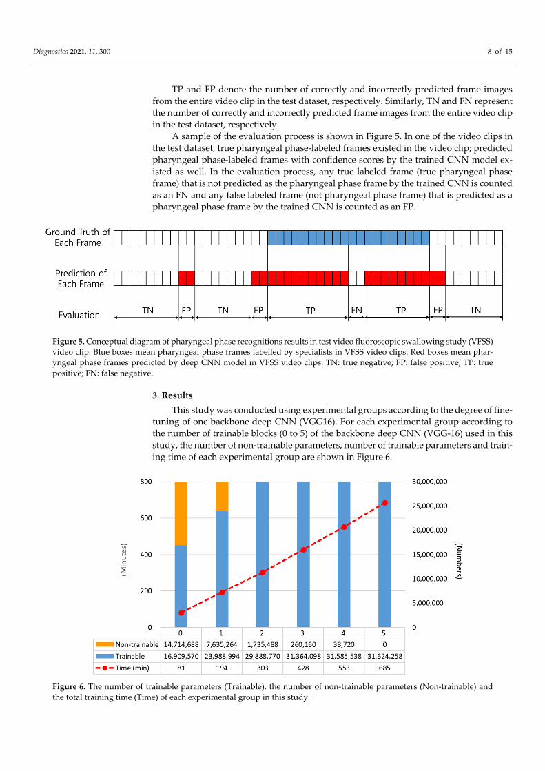
TP and FP denote the number of correctly and incorrectly predicted frame images from the entire video clip in the test dataset, respectively. Similarly, TN and FN represent the number of correctly and incorrectly predicted frame images from the entire video clip in the test dataset, respectively.
A sample of the evaluation process is shown in Figure 5. In one of the video clips in the test dataset, true pharyngeal phase-labeled frames existed in the video clip; predicted pharyngeal phase-labeled frames with confidence scores by the trained CNN model ex-isted as well. In the evaluation process, any true labeled frame (true pharyngeal phase frame) that is not predicted as the pharyngeal phase frame by the trained CNN is counted as an FN and any false labeled frame (not pharyngeal phase frame) that is predicted as a pharyngeal phase frame by the trained CNN is counted as an FP.
Figure 5. Conceptual diagram of pharyngeal phase recognitions results in test video fluoroscopic swallowing study (VFSS) video clip. Blue boxes mean pharyngeal phase frames labelled by specialists in VFSS video clips. Red boxes mean phar-yngeal phase frames predicted by deep CNN model in VFSS video clips. TN: true negative; FP: false positive; TP: true positive; FN: false negative.
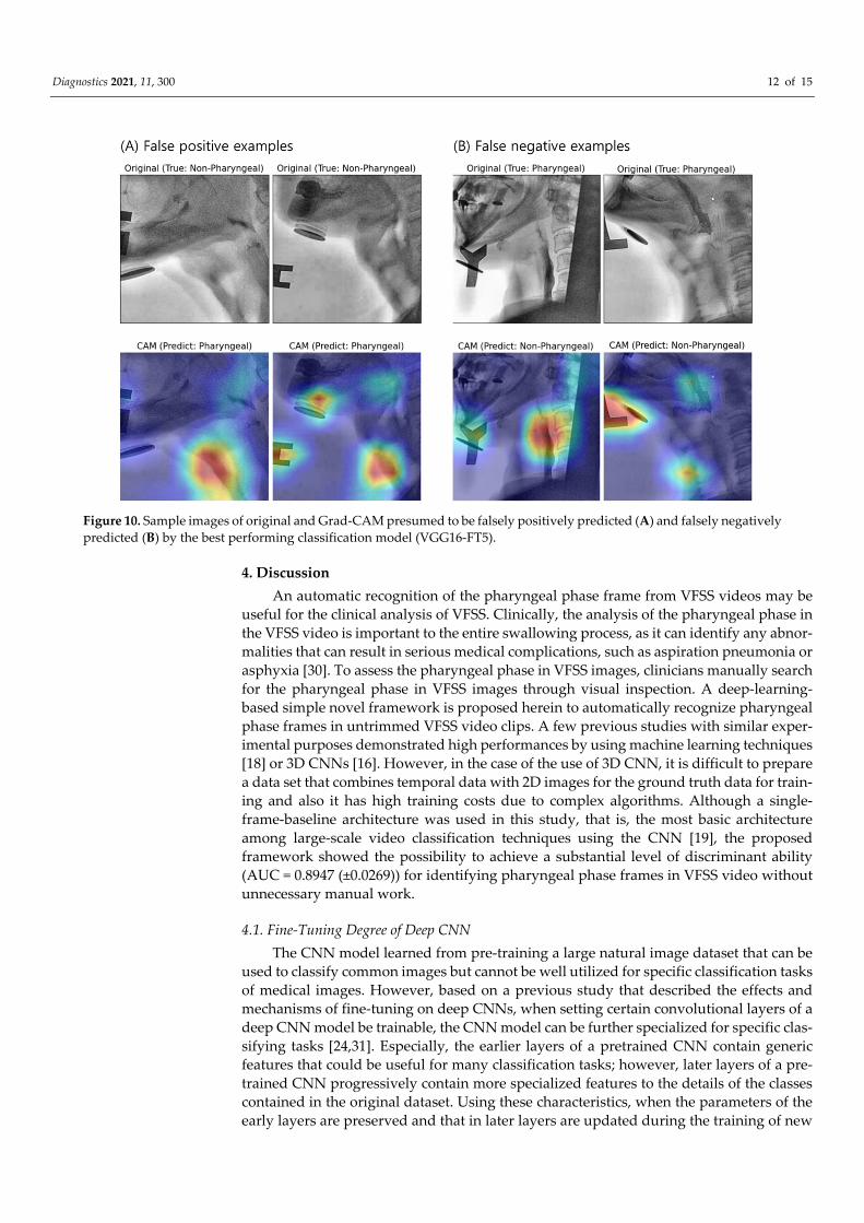
3. Results This study was conducted using experimental groups according to the degree of fine-
tuning of one backbone deep CNN (VGG16). For each experimental group according to the number of trainable blocks (0 to 5) of the backbone deep CNN (VGG-16) used in this study, the number of non-trainable parameters, number of trainable parameters and train-ing time of each experimental group are shown in Figure 6.
Figure 6. The number of trainable parameters (Trainable), the number of non-trainable parameters (Non-trainable) and the total training time (Time) of each experimental group in this study.

Diagnostics 2021, 11, 300 9 of 15
3.1. Classification Performance Table 1, Figures 7 and 8 demonstrate the summarized prediction performance of each
experimental group for recognizing the pharyngeal phase frames in the test VFSS video clips. In particular, Figure 7 depicts the changes of some indexes for model performance, reliability and prediction score according to the numbers of trainable blocks in the deep CNN (VGG-16).
Among all experimental groups, fine-tuned with all blocks of the VGG-16 model (VGG16-FT5) achieved the highest recognizing performance, that is, the accuracy of 93.20 (±1.25)%, sensitivity of 84.57 (±5.19)%, specificity of 94.36 (±1.21)%, FPR of 5.64 (±5.64)%, FNR of 15.43 (±5.19)%, PPV of 67.19 (±4.98)%, NPV of 97.84 (±0.72)%, DOR of 104.9054 (±36.92), AUC of 0.8947 (±0.0269), MCC of 0.716 1 (±0.0482) and Kappa of 0.7093 (±0.0488). All performance metrics values generated through 5-fold cross validation of each experi-mental group are presented in Supplementary Table S1.
Table 1. Performance metrics of experimental groups of this study.
Number of Fine-Tuning Blocks 0 1 2 3 4 5
Accuracy (ACC)
0.8551 (±0.0046)
0.8776 (±0.0214)
0.8918 (±0.0298)
0.9095 (±0.0184)
0.9075 (±0.0176)
0.9320 (±0.0125)
Sensitivity (TPR)
0.0914 (±0.0313)
0.6514 (±0.3040)
0.7429 (±0.2312)
0.8171 (±0.1061)
0.8286 (±0.0833)
0.8457 (±0.0519)
Specificity (TNR)
0.9583 (±0.0079)
0.9081 (±0.0207)
0.9120 (±0.0069)
0.9220 (±0.0129)
0.9181 (±0.0242)
0.9436 (±0.0121)
FPR 0.0417
(±0.0079) 0.0919
(±0.0207) 0.0880
(±0.0069) 0.0780
(±0.0129) 0.0819
(±0.0242) 0.0564
(±0.0121)
FNR 0.9086
(±0.0313) 0.3486
(±0.3040) 0.2571
(±0.2312) 0.1829
(±0.1061) 0.1714
(±0.0833) 0.1543
(±0.0519)
PPV 0.2240 (±0.0437)
0.4580 (±0.1224)
0.5204 (±0.1064)
0.5863 (±0.0576)
0.5835 (±0.0561)
0.6719 (±0.0498)
NPV 0.8864 (±0.0028)
0.9526 (±0.0378)
0.9641 (±0.0308)
0.9740 (±0.0149)
0.9756 (±0.0111)
0.9784 (±0.0072)
DOR 2.29
(±0.64) 32.40
(±23.87) 52.27
(±34.02) 74.00
(±44.04) 63.63
(±20.95) 104.91
(±36.92)
AUC 0.5249 (±0.0127)
0.7798 (±0.1428)
0.8274 (±0.1166)
0.8696 (±0.0546)
0.8734 (±0.0361)
0.8947 (±0.0269)
MCC 0.0739 (±0.0343)
0.4771 (±0.2185)
0.5626 (±0.1803)
0.6432 (±0.0836)
0.6452 (±0.0500)
0.7161 (±0.0482)
Kappa 0.0661
(±0.0323) 0.4616
(±0.2107) 0.5487
(±0.1720) 0.6305
(±0.0796) 0.6297
(±0.0528) 0.7093
(±0.0488)

Diagnostics 2021, 11, 300 10 of 15
Figure 7. The changes of some indexes for model reliability (area under the receiver operating characteristic curve (AUC), Matthews correlation coefficient (MCC) and Kappa) and prediction scores (false positive (FPR), false negative (FNR), positive prediction (PPV) and negative predic-tion (NPV)) according to the numbers of trainable blocks in the deep CNN (VGG-16).
Figure 8. Confusion matrix of best performing classification model (fifth iteration of VGG16-FT5) in this study.

Diagnostics 2021, 11, 300 11 of 15
3.2. Interpretation of Model Decision Using Grad-CAM Figures 9 and 10 show examples of visualized interpretation of predictions using
deep CNN models in this study. In each example, the color heat map present areas that were most affected by the classification of the deep CNN model. Figure 9 shows a repre-sentative example of correctly classified cases for the pharyngeal phase in a VFSS video clip using the VGG16-TF5 CNN model that yielded the best classification performance. Figure 10 shows representative examples of FN and FP classifications, respectively.
Figure 9. Sample frame images of original and gradient-weighted class activation mapping (Grad-CAM) correctly pre-dicted by the best performing classification model (VGG16-FT5) in this study.
Diagnostics 2021, 11, 300 12 of 15
Figure 10. Sample images of original and Grad-CAM presumed to be falsely positively predicted (A) and falsely negatively predicted (B) by the best performing classification model (VGG16-FT5).
4. Discussion An automatic recognition of the pharyngeal phase frame from VFSS videos may be
useful for the clinical analysis of VFSS. Clinically, the analysis of the pharyngeal phase in the VFSS video is important to the entire swallowing process, as it can identify any abnor-malities that can result in serious medical complications, such as aspiration pneumonia or asphyxia [30]. To assess the pharyngeal phase in VFSS images, clinicians manually search for the pharyngeal phase in VFSS images through visual inspection. A deep-learning-based simple novel framework is proposed herein to automatically recognize pharyngeal phase frames in untrimmed VFSS video clips. A few previous studies with similar exper-imental purposes demonstrated high performances by using machine learning techniques [18] or 3D CNNs [16]. However, in the case of the use of 3D CNN, it is difficult to prepare a data set that combines temporal data with 2D images for the ground truth data for train-ing and also it has high training costs due to complex algorithms. Although a single-frame-baseline architecture was used in this study, that is, the most basic architecture among large-scale video classification techniques using the CNN [19], the proposed framework showed the possibility to achieve a substantial level of discriminant ability (AUC = 0.8947 (±0.0269)) for identifying pharyngeal phase frames in VFSS video without unnecessary manual work.
4.1. Fine-Tuning Degree of Deep CNN The CNN model learned from pre-training a large natural image dataset that can be
used to classify common images but cannot be well utilized for specific classification tasks of medical images. However, based on a previous study that described the effects and mechanisms of fine-tuning on deep CNNs, when setting certain convolutional layers of a deep CNN model be trainable, the CNN model can be further specialized for specific clas-sifying tasks [24,31]. Especially, the earlier layers of a pretrained CNN contain generic features that could be useful for many classification tasks; however, later layers of a pre-trained CNN progressively contain more specialized features to the details of the classes contained in the original dataset. Using these characteristics, when the parameters of the early layers are preserved and that in later layers are updated during the training of new

Diagnostics 2021, 11, 300 13 of 15
datasets, the CNN model can be effectively used in new classification tasks. In conclusion, setting the parameters in later layers of pre-trained CNN is trainable through the new dataset can improve the prediction performance and accuracy in the new classification task. This is known as the fin-tuning technique. Although the target medical image and the analysis purpose are different, the results are similar to those of previous studies [32–34] using the transfer learning of a deep CNN via fine-tuning.
As shown in Figure 7, as the trainable parameter increased, model performance (AUC) and model reliability (MCC and Kappa) increased. In particular, it was shown that as the trainable parameter increased, the PPV increased and the FNR decreased, thereby increasing the classification performance of the model. In particular, as the trainable pa-rameter increases, the negative prediction (NPV) or false positive (FPR) hardly changes, whereas the positive prediction (PPV) increases and the false negative (FNR) decreases, thereby increasing the classification performance of the model. Can. This is expected to be due to data imbalance as the number of pharyngeal phase frames among the total number of VFSS video frames is relatively smaller than that of non-pharyngeal phase frames.
4.2. Visual Interpretation Using Grad-CAM Grad-CAM uses the gradient information flowing into the last convolutional layer of
the deep CNN to understand the significance of each neuron for making decisions [35]. For a qualitative evaluation of classification, the Grad-CAM technique was used in this study. In the CNN model, which demonstrated the best classification performance (Figure 9), image feature points for each class were specified for each frame in a VFSS video clip. In particular, it was confirmed that the food mass was automatically recognized as the pharyngeal phase when it was in the pharyngeal cavity. This shows that the CNN auto-matically classifies the pharyngeal phase and the non-pharyngeal phase without prior work, such as object labeling of food bolus in each frame image of VFSS video clips. How-ever, in other swallowing phases (non-swallowing moment, oral phase and esophageal phase), it was classified as a non-pharyngeal phase through the recognition of non-bio-logical markers rather than other biological markers. It is assumed that for images of un-labeled subsets other than the pharyngeal phase, the deep CNN algorithm generated hid-den stratification [36].
When the pharyngeal phase is recognized as a non-pharyngeal phase (False Positive), the pharyngeal cavity was not imaged in the x-ray area as the patient moves and thus it was determined to be a similar part to the pharyngeal cavity (Figure 10A). Even in the pharyngeal phase, when the non-pharyngeal phase (False Negative) was recognized as a large foreign body in the x-ray area, there was a large patient motion, such as a large for-eign body or the jaw being lifted too high unlike other patients (Figure 10B).
4.3. Limitations This study has a limitation in that the sample size estimation method was not applied
as it has only a limited set of data due to the characteristics of medical images that are limited to large-scale data collection. In addition, this study has a limitation in that it does not perform random sampling related to dataset sampling and uses sample of conven-ience that only uses VFSS videos taken at a certain time. The limitation of such a sampling method was mentioned in the previous literature [37], such as an inability to generalize the results of the survey to the population as a whole. Therefore, there is the possibility of under-or over-representation of the population in this study.
5. Conclusions A single-frame-baseline-architecture-based simple CNN for recognizing pharyngeal
phase in untrimmed VFSS video clips is presented here and the following conclusions are drawn. When using deep CNNs for recognizing the pharyngeal phase in VFSS video clips, by applying transfer learning technique to a deep CNN for classification, an appropriate

Diagnostics 2021, 11, 300 14 of 15
fine-tuning degree was required. In addition, in the case of image classification using a deep CNN, classification must be evaluated qualitatively using visual interpretation methods such as the Grad-CAM technique to identify whether an appropriate classifica-tion has occurred based on the correct reason. The single-frame-baseline-architecture-based simple CNN using the factors above demonstrated the possibility of yielding high performances in the full automation of VFSS video analysis. However, this study was con-ducted based on images taken only in one institution and may have limitations compared to studies using multi-center and multiple imaging devices.
Supplementary Materials: The following are available online at www.mdpi.com/2075-4418/11/2/300/s1, Table S1: Performance metrics of experimental groups of this study.
Author Contributions: Conceptualization, K.-S.L., E.L. and S.B.-P.; Data curation, E.L. and B.C.; Formal analysis, K.-S.L. and E.L.; Funding acquisition and S.B.-P.; Investigation, K.-S.L., E.L. and B.C.; Methodology, K.-S.L.; Project administration and S.B.-P.; Resources, E.L. and S.B.-P.; Software, K.-S.L.; Supervision and S.B.-P.; Validation, K.-S.L. and S.B.-P.; Visualization, K.-S.L.; Writing—orig-inal draft, K.-S.L. and E.L.; Writing—review & editing and S.B.-P. All authors have read and agreed to the published version of the manuscript.
Funding: This research was supported by the Basic Science Research Program through the National Research Foundation of Korea (NRF) funded by the Ministry of Education (NRF-2019R1I1A1A01062961) and a Korea University Grant (K2008481).
Institutional Review Board Statement: The study was conducted according to the guidelines of the Declaration of Helsinki and approved by the Institutional Review Board of the Korea University Medical Center (IRB No. 2021AN0019) and carried out according to the guide-lines of the commit-tee.
Informed Consent Statement: Patient consent was waived due to this study was designed as retro-spective study.
Conflicts of Interest: The authors declare no conflict of interest. The funders had no role in the design of the study; in the collection, analyses or interpretation of data; in the writing of the manu-script or in the decision to publish the results.
References 1. Wieseke, A.; Bantz, D.; Siktberg, L.; Dillard, N. Assessment and early diagnosis of dysphagia. Geriatr. Nurs. 2008, 29, 376–383. 2. Gordon, C.; Hewer, R.L.; Wade, D.T. Dysphagia in acute stroke. Br. Med. J. Clin. Res. Ed. 1987, 295, 411–414. 3. Horiguchi, S.; Suzuki, Y. Screening tests in evaluating swallowing function. JMAJ 2011, 54, 31–34. 4. Moraes, D.; Sassi, F.; Mangilli, L.; Zilberstein, B.; De Andrade, C. Clinical prognostic indicators of dysphagia following
prolonged orotracheal intubation in ICU patients. Crit. Care 2013, 17, R243. 5. Heffner, J.E. Swallowing complications after endotracheal extubation: Moving from “whether” to “how”. Chest 2010, 137, 509–
510. 6. Macht, M.; Wimbish, T.; Bodine, C.; Moss, M. ICU-acquired swallowing disorders. Crit. Care Med. 2013, 41, 2396–2405. 7. Martino, R.; Pron, G.; Diamant, N. Screening for oropharyngeal dysphagia in stroke: Insufficient evidence for guidelines.
Dysphagia 2000, 15, 19–30. 8. Cook, I.J.; Kahrilas, P.J. AGA technical review on management of oropharyngeal dysphagia. Gastroenterology 1999, 116, 455–478. 9. Palmer, J.B.; Kuhlemeier, K.V.; Tippett, D.C.; Lynch, C. A protocol for the videofluorographic swallowing study. Dysphagia
1993, 8, 209–214. 10. Shem, K.L.; Castillo, K.; Wong, S.L.; Chang, J.; Kao, M.C.; Kolakowsky-Hayner, S.A. Diagnostic accuracy of bedside swallow
evaluation versus videofluoroscopy to assess dysphagia in individuals with tetraplegia. PMR 2012, 4, 283–289. 11. Zhang, Z.; Coyle, J.L.; Sejdić, E. Automatic hyoid bone detection in fluoroscopic images using deep learning. Sci. Rep. 2018, 8,
1–9. 12. McCullough, G.H.; Wertz, R.T.; Rosenbek, J.C.; Mills, R.H.; Webb, W.G.; Ross, K.B. Inter-and intrajudge reliability for
videofluoroscopic swallowing evaluation measures. Dysphagia 2001, 16, 110–118. 13. Han, H.; Shin, G.; Jun, A.; Park, T.; Ko, D.; Choi, E.; Kim, Y. The relation between the presence of aspiration or penetration and
the clinical indicators of dysphagia in poststroke survivors. Ann. Rehabil. Med. 2016, 40, 88. 14. Tran, D.; Bourdev, L.; Fergus, R.; Torresani, L.; Paluri, M. Learning spatiotemporal features with 3d convolutional networks. In
Proceedings of the IEEE International Conference on Computer Vision, Las Condes, Chile, 11–18 December 2015; pp. 4489–4497.

Diagnostics 2021, 11, 300 15 of 15
15. Zhang, L.; Zhu, G.; Shen, P.; Song, J.; Afaq Shah, S.; Bennamoun, M. Learning spatiotemporal features using 3dcnn and convolutional lstm for gesture recognition. In Proceedings of Proceedings of the IEEE International Conference on Computer Vision Workshops, Venice, Italy, 22–29 October 2017; pp. 3120-3128.
16. Lee, J.T.; Park, E.; Jung, T.-D. Automatic detection of the pharyngeal phase in raw videos for the videofluoroscopic swallowing study using efficient data collection and 3d convolutional networks. Sensors 2019, 19, 3873.
17. Xiong, Y.; Zhao, Y.; Wang, L.; Lin, D.; Tang, X. A pursuit of temporal accuracy in general activity detection. arXiv Prepr. 2017, arXiv:1703.02716.
18. Lee, J.T.; Park, E.; Hwang, J.-M.; Jung, T.-D.; Park, D. Machine learning analysis to automatically measure response time of pharyngeal swallowing reflex in videofluoroscopic swallowing study. Sci. Rep. 2020, 10, 1–9.
19. Karpathy, A.; Toderici, G.; Shetty, S.; Leung, T.; Sukthankar, R.; Fei-Fei, L. Large-scale video classification with convolutional neural networks. In Proceedings of the IEEE Conference on Computer Vision and Pattern Recognition, Columbus, OH, USA, 23–28 June 2014; pp. 1725–1732.
20. Valueva, M.V.; Nagornov, N.; Lyakhov, P.A.; Valuev, G.V.; Chervyakov, N.I. Application of the residue number system to reduce hardware costs of the convolutional neural network implementation. Math. Comput. Simul. 2020, 177, 232–243.
21. Simonyan, K.; Zisserman, A. Very deep convolutional networks for large-scale image recognition. arXiv Prepr. 2014, arXiv:1409.1556.
22. Litjens, G.; Kooi, T.; Bejnordi, B.E.; Setio, A.A.A.; Ciompi, F.; Ghafoorian, M.; Van Der Laak, J.A.; Van Ginneken, B.; Sánchez, C.I. A survey on deep learning in medical image analysis. Med Image Anal. 2017, 42, 60–88.
23. Yosinski, J.; Clune, J.; Bengio, Y.; Lipson, H. How transferable are features in deep neural networks? In Proceedings of Advances in Neural Information Processing Systems, Montreal, QC, Canada, 8–13 December 2014; pp. 3320–3328.
24. Pan, S.J.; Yang, Q. A survey on transfer learning. IEEE Trans. Knowl. Data Eng. 2009, 22, 1345–1359. 25. Stone, M. Cross-validatory choice and assessment of statistical predictions. J. R. Stat. Soc. Ser. B Methodol. 1974, 36, 111–133. 26. Chollet, F. Keras: The python deep learning library. Available online: https://ui.adsabs.harvard.edu/abs/2018ascl.soft06022C
(accessed on 14 November 2020). 27. Abadi, M.; Agarwal, A.; Barham, P.; Brevdo, E.; Chen, Z.; Citro, C.; Corrado, G.S.; Davis, A.; Dean, J.; Devin, M. Tensorflow:
Large-scale machine learning on heterogeneous distributed systems. arXiv Prepr. 2016, arXiv:1603.04467. 28. Ott, D.J.; Pikna, L.A. Clinical and videofluoroscopic evaluation of swallowing disorders. AJR. Am. J. Roentgenol. 1993, 161, 507–
513. 29. Rademaker, A.W.; Pauloski, B.R.; Logemann, J.A.; Shanahan, T.K. Oropharyngeal swallow efficiency as a representative
measure of swallowing function. J. Speech Lang. Hear. Res. 1994, 37, 314–325. 30. Ertekin, C.; Aydogdu, I. Neurophysiology of swallowing. Clin. Neurophysiol. 2003, 114, 2226–2244. 31. Tajbakhsh, N.; Shin, J.Y.; Gurudu, S.R.; Hurst, R.T.; Kendall, C.B.; Gotway, M.B.; Liang, J. Convolutional neural networks for
medical image analysis: Full training or fine tuning? IEEE Trans. Med Imaging 2016, 35, 1299–1312. 32. Xiao, T.; Liu, L.; Li, K.; Qin, W.; Yu, S.; Li, Z. Comparison of transferred deep neural networks in ultrasonic breast masses
discrimination. BioMed Res. Int. 2018, 2018, 4605191. 33. Lee, K.-S.; Jung, S.-K.; Ryu, J.-J.; Shin, S.-W.; Choi, J. Evaluation of Transfer Learning with Deep Convolutional Neural Networks
for Screening Osteoporosis in Dental Panoramic Radiographs. J. Clin. Med. 2020, 9, 392. 34. Lee, K.-S.; Kim, J.Y.; Jeon, E.-T.; Choi, W.S.; Kim, N.H.; Lee, K.Y. Evaluation of Scalability and Degree of Fine-Tuning of Deep
Convolutional Neural Networks for COVID-19 Screening on Chest X-ray Images Using Explainable Deep-Learning Algorithm. J. Pers. Med. 2020, 10, 213.
35. Selvaraju, R.R.; Cogswell, M.; Das, A.; Vedantam, R.; Parikh, D.; Batra, D. Grad-cam: Visual explanations from deep networks via gradient-based localization. In Proceedings of the IEEE International Conference on Computer Vision, Venice, Italy, 22–29 October 2017; pp. 618–626.
36. Oakden-Rayner, L. Exploring large-scale public medical image datasets. Acad. Radiol. 2020, 27, 106–112. 37. Jager, J.; Putnick, D.L.; Bornstein, M.H., II. More than just convenient: The scientific merits of homogeneous convenience
samples. Monogr. Soc. Res. Child. Dev. 2017, 82, 13–30.