Embed Size (px)
Citation preview
ARTÍCULO DE INVESTIGACIÓN REVISTA MEXICANA DE
INGENIERÍA BIOMÉDICAibVol. 36, No. 2, May-Ago 2015, pp. 121-129
dx.doi.org/10.17488/RMIB.36.2.4pdf
Automatic Segmentation of the Cerebellumin Ultrasound Volumes of the Fetal Brain
G. Velásquez Rodríguez1, F. Arámbula Cosío1, M.E. Guzmán Huerta2, L. Camargo Marín2, H.Borboa Olivares2, Boris Escalante Ramírez3
1Laboratorio de Imagenología Biomédica, Centro de Ciencias Aplicadas y Desarrollo Tecnológico, UniversidadNacional Autónoma de México (UNAM).2Instituto Nacional de Perinatología, Ciudad de México.3Departamento de Procesamiento de Señales, Facultad de Ingeniería, Universidad Nacional Autónoma de México(UNAM).
ABSTRACTThe size of the cerebellum in ultrasound volumes of the fetal brain has shown a high correlation with gestationalage, which makes it a valuable feature to detect fetal growth restrictions. Manual annotation of the 3D surface of thecerebellum in an ultrasound volume is a time consuming task, which needs to be performed by a highly trained expert.In order to assist the experts in the evaluation of cerebellar dimensions, we developed an automatic scheme for thesegmentation of the 3D surface of the cerebellum in ultrasound volumes, using a spherical harmonics model. In thiswork we present our validation results on 10 ultrasound volumes in which we have obtained an adequate accuracyin the segmentation of the cerebellum (mean Dice coefficient of 0.689). The method reported shows potential toeffectively assist the experts in the assessment of fetal growth in ultrasound volumes.Keywords: 3D fetal ultrasound segmentation, fetal cerebellum, statistical shape models, sphericalharmonics
Correspondencia:Fernando Arámbula CosíoCorreo electrónico: [email protected]
Fecha de recepción:15 de enero de 2015
Fecha de aceptación:20 de abril de 2015
122 Revista Mexicana de Ingeniería Biomédica · volumen 36 · número 2 · May-Ago, 2015
RESUMENEl tamaño del cerebelo, en un volumen de ultrasonido del cerebro fetal, ha mostrado una alta correlación con laedad gestacional, lo que hace importante a esta medición para la detección de restricciones del crecimiento del feto.La anotación manual de la superficie 3D del cerebelo en un volumen de ultrasonido es una tarea demandante, quedebe ser realizada por un experto. Con el propósito de apoyar a los expertos en la evaluación de las dimensionesdel cerebelo fetal, hemos desarrollado un método automático para la segmentación de la superficie 3D del cerebeloen volúmenes de ultrasonido, utilizando un modelo de harmónicos esféricos (spherical harmonics). En este trabajopresentamos los resultados de una evaluación del método automático en 10 volúmenes de ultrasonido con los quehemos obtenido un valor adecuado de exactitud (coeficiente promedio de Dice de 0.689). El método reportado tienepotencial para asistir de manera efectiva a los expertos en la evaluación del crecimiento fetal, utilizando volúmenesde ultrasonido.Palabras clave: segmentación 3D de ultrasonido fetal, cerebelo fetal, modelos estadísticos de formas,harmónicos esféricos.
INTRODUCTION
Cerebellar volumes obtained by multiplanar,and the Virtual Organ Computer AidedAnalysis (VOCAL) techniques have showna high correlation with gestational ageand might have statistically significantdifferences between different ethnic groups[1]. Cerebellar volumes evaluated bymultiplanar and VOCAL techniques haveshown differences between growth restrictedfetuses and also adequacy for the assessmentof the gestational age [2]. In [3],and [4] is evaluated the growth of thecerebellum in normal pregnancy, using themultiplanar technique. Both techniques(VOCAL and multiplanar evaluation) areuseful for fetal evaluation of brain structures,however both have the necessity of manualdelineation by an experienced operator,which is considered a disadvantage respectto automated segmentation techniques dueto the low reproducibility and agreementobtained by manual segmentation techniques.In order to ease the task for the experts and,more importantly, to contribute to improvedreproducibility in the measurement of thecerebellar volume, we have developed two
different schemes for automatic segmentationof the cerebellum in ultrasound volumes.
Previous work on the automaticsegmentation of fetal brain structures hasbeen reported in Yaqub et al., [12] were arandom decision forest classifier was trainedto automatically segment: the choroid plexus,the lateral posterior ventricle cavity, thecavum septum pellucidi, and the cerebellumin 3D ultrasound. A mean Dice coefficient of0.63 was reported for the segmentation of thecerebellum.
In Gutierrez et al., [5] was reportedthe development of a point distributionmodel (PDM) of the cerebellum which isautomatically adjusted to an ultrasoundvolume. The model is able to automaticallysegment the 3D surface of the cerebellumwith good accuracy (mean Dice coefficientof 0.8) with a mean processing time of 50s. Although good results have been achievedwith the PDM, in this work we explore the useof a spherical harmonic (SPHARM) modelof the cerebellum for automatic segmentationof the structure in ultrasound volumes.SPHARMs have methodological advantagesfor the construction of statistical deformablemodels of 3D shapes: There is no need
Velásquez Rodríguez et al. Automatic segmentation of the cerebellum in ultrasound volumes of the fetal brain 123
for extensive annotation of landmark points;accurate shape modeling is achieved throughprincipal components analysis of a set ofnormalized 3D shape examples. In the restof this work is reported the developmentof our SPHARM of the cerebellum and itsuse for the automatic segmentation of thestructure in ultrasound volumes. In section 2we report the construction of the SPHARMof the cerebellum trained from a set of 9annotated ultrasound volumes. In section 3we describe the method used to automaticallyadjust the SPHARM model to an ultrasoundvolume. In section 4 are reported the resultsfrom automatic adjustment of the SPHARMof the cerebellum to a set of 10 different non-training ultrasound volumes, using the leave-one-out method. In section 5 we presentthe discussion and conclusions of the workreported.
SPHARM MODEL OF THECEREBELLUM
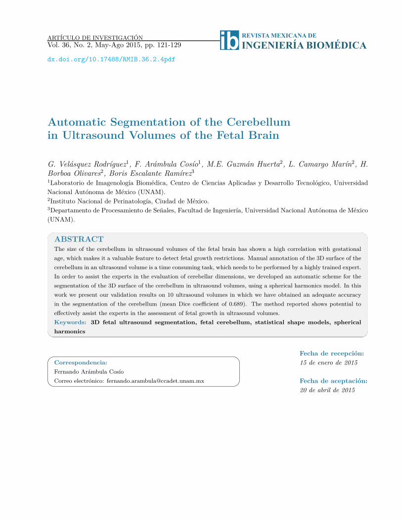
Our spherical harmonics (SPHARM) modelof the cerebellum was trained with 9cerebellum shapes annotated by an expert inthe corresponding ultrasound volumes. Eachshape was discretized as shown in Fig. 1.
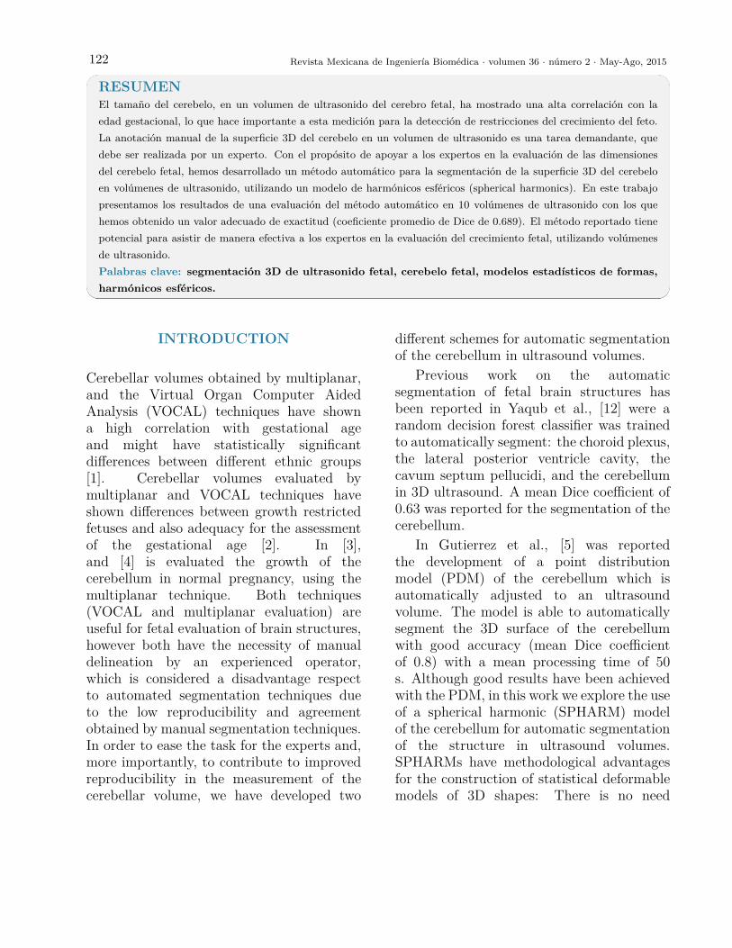
The corresponding triangular mesh wasgenerated for each shape using marchingcubes [6]. An example of a mesh is shownin Fig.2.
In order to build the SPHARM model,each mesh of the training set is modeled withspherical harmonic functions as follows [7]:
2
In Gutierrez et al., [5] was reported the development of a point distribution model (PDM) of the cerebellum which is automatically adjusted to an ultrasound volume. The model is able to automatically segment the 3D surface of the cerebellum with good accuracy (mean Dice coefficient of 0.8) with a mean processing time of 50 s. Although good results have been achieved with the PDM, in this work we explore the use of a spherical harmonic (SPHARM) model of the cerebellum for automatic segmentation of the structure in ultrasound volumes. SPHARMs have methodological advantages for the construction of statistical deformable models of 3D shapes: There is no need for extensive annotation of landmark points; accurate shape modeling is achieved through principal components analysis of a set of normalized 3D shape examples. In the rest of this work is reported the development of our SPHARM of the cerebellum and its use for the automatic segmentation of the structure in ultrasound volumes. In section 2 we report the construction of the SPHARM of the cerebellum trained from a set of 9 annotated ultrasound volumes. In section 3 we describe the method used to automatically adjust the SPHARM model to an ultrasound volume. In section 4 are reported the results from automatic adjustment of the SPHARM of the cerebellum to a set of 10 different non-training ultrasound volumes, using the leave-one-out method. In section 5 we present the discussion and conclusions of the work reported.
2 SPHARM MODEL OF THE CEREBELLUM
Our spherical harmonics (SPHARM) model of the cerebellum was trained with 9 cerebellum
shapes annotated by an expert in the corresponding ultrasound volumes. Each shape was discretized as shown in Fig. 1.
Fig. 1. Discretized cerebellum example The corresponding triangular mesh was generated for each shape using marching cubes [6]. An
example of a mesh is shown in Fig.2.
Figure 1. Discretized cerebellum example.
3
Fig.2. Triangular mesh of a cerebellum example In order to build the SPHARM model, each mesh of the training set is modeled with spherical
harmonic functions as follows [7]: Spherical harmonic functions of order $m$ and degree $l$: $Y_l^m$, $-l\le m \le l$ are defined in
$\theta \in [0,\pi]$, $\varphi\in [0,2\pi )$ as:
𝑌!! 𝜃,𝜑 = !!!!!!
!!! !!!! !
𝑃!! cos 𝜃 𝑒!"# (1)
where: $P_l^m$ is the associated Legendre polynomial:
𝑃!! 𝑤 = !! !
!!!! (1 − 𝑤!)
!! !!!!
!"!!! (𝑤! − 1)! (2) where: $w$ is $\cos(\theta )$
To define a surface three SPHARM functions are used, one for each coordinate of the points in the surface:
𝑣 𝜃,𝜑 = (𝑥( 𝜃,𝜑), 𝑦( 𝜃,𝜑), 𝑧( 𝜃,𝜑) )! (3)
The surface is then described by the vector valued function:
𝑣 𝜃,𝜑 = 𝑐!!𝑌!! 𝜃,𝜑!
!!!!
!"#$
!!!
(4)
where the coefficients are 3D vectors corresponding to the 3 coordinate functions in (3):
Figure 2. Triangular mesh of a cerebellum example.
Spherical harmonic functions of order mand degree l: Y m
l , −l ≤ m ≤ l are defined inθ ∈ [0, π], ϕ ∈ [0, 2π) as:
Y ml (θ, ϕ) =
√√√√2l + 14π
(l −m)!(l +m)!)P
ml (cos(θ))eimϕ
(1)
where: Pml is the associated Legendre
polynomial:
Pml (w) = (−1)m
2ll! (1− w2)m2dm+1
dwm+1 (w2 − 1)l
(2)
where: w is cos(θ).To define a surface three SPHARM
functions are used, one for each coordinateof the points in the surface:
v(θ, ϕ) = (x(θ, ϕ), y(θ, ϕ), z(θ, ϕ))T (3)
The surface is then described by the vector-valued function:
v(θ, ϕ) =Lmax∑l=0
l∑m=−l
cml Yml (θ, ϕ) (4)
where the coefficients are 3D vectorscorresponding to the 3 coordinate functionsin (3):
cml = (clxm, clym, clzm)T (5)
The coefficients are obtained using a leastsquares adjustment to a given shape mesh,as described in [7].
124 Revista Mexicana de Ingeniería Biomédica · volumen 36 · número 2 · May-Ago, 2015
4
𝑐!! = (𝑐!"! , 𝑐!"! , 𝑐!"!)! (5)
The coefficients are obtained using a least squares adjustment to a given shape mesh, as described in [7]. Using its decomposition in spherical harmonics we can model the 3D surface corresponding to
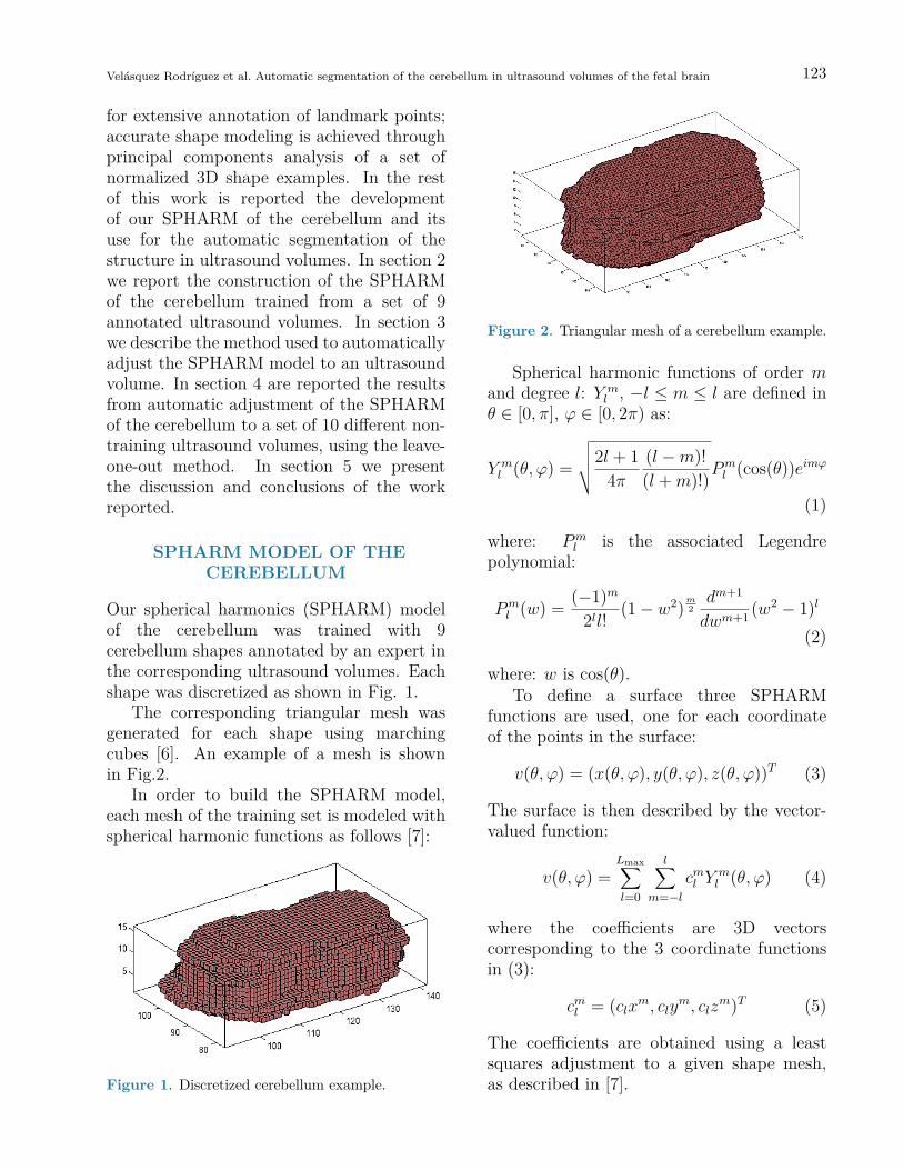
each cerebellum volume in the training set. In Fig. 3 is illustrated an SPHARM model of one cerebellum, we can observe how shape details increase with the degree ($L_{\max}$) of the SPHARM.
Fig.3. SPHARM of the cerebellum for an increasing degree ($L_{\max}$): A) $L_{\max}$ =1; B) $L_{\max}$ = 7; C) $L_{\max}$ =15.
Once we have a set of SPHARM coefficients for each shape of the cerebellum training set, a
principal component analysis (PCA) of the shape parameters is performed through registration of all
A B
C
Figure 3. SPHARM of the cerebellum for an increasing degree (Lmax): A) Lmax =1; B) Lmax = 7; C) Lmax
=15.
Using its decomposition in sphericalharmonics we can model the 3D surfacecorresponding to each cerebellum volume inthe training set. In Fig. 3 is illustrated anSPHARM model of one cerebellum, we canobserve how shape details increase with thedegree (Lmax) of the SPHARM.
Once we have a set of SPHARMcoefficients for each shape of the cerebellumtraining set, a principal component analysis(PCA) of the shape parameters is performedthrough registration of all SPHARM models,using a few corresponding landmark points oneach shape [7], for the cerebellum we used 6landmark points. The Iterative Closest Pointmethod [11] is used to register the shapes inEuclidean space, followed by registration inparameter space through minimization of the
Root Mean Square Distance (RMSD) [7]:
RMSD =
√√√√14π
Lmax∑L=0
l∑m=−1
‖c1− c2‖2 (6)
where: c1 and c2 are two SPHARMparameterizations to be registered.
Principal Component Analysis isperformed on the parameters (ci, eq.5) of allthe examples in the registered training set,to calculate the mean shape and the mainmodes of shape variation. From which it ispossible to generate new shapes of the classof the training set (i.e. cerebellums) usingEq. 7.
Sα = Sµ +M∑i=1
αiSi (7)
where: Sµ is the mean shape.Si, are the principal component vectors
Velásquez Rodríguez et al. Automatic segmentation of the cerebellum in ultrasound volumes of the fetal brain 125
of the training set and αi are the shapeadjustment parameters, with each αi withinthe range:
3√λi < αi < 3
√λi
λi is the corresponding eigenvalue of eachprincipal component Si.
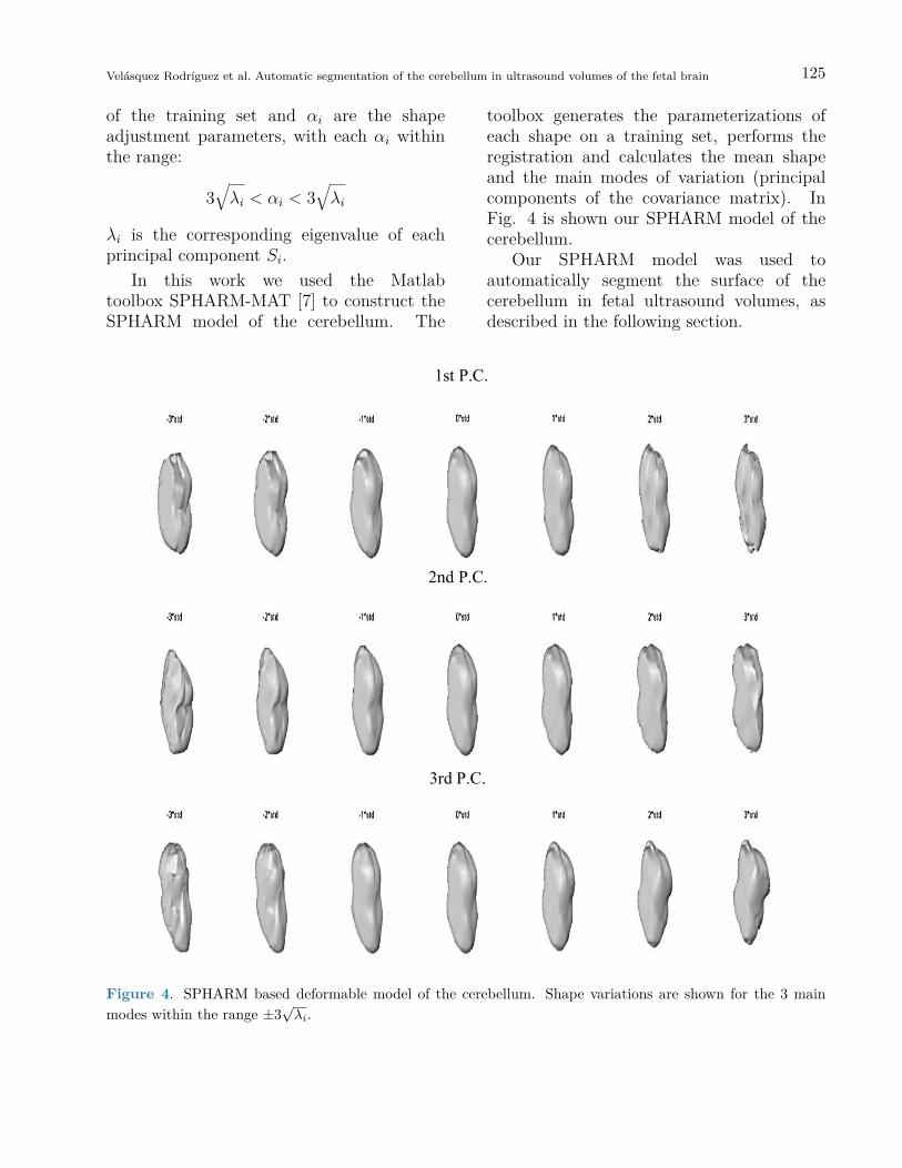
In this work we used the Matlabtoolbox SPHARM-MAT [7] to construct theSPHARM model of the cerebellum. The
toolbox generates the parameterizations ofeach shape on a training set, performs theregistration and calculates the mean shapeand the main modes of variation (principalcomponents of the covariance matrix). InFig. 4 is shown our SPHARM model of thecerebellum.
Our SPHARM model was used toautomatically segment the surface of thecerebellum in fetal ultrasound volumes, asdescribed in the following section.
6
Fig. 4. SPHARM based deformable model of the cerebellum. Shape variations are shown for the 3 main
modes within the range $\pm 3 \sqrt{\lambda_i}$ Our SPHARM model was used to automatically segment the surface of the cerebellum in fetal ultrasound volumes, as described in the following section. 3 AUTOMATIC ADJUSTMENT OF THE SPHARM MODEL TO THE CEREBELLUM IN AN ULTRASOUND VOLUME
1st P.C.
2nd P.C.
3rd P.C.
Figure 4. SPHARM based deformable model of the cerebellum. Shape variations are shown for the 3 mainmodes within the range ±3
√λi.
126 Revista Mexicana de Ingeniería Biomédica · volumen 36 · número 2 · May-Ago, 2015
AUTOMATIC ADJUSTMENT OFTHE SPHARM MODEL TO THE
CEREBELLUM IN ANULTRASOUND VOLUME
We used the method reported by Ahmadi etal [8] for the automatic adjustment of ourSPHARM model to the fetal cerebellum inan ultrasound volume. The method is basedon the optimization of the following local(gray level) energy function, through gradientdescent.
E(S) =∫
int S
fidx+∫
ext S
fedx (8)
where:
fi = (I − ci(x)2), where (9)
ci(x) =∫int S Bε(x)I(x)dx∫int S Bε(x)dx
fe = (I − ce(x)2), where (10)
ce(x) =∫ext S Bε(x)I(x)dx∫ext S Bε(x)dx
Bε is an sphere of radius ε used to calculatethe values ci and ce for each position x of I.The function E(S) is minimum at the surfacewhich separates two homogeneous regionswith significantly different mean grey levels.The gradient of E(S) is given by Eq. 11.
∂
∂αjE(Sα) = ∂E
∂S
∂S
∂αj=
∫S(fi − fe)N · Sjds
(11)where: N is surface normal and Sj the vertex-wise cartesian deformations for the j-th shapeparameter.
Together, these partial derivatives yieldthe gradient of E with respect to the shapevector α, which we denote by ∇αE .Thecorresponding discrete equation is:∫S(fi − fe)NSjds ≈
N∑k=1
[fi − fe]k[N ]k · [Sj]k
(12)
where: k denotes the vertex number and [·]k
denotes the evaluation at vertex k.Eq. 12 is evaluated for all the vertices of
the mesh to find optimum values of the shapeparameters (αi) - which accurately fit thecerebellum in an ultrasound volume - throughgradient descent.
TESTS AND RESULTS
We used 10 different ultrasound volumesacquired in an axial plane using a Voluson730 Expert from General Electric, with a 4-8MHz 3D probe. All volumes were acquiredwith informed consent of the patients atthe National Institute of Perinatology inMéxico City. The cerebellum was annotatedon 10 corresponding volumes by an expertsonographer.
The automatic segmentation of thecerebellum was evaluated on the trainingset using the leave-one-out method. Ninevolumes were used for training the SPHARMof the cerebellum with validation ofautomatic segmentation on the volume leftout. This was repeated 10 times, validatingthe automatic segmentation on each of the 10fetal ultrasound volumes. The Dice SimilarityCoefficient (DSC) [9] was used to measurethe accuracy of the automatic SPHARMsegmentation as compared against manualexpert annotations.
DSC = 2TP(2TP + FP + FN) (13)
where: TP corresponds to the number of truecerebellum voxels,
FP corresponds to the number of voxelswrongly included as cerebellum and
FN corresponds to the number ofcerebellum voxels wrongly left out of thesegmentation.
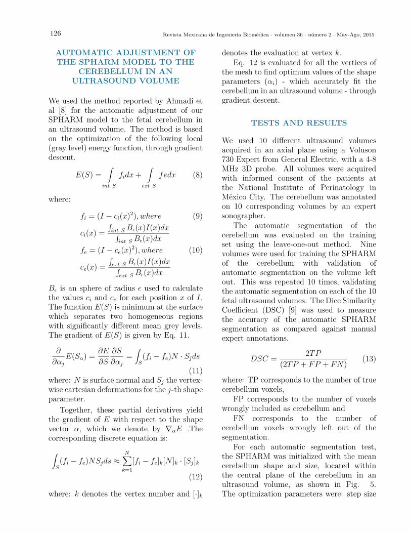
For each automatic segmentation test,the SPHARM was initialized with the meancerebellum shape and size, located withinthe central plane of the cerebellum in anultrasound volume, as shown in Fig. 5.The optimization parameters were: step size
Velásquez Rodríguez et al. Automatic segmentation of the cerebellum in ultrasound volumes of the fetal brain 127
8
automatic segmentation on the volume left out. This was repeated 10 times, validating the automatic segmentation on each of the 10 fetal ultrasound volumes. The Dice Similarity Coefficient (DSC) [9] was used to measure the accuracy of the automatic SPHARM segmentation as compared against manual expert annotations.
𝐷𝑆𝐶 =2 𝑇𝑃
(2𝑇𝑃 + 𝐹𝑃 + 𝐹𝑁) (13)
where: TP corresponds to the number of true cerebellum voxels, FP corresponds to the number of voxels wrongly included as cerebellum and FN corresponds to the number of cerebellum voxels wrongly left out of the segmentation.
For each automatic segmentation test, the SPHARM was initialized with the mean cerebellum
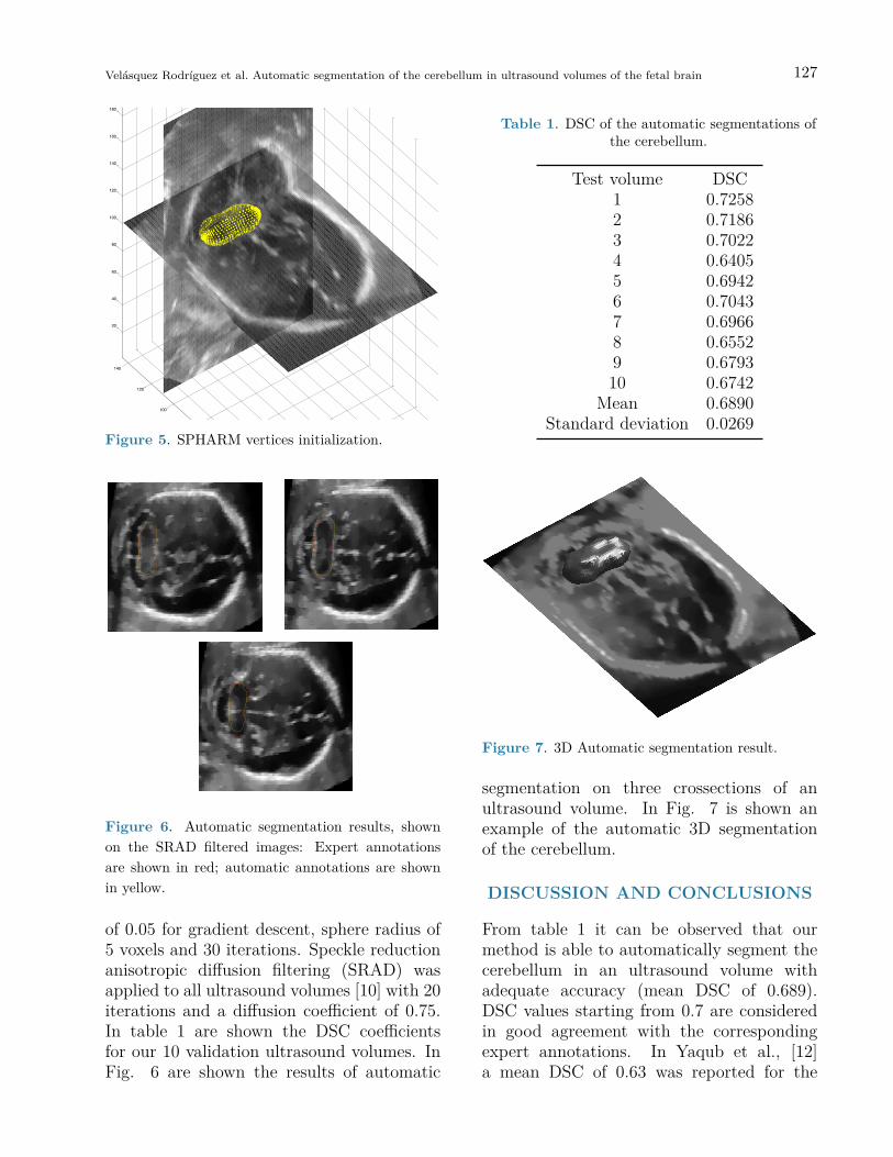
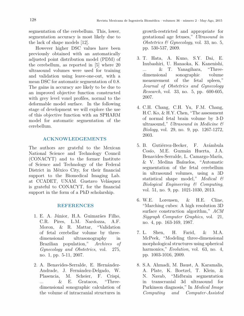
shape and size, located within the central plane of the cerebellum in an ultrasound volume, as shown in Fig. 5. The optimization parameters were: step size of 0.05 for gradient descent, sphere radius of 5 voxels and 30 iterations. Speckle reduction anisotropic diffusion filtering (SRAD) was applied to all ultrasound volumes [10] with 20 iterations and a diffusion coefficient of 0.75. In table 1 are shown the DSC coefficients for our 10 validation ultrasound volumes. In Fig. 6 are shown the results of automatic segmentation on three crossections of an ultrasound volume. In Fig. 7 is shown an example of the automatic 3D segmentation of the cerebellum.
Fig. 5. SPHARM vertices initialization
0
10
20
30
40
50
60
70
80
90
100
20
40
60
80
100
120
140
20
40
60
80
100
120
140
160
180
Figure 5. SPHARM vertices initialization.
10
Fig. 6 Automatic segmentation results, shown on the SRAD filtered images: Expert annotations
are shown in red; automatic annotations are shown in yellow.
Figure 6. Automatic segmentation results, shownon the SRAD filtered images: Expert annotationsare shown in red; automatic annotations are shownin yellow.
of 0.05 for gradient descent, sphere radius of5 voxels and 30 iterations. Speckle reductionanisotropic diffusion filtering (SRAD) wasapplied to all ultrasound volumes [10] with 20iterations and a diffusion coefficient of 0.75.In table 1 are shown the DSC coefficientsfor our 10 validation ultrasound volumes. InFig. 6 are shown the results of automatic
Table 1. DSC of the automatic segmentations ofthe cerebellum.
Test volume DSC1 0.72582 0.71863 0.70224 0.64055 0.69426 0.70437 0.69668 0.65529 0.679310 0.6742
Mean 0.6890Standard deviation 0.0269
11
Figure 7. 3D Automatic segmentation result.
5 DISCUSSION AND CONCLUSIONS
From table 1 it can be observed that our method is able to automatically segment the cerebellum in
an ultrasound volume with adequate accuracy (mean DSC of 0.689). DSC values starting from 0.7 are considered in good agreement with the corresponding expert annotations. In Yaqub et al., [12] a mean DSC of 0.63 was reported for the segmentation of the cerebellum. This, lower, segmentation accuracy is most likely due to the lack of shape models [12].
However higher DSC values have been previously obtained with an automatically adjusted point
distribution model (PDM) of the cerebellum, as reported in [5] where 20 ultrasound volumes were used for training and validation using leave-one-out, with a mean DSC for automatic segmentation of 0.8. The gains in accuracy are likely to be due to an improved objective function constructed with grey level voxel profiles, normal to the deformable model surface. In the following stage of development we will explore the use of this objective function with an SPHARM model for automatic segmentation of the cerebellum.
ACKNOWLEDGEMENTS
The authors are grateful to the Mexican National Science and Technology Council (CONACYT) and to the former Institute of Science and Technology of the Federal District in México City, for their financial support to the Biomedical Imaging Lab. at CCADET, UNAM. Gustavo Velásquez is grateful to CONACYT, for the financial support in the form of a PhD scholarship.
REFERENCES
\item E. A. Júnior, H.A. Guimarães Filho, C.R. Pires, L.M. Nardozza, A.F. Moron, \& R. Mattar, ``Validation of fetal cerebellar volume by three-dimensional ultrasonography in Brazilian population,’’ {\it Archives of Gynecology and Obstetrics}, vol. 275, no. 1, pp. 5-11, 2007.
Figure 7. 3D Automatic segmentation result.
segmentation on three crossections of anultrasound volume. In Fig. 7 is shown anexample of the automatic 3D segmentationof the cerebellum.
DISCUSSION AND CONCLUSIONS
From table 1 it can be observed that ourmethod is able to automatically segment thecerebellum in an ultrasound volume withadequate accuracy (mean DSC of 0.689).DSC values starting from 0.7 are consideredin good agreement with the correspondingexpert annotations. In Yaqub et al., [12]a mean DSC of 0.63 was reported for the
128 Revista Mexicana de Ingeniería Biomédica · volumen 36 · número 2 · May-Ago, 2015
segmentation of the cerebellum. This, lower,segmentation accuracy is most likely due tothe lack of shape models [12].
However higher DSC values have beenpreviously obtained with an automaticallyadjusted point distribution model (PDM) ofthe cerebellum, as reported in [5] where 20ultrasound volumes were used for trainingand validation using leave-one-out, with amean DSC for automatic segmentation of 0.8.The gains in accuracy are likely to be due toan improved objective function constructedwith grey level voxel profiles, normal to thedeformable model surface. In the followingstage of development we will explore the useof this objective function with an SPHARMmodel for automatic segmentation of thecerebellum.
ACKNOWLEDGEMENTS
The authors are grateful to the MexicanNational Science and Technology Council(CONACYT) and to the former Instituteof Science and Technology of the FederalDistrict in México City, for their financialsupport to the Biomedical Imaging Lab.at CCADET, UNAM. Gustavo Velásquezis grateful to CONACYT, for the financialsupport in the form of a PhD scholarship.
REFERENCES
1. E. A. Júnior, H.A. Guimarães Filho,C.R. Pires, L.M. Nardozza, A.F.Moron, & R. Mattar, “Validationof fetal cerebellar volume by three-dimensional ultrasonography inBrazilian population,” Archives ofGynecology and Obstetrics, vol. 275,no. 1, pp. 5-11, 2007.
2. A. Benavides-Serralde, E. Hernández-Andrade, J. Fernández-Delgado, W.Plasencia, M. Scheier, F. Crispi,... & E. Gratacos, “Three-dimensional sonographic calculation ofthe volume of intracranial structures in
growth-restricted and appropriate forgestational age fetuses,” Ultrasound inObstetrics & Gynecology, vol. 33, no. 5,pp. 530-537, 2009.
3. T. Hata, A. Kuno, S.Y. Dai, E.Inubashiri, U. Hanaoka, K. Kanenishi,... & T. Yanagihara, “Three-dimensional sonographic volumemeasurement of the fetal spleen,”Journal of Obstetrics and GynecologyResearch, vol. 33, no. 5, pp. 600-605,2007.
4. C.H. Chang, C.H. Yu, F.M. Chang,H.C. Ko, & H.Y. Chen, “The assessmentof normal fetal brain volume by 3-Dultrasound,” Ultrasound in Medicine &Biology, vol. 29, no. 9, pp. 1267-1272,2003.
5. B. Gutiérrez-Becker, F. ArámbulaCosío, M.E. Guzmán Huerta, J.A.Benavides-Serralde, L. Camargo-Marín,& V. Medina Bañuelos, “Automaticsegmentation of the fetal cerebellumin ultrasound volumes, using a 3Dstatistical shape model,” Medical &Biological Engineering & Computing,vol. 51, no. 9, pp. 1021-1030, 2013.
6. W.E. Lorensen, & H.E. Cline,“Marching cubes: A high resolution 3Dsurface construction algorithm,” ACMSiggraph Computer Graphics, vol. 21,no. 4, pp. 163-169, 1987.
7. L. Shen, H. Farid, & M.A.McPeek, “Modeling three-dimensionalmorphological structures using sphericalharmonics,” Evolution, vol. 63, no. 4,pp. 1003-1016, 2009.
8. S.A. Ahmadi, M. Baust, A. Karamalis,A. Plate, K. Boetzel, T. Klein, &N. Navab, “Midbrain segmentationin transcranial 3d ultrasound forParkinson diagnosis,” In Medical ImageComputing and Computer-Assisted
Velásquez Rodríguez et al. Automatic segmentation of the cerebellum in ultrasound volumes of the fetal brain 129
Intervention-MICCAI 2011, pp. 362-369. Springer Berlin Heidelberg, 2011.
9. L.R. Dice, “Measures of the amountof ecologic association between species,”Ecology, vol. 26, no. 3, pp. 297-302,1945.
10. Y. Yu, J.A. Molloy, & S.T. Acton,“Three-dimensional speckle reducinganisotropic diffusion,” In Signals,Systems and Computers, 2004.Conference Record of the Thirty-Seventh Asilomar Conference on, vol.2, pp. 1987-1991, IEEE, 2003.
11. P.J. Besl, N.D. McKay, “A methodfor registration of 3-D Shapes,” IEEETrans. on Pattern Analysis andMachine Intelligence, vol. 14, no. 2,pp. 239-256, 1992.
12. M. Yaqub, R. Cuignet, R. Napolitano,D. Roundhill, A. Papageorghiou,R. Ardon, J.A. Noble, “Volumetricsegmentation of key fetal brainstructures in 3D ultrasound,” InMachine Learning in Medical Imaging2013, LNCS 8184, pp. 25-32, 2013.
ib


![Segmentation of Carotid Artery Ultrasound Images …...Abdel-Dayem et al. [11] integrated multi-resolution-analysis with their watershed-based segmentation scheme [9] to reduce the](https://img.pdfslide.net/doc/110x75/5ea13b984564951e98585a91/segmentation-of-carotid-artery-ultrasound-images-abdel-dayem-et-al-11-integrated.jpg)