Embed Size (px)
Citation preview
University of Birmingham
Autonomic function and rheumatoid arthritis--asystematic reviewAdlan, Ahmed M; Lip, Gregory Y H; Paton, Julian F R; Kitas, George D; Fisher, James P
DOI:10.1016/j.semarthrit.2014.06.003
License:None: All rights reserved
Document VersionPeer reviewed version
Citation for published version (Harvard):Adlan, AM, Lip, GYH, Paton, JFR, Kitas, GD & Fisher, JP 2014, 'Autonomic function and rheumatoid arthritis--asystematic review', Seminars in arthritis and rheumatism, vol. 44, no. 3, pp. 283-304.https://doi.org/10.1016/j.semarthrit.2014.06.003
Link to publication on Research at Birmingham portal
Publisher Rights Statement:NOTICE: this is the author’s version of a work that was accepted for publication in Seminars in Arthritis and Rheumatism. Changes resultingfrom the publishing process, such as peer review, editing, corrections, structural formatting, and other quality control mechanisms may notbe reflected in this document. Changes may have been made to this work since it was submitted for publication. A definitive version wassubsequently published in Seminars in Arthritis and Rheumatism, Vol 44, Issue 3, December 2014. DOI: 10.1016/j.semarthrit.2014.06.003
Eligibility for repository checked February 2015
General rightsUnless a licence is specified above, all rights (including copyright and moral rights) in this document are retained by the authors and/or thecopyright holders. The express permission of the copyright holder must be obtained for any use of this material other than for purposespermitted by law.
•Users may freely distribute the URL that is used to identify this publication.•Users may download and/or print one copy of the publication from the University of Birmingham research portal for the purpose of privatestudy or non-commercial research.•User may use extracts from the document in line with the concept of ‘fair dealing’ under the Copyright, Designs and Patents Act 1988 (?)•Users may not further distribute the material nor use it for the purposes of commercial gain.
Where a licence is displayed above, please note the terms and conditions of the licence govern your use of this document.
When citing, please reference the published version.
Take down policyWhile the University of Birmingham exercises care and attention in making items available there are rare occasions when an item has beenuploaded in error or has been deemed to be commercially or otherwise sensitive.
If you believe that this is the case for this document, please contact [email protected] providing details and we will remove access tothe work immediately and investigate.
Download date: 03. Oct. 2021
Author's Accepted Manuscript
Autonomic Function and Rheumatoid Arthri-tis - A systematic Review
Ahmed M. Adlan, Gregory Y.H. Lip, Julian F.R.Paton, George D. Kitas, James P. Fisher
PII: S0049-0172(14)00159-0DOI: http://dx.doi.org/10.1016/j.semarthrit.2014.06.003Reference: YSARH50828
To appear in: Seminars in Arthritis and Rheumatism
Cite this article as: Ahmed M. Adlan, Gregory Y.H. Lip, Julian F.R. Paton, GeorgeD. Kitas, James P. Fisher, Autonomic Function and Rheumatoid Arthritis - Asystematic Review, Seminars in Arthritis and Rheumatism, http://dx.doi.org/10.1016/j.semarthrit.2014.06.003
This is a PDF file of an unedited manuscript that has been accepted forpublication. As a service to our customers we are providing this early version ofthe manuscript. The manuscript will undergo copyediting, typesetting, andreview of the resulting galley proof before it is published in its final citable form.Please note that during the production process errors may be discovered whichcould affect the content, and all legal disclaimers that apply to the journalpertain.
www.elsevier.com/locate/semarthrit
AUTONOMIC FUNCTION AND RHEUMATOID ARTHRITIS - A SYSTEMATIC
REVIEW
Ahmed M Adlan1 ; Gregory Y H Lip2 ; Julian F R Paton3 ; George D Kitas4; James P Fisher1
1 College of Life and Environmental Sciences, University of Birmingham, Edgbaston,
Birmingham, B15 2TT; 2 University of Birmingham Centre of Cardiovascular Sciences, City
Hospital, Birmingham, B18 7QH; 3 School of Physiology & Pharmacology, Bristol Heart
Institute, Medical Sciences Building, University of Bristol, Bristol, BS8 1TD; 4 Department
of Rheumatology, Dudley Group NHS Foundation Trust, Russells Hall Hospital, Dudley,
West Midlands, DY1 2HQ, UK
Corresponding author: Dr Ahmed M Adlan, College of Life and Environmental Sciences,
University of Birmingham, Edgbaston, Birmingham, B15 2TT, UK. Tel +44 121 4147272;
Fax +44 121 4144121; Email [email protected]
Source of support: This work was supported by a grant from Arthritis Research UK (grant
number 196633).
Key words: Rheumatoid arthritis; cardiovascular; inflammation; nervous; physiology;
systematic review; autonomic; autonomic function
Word count: 4447
2
ABSTRACT
Objectives Rheumatoid arthritis (RA) is a chronic inflammatory condition with increased all-
cause and cardiovascular mortality. Accumulating evidence indicates that the immune and
autonomic nervous systems (ANS) are major contributors to the pathogenesis of
cardiovascular disease. We performed the first systematic literature review to determine the
prevalence and nature of ANS dysfunction in RA and whether there is a causal relationship
between inflammation and ANS function.
Methods Electronic databases (Medline, Central and Cochrane Library) were searched for
studies of RA patients where autonomic function was assessed.
Results Forty studies in total were included. ANS function was assessed by clinical
cardiovascular reflex tests (CCTs)(n=18), heart rate variability (HRV)(n=15), catecholamines
(n=5), biomarkers of sympathetic activity (n=5), sympathetic skin responses (n=5), cardiac
baroreflex sensitivity (cBRS) (n=2) and pupillary light reflexes (n=2). 9 small studies
reported a ~60% (median, range 20-86%) prevalence of ANS dysfunction (defined by
abnormal CCTs) in RA. 73% of studies (n=27/37) reported at least one abnormality in ANS
function: parasympathetic dysfunction (n=20/26, 77%), sympathetic dysfunction (n=16/30,
53%) or reduced cBRS (n=1/2, 50%). An association between increased inflammation and
ANS dysfunction was found (n=7/19, 37%) although causal relationships could not be
elucidated from the studies available to date.
Conclusions ANS dysfunction is prevalent in ~60% of RA patients. The main pattern of
dysfunction is impairment of cardiovascular reflexes and altered HRV indicative of reduced
cardiac parasympathetic (strong evidence) and elevated cardiac sympathetic activity (limited
evidence). The literature to date is underpowered to determine causal relationships between
inflammation and ANS dysfunction in RA.
3
INTRODUCTION
Rheumatoid arthritis (RA) is a chronic inflammatory condition predominantly
affecting the synovial joints but leading to extra-articular manifestations. The increased
cardiovascular mortality in RA patients (by up to 50%)(1-4) is not fully explained by the
presence of traditional risk factors and remains an important research focus.(3, 5-13)
The autonomic nervous system (ANS) plays a critical role in the normal regulation of
cardiovascular disease through its effects on the heart, peripheral vasculature and kidneys
(Fig. 1).(14) The ANS is broadly comprised of the sympathetic and parasympathetic branches
which work independently or in counter-balance to ensure homeostasis is maintained.
Accumulating evidence indicates that altered ANS function contributes to the pathogenesis of
cardiovascular disease (15, 16) and is an important predictor of cardiovascular mortality.(14,
17-19) Indeed, recent animal studies have demonstrated mechanistic and reciprocating links
between inflammation and ANS dysfunction.(20-26) Elevations in circulating pro-
inflammatory cytokines increase sympathetic activity (20, 21), reduce cardiovagal baroreflex
sensitivity (22) and reduce heart rate variability (HRV) derived indices of cardiac
parasympathetic activity (Fig. 1) (26); these are all features of ANS dysfunction associated
with cardiovascular disease and increased mortality in humans.(14, 17-19) Therefore,
determining ANS function in RA may provide prognostic benefit as well as improve
understanding of underlying pathological mechanisms, and hence new improved therapeutic
strategies.
Assessing ANS function – an overview
There are various clinical and research techniques that can be used to assess ANS
function (Table 1); each with their relative merits and limitations.(27-43)
4
Clinical cardiovascular reflex tests (e.g. heart rate or blood pressure responses to
orthostasis) allow for simple, quick and non-invasive detection of autonomic dysfunction
with the additional benefit of grading severity.(28) These reflex tests however are unable to
diagnose the cause of autonomic dysfunction, and hence should be interpreted within the
clinical context.
HRV is a useful, non-invasive research tool that provides an indirect assessment of
cardiac ANS function.(35) Cyclical fluctuations in resting heart rate are caused by cardiac
parasympathetic and sympathetic influences and modulated by baroreflex mechanisms.
Statistically derived indices of HRV can indicate the contribution of these parasympathetic
and sympathetic influences (38, 44), although the physiological interpretation of HRV
metrics is an issue of debate.(45) Despite guidelines for HRV assessment and interpretation
(Task Force of the European Society and the North American Society of Pacing and
Electrophysiology 1996) there is variability in methodology and a lack of normative data;(35)
which needs to be considered when comparing results between studies.
Plasma or urinary catecholamines provide an estimate of global sympathetic activity
but cannot delineate regional variations in sympathetic activity. Measured levels of
catecholamines reflect metabolism and clearance, as well as resting sympathetic tone or
release and are affected by numerous confounding factors (including medications, diurnal
variation and concomitant diseases) that can make interpretation difficult.(37, 38) Other
blood biomarkers of sympathetic activity (e.g. neuropeptide Y) have similar limitations.(30,
41) Norepinephrine spillover studies, unlike plasma or urinary measurement, can assess
organ-specific sympathetic activity but are invasive, expensive and technically
challenging.(37, 38) Pharmacological agents (e.g. adrenoreceptor antagonists or
sympathomimetics) interrogate the ANS system to characterise the precise mechanisms of
ANS dysfunction but are invasive and carry inherent risk.(37)
5
Cardiovascular baroreflex sensitivity assesses cardiovascular control mechanisms that
are important for beat-to-beat regulation of blood pressure. Baroreflex assessment involves
simultaneous measurement of heart rate (HR) and blood pressure (BP) while subjects are
resting quietly (e.g. spontaneous methods), and during perturbations of BP either by non-
invasive procedures (e.g. Valsalva’s manoeuvre, lower body negative pressure or neck
suction pressure) or pharmacological agents (e.g. phenylephrine infusion).(27, 37) The
relative strengths and weakness of the methods used for assessing baroreflex function have
been reviewed elsewhere.(46)
The microneurography technique uses tungsten microelectrodes to make intra-neural
recordings (typically from the peroneal nerve) of sympathetic outflow to the muscle (blood
vessel vasoconstrictor impulses) or skin.(37, 38) Muscle sympathetic nerve activity correlates
well with cardiac sympathetic activity; is reproducible and well-tolerated in numerous disease
populations; and allows quantification of resting activity and response to various stimuli. Its
technically challenging nature is the main limitation of this procedure.(38)
Cardiac sympathetic imaging is a minimally invasive research technique that allows
for visualisation of various imaging agents (e.g. radio-labelled sympathomimetic amines)
using single photon emission computed tomography.(37, 38) This technique has been used in
cardiovascular disease and demonstrated prognostic significance; however its use is limited
due to expense and lack of availability.(37) Other assessments such as pupillary light reflex
responses(34) or sympathetic skin responses(32, 36) can provide an estimation of autonomic
dysfunction; however their significance in cardiac autonomic function is not clear.
In this article, we performed the first systematic literature review on ANS function in
RA to: i) investigate whether there is sufficient evidence to determine if patients with RA
have altered ANS function; ii) determine the prevalence and nature of any autonomic
6
dysregulation in patients with RA; iii) elucidate whether there is a causal relationship
between systemic inflammation (e.g. clinical markers of disease activity, elevated
concentrations of specific circulating pro-inflammatory molecules) and ANS dysfunction in
RA.
METHODS
Using the Preferred Reporting Items for Systematic Reviews and Meta-Analyses
(PRISMA) guidelines,(47) electronic databases (Medline, Central and Cochrane Library)
were searched to identify articles between January 1974 and June 2013, in English. The
search term “rheumatoid arthritis” was used in combination with each of the following terms
(incorporating common assessments of ANS function, Table 1): “autonomic”, “sympathetic”,
“parasympathetic”, “vagal”, “heart rate variability”, “baroreflex”, “catecholamine”,
“epinephrine”, “norepinephrine”, “adrenaline”, “acetylcholine”, “noradrenaline”,
“cardiovascular battery”, “Ewing”, “Valsalva”, “hand grip”, “cold pressor”, “orthostasis”
and “tilt”.
6350 citations were identified and the summaries and/or abstracts were screened for
relevance; clinical studies of adults with RA where at least one aspect of ANS function was
assessed were deemed relevant. Following removal of duplicate and irrelevant articles 44 full
articles were accessed. Irrelevant articles included those that were non-original research (e.g.
review articles, editorials, letters etc.), non-RA and animal studies. The following eligibility
criteria were applied: articles written in the English language; involving adults with RA; at
least one known parameter of ANS function assessed and reported; and an attempt to assess
the association between inflammation and ANS function either by inclusion of a non-RA
control group, by statistical analysis within a cohort of RA patients, or by intervention with
anti-inflammatory therapy. Four articles were excluded as they failed to meet the eligibility
7
criteria (association between inflammation and ANS function not assessed and did not
include a non-RA control group). In total 40 articles were included in the review (Fig. 2).
Data extraction was performed by one of the authors (A.M.A.). A quality assessment
was made for each study by adapting a known quality assessment tool (see Appendix 1).(48)
The following indices were assessed: study design, inclusion/exclusion criteria, disease
characteristics, standardised testing conditions (e.g. time of test, subject position),
standardised methodology for autonomic assessment (e.g. adhering to published guidelines),
quality of autonomic assessment tool (e.g. more than one technique used), appropriate sample
size (e.g. use of power calculations to determine sample size), appropriate statistical tests
(e.g. adjustment made for group differences), and associations between ANS function and
inflammation tested. Each index was graded between 0-2, and the total points added to give a
final score between 0-18. A percentage was calculated to give a Quality Index Score (QIS).
The quality assessment was performed by two authors (A.M.A. and J.P.F.) and disagreements
were discussed until a consensus was reached. Each study was placed into one (or more)
category representing parasympathetic function, sympathetic function and cardiac baroreflex
sensitivity and scored as either normal or abnormal. At least one abnormal parameter of
autonomic function was required to qualify as an abnormal study (i.e. no studies could be
classified as both normal and abnormal in a single domain). Furthermore each study was
classified according to the type of autonomic function test performed and placed into one
category if comparisons were made between rheumatoid arthritis patients and controls: RA
worse than control, no difference or RA better than control. Due to the large heterogeneity in
the patient characteristics, tools of ANS assessment employed and parameters reported, no
meta-analysis was performed.

8
RESULTS
Forty articles were included in the review.(49-88) Thirty-six studies were case-
control, cross-sectional, observational (Table 2A), of which 3 had an interventional arm
(Table 2B); 3 were cohort studies, of which 1 was cross-sectional; and 1 study utilized a
randomized, placebo-controlled, single-blind, cross-over design (Table 2B).
In all but six studies the diagnosis of RA was based on the 1987 revised criteria of the
American Rheumatism Association.(89) Approximately 80% of patients studied were female
with a mean age of ~50 years (estimated calculation from reported values). Mean reported
disease duration (from 26 studies) was ~9 years; 4 studies included RA patients diagnosed <2
years. Twenty three (of forty) studies reported RA medications which included disease
modified anti-rheumatic drugs of which methotrexate was the most common. Other
medications and co-morbidities were only reported in a few studies; but most studies (30 of
40) excluded patients with conditions or medications affecting the ANS (e.g., diabetes
mellitus, neurological disease, hypertension, heart failure, vaso-active drugs).
Assessment of ANS Function
Eighteen studies utilized clinical cardiovascular tests (CCTs) of ANS function;(51,
53, 54, 58, 60, 62, 69, 74-83, 85) 15 studies assessed heart rate variability (HRV)(49-51, 55,
57, 59, 61, 65, 68, 71, 73, 77, 86-88) of which 5 assessed HRV in combination with clinical
cardiovascular reactivity;(51, 68, 77, 86, 87) and 16 studies used other methods of assessing
ANS function including catecholamines (n=5),(66, 67, 84, 86, 87) biomarkers (n=5),(56, 63,
64, 86, 87) sympathetic skin responses (SSR)(n=5),(60, 62, 69, 70, 82) cardiac baroreflex
sensitivity (cBRS)(n=2)(50, 51) and pupillary light reflexes (PLR)(n=2).(52, 80) Studies
assessed either one (n=30), two (n=8) or three (n=2) parameters of ANS function.
9
Assessments of ANS function undertaken in RA patients can be broadly categorised
into: parasympathetic activity;(27) sympathetic activity;(38) and cBRS.(37) Resting activity
was assessed in addition to the response to stimuli. For the purposes of this review ANS
dysfunction is defined as either: abnormality in CCTs; impaired HRV and/or disrupted
sympatho-vagal balance; reduced cBRS; altered concentrations of catecholamines or
biomarkers of sympathetic activity; impairment in SSR; impairment in PLR; abnormalities in
the above parameters occurring either at rest or following various stimuli.
Prevalence of ANS dysfunction
73% of studies (n=27/37) reported at least one abnormality in ANS function in RA
patients. Nine studies reported the prevalence of ANS dysfunction, determined from
abnormal CCTs, in RA patients with varying results (median prevalence 60%, range 33-86%)
(see Appendix 2).(51, 54, 58, 60, 62, 77, 80, 81, 83). The wide range in prevalence is
reflective of variations in criteria for ANS dysfunction; numbers of patients included in
studies (n=10-50); and assessments of ANS function performed. CCTs, unlike many others
assessments of ANS function have validated reference values and established criteria for
detection of abnormalities and classification of the severity of dysfunction (mild, moderate or
severe).(28)
Parasympathetic dysfunction
Parasympathetic activity in RA patients was assessed by 25 case-control, cross-
sectional observational studies and 1 cohort study using: CCTs (n=14) with HR responses to
deep breathing(51, 53, 58, 62, 74-83) and/or orthostasis(51, 53, 58, 74-81, 83) and/or
10
Valsalva’s manoeuvre)(51, 53, 58, 76, 78-81, 83); HRV (n=13) with time domain(49, 59, 61,
68, 71, 77, 88) and/or frequency domain parameters,(51, 55, 59, 61, 68, 86-88) respiratory
sinus arrhythmia (RSA)(57) or heart rate turbulence (HRT)(50); and the PLR (n=2)(52, 80)
with constriction and/or maximum velocity latency (Table 2).
Of the 26 cross-sectional studies assessing parasympathetic activity, approximately
77% reported parasympathetic dysfunction (Table 3). The main pattern of parasympathetic
dysfunction included impaired clinical cardiovascular reflexes (85%) and abnormal HRV
indices (62%)(Table 4). When studies of low quality were excluded (QIS less than 50%)
most studies using CCTs found parasympathetic dysfunction (7 of 8) which was supported by
abnormal HRV in most studies (7 of 12). Most of the studies that failed to demonstrate an
abnormality in parasympathetic function assessed females only (n=5/7) who were relatively
young (mean age range 31-56 years); a demographic known to have elevated HRV indices of
parasympathetic activity possibly reflecting the effects of oestrogen.(90-93)
For example, Piha et al(78) found a higher resting HR in 43 female RA patients (mean
age 49 years) compared to 69 female controls (mean age 43 years) which may suggest
reduced resting parasympathetic activity in the RA group. They reported impaired HR
(parasympathetic) responses to orthostasis and Valsalva’s manoeuvre in RA patients, which
was statistically non-significant when age and resting HR were used as co-variates. Although
elevations in resting HR may be a result of autonomic dysfunction other factors are known to
contribute (e.g. anaemia, infection, anxiety, medications).
Avsar et al(50) reported no difference in HRT in 26 RA patients (18 females, mean
age 56 years) compared to 26 well matched healthy controls. HRT assesses the autonomic
response to ventricular premature complexes (VPC) (Table 1) and hence there is a selection
bias inherent to this technique; the ANS function of subjects without VPCs cannot be
11
assessed. Secondly, no power calculation was reported and larger studies (>100 patients)
were required to predict cardiovascular risk using HRT.(40)
Sympathetic dysfunction
Sympathetic activity in RA patients was assessed by 29 case-control, cross-sectional
observational studies and 1 cohort study using CCTs (n=13) with BP responses to
orthostasis(51, 53, 54, 74-77, 79-81) and/or handgrip(51, 54, 79, 81) and/or cold pressor
tests(54) and/or mental stress(60, 69, 85); HRV (n=10) with frequency domain
parameters,(51, 55, 59, 61, 68, 77, 86-88) pre-ejection period (PEP)(57); biomarkers of
sympathetic activity (n=5) with plasma neuropeptide Y (NPY)(63, 64, 72, 86), serum
chromogranin(56); SSR (n=5)(60, 62, 69, 70, 82); catecholamines (n=4) with plasma(67, 86,
87) or urinary(66) epinephrine (EPI), norepinephrine (NE); PLR (n=1) with maximal area in
darkness.(80)
Of the 30 studies assessing sympathetic activity over half reported sympathetic
dysfunction (Table 3). The main pattern of sympathetic dysfunction included impaired
clinical cardiovascular reflexes (67%), whilst HRV parameters of sympathetic activity were
normal in the majority of studies (70%)(Table 4). When studies of low quality were excluded
(QIS less than 50%) most studies using CCTs found sympathetic dysfunction (6 of 9)
however this was not supported by abnormal HRV in the majority of studies (2 of 10).
The majority of studies that failed to demonstrate sympathetic dysfunction in RA
patients were of predominantly pre-menopausal women, which as discussed previously may
cause confounding results. Other possible explanations for negative findings include: failure
to control for medications that are known to have an effect on the ANS(85); underpowered
12
studies(63, 75); selection bias when matching controls to RA patients(75); and limitations
inherent to ANS assessments for example lack of standardised testing conditions (see
introduction).
Baroreflex sensitivity
Of the two cross-sectional, case-control, observational studies(50, 51) assessing cBRS
one reported abnormality in RA compared to controls (Tables 3, 4).(51) Aydemir et al(51)
reported a lower resting cBRS (using the sequence technique) in 36 RA patients (30 females,
mean age 49 years) compared to 40 age and gender matched controls.(51) Avsar et al found
no difference in HRT in 26 RA patients (mean age 56±10 years, 18 female) and 26 age and
sex matched healthy controls (mean age 55 years, 18 females).(50)
Time course of ANS dysfunction
Three studies assessed patients with early RA (duration<2years); (57, 60, 63) and in 2
studies sympathetic dysfunction was reported (increased resting sympathetic activity and
impaired sympathetic responses to mental stress).(57, 60) These few studies suggest that
ANS dysfunction in RA may not necessarily be a consequence of long-term disease and
inflammatory burden.
Dekkers et al(57) found no difference in respiratory sinus arrhythmia (RSA), a marker
of parasympathetic activity in 25 RA patients (19 females, mean age 55 years) compared to
well matched healthy controls. RA patients included in this study had a low erythrocyte
sedimentation rate (ESR, mean 15 mm/1st hour) and a disease duration <2 years, suggesting
that parasympathetic dysfunction may be a late phenomenon. They also reported increased
13
sympathetic activity (PEP) in RA patients compared to controls suggesting that sympathetic
dysfunction may precede parasympathetic dysfunction.
Inflammation and ANS dysfunction
Observational studies
Twenty four studies reported at least one marker of disease severity including ESR
(n=19; range 14-61 mm/1st hour)(49, 51, 53, 57, 59, 60, 63, 66, 67, 71, 73, 75, 77, 78, 80-82,
84, 85), CRP (n=12; 5-380 mg/L)(51, 53, 59, 61, 67, 68, 71, 73, 80, 82, 85, 87) and disease
activity score (DAS or DAS28; a clinical index comprising of number of swollen and tender
joints, acute phase response typically CRP or ESR, and general health)(94)(n=8; 6 moderate
and 2 severe)(49, 51, 55, 61, 65, 68, 85, 87). ANS dysfunction was reported more frequently
in those studies with higher CRP values (5 v 2; CRP≥14.5 v <14.5 mg/L) and mainly
comprised of parasympathetic dysfunction: reduced HRV indices of cardiac parasympathetic
control (n=3)(59, 61, 71); and impaired heart rate responses to deep breathing, orthostasis and
Valsalva’s manoeuvre (n=1)(80).
Approximately one third of studies (n=7/19) reported an association between ANS
function and inflammation: CCTs (n=2/9); HRV (n=3/5); biomarkers of sympathetic activity
(n=1/2); and PLR (n=1/1) (Table 5). When low quality studies were excluded (QIS less than
50%) only 5 of 14 studies found an association.
In 7 more recent studies (≥1993) using CCTs,(51, 60, 75, 78, 79, 81, 83) no
significant correlation was found in RA patients between ANS function and any of the
following: ESR, CRP, the Ritchie articular index (assessment of joint tenderness and
swelling), the presence of an inflammatory syndrome (not defined), DAS28 (an updated
14
version of DAS with clinical assessment of 28 joints), disease duration, presence of
rheumatoid factor or articular damage on radiograph.
Yadav et al(88) studied 45 RA patients (41 females, mean age 41 years) and found a
significant positive correlation between DAS28 and a parasympathetic index of HRV.
Anichkov et al(49) also found a correlation between 24-hour HRV parameters of
parasympathetic function and markers of disease severity and inflammation such as number
of swollen joints, Ritchie articular index, disease activity score and leucocyte count. Dekkers
et al(57) (described earlier in review) reported that higher sympathetic activity (determined
from PEP) was associated with higher disease activity (ESR and Thompson joint score).
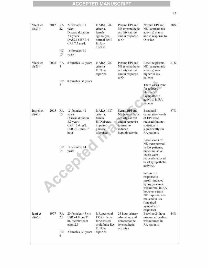
Two studies found no significant correlation between catecholamines and
inflammatory indices. Vlcek et al(87) found no significant correlation between plasma
catecholamines and inflammation (CRP, DAS28-CRP). Van Middendorp et al(84) found no
correlation between 24 hour urinary noradrenaline excretion and markers of inflammation
(ESR or interleukin-6) in a cohort of 60 RA patients (38 females, mean age 59 years). Igari et
al(66) in a sub-study of 6 RA patients who underwent synovectomy found that 24 hour
urinary adrenaline and noradrenaline significantly decreased 2 weeks following
synovectomy. Although the investigators did not assess inflammatory markers following
synovectomy, it may be assumed that local joint inflammation would have been reduced
following synovectomy and hence possibly removing the stimulus for sympathetic activation.
Barendregt et al(52) found that ESR levels were higher in the group with
parasympathetic dysfunction (abnormal PLR in the RA group with ocular dryness) compared
to those without (although significance values were not reported).
15
Interventional studies
Two studies investigated HRV in RA patients receiving tumour necrosis factor (TNF)
alpha inhibitor therapy.(65, 73) Holman et al(65) studied 33 patients (25 with RA, 8 with
psoriatic arthritis) before treatment with TNF-alpha inhibitor therapy and assessed clinical
response to treatment (using American College of Rheumatology criteria ACR20/50/70 and
DAS28) at various time points up to one year. They found that low HRV indices, reduced
parasympathetic and increased sympathetic activity were predictors of poor response to TNF-
alpha inhibitor therapy. However the study may have been underpowered as they found no
direct correlation between baseline autonomic function and change in DAS28 score following
TNF-alpha inhibitor therapy. Despite limitations of the study (one third of patients
discontinued therapy by one year; use and dosage of other medications were not controlled;
small numbers of RA patients) these results suggest that HRV and sympatho-parasympathetic
balance may play an important role in disease activity.
Two studies assessed plasma NPY levels before and after TNF-alpha inhibitor
therapy. In a study of 16 female RA patients Kopec-Medrek et al(72) found that infliximab
(TNF-alpha inhibitor) significantly reduced inflammation (CRP, ESR) but did not reduce
sympathetic activity (plasma NPY). In fact, plasma NPY concentrations rose to a peak after 6
infusions of infliximab and fell to baseline levels 8 weeks after the ninth (final) infusion. The
authors did however report a positive correlation between plasma NPY concentrations and
CRP (Kendall tau coefficient=0.506, P<0.006) and DAS28 (Kendall tau coefficient=0.393,
P<0.033) at baseline, indicating that plasma NPY may reflect inflammatory status.
Harle et al (64) found that in a cohort of RA patients, adalimumab (TNF-alpha
inhibitor) had no effect on serum NPY levels despite good clinical response. They reported
16
higher plasma NPY concentrations in RA patients with previous prednisolone use only,
indicating a possible interaction effect with the hypothalamic-pituitary-adrenal axis.
DISCUSSION
The results of this systematic literature review indicate that ANS dysfunction is
prevalent in ~60% (33-86%) of RA patients as determined from observational studies of
small sample size (10-50 patients). Stronger evidence (from large prospective cohort studies)
is required to confidently determine the true prevalence of autonomic dysfunction in RA.
HRV is probably the most feasible ANS assessment in such a large population. Few studies
have assessed patients with early RA (duration<2years) but have shown that ANS
dysfunction occurs early in RA and is not necessarily an effect of long-term disease and
inflammatory burden. More studies of RA patients with early disease are clearly needed and
if possible ANS assessment preceding the onset of RA, to determine whether altered ANS
function predisposes to developing RA.
Studies using CCTs in RA have shown reduced resting parasympathetic activity and
impairment in both sympathetic and parasympathetic reflex responses. Strong evidence from
good quality HRV data supports these findings with the majority demonstrating low HRV
reflecting reduced resting parasympathetic activity. In addition there is limited evidence for
elevated resting sympathetic activity with the majority of good quality HRV data failing to
detect abnormal sympathetic function in RA. Studies employing other methods of ANS
assessment have shown conflicting results, which may reflect their inherent limitations. There
is a lack of evidence from the literature to date to determine causal relationships between
systemic inflammation and autonomic dysfunction. The available literature is too small to be
clear whether the lack of evidence represents a lack of relationship or simply inadequate
power. Only two studies assessed the effects of anti-inflammatory therapy on ANS function
17
and failed to demonstrate an effect. However, their results suggest that plasma NPY may not
be a reliable method of assessing sympathetic activity particularly as the effects of steroids on
NPY are not known. Further interventional studies are needed to elucidate causation. The
most feasible and ethical study design would be to assess ANS function in RA patients prior
to and after anti-inflammatory therapy. This could be achieved for example with HRV
assessments using a 24-hour electrocardiograph holter. Although HRV is not routinely used
in clinical practice one study suggested a possible clinical role. Holman et al (65) found that
low HRV in RA patients predicted a poor response to TNF-alpha inhibitor therapy indicating
a possible benefit in determining ANS status prior to initiation of biologic agents. What
remains unknown however is whether therapy to improve HRV in these patients would
improve their response to anti-inflammatory agents.
Less than half the studies demonstrated an association between increased
inflammation and ANS dysfunction (mainly CCTs and HRV), consistent with the results of
recent animal studies.(20-22) The lack of associations in the remaining studies may be simply
due to a lack of statistical power; the majority of studies in our review did not report a power
calculation. Another possible explanation may be the relatively low inflammatory status of
patients tested. CRP, ESR and DAS (reported in less than two thirds of studies) were only
modestly elevated although it is unclear whether cumulative inflammatory burden can be
determined from assessment at a single time point.. Another explanation for a lack of
association between inflammation and ANS function in the studies included in our review
may be the subtle nature of autonomic dysfunction present in RA or simply the inappropriate
choice of immune markers assessed.
The main limitations of this review are the types and number of ANS tests employed
in RA patients, with the majority of studies making only one assessment of ANS function.
ANS function is complex and multi-faceted and hence a comprehensive assessment is
18
required in order to fully categorise the presence of dysfunction. Future studies should
include a greater variety of tests including arterial baroreflex assessment, with attempts to
measure resting ANS function and response to stimuli. Larger sample sizes are required to
confirm the prevalence of ANS in RA, and in order to ensure that statistical power is
achieved.
Future studies in RA should aim to characterise the inflammatory profile of patients
studied so that causal links between inflammation and ANS dysfunction can be determined.
The effects of RA medications on ANS function is not fully known and is a difficult
confounding factor to control for, especially as RA patients often require medications to
induce and maintain remission of disease. One study showed that infliximab infusion (TNF-
alpha inhibitor therapy) caused an acute reduction in HRV and sympathetic activity compared
to a placebo. The effects of other RA medications on the ANS tests employed to date are
unknown although studies of healthy subjects may be the most ethically acceptable way to
investigate this.
Another difficulty is discerning between the effects of RA and concomitant co-
morbidities or medications on ANS function. Although many studies excluded RA patients
with conditions or medications affecting the ANS system, cardiovascular disease (CVD)
remains under-diagnosed in this population.(6, 8, 11) Cardiac imaging (e.g. echocardiography
or magnetic resonance imaging) to identify such patients and the possible inclusion of a
cardiovascular disease control group may help tackle this problem.
In conclusion, the evidence to date supports that ANS dysfunction is a feature of RA
although not universally found in all patients. The profile of ANS dysfunction found in RA
patients (low HRV, reduced parasympathetic activity and elevated sympathetic activity) is
associated with increased cardiovascular and mortality risk and may help to explain the
19
increased risk in RA patients. Furthermore, this pattern of ANS dysfunction supports the
findings from animal studies and may be a consequence of high inflammatory burden.
Although associations between inflammation and ANS dysfunction are present in RA
patients, the available literature is too small and underpowered to be clear about causality.
Further studies are required to: determine the true prevalence of ANS dysfunction in RA,
characterise RA patients who have altered ANS function; determine the prognostic role of
ANS assessments in predicting cardiovascular and mortality risk; assess the effects of
biologic agents on ANS function; consider the role of therapeutic strategies targeting the
ANS in RA patients to help control disease activity or improve response to biologic agents.
ACKNOWLEDGEMENTS
None.
TABLES
Table 1. Definition of ANS assessments included in the review
Table 2. Characteristics of studies included in the review
Table 3. Results Summary: Number of studies with abnormal autonomic function in
rheumatoid arthritis patients from observational studies
Table 4. Results Summary: Outcome of autonomic assessments from case-control studies
Table 5. Results Summary: Outcome of associations between autonomic function and
inflammation in RA
20
Supplementary data
Appendix 1. Quality index score assessment tool criteria
Appendix 2. Prevalence of autonomic nervous system dysfunction in rheumatoid arthritis
21
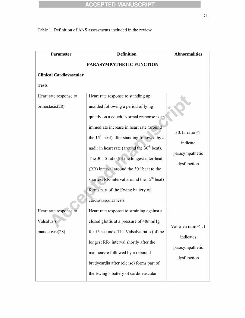
Table 1. Definition of ANS assessments included in the review
Parameter Definition Abnormalities
PARASYMPATHETIC FUNCTION
Clinical Cardiovascular
Tests
Heart rate response to
orthostasis(28)
Heart rate response to standing up
unaided following a period of lying
quietly on a couch. Normal response is an
immediate increase in heart rate (around
the 15th beat) after standing followed by a
nadir in heart rate (around the 30th beat).
The 30:15 ratio (of the longest inter-beat
(RR) interval around the 30th beat to the
shortest RR-interval around the 15th beat)
forms part of the Ewing battery of
cardiovascular tests.
30:15 ratio ≤1
indicate
parasympathetic
dysfunction
Heart rate response to
Valsalva’s
manoeuvre(28)
Heart rate response to straining against a
closed glottis at a pressure of 40mmHg
for 15 seconds. The Valsalva ratio (of the
longest RR- interval shortly after the
manoeuvre followed by a rebound
bradycardia after release) forms part of
the Ewing’s battery of cardiovascular
Valsalva ratio ≤1.1
indicates
parasympathetic
dysfunction
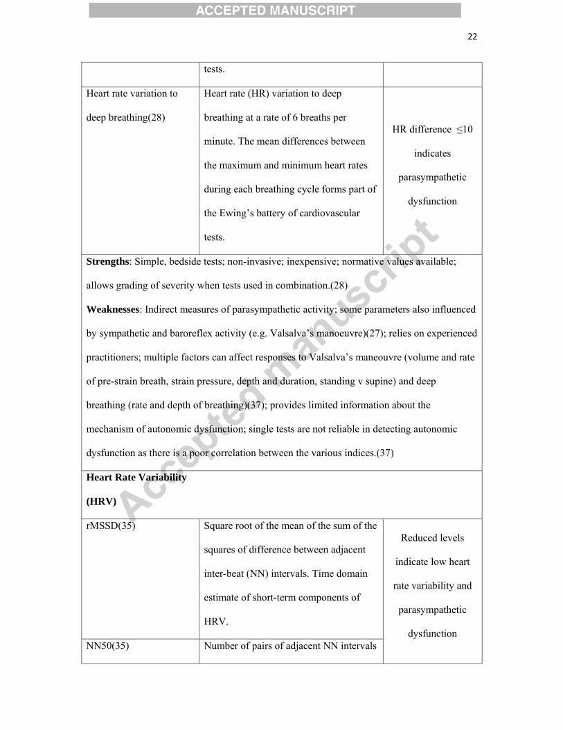
22
tests.
Heart rate variation to
deep breathing(28)
Heart rate (HR) variation to deep
breathing at a rate of 6 breaths per
minute. The mean differences between
the maximum and minimum heart rates
during each breathing cycle forms part of
the Ewing’s battery of cardiovascular
tests.
HR difference ≤10
indicates
parasympathetic
dysfunction
Strengths: Simple, bedside tests; non-invasive; inexpensive; normative values available;
allows grading of severity when tests used in combination.(28)
Weaknesses: Indirect measures of parasympathetic activity; some parameters also influenced
by sympathetic and baroreflex activity (e.g. Valsalva’s manoeuvre)(27); relies on experienced
practitioners; multiple factors can affect responses to Valsalva’s maneouvre (volume and rate
of pre-strain breath, strain pressure, depth and duration, standing v supine) and deep
breathing (rate and depth of breathing)(37); provides limited information about the
mechanism of autonomic dysfunction; single tests are not reliable in detecting autonomic
dysfunction as there is a poor correlation between the various indices.(37)
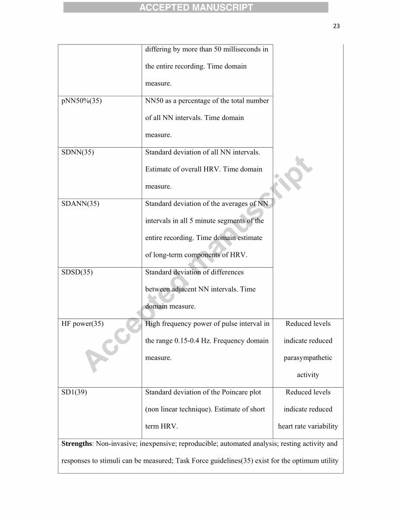
Heart Rate Variability
(HRV)
rMSSD(35) Square root of the mean of the sum of the
squares of difference between adjacent
inter-beat (NN) intervals. Time domain
estimate of short-term components of
HRV.
Reduced levels
indicate low heart
rate variability and
parasympathetic
dysfunction NN50(35) Number of pairs of adjacent NN intervals
23
differing by more than 50 milliseconds in
the entire recording. Time domain
measure.
pNN50%(35) NN50 as a percentage of the total number
of all NN intervals. Time domain
measure.
SDNN(35) Standard deviation of all NN intervals.
Estimate of overall HRV. Time domain
measure.
SDANN(35) Standard deviation of the averages of NN
intervals in all 5 minute segments of the
entire recording. Time domain estimate
of long-term components of HRV.
SDSD(35) Standard deviation of differences
between adjacent NN intervals. Time
domain measure.
HF power(35) High frequency power of pulse interval in
the range 0.15-0.4 Hz. Frequency domain
measure.
Reduced levels
indicate reduced
parasympathetic
activity
SD1(39) Standard deviation of the Poincare plot
(non linear technique). Estimate of short
term HRV.
Reduced levels
indicate reduced
heart rate variability
Strengths: Non-invasive; inexpensive; reproducible; automated analysis; resting activity and
responses to stimuli can be measured; Task Force guidelines(35) exist for the optimum utility
24
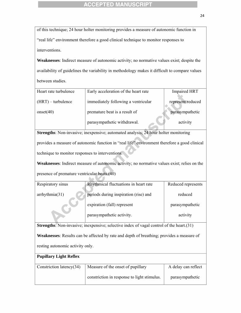
of this technique; 24 hour holter monitoring provides a measure of autonomic function in
“real life” environment therefore a good clinical technique to monitor responses to
interventions.
Weaknesses: Indirect measure of autonomic activity; no normative values exist; despite the
availability of guidelines the variability in methodology makes it difficult to compare values
between studies.
Heart rate turbulence
(HRT) – turbulence
onset(40)
Early acceleration of the heart rate
immediately following a ventricular
premature beat is a result of
parasympathetic withdrawal.
Impaired HRT
represent reduced
parasympathetic
activity
Strengths: Non-invasive; inexpensive; automated analysis; 24 hour holter monitoring
provides a measure of autonomic function in “real life” environment therefore a good clinical
technique to monitor responses to interventions.
Weaknesses: Indirect measure of autonomic activity; no normative values exist; relies on the
presence of premature ventricular beats.(40)
Respiratory sinus
arrhythmia(31)
Rhythmical fluctuations in heart rate
periods during inspiration (rise) and
expiration (fall) represent
parasympathetic activity.
Reduced represents
reduced
parasympathetic
activity
Strengths: Non-invasive; inexpensive; selective index of vagal control of the heart.(31)
Weaknesses: Results can be affected by rate and depth of breathing; provides a measure of
resting autonomic activity only.
Pupillary Light Reflex
Constriction latency(34) Measure of the onset of pupillary
constriction in response to light stimulus.
A delay can reflect
parasympathetic
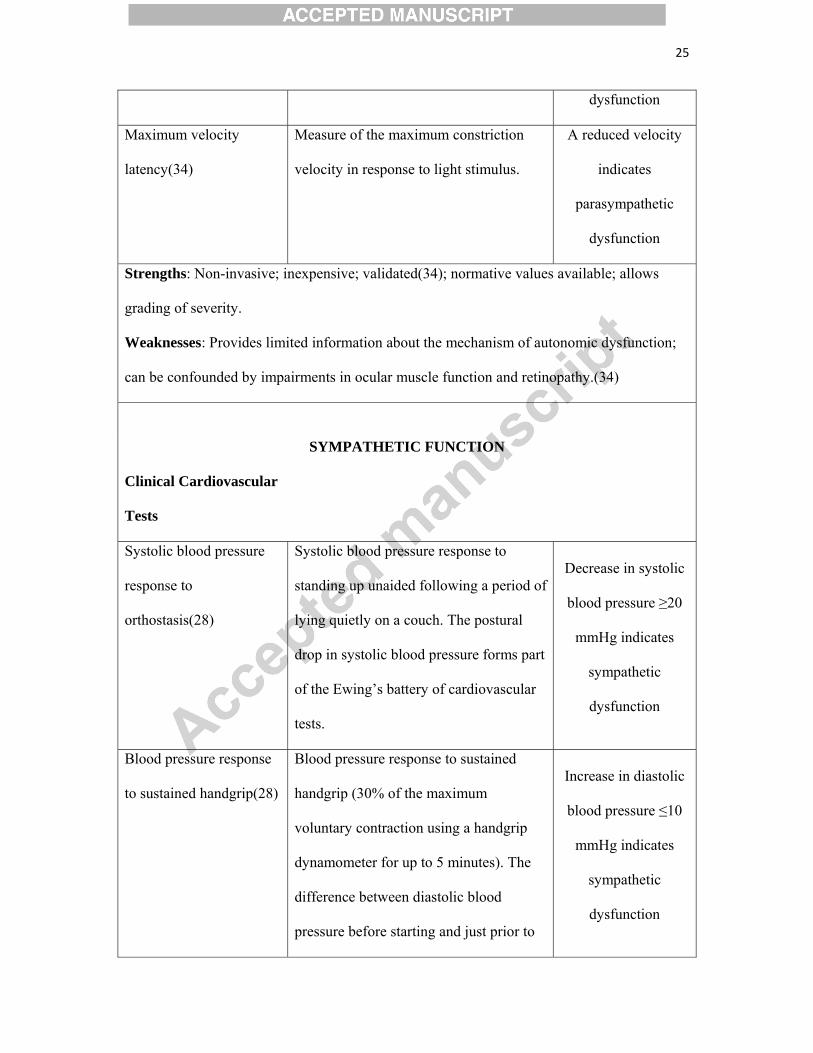
25
dysfunction
Maximum velocity
latency(34)
Measure of the maximum constriction
velocity in response to light stimulus.
A reduced velocity
indicates
parasympathetic
dysfunction
Strengths: Non-invasive; inexpensive; validated(34); normative values available; allows
grading of severity.
Weaknesses: Provides limited information about the mechanism of autonomic dysfunction;
can be confounded by impairments in ocular muscle function and retinopathy.(34)
SYMPATHETIC FUNCTION
Clinical Cardiovascular
Tests
Systolic blood pressure
response to
orthostasis(28)
Systolic blood pressure response to
standing up unaided following a period of
lying quietly on a couch. The postural
drop in systolic blood pressure forms part
of the Ewing’s battery of cardiovascular
tests.
Decrease in systolic
blood pressure ≥20
mmHg indicates
sympathetic
dysfunction
Blood pressure response
to sustained handgrip(28)
Blood pressure response to sustained
handgrip (30% of the maximum
voluntary contraction using a handgrip
dynamometer for up to 5 minutes). The
difference between diastolic blood
pressure before starting and just prior to
Increase in diastolic
blood pressure ≤10
mmHg indicates
sympathetic
dysfunction
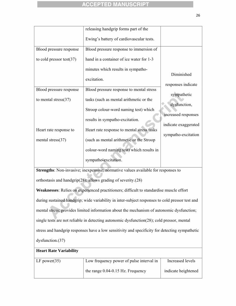
26
releasing handgrip forms part of the
Ewing’s battery of cardiovascular tests.
Blood pressure response
to cold pressor test(37)
Blood pressure response to immersion of
hand in a container of ice water for 1-3
minutes which results in sympatho-
excitation. Diminished
responses indicate
sympathetic
dysfunction,
increased responses
indicate exaggerated
sympatho-excitation
Blood pressure response
to mental stress(37)
Blood pressure response to mental stress
tasks (such as mental arithmetic or the
Stroop colour-word naming test) which
results in sympatho-excitation.
Heart rate response to
mental stress(37)
Heart rate response to mental stress tasks
(such as mental arithmetic or the Stroop
colour-word naming test) which results in
sympatho-excitation.
Strengths: Non-invasive; inexpensive; normative values available for responses to
orthostasis and handgrip(28); allows grading of severity.(28)
Weaknesses: Relies on experienced practitioners; difficult to standardise muscle effort
during sustained handgrip; wide variability in inter-subject responses to cold pressor test and
mental stress; provides limited information about the mechanism of autonomic dysfunction;
single tests are not reliable in detecting autonomic dysfunction(28); cold pressor, mental
stress and handgrip responses have a low sensitivity and specificity for detecting sympathetic
dysfunction.(37)
Heart Rate Variability
LF power(35) Low frequency power of pulse interval in
the range 0.04-0.15 Hz. Frequency
Increased levels
indicate heightened
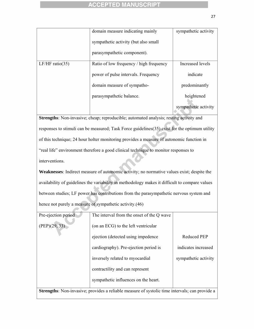
27
domain measure indicating mainly
sympathetic activity (but also small
parasympathetic component).
sympathetic activity
LF/HF ratio(35) Ratio of low frequency / high frequency
power of pulse intervals. Frequency
domain measure of sympatho-
parasympathetic balance.
Increased levels
indicate
predominantly
heightened
sympathetic activity
Strengths: Non-invasive; cheap; reproducible; automated analysis; resting activity and
responses to stimuli can be measured; Task Force guidelines(35) exist for the optimum utility
of this technique; 24 hour holter monitoring provides a measure of autonomic function in
“real life” environment therefore a good clinical technique to monitor responses to
interventions.
Weaknesses: Indirect measure of autonomic activity; no normative values exist; despite the
availability of guidelines the variability in methodology makes it difficult to compare values
between studies; LF power has contributions from the parasympathetic nervous system and
hence not purely a measure of sympathetic activity.(46)
Pre-ejection period
(PEP)(29, 33)
The interval from the onset of the Q wave
(on an ECG) to the left ventricular
ejection (detected using impedence
cardiography). Pre-ejection period is
inversely related to myocardial
contractility and can represent
sympathetic influences on the heart.
Reduced PEP
indicates increased
sympathetic activity
Strengths: Non-invasive; provides a reliable measure of systolic time intervals; can provide a
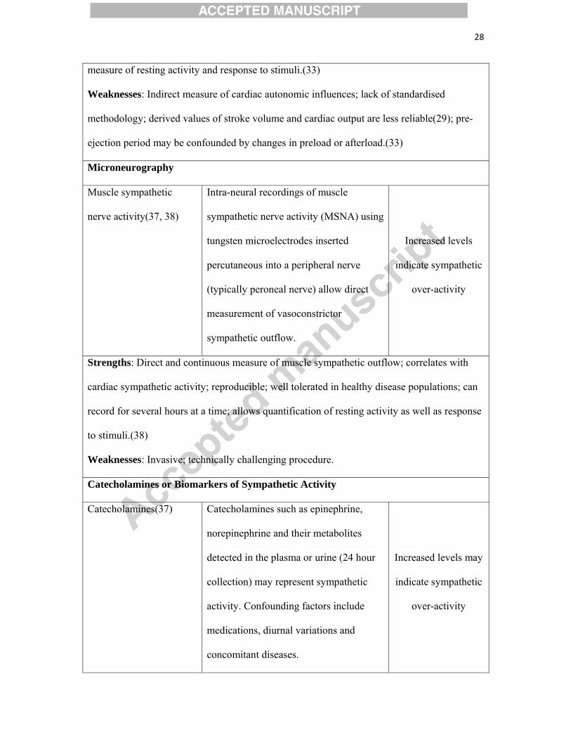
28
measure of resting activity and response to stimuli.(33)
Weaknesses: Indirect measure of cardiac autonomic influences; lack of standardised
methodology; derived values of stroke volume and cardiac output are less reliable(29); pre-
ejection period may be confounded by changes in preload or afterload.(33)
Microneurography
Muscle sympathetic
nerve activity(37, 38)
Intra-neural recordings of muscle
sympathetic nerve activity (MSNA) using
tungsten microelectrodes inserted
percutaneous into a peripheral nerve
(typically peroneal nerve) allow direct
measurement of vasoconstrictor
sympathetic outflow.
Increased levels
indicate sympathetic
over-activity
Strengths: Direct and continuous measure of muscle sympathetic outflow; correlates with
cardiac sympathetic activity; reproducible; well tolerated in healthy disease populations; can
record for several hours at a time; allows quantification of resting activity as well as response
to stimuli.(38)
Weaknesses: Invasive; technically challenging procedure.
Catecholamines or Biomarkers of Sympathetic Activity
Catecholamines(37) Catecholamines such as epinephrine,
norepinephrine and their metabolites
detected in the plasma or urine (24 hour
collection) may represent sympathetic
activity. Confounding factors include
medications, diurnal variations and
concomitant diseases.
Increased levels may
indicate sympathetic
over-activity
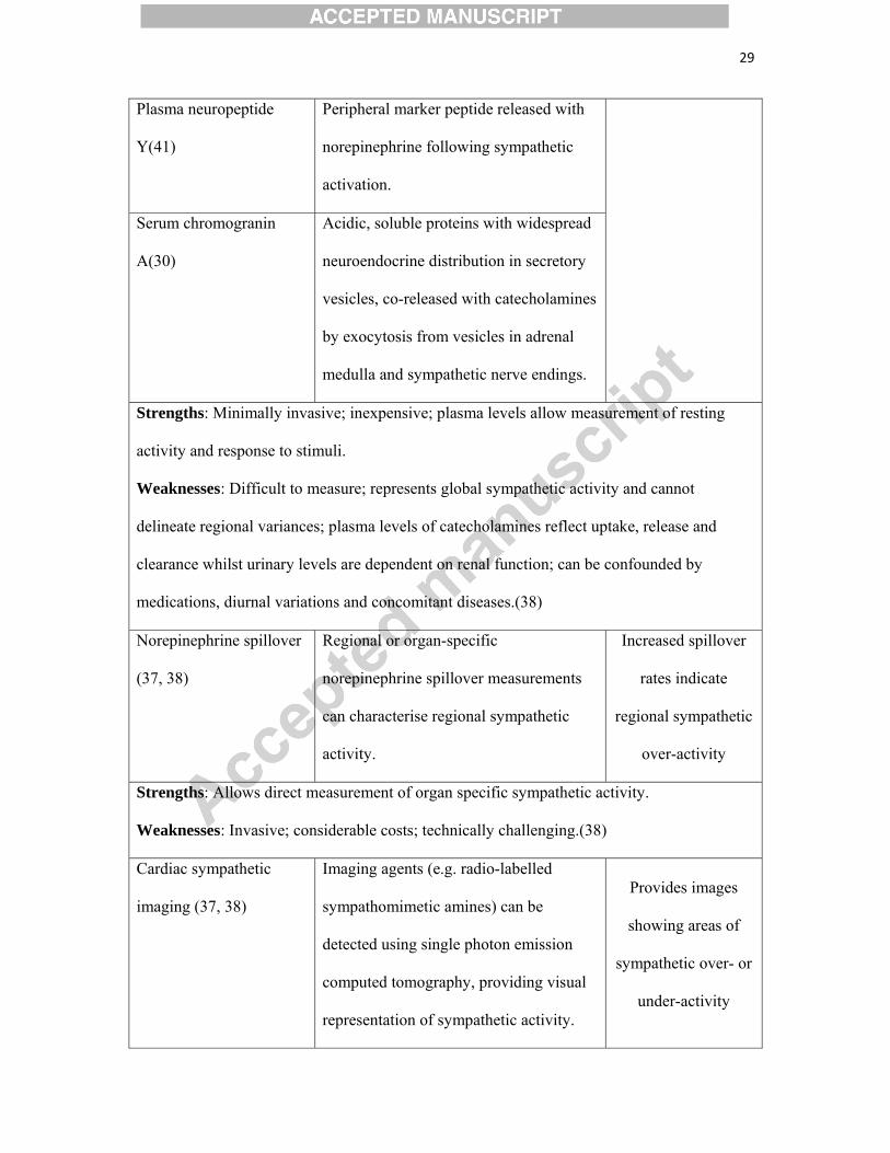
29
Plasma neuropeptide
Y(41)
Peripheral marker peptide released with
norepinephrine following sympathetic
activation.
Serum chromogranin
A(30)
Acidic, soluble proteins with widespread
neuroendocrine distribution in secretory
vesicles, co-released with catecholamines
by exocytosis from vesicles in adrenal
medulla and sympathetic nerve endings.
Strengths: Minimally invasive; inexpensive; plasma levels allow measurement of resting
activity and response to stimuli.
Weaknesses: Difficult to measure; represents global sympathetic activity and cannot
delineate regional variances; plasma levels of catecholamines reflect uptake, release and
clearance whilst urinary levels are dependent on renal function; can be confounded by
medications, diurnal variations and concomitant diseases.(38)
Norepinephrine spillover
(37, 38)
Regional or organ-specific
norepinephrine spillover measurements
can characterise regional sympathetic
activity.
Increased spillover
rates indicate
regional sympathetic
over-activity
Strengths: Allows direct measurement of organ specific sympathetic activity.
Weaknesses: Invasive; considerable costs; technically challenging.(38)
Cardiac sympathetic
imaging (37, 38)
Imaging agents (e.g. radio-labelled
sympathomimetic amines) can be
detected using single photon emission
computed tomography, providing visual
representation of sympathetic activity.
Provides images
showing areas of
sympathetic over- or
under-activity
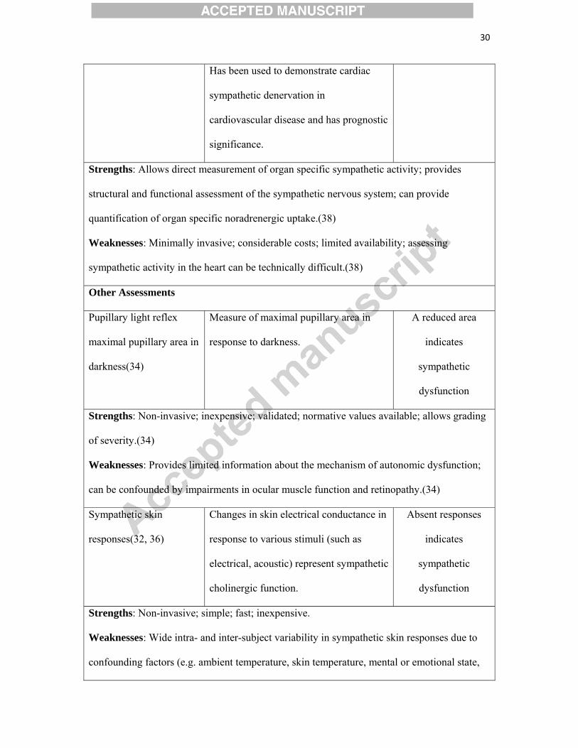
30
Has been used to demonstrate cardiac
sympathetic denervation in
cardiovascular disease and has prognostic
significance.
Strengths: Allows direct measurement of organ specific sympathetic activity; provides
structural and functional assessment of the sympathetic nervous system; can provide
quantification of organ specific noradrenergic uptake.(38)
Weaknesses: Minimally invasive; considerable costs; limited availability; assessing
sympathetic activity in the heart can be technically difficult.(38)
Other Assessments
Pupillary light reflex
maximal pupillary area in
darkness(34)
Measure of maximal pupillary area in
response to darkness.
A reduced area
indicates
sympathetic
dysfunction
Strengths: Non-invasive; inexpensive; validated; normative values available; allows grading
of severity.(34)
Weaknesses: Provides limited information about the mechanism of autonomic dysfunction;
can be confounded by impairments in ocular muscle function and retinopathy.(34)
Sympathetic skin
responses(32, 36)
Changes in skin electrical conductance in
response to various stimuli (such as
electrical, acoustic) represent sympathetic
cholinergic function.
Absent responses
indicates
sympathetic
dysfunction
Strengths: Non-invasive; simple; fast; inexpensive.
Weaknesses: Wide intra- and inter-subject variability in sympathetic skin responses due to
confounding factors (e.g. ambient temperature, skin temperature, mental or emotional state,
31
habituation with repeated stimuli); low sensitivity and specificity; poor correlation with other
autonomic assessments (e.g. sudomotor dysfunction).(36)
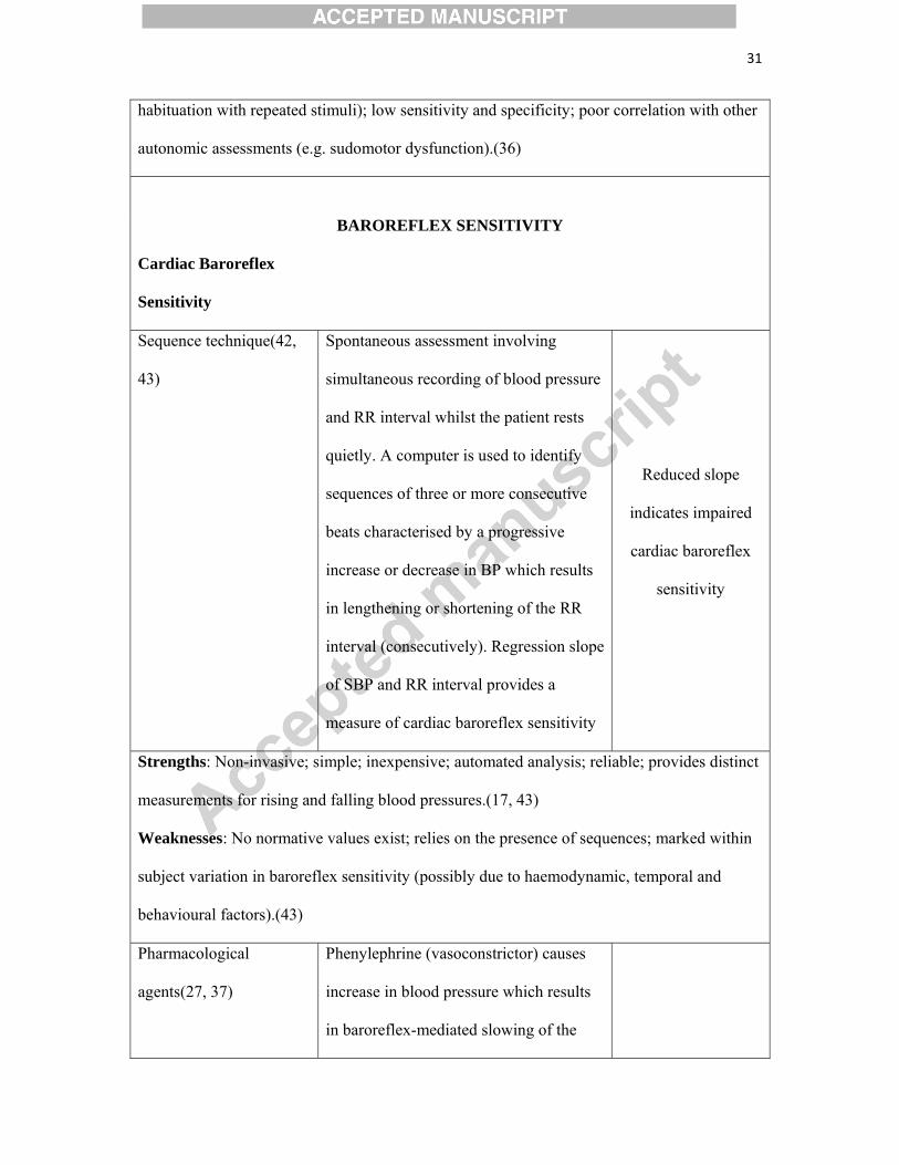
BAROREFLEX SENSITIVITY
Cardiac Baroreflex
Sensitivity
Sequence technique(42,
43)
Spontaneous assessment involving
simultaneous recording of blood pressure
and RR interval whilst the patient rests
quietly. A computer is used to identify
sequences of three or more consecutive
beats characterised by a progressive
increase or decrease in BP which results
in lengthening or shortening of the RR
interval (consecutively). Regression slope
of SBP and RR interval provides a
measure of cardiac baroreflex sensitivity
Reduced slope
indicates impaired
cardiac baroreflex
sensitivity
Strengths: Non-invasive; simple; inexpensive; automated analysis; reliable; provides distinct
measurements for rising and falling blood pressures.(17, 43)
Weaknesses: No normative values exist; relies on the presence of sequences; marked within
subject variation in baroreflex sensitivity (possibly due to haemodynamic, temporal and
behavioural factors).(43)
Pharmacological
agents(27, 37)
Phenylephrine (vasoconstrictor) causes
increase in blood pressure which results
in baroreflex-mediated slowing of the
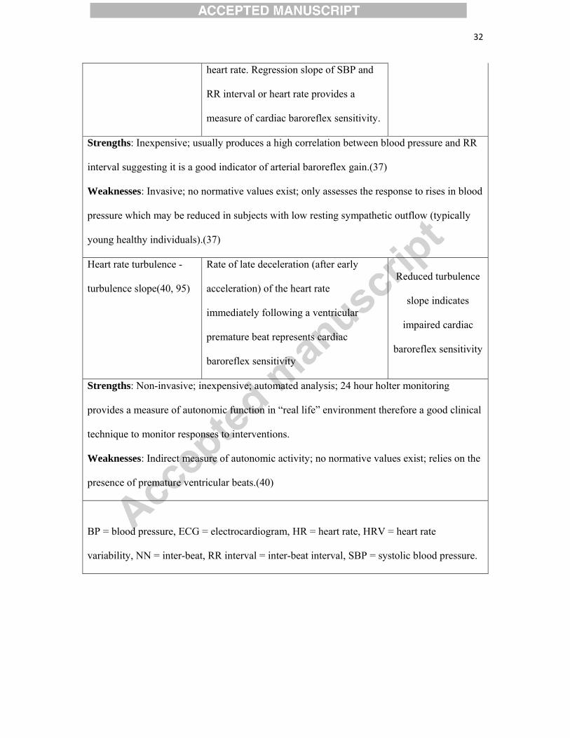
32
heart rate. Regression slope of SBP and
RR interval or heart rate provides a
measure of cardiac baroreflex sensitivity.
Strengths: Inexpensive; usually produces a high correlation between blood pressure and RR
interval suggesting it is a good indicator of arterial baroreflex gain.(37)
Weaknesses: Invasive; no normative values exist; only assesses the response to rises in blood
pressure which may be reduced in subjects with low resting sympathetic outflow (typically
young healthy individuals).(37)
Heart rate turbulence -
turbulence slope(40, 95)
Rate of late deceleration (after early
acceleration) of the heart rate
immediately following a ventricular
premature beat represents cardiac
baroreflex sensitivity
Reduced turbulence
slope indicates
impaired cardiac
baroreflex sensitivity
Strengths: Non-invasive; inexpensive; automated analysis; 24 hour holter monitoring
provides a measure of autonomic function in “real life” environment therefore a good clinical
technique to monitor responses to interventions.
Weaknesses: Indirect measure of autonomic activity; no normative values exist; relies on the
presence of premature ventricular beats.(40)
BP = blood pressure, ECG = electrocardiogram, HR = heart rate, HRV = heart rate
variability, NN = inter-beat, RR interval = inter-beat interval, SBP = systolic blood pressure.
33
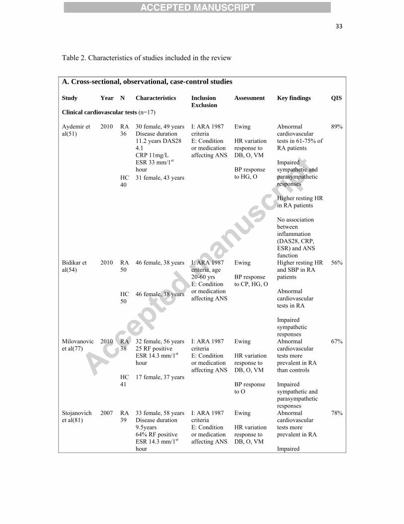
Table 2. Characteristics of studies included in the review
A. Cross-sectional, observational, case-control studies Study Year N Characteristics Inclusion
Exclusion Assessment Key findings QIS
Clinical cardiovascular tests (n=17) Aydemir et al(51)
2010 RA 36
30 female, 49 years Disease duration 11.2 years DAS28 4.1 CRP 11mg/L ESR 33 mm/1st hour
I: ARA 1987 criteria E: Condition or medication affecting ANS
Ewing HR variation response to DB, O, VM BP response to HG, O
Abnormal cardiovascular tests in 61-75% of RA patients Impaired sympathetic and parasympathetic responses Higher resting HR in RA patients No association between inflammation (DAS28, CRP, ESR) and ANS function
89%
HC 40
31 female, 43 years
Bidikar et al(54)
2010 RA 50
46 female, 38 years I: ARA 1987 criteria, age 20-60 yrs E: Condition or medication affecting ANS
Ewing BP response to CP, HG, O
Higher resting HR and SBP in RA patients Abnormal cardiovascular tests in RA Impaired sympathetic responses
56%
HC 50
46 female, 38 years
Milovanovic et al(77)
2010 RA 38
32 female, 56 years 25 RF positive ESR 14.3 mm/1st hour
I: ARA 1987 criteria E: Condition or medication affecting ANS
Ewing HR variation response to DB, O, VM BP response to O
Abnormal cardiovascular tests more prevalent in RA than controls Impaired sympathetic and parasympathetic responses
67%
HC 41
17 female, 37 years
Stojanovich et al(81)
2007 RA 39
33 female, 58 years Disease duration 9.5years 64% RF positive ESR 14.3 mm/1st hour
I: ARA 1987 criteria E: Condition or medication affecting ANS
Ewing HR variation response to DB, O, VM
Abnormal cardiovascular tests more prevalent in RA Impaired
78%
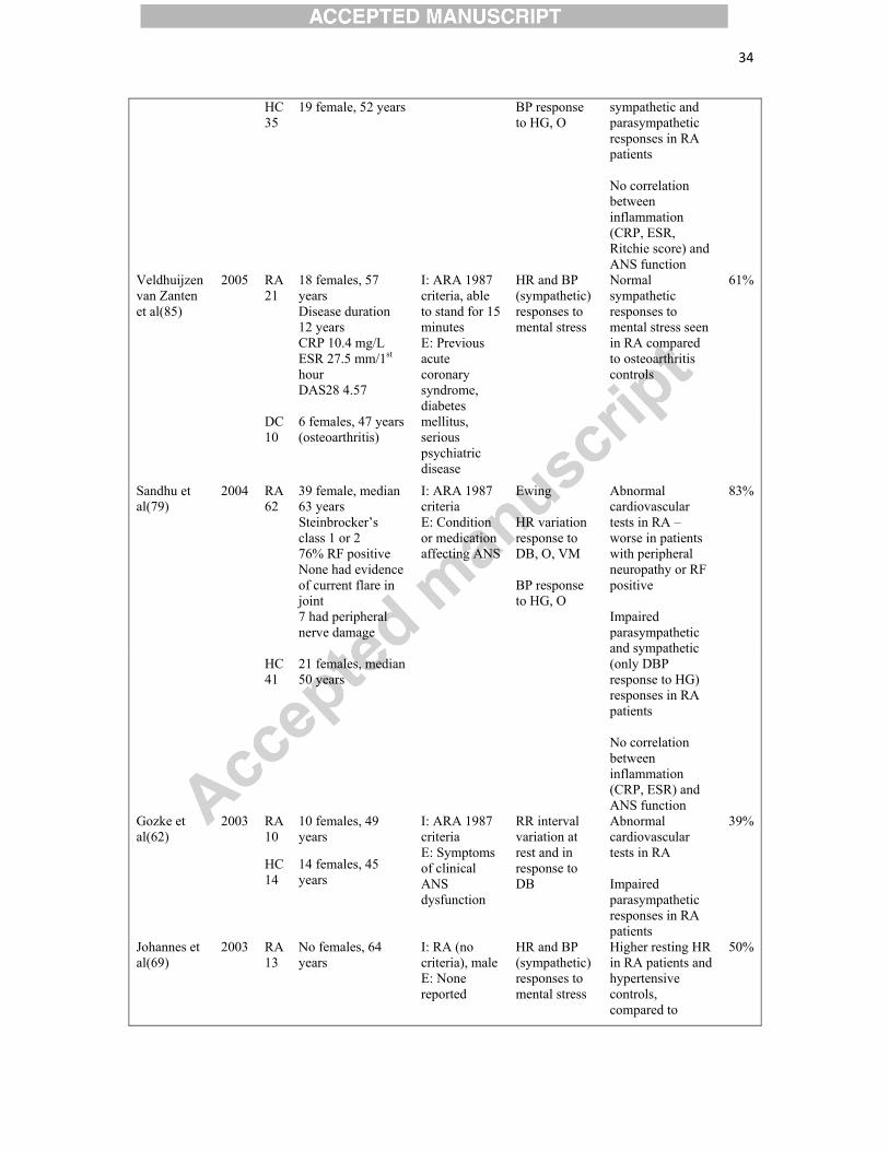
34
HC 35
19 female, 52 years
BP response to HG, O
sympathetic and parasympathetic responses in RA patients No correlation between inflammation (CRP, ESR, Ritchie score) and ANS function
Veldhuijzen van Zanten et al(85)
2005 RA 21
18 females, 57 years Disease duration 12 years CRP 10.4 mg/L ESR 27.5 mm/1st hour DAS28 4.57
I: ARA 1987 criteria, able to stand for 15 minutes E: Previous acute coronary syndrome, diabetes mellitus, serious psychiatric disease
HR and BP (sympathetic) responses to mental stress
Normal sympathetic responses to mental stress seen in RA compared to osteoarthritis controls
61%
DC 10
6 females, 47 years (osteoarthritis)
Sandhu et al(79)
2004 RA 62
39 female, median 63 years Steinbrocker’s class 1 or 2 76% RF positive None had evidence of current flare in joint 7 had peripheral nerve damage
I: ARA 1987 criteria E: Condition or medication affecting ANS
Ewing HR variation response to DB, O, VM BP response to HG, O
Abnormal cardiovascular tests in RA – worse in patients with peripheral neuropathy or RF positive Impaired parasympathetic and sympathetic (only DBP response to HG) responses in RA patients No correlation between inflammation (CRP, ESR) and ANS function
83%
HC 41
21 females, median 50 years
Gozke et al(62)
2003 RA 10
10 females, 49 years
I: ARA 1987 criteria E: Symptoms of clinical ANS dysfunction
RR interval variation at rest and in response to DB
Abnormal cardiovascular tests in RA Impaired parasympathetic responses in RA patients
39%
HC 14
14 females, 45 years
Johannes et al(69)
2003 RA 13
No females, 64 years
I: RA (no criteria), male E: None reported
HR and BP (sympathetic) responses to mental stress
Higher resting HR in RA patients and hypertensive controls, compared to
50%
35
HC 30
No females, 39 years
healthy Lower resting DBP in RA patients compared to hypertensive and healthy controls Higher BP (sympathetic) response to mental stress in RA patients compared to hypertensive and healthy controls
DC 53
No females, 49 years (Hypertensive)
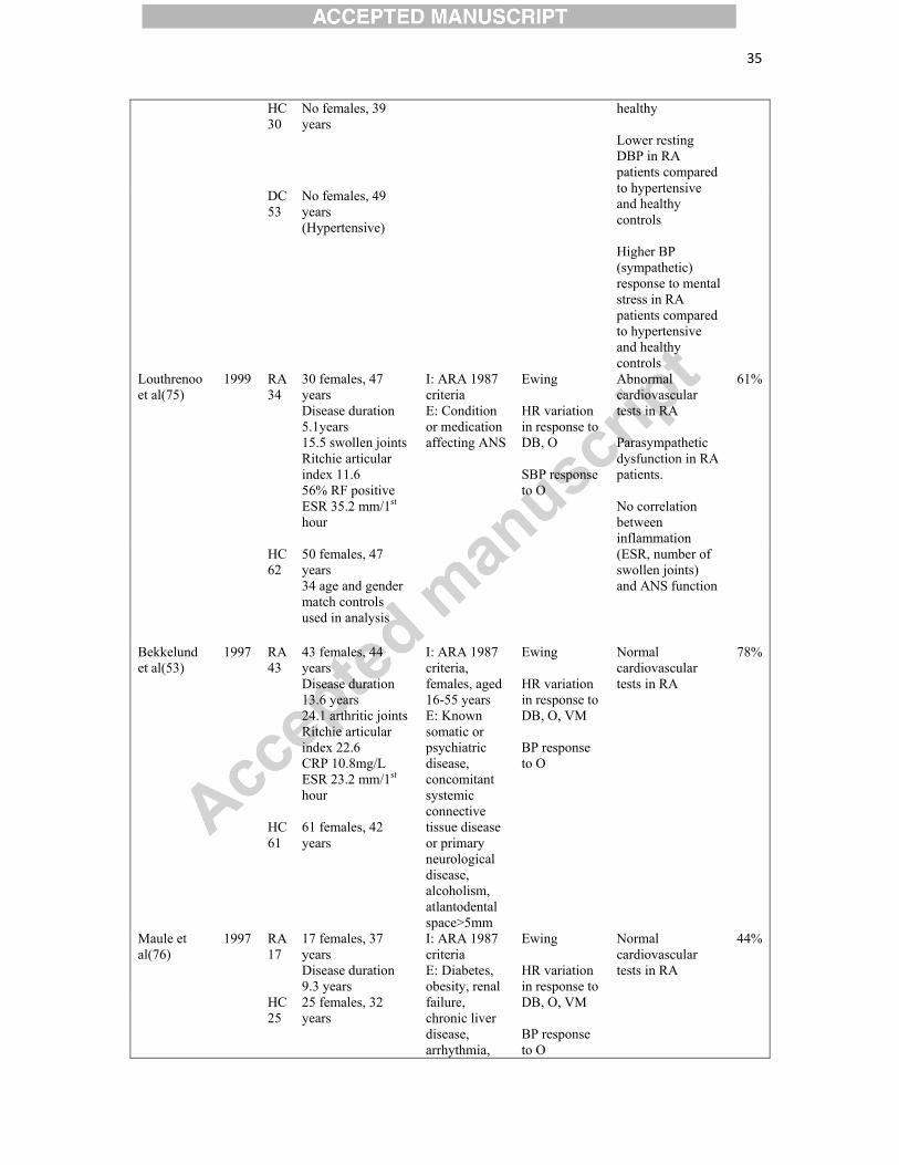
Louthrenoo et al(75)
1999 RA 34
30 females, 47 years Disease duration 5.1years 15.5 swollen joints Ritchie articular index 11.6 56% RF positive ESR 35.2 mm/1st hour
I: ARA 1987 criteria E: Condition or medication affecting ANS
Ewing HR variation in response to DB, O SBP response to O
Abnormal cardiovascular tests in RA Parasympathetic dysfunction in RA patients. No correlation between inflammation (ESR, number of swollen joints) and ANS function
61%
HC 62
50 females, 47 years 34 age and gender match controls used in analysis
Bekkelund et al(53)
1997 RA 43
43 females, 44 years Disease duration 13.6 years 24.1 arthritic joints Ritchie articular index 22.6 CRP 10.8mg/L ESR 23.2 mm/1st hour
I: ARA 1987 criteria, females, aged 16-55 years E: Known somatic or psychiatric disease, concomitant systemic connective tissue disease or primary neurological disease, alcoholism, atlantodental space>5mm
Ewing HR variation in response to DB, O, VM BP response to O
Normal cardiovascular tests in RA
78%
HC 61
61 females, 42 years
Maule et al(76)
1997 RA 17
17 females, 37 years Disease duration 9.3 years
I: ARA 1987 criteria E: Diabetes, obesity, renal failure, chronic liver disease, arrhythmia,
Ewing HR variation in response to DB, O, VM BP response to O
Normal cardiovascular tests in RA
44%
HC 25
25 females, 32 years
36
anaemia, anti-hypertensive therapy
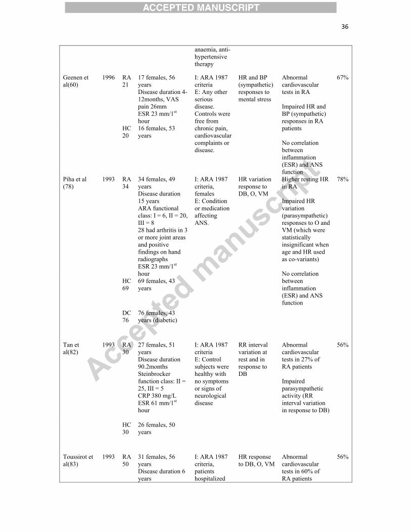
Geenen et al(60)
1996 RA 21
17 females, 56 years Disease duration 4-12months, VAS pain 26mm ESR 23 mm/1st hour
I: ARA 1987 criteria E: Any other serious disease. Controls were free from chronic pain, cardiovascular complaints or disease.
HR and BP (sympathetic) responses to mental stress
Abnormal cardiovascular tests in RA Impaired HR and BP (sympathetic) responses in RA patients No correlation between inflammation (ESR) and ANS function
67%
HC 20
16 females, 53 years
Piha et al (78)
1993 RA 34
34 females, 49 years Disease duration 15 years ARA functional class: I = 6, II = 20, III = 8 28 had arthritis in 3 or more joint areas and positive findings on hand radiographs ESR 23 mm/1st hour
I: ARA 1987 criteria, females E: Condition or medication affecting ANS.
HR variation response to DB, O, VM
Higher resting HR in RA Impaired HR variation (parasympathetic) responses to O and VM (which were statistically insignificant when age and HR used as co-variants) No correlation between inflammation (ESR) and ANS function
78%
HC 69
69 females, 43 years
DC 76
76 females, 43 years (diabetic)
Tan et al(82)
1993 RA 30
27 females, 51 years Disease duration 90.2months Steinbrocker function class: II = 25, III = 5 CRP 380 mg/L ESR 61 mm/1st hour
I: ARA 1987 criteria E: Control subjects were healthy with no symptoms or signs of neurological disease
RR interval variation at rest and in response to DB
Abnormal cardiovascular tests in 27% of RA patients Impaired parasympathetic activity (RR interval variation in response to DB)
56%
HC 30
26 females, 50 years
Toussirot et al(83)
1993 RA 50
31 females, 56 years Disease duration 6 years
I: ARA 1987 criteria, patients hospitalized
HR response to DB, O, VM
Abnormal cardiovascular tests in 60% of RA patients
56%

37
52% RF positive 52% inflammatory syndrome (not clearly defined)
with a flare or for therapeutic adjustment E: Condition or medication affecting ANS
Impaired parasympathetic responses (HR response to VM only) in RA patients No correlation between inflammation (inflammatory syndrome, articular damage on radiograph) and ANS function
HC 82
53 females, 47 years
Leden et al(74)
1983 RA 17
12 females, 56 years Disease duration 20 years 14 seropositive Steinbrocker’s function class: II = 6, III = 8, IV = 2. All had erosions
I: ARA 1987 criteria admitted for reconstruction joint surgery E: Respiratory disease, abnormal creatinine or proteinuria
BP response to O HR variation response to DB, O
Normal cardiovascular tests in RA patients overall Sub-group showed significant impairment in cardiovascular tests in RA patients with a high v low (7 v 10) disease severity score Impaired parasympathetic (HR variation response to DB and O) and sympathetic responses (BP response to O) found in RA patients with high disease severity score v controls
44%
HC 24
8 females, 53 years
Edmonds et al(58)
1979 RA 27
55 years I: Ropes et al 1958 criteria, normotensive E: Cardiac failure, anaemia, medications affecting cardiac rhythm
Ewing HR variation response to DB, O, VM
Higher proportion of abnormal cardiovascular tests in RA Impaired parasympathetic responses in RA patients Mean ESR higher in RA patients with abnormal HR variation response to O
39%
HC 13
51 years (old healthy)
HC 15
25 years (young healthy)
DC 13
54 years (osteoarthritis)
38
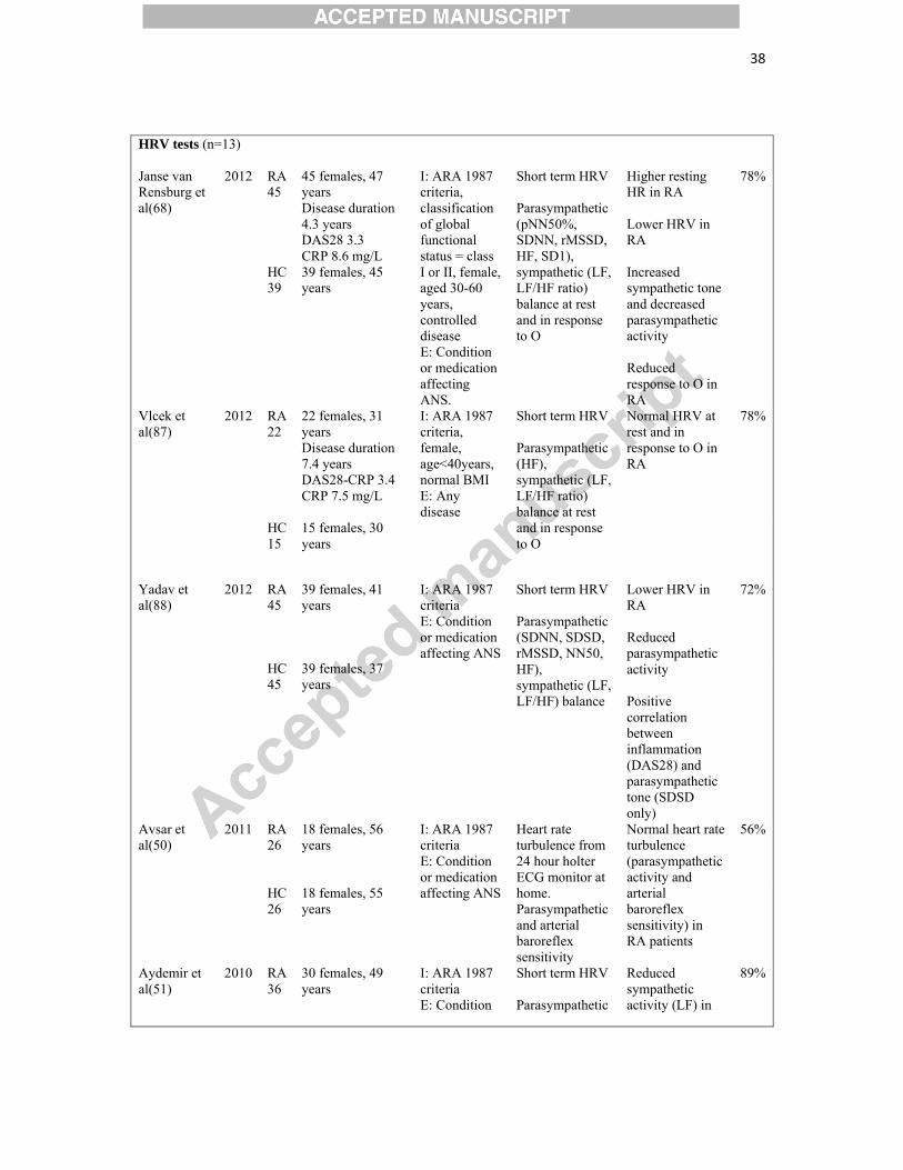
HRV tests (n=13) Janse van Rensburg et al(68)
2012 RA 45
45 females, 47 years Disease duration 4.3 years DAS28 3.3 CRP 8.6 mg/L
I: ARA 1987 criteria, classification of global functional status = class I or II, female, aged 30-60 years, controlled disease E: Condition or medication affecting ANS.
Short term HRV Parasympathetic (pNN50%, SDNN, rMSSD, HF, SD1), sympathetic (LF, LF/HF ratio) balance at rest and in response to O
Higher resting HR in RA Lower HRV in RA Increased sympathetic tone and decreased parasympathetic activity Reduced response to O in RA
78%
HC 39
39 females, 45 years
Vlcek et al(87)
2012 RA 22
22 females, 31 years Disease duration 7.4 years DAS28-CRP 3.4 CRP 7.5 mg/L
I: ARA 1987 criteria, female, age<40years, normal BMI E: Any disease
Short term HRV Parasympathetic (HF), sympathetic (LF, LF/HF ratio) balance at rest and in response to O
Normal HRV at rest and in response to O in RA
78%
HC 15
15 females, 30 years
Yadav et al(88)
2012 RA 45
39 females, 41 years
I: ARA 1987 criteria E: Condition or medication affecting ANS
Short term HRV Parasympathetic (SDNN, SDSD, rMSSD, NN50, HF), sympathetic (LF, LF/HF) balance
Lower HRV in RA Reduced parasympathetic activity Positive correlation between inflammation (DAS28) and parasympathetic tone (SDSD only)
72%
HC 45
39 females, 37 years
Avsar et al(50)
2011 RA 26
18 females, 56 years
I: ARA 1987 criteria E: Condition or medication affecting ANS
Heart rate turbulence from 24 hour holter ECG monitor at home. Parasympathetic and arterial baroreflex sensitivity
Normal heart rate turbulence (parasympathetic activity and arterial baroreflex sensitivity) in RA patients
56%
HC 26
18 females, 55 years
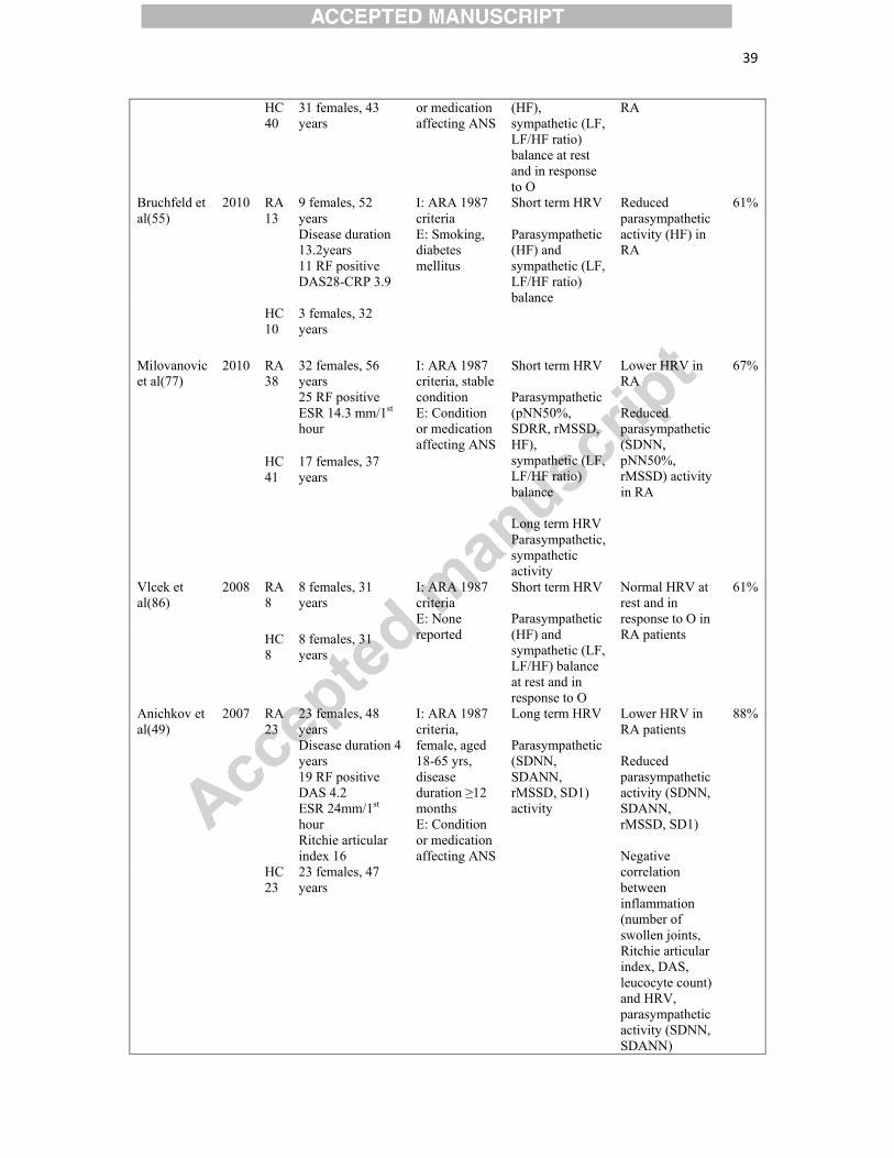
Aydemir et al(51)
2010 RA 36
30 females, 49 years
I: ARA 1987 criteria E: Condition
Short term HRV Parasympathetic
Reduced sympathetic activity (LF) in
89%
39
HC 40
31 females, 43 years
or medication affecting ANS
(HF), sympathetic (LF, LF/HF ratio) balance at rest and in response to O
RA
Bruchfeld et al(55)
2010 RA 13
9 females, 52 years Disease duration 13.2years 11 RF positive DAS28-CRP 3.9
I: ARA 1987 criteria E: Smoking, diabetes mellitus
Short term HRV Parasympathetic (HF) and sympathetic (LF, LF/HF ratio) balance
Reduced parasympathetic activity (HF) in RA
61%
HC 10
3 females, 32 years
Milovanovic et al(77)
2010 RA 38
32 females, 56 years 25 RF positive ESR 14.3 mm/1st hour
I: ARA 1987 criteria, stable condition E: Condition or medication affecting ANS
Short term HRV Parasympathetic (pNN50%, SDRR, rMSSD, HF), sympathetic (LF, LF/HF ratio) balance Long term HRV Parasympathetic, sympathetic activity
Lower HRV in RA Reduced parasympathetic (SDNN, pNN50%, rMSSD) activity in RA
67%
HC 41
17 females, 37 years
Vlcek et al(86)
2008 RA 8
8 females, 31 years
I: ARA 1987 criteria E: None reported
Short term HRV Parasympathetic (HF) and sympathetic (LF, LF/HF) balance at rest and in response to O
Normal HRV at rest and in response to O in RA patients
61%
HC 8
8 females, 31 years
Anichkov et al(49)
2007 RA 23
23 females, 48 years Disease duration 4 years 19 RF positive DAS 4.2 ESR 24mm/1st hour Ritchie articular index 16
I: ARA 1987 criteria, female, aged 18-65 yrs, disease duration ≥12 months E: Condition or medication affecting ANS
Long term HRV Parasympathetic (SDNN, SDANN, rMSSD, SD1) activity
Lower HRV in RA patients Reduced parasympathetic activity (SDNN, SDANN, rMSSD, SD1) Negative correlation between inflammation (number of swollen joints, Ritchie articular index, DAS, leucocyte count) and HRV, parasympathetic activity (SDNN, SDANN)
88%
HC 23
23 females, 47 years
40
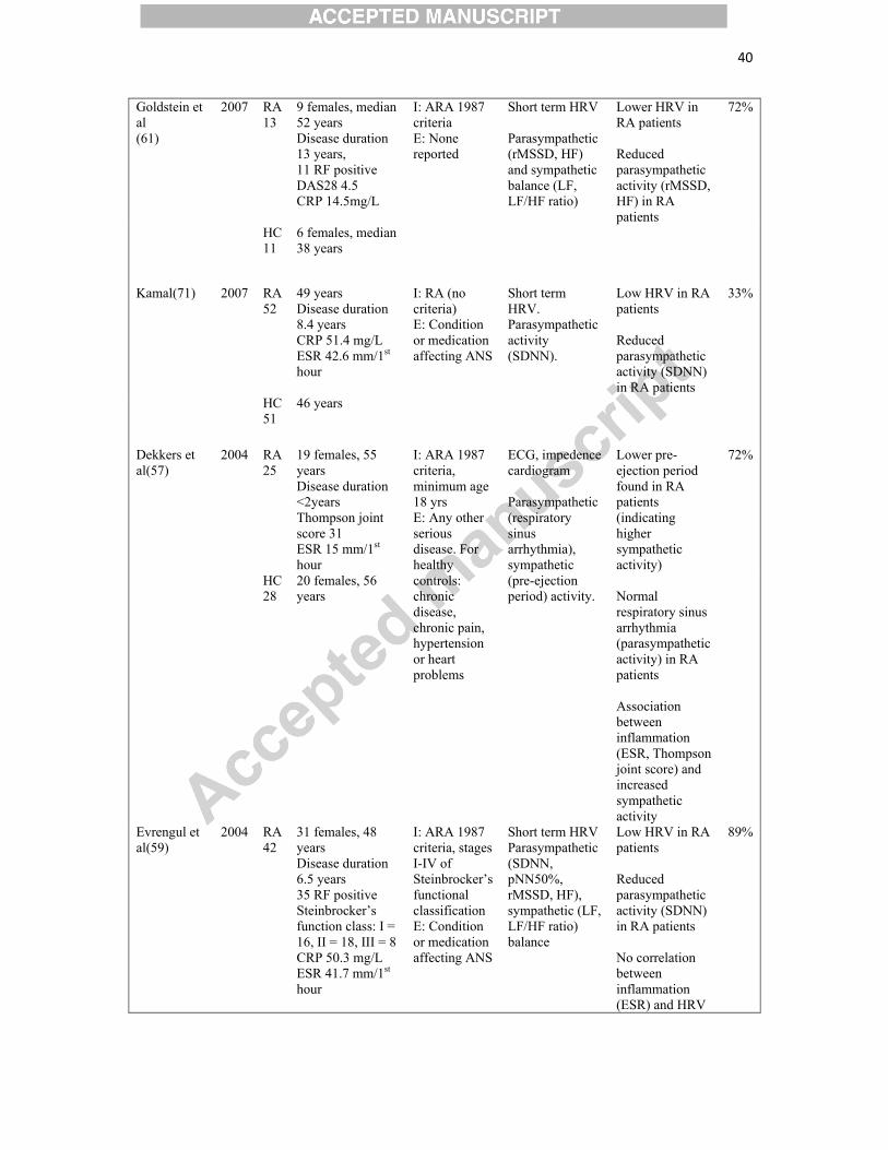
Goldstein et al (61)
2007 RA 13
9 females, median 52 years Disease duration 13 years, 11 RF positive DAS28 4.5 CRP 14.5mg/L
I: ARA 1987 criteria E: None reported
Short term HRV Parasympathetic (rMSSD, HF) and sympathetic balance (LF, LF/HF ratio)
Lower HRV in RA patients Reduced parasympathetic activity (rMSSD, HF) in RA patients
72%
HC 11
6 females, median 38 years
Kamal(71) 2007 RA 52
49 years Disease duration 8.4 years CRP 51.4 mg/L ESR 42.6 mm/1st hour
I: RA (no criteria) E: Condition or medication affecting ANS
Short term HRV. Parasympathetic activity (SDNN).
Low HRV in RA patients Reduced parasympathetic activity (SDNN) in RA patients
33%
HC 51
46 years
Dekkers et al(57)
2004 RA 25
19 females, 55 years Disease duration <2years Thompson joint score 31 ESR 15 mm/1st hour
I: ARA 1987 criteria, minimum age 18 yrs E: Any other serious disease. For healthy controls: chronic disease, chronic pain, hypertension or heart problems
ECG, impedence cardiogram Parasympathetic (respiratory sinus arrhythmia), sympathetic (pre-ejection period) activity.
Lower pre-ejection period found in RA patients (indicating higher sympathetic activity) Normal respiratory sinus arrhythmia (parasympathetic activity) in RA patients Association between inflammation (ESR, Thompson joint score) and increased sympathetic activity
72%
HC 28
20 females, 56 years
Evrengul et al(59)
2004 RA 42
31 females, 48 years Disease duration 6.5 years 35 RF positive Steinbrocker’s function class: I = 16, II = 18, III = 8 CRP 50.3 mg/L ESR 41.7 mm/1st hour
I: ARA 1987 criteria, stages I-IV of Steinbrocker’s functional classification E: Condition or medication affecting ANS
Short term HRV Parasympathetic (SDNN, pNN50%, rMSSD, HF), sympathetic (LF, LF/HF ratio) balance
Low HRV in RA patients Reduced parasympathetic activity (SDNN) in RA patients No correlation between inflammation (ESR) and HRV
89%
41
HC 44
31 females, 45 years
parameters
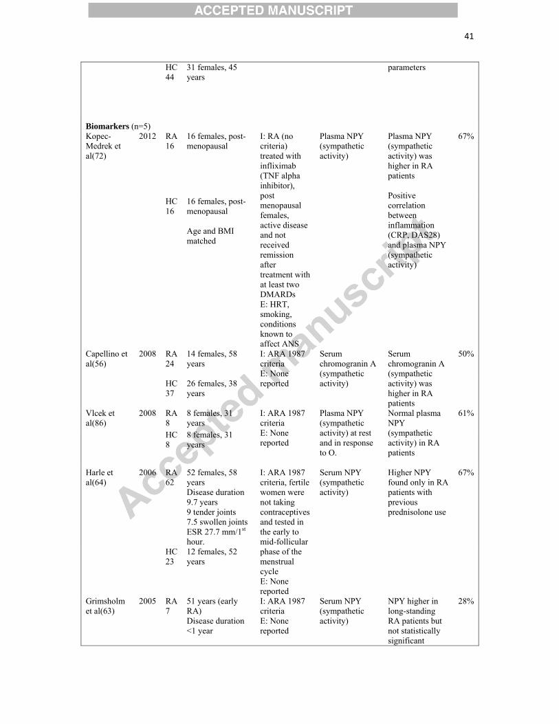
Biomarkers (n=5) Kopec-Medrek et al(72)
2012 RA 16
16 females, post-menopausal
I: RA (no criteria) treated with infliximab (TNF alpha inhibitor), post menopausal females, active disease and not received remission after treatment with at least two DMARDs E: HRT, smoking, conditions known to affect ANS
Plasma NPY (sympathetic activity)
Plasma NPY (sympathetic activity) was higher in RA patients Positive correlation between inflammation (CRP, DAS28) and plasma NPY (sympathetic activity)
67%
HC 16
16 females, post-menopausal Age and BMI matched
Capellino et al(56)
2008 RA 24
14 females, 58 years
I: ARA 1987 criteria E: None reported
Serum chromogranin A (sympathetic activity)
Serum chromogranin A (sympathetic activity) was higher in RA patients
50%
HC 37
26 females, 38 years
Vlcek et al(86)
2008 RA 8
8 females, 31 years
I: ARA 1987 criteria E: None reported
Plasma NPY (sympathetic activity) at rest and in response to O.
Normal plasma NPY (sympathetic activity) in RA patients
61%
HC 8
8 females, 31 years
Harle et al(64)
2006 RA 62
52 females, 58 years Disease duration 9.7 years 9 tender joints 7.5 swollen joints ESR 27.7 mm/1st hour.
I: ARA 1987 criteria, fertile women were not taking contraceptives and tested in the early to mid-follicular phase of the menstrual cycle E: None reported
Serum NPY (sympathetic activity)
Higher NPY found only in RA patients with previous prednisolone use
67%
HC 23
12 females, 52 years
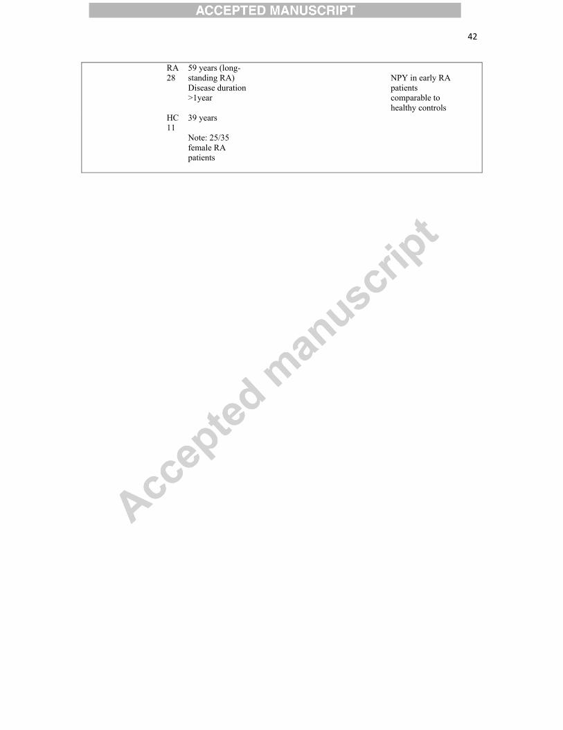
Grimsholm et al(63)
2005 RA 7
51 years (early RA) Disease duration <1 year
I: ARA 1987 criteria E: None reported
Serum NPY (sympathetic activity)
NPY higher in long-standing RA patients but not statistically significant
28%
42
RA 28
59 years (long-standing RA) Disease duration >1year
NPY in early RA patients comparable to healthy controls
HC 11
39 years Note: 25/35 female RA patients
43
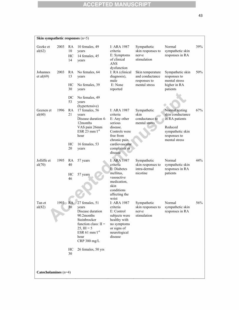
Skin sympathetic responses (n=5) Gozke et al(62)
2003 RA 10
10 females, 49 years
I: ARA 1987 criteria E: Symptoms of clinical ANS dysfunction
Sympathetic skin responses to nerve stimulation
Normal sympathetic skin responses in RA
39%
HC 14
14 females, 45 years
Johannes et al(69)
2003 RA 13
No females, 64 years
I: RA (clinical diagnosis), male E: None reported
Skin temperature and conductance responses to mental stress
Sympathetic skin responses to mental stress higher in RA patients
50%
HC 30
No females, 39 years
DC 53
No females, 49 years (hypertensive)
Geenen et al(60)
1996 RA 21
17 females, 56 years Disease duration 4-12months VAS pain 26mm ESR 23 mm/1st hour
I: ARA 1987 criteria E: Any other serious disease. Controls were free from chronic pain, cardiovascular complaints or disease
Sympathetic skin conductance to mental stress
Normal resting skin conductance in RA patients Reduced sympathetic skin responses to mental stress
67%
HC 20
16 females, 53 years
Jolliffe et al(70)
1995 RA 40
57 years I: ARA 1987 criteria E: Diabetes mellitus, vasoactive medication, skin conditions affecting the wrist
Sympathetic skin responses to intra-dermal nicotine
Normal sympathetic skin responses in RA patients
44%
HC 46
57 years
Tan et al(82)
1993 RA 30
27 females, 51 years Disease duration 90.2months Steinbrocker function class: II = 25, III = 5 ESR 61 mm/1st hour CRP 380 mg/L
I: ARA 1987 criteria E: Control subjects were healthy with no symptoms or signs of neurological disease
Sympathetic skin responses to nerve stimulation
Normal sympathetic skin responses in RA
56%
HC 30
26 females, 50 yrs
Catecholamines (n=4)
44
Vlcek et al(87)
2012 RA 22
22 females, 31 years Disease duration 7.4 years DAS28-CRP 3.4 CRP 7.5 mg/L
I: ARA 1987 criteria, female, age<40yrs, normal BMI E: Any disease
Plasma EPI and NE (sympathetic activity) at rest and in response to O
Normal EPI and NE (sympathetic activity) at rest and in response to O in RA
78%
HC 15
15 females, 30 years
Vlcek et al(86)
2008 RA 8
8 females, 31 years I: ARA 1987 criteria E: None reported
Plasma EPI and NE (sympathetic activity) at rest and in response to O
Baseline plasma NE (sympathetic activity) was higher in RA patients There was a trend for reduced plasma NE (sympathetic activity) in RA patients
61%
HC 8
8 females, 31 years
Imrich et al(67)
2005 RA 15
15 females, 41 years Disease duration 8.2 years CRP 15.4mg/L ESR 20.3 mm/1st hour
I: ARA 1987 criteria, female E: Diabetes, impaired glucose tolerance
Serum EPI and NE (sympathetic activity) at rest and in response to insulin-induced hypoglycaemia
Basal and cumulative levels of EPI were reduced (but not statistically significantly) in RA patients Basal levels of NE were normal in RA patients, but cumulative levels were reduced (reduced basal sympathetic activity). Serum EPI response to insulin-induced hypoglycaemia was normal in RA however serum NE response was reduced in RA (impaired sympathetic response)
67%
HC 14
14 females, 44 years
Igari et al(66)
1977 RA 22
20 females, 45 yrs ESR 44.8mm/1st hr, Steinbrocker class 2.5
I: Ropes et al 1958 criteria for classical or definite RA E: None reported
24 hour urinary adrenaline and noradrenaline (sympathetic activity)
Baseline 24 hour urinary adrenaline was reduced in RA patients
44%
HC 6
2 females, 33 years
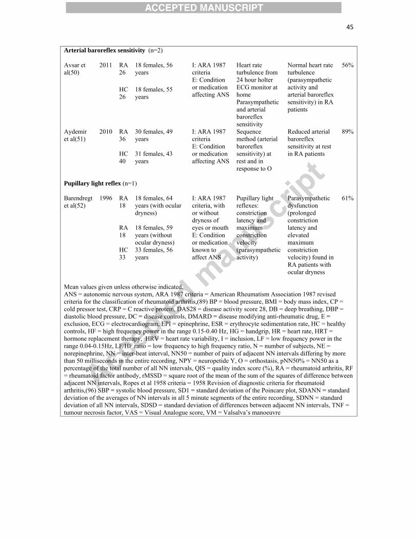
45
Arterial baroreflex sensitivity (n=2) Avsar et al(50)
2011 RA 26
18 females, 56 years
I: ARA 1987 criteria E: Condition or medication affecting ANS
Heart rate turbulence from 24 hour holter ECG monitor at home Parasympathetic and arterial baroreflex sensitivity
Normal heart rate turbulence (parasympathetic activity and arterial baroreflex sensitivity) in RA patients
56%
HC 26
18 females, 55 years
Aydemir et al(51)
2010 RA 36
30 females, 49 years
I: ARA 1987 criteria E: Condition or medication affecting ANS
Sequence method (arterial baroreflex sensitivity) at rest and in response to O
Reduced arterial baroreflex sensitivity at rest in RA patients
89%
HC 40
31 females, 43 years
Pupillary light reflex (n=1) Barendregt et al(52)
1996 RA 18
18 females, 64 years (with ocular dryness)
I: ARA 1987 criteria, with or without dryness of eyes or mouth E: Condition or medication known to affect ANS
Pupillary light reflexes: constriction latency and maximum constriction velocity (parasympathetic activity)
Parasympathetic dysfunction (prolonged constriction latency and elevated maximum constriction velocity) found in RA patients with ocular dryness
61%
RA 18
18 females, 59 years (without ocular dryness)
HC 33
33 females, 56 years
Mean values given unless otherwise indicated. ANS = autonomic nervous system, ARA 1987 criteria = American Rheumatism Association 1987 revised criteria for the classification of rheumatoid arthritis,(89) BP = blood pressure, BMI = body mass index, CP = cold pressor test, CRP = C reactive protein, DAS28 = disease activity score 28, DB = deep breathing, DBP = diastolic blood pressure, DC = disease controls, DMARD = disease modifying anti-rheumatic drug, E = exclusion, ECG = electrocardiogram, EPI = epinephrine, ESR = erythrocyte sedimentation rate, HC = healthy controls, HF = high frequency power in the range 0.15-0.40 Hz, HG = handgrip, HR = heart rate, HRT = hormone replacement therapy, HRV = heart rate variability, I = inclusion, LF = low frequency power in the range 0.04-0.15Hz, LF/HF ratio = low frequency to high frequency ratio, N = number of subjects, NE = norepinephrine, NN = inter-beat interval, NN50 = number of pairs of adjacent NN intervals differing by more than 50 milliseconds in the entire recording, NPY = neuropetide Y, O = orthostasis, pNN50% = NN50 as a percentage of the total number of all NN intervals, QIS = quality index score (%), RA = rheumatoid arthritis, RF = rheumatoid factor antibody, rMSSD = square root of the mean of the sum of the squares of difference between adjacent NN intervals, Ropes et al 1958 criteria = 1958 Revision of diagnostic criteria for rheumatoid arthritis,(96) SBP = systolic blood pressure, SD1 = standard deviation of the Poincare plot, SDANN = standard deviation of the averages of NN intervals in all 5 minute segments of the entire recording, SDNN = standard deviation of all NN intervals, SDSD = standard deviation of differences between adjacent NN intervals, TNF = tumour necrosis factor, VAS = Visual Analogue score, VM = Valsalva’s manoeuvre
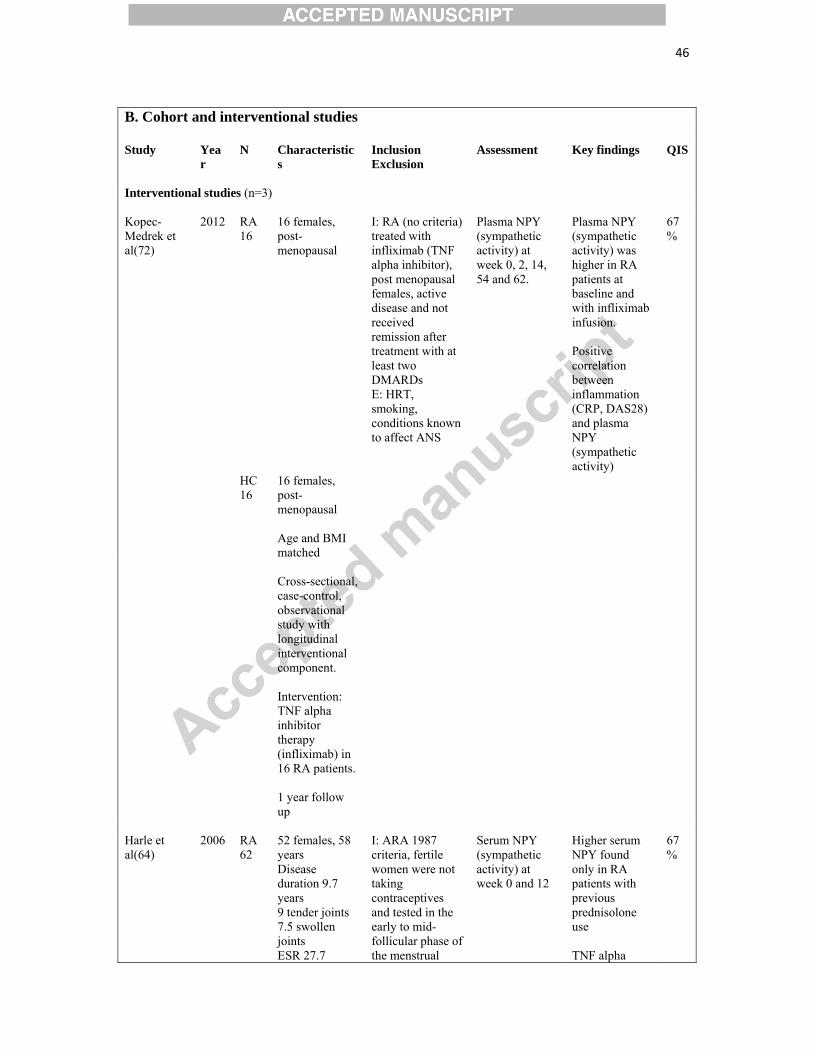
46
B. Cohort and interventional studies Study Yea
r N Characteristic
s Inclusion Exclusion
Assessment Key findings QIS
Interventional studies (n=3) Kopec-Medrek et al(72)
2012 RA 16
16 females, post-menopausal
I: RA (no criteria) treated with infliximab (TNF alpha inhibitor), post menopausal females, active disease and not received remission after treatment with at least two DMARDs E: HRT, smoking, conditions known to affect ANS
Plasma NPY (sympathetic activity) at week 0, 2, 14, 54 and 62.
Plasma NPY (sympathetic activity) was higher in RA patients at baseline and with infliximab infusion. Positive correlation between inflammation (CRP, DAS28) and plasma NPY (sympathetic activity)
67%
HC 16
16 females, post-menopausal Age and BMI matched
Cross-sectional, case-control, observational study with longitudinal interventional component. Intervention: TNF alpha inhibitor therapy (infliximab) in 16 RA patients. 1 year follow up
Harle et al(64)
2006 RA 62
52 females, 58 years Disease duration 9.7 years 9 tender joints 7.5 swollen joints ESR 27.7
I: ARA 1987 criteria, fertile women were not taking contraceptives and tested in the early to mid-follicular phase of the menstrual
Serum NPY (sympathetic activity) at week 0 and 12
Higher serum NPY found only in RA patients with previous prednisolone use TNF alpha
67%
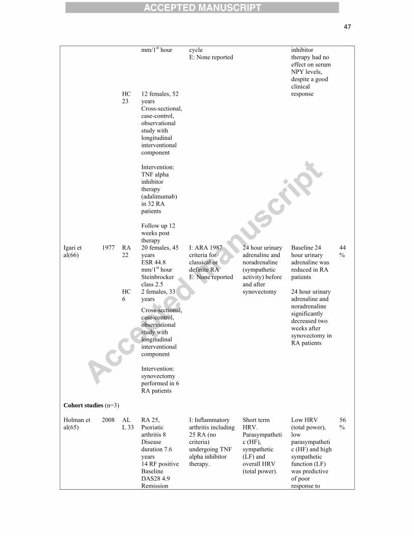
47
mm/1st hour cycle E: None reported
inhibitor therapy had no effect on serum NPY levels, despite a good clinical response HC
23 12 females, 52 years
Cross-sectional, case-control, observational study with longitudinal interventional component Intervention: TNF alpha inhibitor therapy (adalimumab) in 32 RA patients Follow up 12 weeks post therapy
Igari et al(66)
1977 RA 22
20 females, 45 years ESR 44.8 mm/1st hour Steinbrocker class 2.5
I: ARA 1987 criteria for classical or definite RA E: None reported
24 hour urinary adrenaline and noradrenaline (sympathetic activity) before and after synovectomy
Baseline 24 hour urinary adrenaline was reduced in RA patients 24 hour urinary adrenaline and noradrenaline significantly decreased two weeks after synovectomy in RA patients
44%
HC 6
2 females, 33 years
Cross-sectional, case-control, observational study with longitudinal interventional component Intervention: synovectomy performed in 6 RA patients
Cohort studies (n=3) Holman et al(65)
2008 ALL 33
RA 25, Psoriatic arthritis 8 Disease duration 7.6 years 14 RF positive Baseline DAS28 4.9 Remission
I: Inflammatory arthritis including 25 RA (no criteria) undergoing TNF alpha inhibitor therapy.
Short term HRV. Parasympathetic (HF), sympathetic (LF) and overall HRV (total power).
Low HRV (total power), low parasympathetic (HF) and high sympathetic function (LF) was predictive of poor response to
56%
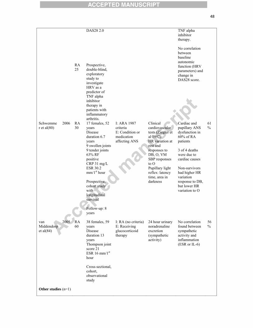
48
DAS28 2.0 TNF alpha inhibitor therapy. No correlation between baseline autonomic function (HRV parameters) and change in DAS28 score.
RA 25
Prospective, double-blind, exploratory study to investigate HRV as a predictor of TNF alpha inhibitor therapy in patients with inflammatory arthritis.
Schwemmer et al(80)
2006 RA 30
17 females, 52 years Disease duration 6.7 years 9 swollen joints 9 tender joints 63% RF positive CRP 31 mg/L ESR 30.2 mm/1st hour Prospective, cohort study with longitudinal survival Follow-up: 8 years
I: ARA 1987 criteria E: Condition or medication affecting ANS
Clinical cardiovascular tests (Ziegler et al 1992) HR variation at rest and responses to DB, O, VM SBP responses to O Pupillary light reflex: latency time, area in darkness
Cardiac and pupillary ANS dysfunction in 60% of RA patients 3 of 4 deaths were due to cardiac causes Non-survivors had higher HR variation response to DB, but lower HR variation to O
61%
van Middendorp et al(84)
2005 RA 60
38 females, 59 years Disease duration 13 years Thompson joint score 21 ESR 16 mm/1st hour Cross-sectional, cohort, observational study
I: RA (no criteria) E: Receiving glucocorticoid therapy
24 hour urinary noradrenaline excretion (sympathetic activity)
No correlation found between sympathetic activity and inflammation (ESR or IL-6)
56%
Other studies (n=1)
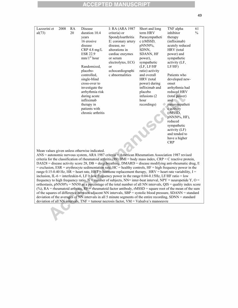
49
Lazzerini et al(73)
2008 RA 20
Disease duration 10.4 years 16 erosive disease CRP 4.8 mg/L ESR 22.9 mm/1st hour Randomized, placebo-controlled, single-blind cross-over to investigate the arrhythmia risk during acute infliximab therapy in patients with chronic arthritis
I: RA (ARA 1987 criteria) or Spondyloarthritis E: coronary artery disease, no alterations in cardiac enzymes or serum electrolytes, ECG or echocardiographic abnormalities
Short and long term HRV Parasympathetic (rMSSD, pNN50%, SDNN, SDANN, HF power), sympathetic (LF, LF/HF ratio) activity and overall HRV (total power) during infliximab and placebo infusions (2 hour recordings)
TNF alpha inhibitor therapy (infliximab) acutely reduced HRV (total power) and sympathetic activity (LF, LF/HF) Patients who developed new-onset arrhythmia had reduced HRV (total power) and parasympathetic activity (rMSSD, pNN50%, HF), reduced sympathetic activity (LF) and tended to have a higher CRP
61%
Mean values given unless otherwise indicated. ANS = autonomic nervous system, ARA 1987 criteria = American Rheumatism Association 1987 revised criteria for the classification of rheumatoid arthritis,(89) BMI = body mass index, CRP = C reactive protein, DAS28 = disease activity score 28, DB = deep breathing, DMARD = disease modifying anti-rheumatic drug, E = exclusion, ESR = erythrocyte sedimentation rate, HC = healthy controls, HF = high frequency power in the range 0.15-0.40 Hz, HR = heart rate, HRT = hormone replacement therapy, HRV = heart rate variability, I = inclusion, IL-6 = interleukin-6, LF = low frequency power in the range 0.04-0.15Hz, LF/HF ratio = low frequency to high frequency ratio, N = number of subjects, NN= inter-beat interval, NPY = neuropetide Y, O = orthostasis, pNN50% = NN50 as a percentage of the total number of all NN intervals, QIS = quality index score (%), RA = rheumatoid arthritis, RF = rheumatoid factor antibody, rMSSD = square root of the mean of the sum of the squares of difference between adjacent NN intervals, SBP = systolic blood pressure, SDANN = standard deviation of the averages of NN intervals in all 5 minute segments of the entire recording, SDNN = standard deviation of all NN intervals, TNF = tumour necrosis factor, VM = Valsalva’s manoeuvre
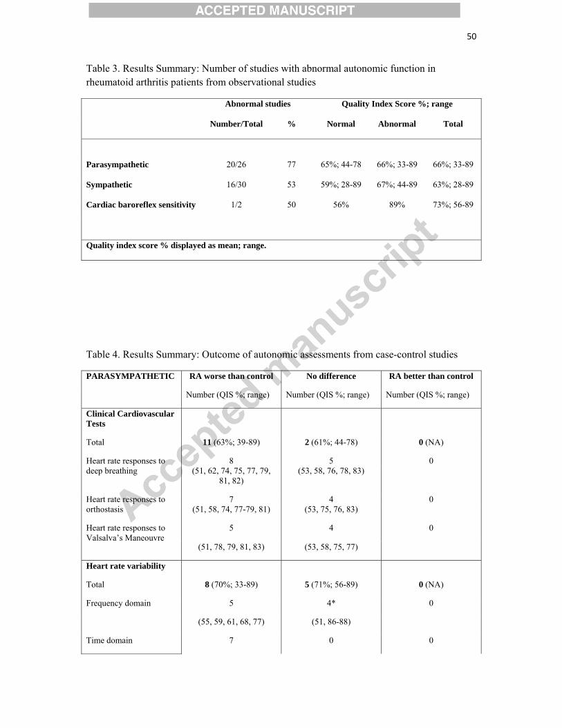
50
Table 3. Results Summary: Number of studies with abnormal autonomic function in rheumatoid arthritis patients from observational studies
Abnormal studies Quality Index Score %; range
Number/Total % Normal Abnormal Total
Parasympathetic 20/26 77 65%; 44-78 66%; 33-89 66%; 33-89
Sympathetic 16/30 53 59%; 28-89 67%; 44-89 63%; 28-89
Cardiac baroreflex sensitivity 1/2 50 56% 89% 73%; 56-89
Quality index score % displayed as mean; range.
Table 4. Results Summary: Outcome of autonomic assessments from case-control studies
PARASYMPATHETIC RA worse than control No difference RA better than control
Number (QIS %; range) Number (QIS %; range) Number (QIS %; range)
Clinical Cardiovascular Tests
Total 11 (63%; 39-89) 2 (61%; 44-78) 0 (NA)
Heart rate responses to deep breathing
8 5 0 (51, 62, 74, 75, 77, 79,
81, 82) (53, 58, 76, 78, 83)
Heart rate responses to orthostasis
7 4 0 (51, 58, 74, 77-79, 81) (53, 75, 76, 83)
Heart rate responses to Valsalva’s Maneouvre
5 4 0
(51, 78, 79, 81, 83) (53, 58, 75, 77)
Heart rate variability
Total 8 (70%; 33-89) 5 (71%; 56-89) 0 (NA)
Frequency domain 5 4* 0
(55, 59, 61, 68, 77) (51, 86-88)
Time domain 7 0 0
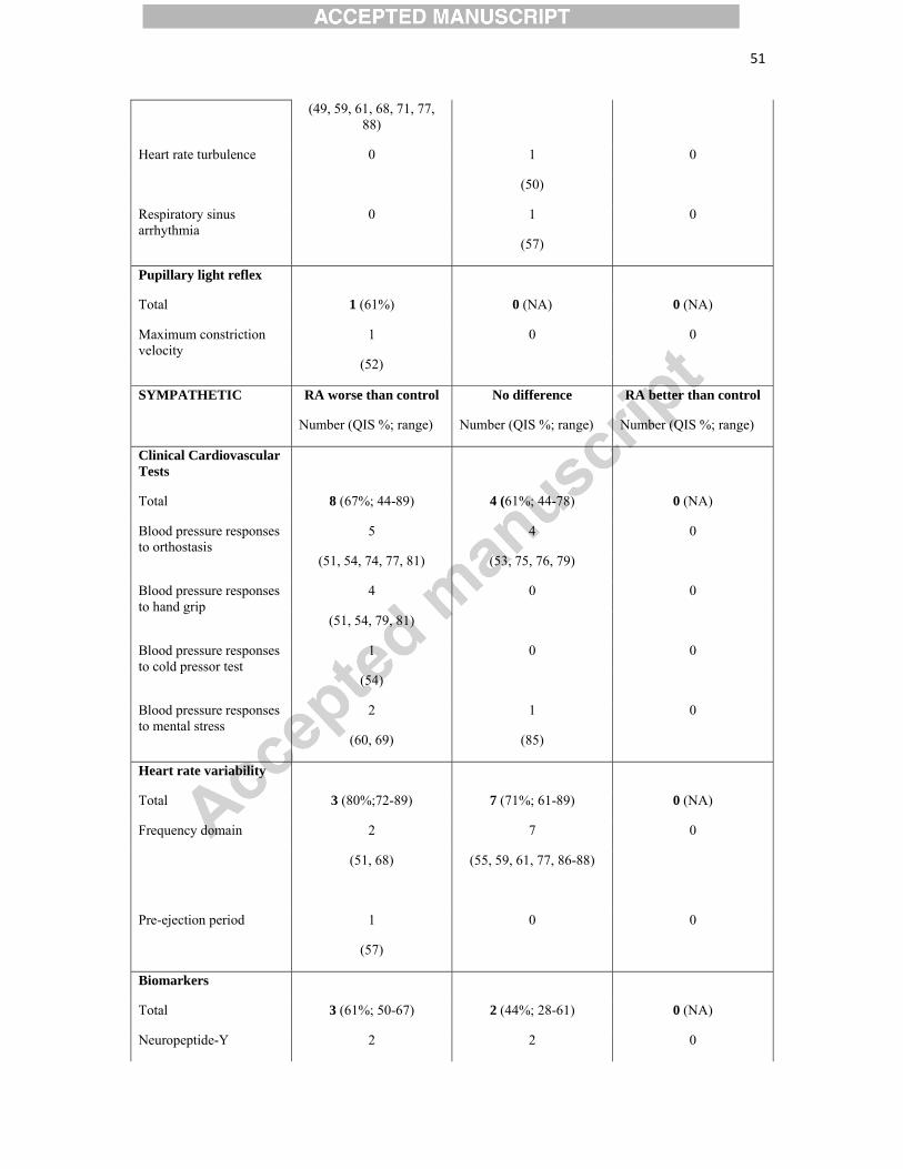
51
(49, 59, 61, 68, 71, 77, 88)
Heart rate turbulence 0 1 0
(50)
Respiratory sinus arrhythmia
0 1 0
(57)
Pupillary light reflex
Total 1 (61%) 0 (NA) 0 (NA)
Maximum constriction velocity
1 0 0
(52)
SYMPATHETIC RA worse than control No difference RA better than control
Number (QIS %; range) Number (QIS %; range) Number (QIS %; range)
Clinical Cardiovascular Tests
Total 8 (67%; 44-89) 4 (61%; 44-78) 0 (NA)
Blood pressure responses to orthostasis
5 4 0
(51, 54, 74, 77, 81) (53, 75, 76, 79)
Blood pressure responses to hand grip
4 0 0
(51, 54, 79, 81)
Blood pressure responses to cold pressor test
1 0 0
(54)
Blood pressure responses to mental stress
2 1 0
(60, 69) (85)
Heart rate variability
Total 3 (80%;72-89) 7 (71%; 61-89) 0 (NA)
Frequency domain 2 7 0
(51, 68)
(55, 59, 61, 77, 86-88)
Pre-ejection period 1 0 0
(57)
Biomarkers
Total 3 (61%; 50-67) 2 (44%; 28-61) 0 (NA)
Neuropeptide-Y 2 2 0
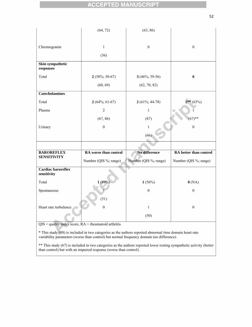
52
(64, 72)
(63, 86)
Chromogranin 1 0 0
(56)
Skin sympathetic responses
Total 2 (58%; 50-67) 3 (46%; 39-56) 0
(60, 69) (62, 70, 82)
Catecholamines
Total 2 (64%; 61-67) 2 (61%; 44-78) 1** (63%)
Plasma 2 1 1
(67, 86) (87) (67)**
Urinary 0 1 0
(66)
BAROREFLEX SENSITIVITY
RA worse than control No difference RA better than control
Number (QIS %; range) Number (QIS %; range) Number (QIS %; range)
Cardiac baroreflex sensitivity
Total 1 (89%) 1 (56%) 0 (NA)
Spontaneous 1 0 0
(51)
Heart rate turbulence 0 1 0
(50)
QIS = quality index score, RA = rheumatoid arthritis.
* This study (88) is included in two categories as the authors reported abnormal time domain heart rate variability parameters (worse than control) but normal frequency domain (no difference).
** This study (67) is included in two categories as the authors reported lower resting sympathetic activity (better than control) but with an impaired response (worse than control).
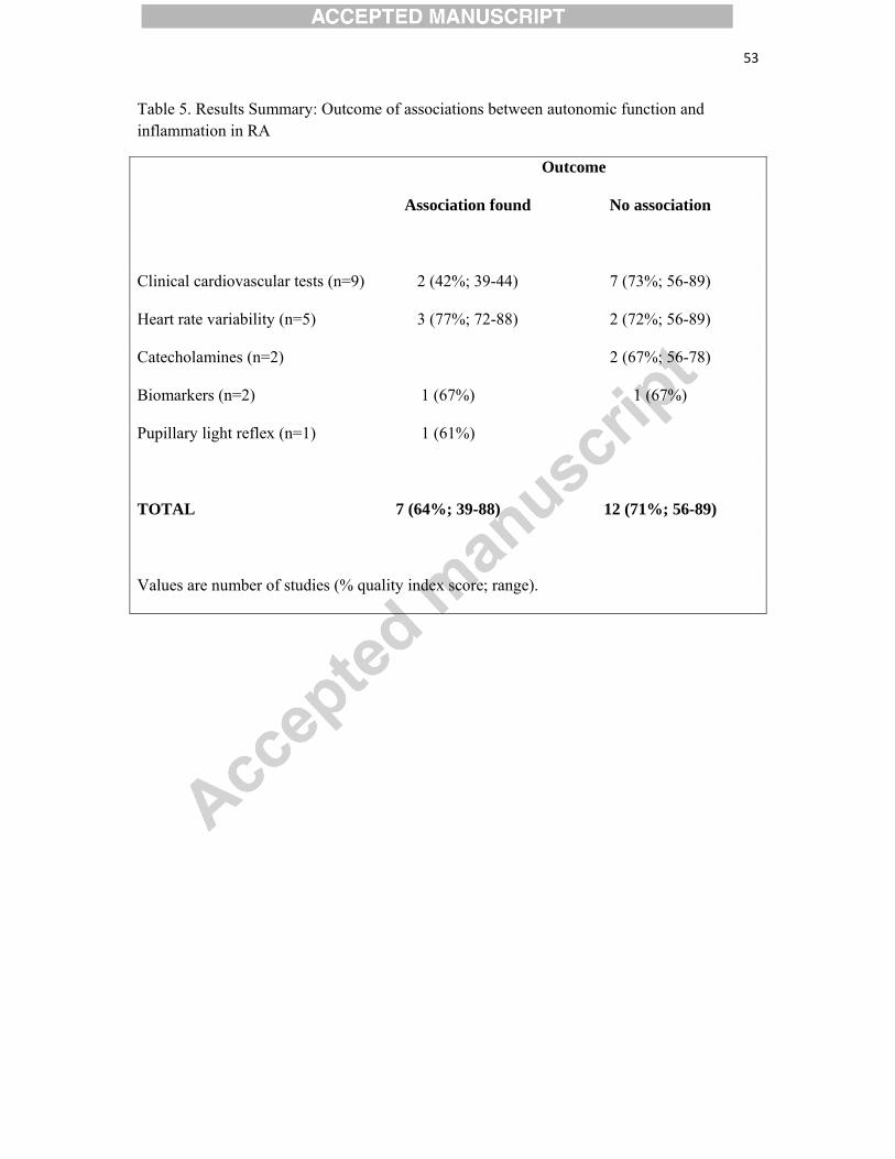
53
Table 5. Results Summary: Outcome of associations between autonomic function and inflammation in RA
Outcome
Association found No association
Clinical cardiovascular tests (n=9) 2 (42%; 39-44) 7 (73%; 56-89)
Heart rate variability (n=5) 3 (77%; 72-88) 2 (72%; 56-89)
Catecholamines (n=2) 2 (67%; 56-78)
Biomarkers (n=2) 1 (67%) 1 (67%)
Pupillary light reflex (n=1) 1 (61%)
TOTAL 7 (64%; 39-88) 12 (71%; 56-89)
Values are number of studies (% quality index score; range).
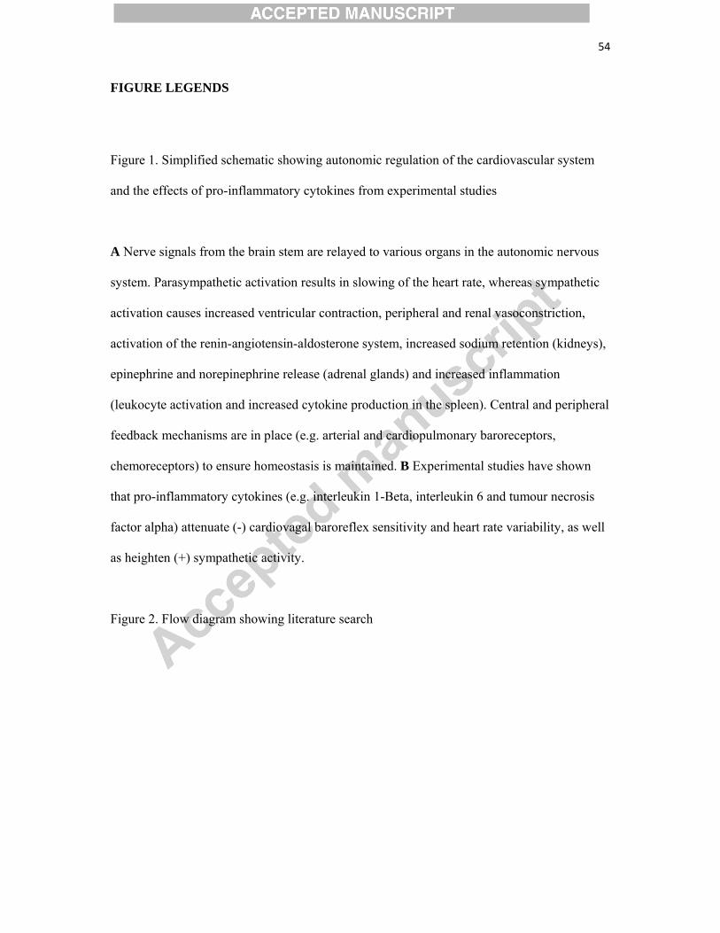
54
FIGURE LEGENDS
Figure 1. Simplified schematic showing autonomic regulation of the cardiovascular system
and the effects of pro-inflammatory cytokines from experimental studies
A Nerve signals from the brain stem are relayed to various organs in the autonomic nervous
system. Parasympathetic activation results in slowing of the heart rate, whereas sympathetic
activation causes increased ventricular contraction, peripheral and renal vasoconstriction,
activation of the renin-angiotensin-aldosterone system, increased sodium retention (kidneys),
epinephrine and norepinephrine release (adrenal glands) and increased inflammation
(leukocyte activation and increased cytokine production in the spleen). Central and peripheral
feedback mechanisms are in place (e.g. arterial and cardiopulmonary baroreceptors,
chemoreceptors) to ensure homeostasis is maintained. B Experimental studies have shown
that pro-inflammatory cytokines (e.g. interleukin 1-Beta, interleukin 6 and tumour necrosis
factor alpha) attenuate (-) cardiovagal baroreflex sensitivity and heart rate variability, as well
as heighten (+) sympathetic activity.
Figure 2. Flow diagram showing literature search
55
REFERENCES
1. Solomon DH, Karlson EW, Rimm EB, Cannuscio CC, Mandl LA, Manson JE, et al. Cardiovascular morbidity and mortality in women diagnosed with rheumatoid arthritis. Circulation. 2003;107(9):1303‐7. 2. Maradit‐Kremers H, Nicola PJ, Crowson CS, Ballman KV, Gabriel SE. Cardiovascular death in rheumatoid arthritis: a population‐based study. Arthritis and rheumatism. 2005;52(3):722‐32. 3. Maradit‐Kremers H, Crowson CS, Nicola PJ, Ballman KV, Roger VL, Jacobsen SJ, et al. Increased unrecognized coronary heart disease and sudden deaths in rheumatoid arthritis: a population‐based cohort study. Arthritis and rheumatism. 2005;52(2):402‐11. 4. Kapetanovic MC, Lindqvist E, Geborek P, Saxne T, Eberhard K. Long‐term mortality rate in rheumatoid arthritis patients with disease onset in the 1980s. Scandinavian journal of rheumatology. 2011;40(6):433‐8. 5. Alkaabi JK, Ho M, Levison R, Pullar T, Belch JJ. Rheumatoid arthritis and macrovascular disease. Rheumatology. 2003;42(2):292‐7. 6. Kitas GD, Erb N. Tackling ischaemic heart disease in rheumatoid arthritis. Rheumatology. 2003;42(5):607‐13. 7. Stevens RJ, Douglas KM, Saratzis AN, Kitas GD. Inflammation and atherosclerosis in rheumatoid arthritis. Expert reviews in molecular medicine. 2005;7(7):1‐24. 8. Bhatia GS, Sosin MD, Patel JV, Grindulis KA, Khattak FH, Hughes EA, et al. Left ventricular systolic dysfunction in rheumatoid disease: an unrecognized burden? Journal of the American College of Cardiology. 2006;47(6):1169‐74. 9. Arosio E, De Marchi S, Rigoni A, Prior M, Delva P, Lechi A. Forearm haemodynamics, arterial stiffness and microcirculatory reactivity in rheumatoid arthritis. Journal of hypertension. 2007;25(6):1273‐8. 10. Panoulas VF, Toms TE, Metsios GS, Stavropoulos‐Kalinoglou A, Kosovitsas A, Milionis HJ, et al. Target organ damage in patients with rheumatoid arthritis: the role of blood pressure and heart rate. Atherosclerosis. 2010;209(1):255‐60. 11. Panoulas VF, Douglas KM, Milionis HJ, Stavropoulos‐Kalinglou A, Nightingale P, Kita MD, et al. Prevalence and associations of hypertension and its control in patients with rheumatoid arthritis. Rheumatology. 2007;46(9):1477‐82. 12. Panoulas VF, Douglas KM, Milionis HJ, Nightingale P, Kita MD, Klocke R, et al. Serum uric acid is independently associated with hypertension in patients with rheumatoid arthritis. Journal of human hypertension. 2008;22(3):177‐82. 13. Straub RH, Cutolo M, Buttgereit F, Pongratz G. Energy regulation and neuroendocrine‐immune control in chronic inflammatory diseases. Journal of internal medicine. 2010;267(6):543‐60. 14. Fisher JP, Young CN, Fadel PJ. Central sympathetic overactivity: maladies and mechanisms. Autonomic neuroscience : basic & clinical. 2009;148(1‐2):5‐15. 15. Abboud FM, Harwani SC, Chapleau MW. Autonomic neural regulation of the immune system: implications for hypertension and cardiovascular disease. Hypertension. 2012;59(4):755‐62. 16. Fisher JP, Paton JF. The sympathetic nervous system and blood pressure in humans: implications for hypertension. Journal of human hypertension. 2012;26(8):463‐75. 17. La Rovere MT, Bigger JT, Jr., Marcus FI, Mortara A, Schwartz PJ. Baroreflex sensitivity and heart‐rate variability in prediction of total cardiac mortality after myocardial infarction. ATRAMI (Autonomic Tone and Reflexes After Myocardial Infarction) Investigators. Lancet. 1998;351(9101):478‐84. 18. Barretto AC, Santos AC, Munhoz R, Rondon MU, Franco FG, Trombetta IC, et al. Increased muscle sympathetic nerve activity predicts mortality in heart failure patients. International journal of cardiology. 2009;135(3):302‐7.
56
19. Cohn JN, Levine TB, Olivari MT, Garberg V, Lura D, Francis GS, et al. Plasma norepinephrine as a guide to prognosis in patients with chronic congestive heart failure. The New England journal of medicine. 1984;311(13):819‐23. 20. Helwig BG, Craig RA, Fels RJ, Blecha F, Kenney MJ. Central nervous system administration of interleukin‐6 produces splenic sympathoexcitation. Autonomic neuroscience : basic & clinical. 2008;141(1‐2):104‐11. 21. Niijima A, Hori T, Aou S, Oomura Y. The effects of interleukin‐1 beta on the activity of adrenal, splenic and renal sympathetic nerves in the rat. Journal of the autonomic nervous system. 1991;36(3):183‐92. 22. Takagishi M, Waki H, Bhuiyan ME, Gouraud SS, Kohsaka A, Cui H, et al. IL‐6 microinjected in the nucleus tractus solitarii attenuates cardiac baroreceptor reflex function in rats. American journal of physiology Regulatory, integrative and comparative physiology. 2010;298(1):R183‐90. 23. Waki H, Gouraud SS, Maeda M, Paton JF. Gene expression profiles of major cytokines in the nucleus tractus solitarii of the spontaneously hypertensive rat. Autonomic neuroscience : basic & clinical. 2008;142(1‐2):40‐4. 24. Grebe KM, Takeda K, Hickman HD, Bailey AL, Embry AC, Bennink JR, et al. Cutting edge: Sympathetic nervous system increases proinflammatory cytokines and exacerbates influenza A virus pathogenesis. Journal of immunology. 2010;184(2):540‐4. 25. Templeton A, Nguyen G, Ash JD, Straub RH, Carr DJ. Chemical sympathectomy increases susceptibility to ocular herpes simplex virus type 1 infection. Journal of neuroimmunology. 2008;197(1):37‐46. 26. Fairchild KD, Saucerman JJ, Raynor LL, Sivak JA, Xiao Y, Lake DE, et al. Endotoxin depresses heart rate variability in mice: cytokine and steroid effects. American journal of physiology Regulatory, integrative and comparative physiology. 2009;297(4):R1019‐27. 27. Eckberg DL. Parasympathetic cardiovascular control in human disease: a critical review of methods and results. The American journal of physiology. 1980;239(5):H581‐93. 28. Ewing DJ, Martyn CN, Young RJ, Clarke BF. The value of cardiovascular autonomic function tests: 10 years experience in diabetes. Diabetes care. 1985;8(5):491‐8. 29. Sherwood A, Allen MT, Fahrenberg J, Kelsey RM, Lovallo WR, van Doornen LJ. Methodological guidelines for impedance cardiography. Psychophysiology. 1990;27(1):1‐23. 30. Takiyyuddin MA, Cervenka JH, Hsiao RJ, Barbosa JA, Parmer RJ, O'Connor DT. Chromogranin A. Storage and release in hypertension. Hypertension. 1990;15(3):237‐46. 31. Berntson GG, Cacioppo JT, Quigley KS. Respiratory sinus arrhythmia: autonomic origins, physiological mechanisms, and psychophysiological implications. Psychophysiology. 1993;30(2):183‐96. 32. Schondorf R. New investigations of autonomic nervous system function. Journal of clinical neurophysiology : official publication of the American Electroencephalographic Society. 1993;10(1):28‐38. 33. Cacioppo JT, Berntson GG, Binkley PF, Quigley KS, Uchino BN, Fieldstone A. Autonomic cardiac control. II. Noninvasive indices and basal response as revealed by autonomic blockades. Psychophysiology. 1994;31(6):586‐98. 34. Straub RH, Thies U, Jeron A, Palitzsch KD, Scholmerich J. Valid parameters for investigation of the pupillary light reflex in normal and diabetic subjects shown by factor analysis and partial correlation. Diabetologia. 1994;37(4):414‐9. 35. Heart rate variability: standards of measurement, physiological interpretation and clinical use. Task Force of the European Society of Cardiology and the North American Society of Pacing and Electrophysiology. Circulation. 1996;93(5):1043‐65. 36. Vetrugno R, Liguori R, Cortelli P, Montagna P. Sympathetic skin response: basic mechanisms and clinical applications. Clinical autonomic research : official journal of the Clinical Autonomic Research Society. 2003;13(4):256‐70.
57
37. Freeman R. Assessment of cardiovascular autonomic function. Clinical neurophysiology : official journal of the International Federation of Clinical Neurophysiology. 2006;117(4):716‐30. 38. Grassi G, Esler M. How to assess sympathetic activity in humans. Journal of hypertension. 1999;17(6):719‐34. 39. Brennan M, Palaniswami M, Kamen P. Poincare plot interpretation using a physiological model of HRV based on a network of oscillators. American journal of physiology Heart and circulatory physiology. 2002;283(5):H1873‐86. 40. Schmidt G, Malik M, Barthel P, Schneider R, Ulm K, Rolnitzky L, et al. Heart‐rate turbulence after ventricular premature beats as a predictor of mortality after acute myocardial infarction. Lancet. 1999;353(9162):1390‐6. 41. Morris MJ, Russell AE, Kapoor V, Cain MD, Elliott JM, West MJ, et al. Increases in plasma neuropeptide Y concentrations during sympathetic activation in man. Journal of the autonomic nervous system. 1986;17(2):143‐9. 42. Parati G, Di Rienzo M, Omboni S, Ulian L, Mancia G. Blood pressure variability over 24 hours: its different components and its relationship to the arterial baroreflex. Journal of sleep research. 1995;4(S1):21‐9. 43. Parati G, Di Rienzo M, Bertinieri G, Pomidossi G, Casadei R, Groppelli A, et al. Evaluation of the baroreceptor‐heart rate reflex by 24‐hour intra‐arterial blood pressure monitoring in humans. Hypertension. 1988;12(2):214‐22. 44. Kingwell BA, Thompson JM, Kaye DM, McPherson GA, Jennings GL, Esler MD. Heart rate spectral analysis, cardiac norepinephrine spillover, and muscle sympathetic nerve activity during human sympathetic nervous activation and failure. Circulation. 1994;90(1):234‐40. 45. Eckberg DL. Sympathovagal balance: a critical appraisal. Circulation. 1997;96(9):3224‐32. 46. Freeman R, Chapleau MW. Testing the autonomic nervous system. Handbook of clinical neurology. 2013;115:115‐36. 47. Liberati A, Altman DG, Tetzlaff J, Mulrow C, Gotzsche PC, Ioannidis JP, et al. The PRISMA statement for reporting systematic reviews and meta‐analyses of studies that evaluate healthcare interventions: explanation and elaboration. Bmj. 2009;339:b2700. 48. Jadad AR, Moore RA, Carroll D, Jenkinson C, Reynolds DJ, Gavaghan DJ, et al. Assessing the quality of reports of randomized clinical trials: is blinding necessary? Controlled clinical trials. 1996;17(1):1‐12. 49. Anichkov DA, Shostak NA, Ivanov DS. Heart rate variability is related to disease activity and smoking in rheumatoid arthritis patients. Int J Clin Pract. 2007;61(5):777‐83. 50. Avsar A, Onrat E, Evcik D, Celik A, Kilit C, Kara Gunay N, et al. Cardiac autonomic function in patients with rheumatoid arthritis: heart rate turbulence analysis. Anadolu kardiyoloji dergisi : AKD = the Anatolian journal of cardiology. 2011;11(1):11‐5. 51. Aydemir M, Yazisiz V, Basarici I, Avci AB, Erbasan F, Belgi A, et al. Cardiac autonomic profile in rheumatoid arthritis and systemic lupus erythematosus. Lupus. 2010;19(3):255‐61. 52. Barendregt PJ, van der Heijde GL, Breedveld FC, Markusse HM. Parasympathetic dysfunction in rheumatoid arthritis patients with ocular dryness. Annals of the rheumatic diseases. 1996;55(9):612‐5. 53. Bekkelund SI, Jorde R, Husby G, Mellgren SI. Autonomic nervous system function in rheumatoid arthritis. A controlled study. The Journal of rheumatology. 1996;23(10):1710‐4. 54. Bidikar MP, Ichaporia RB. Autonomic (sympathetic) nervous system involvement in rheumatoid arthiritis patients. Indian journal of physiology and pharmacology. 2010;54(1):73‐9. 55. Bruchfeld A, Goldstein RS, Chavan S, Patel NB, Rosas‐Ballina M, Kohn N, et al. Whole blood cytokine attenuation by cholinergic agonists ex vivo and relationship to vagus nerve activity in rheumatoid arthritis. Journal of internal medicine. 2010;268(1):94‐101. 56. Capellino S, Lowin T, Angele P, Falk W, Grifka J, Straub RH. Increased chromogranin A levels indicate sympathetic hyperactivity in patients with rheumatoid arthritis and systemic lupus erythematosus. The Journal of rheumatology. 2008;35(1):91‐9.
58
57. Dekkers JC, Geenen R, Godaert GL, Bijlsma JW, van Doornen LJ. Elevated sympathetic nervous system activity in patients with recently diagnosed rheumatoid arthritis with active disease. Clinical and experimental rheumatology. 2004;22(1):63‐70. 58. Edmonds ME, Jones TC, Saunders WA, Sturrock RD. Autonomic neuropathy in rheumatoid arthritis. British medical journal. 1979;2(6183):173‐5. 59. Evrengul H, Dursunoglu D, Cobankara V, Polat B, Seleci D, Kabukcu S, et al. Heart rate variability in patients with rheumatoid arthritis. Rheumatology international. 2004;24(4):198‐202. 60. Geenen R, Godaert GL, Jacobs JW, Peters ML, Bijlsma JW. Diminished autonomic nervous system responsiveness in rheumatoid arthritis of recent onset. The Journal of rheumatology. 1996;23(2):258‐64. 61. Goldstein RS, Bruchfeld A, Yang L, Qureshi AR, Gallowitsch‐Puerta M, Patel NB, et al. Cholinergic anti‐inflammatory pathway activity and High Mobility Group Box‐1 (HMGB1) serum levels in patients with rheumatoid arthritis. Molecular medicine. 2007;13(3‐4):210‐5. 62. Gozke E, Erdogan N, Akyuz G, Turan B, Akyuz E, Us O. Sympathetic skin response and R‐R interval variation in cases with rheumatoid arthritis. Electromyography and clinical neurophysiology. 2003;43(2):81‐4. 63. Grimsholm O, Rantapaa‐Dahlqvist S, Forsgren S. Levels of gastrin‐releasing peptide and substance P in synovial fluid and serum correlate with levels of cytokines in rheumatoid arthritis. Arthritis research & therapy. 2005;7(3):R416‐26. 64. Harle P, Straub RH, Wiest R, Mayer A, Scholmerich J, Atzeni F, et al. Increase of sympathetic outflow measured by neuropeptide Y and decrease of the hypothalamic‐pituitary‐adrenal axis tone in patients with systemic lupus erythematosus and rheumatoid arthritis: another example of uncoupling of response systems. Annals of the rheumatic diseases. 2006;65(1):51‐6. 65. Holman AJ, Ng E. Heart rate variability predicts anti‐tumor necrosis factor therapy response for inflammatory arthritis. Autonomic neuroscience : basic & clinical. 2008;143(1‐2):58‐67. 66. Igari T, Takeda M, Obara K, Ono S. Catecholamine metabolism in the patients with rheumatoid arthritis. The Tohoku journal of experimental medicine. 1977;122(1):9‐20. 67. Imrich R, Rovensky J, Malis F, Zlnay M, Killinger Z, Kvetnansky R, et al. Low levels of dehydroepiandrosterone sulphate in plasma, and reduced sympathoadrenal response to hypoglycaemia in premenopausal women with rheumatoid arthritis. Annals of the rheumatic diseases. 2005;64(2):202‐6. 68. Janse van Rensburg DC, Ker JA, Grant CC, Fletcher L. Autonomic impairment in rheumatoid arthritis. International journal of rheumatic diseases. 2012;15(4):419‐26. 69. Johannes B, Salnitski VP, Thieme K, Kirsch KA. Differences in the autonomic reactivity pattern to psychological load in patients with hypertension and rheumatic diseases. Aviakosmicheskaia i ekologicheskaia meditsina = Aerospace and environmental medicine. 2003;37(1):28‐42. 70. Jolliffe VA, Anand P, Kidd BL. Assessment of cutaneous sensory and autonomic axon reflexes in rheumatoid arthritis. Annals of the rheumatic diseases. 1995;54(4):251‐5. 71. Kamal A. Assessment of autonomic function in patients with rheumatoid arthritis using spectral analysis and approximate entropy method. Neurosciences. 2007;12(2):136‐9. 72. Kopec‐Medrek M, Kotulska A, Widuchowska M, Adamczak M, Wiecek A, Kucharz EJ. Plasma leptin and neuropeptide Y concentrations in patients with rheumatoid arthritis treated with infliximab, a TNF‐alpha antagonist. Rheumatology international. 2012;32(11):3383‐9. 73. Lazzerini PE, Acampa M, Hammoud M, Maffei S, Capecchi PL, Selvi E, et al. Arrhythmic risk during acute infusion of infliximab: a prospective, single‐blind, placebo‐controlled, crossover study in patients with chronic arthritis. The Journal of rheumatology. 2008;35(10):1958‐65. 74. Leden I, Eriksson A, Lilja B, Sturfelt G, Sundkvist G. Autonomic nerve function in rheumatoid arthritis of varying severity. Scandinavian journal of rheumatology. 1983;12(2):166‐70. 75. Louthrenoo W, Ruttanaumpawan P, Aramrattana A, Sukitawut W. Cardiovascular autonomic nervous system dysfunction in patients with rheumatoid arthritis and systemic lupus erythematosus. QJM : monthly journal of the Association of Physicians. 1999;92(2):97‐102.
59
76. Maule S, Quadri R, Mirante D, Pellerito RA, Marucco E, Marinone C, et al. Autonomic nervous dysfunction in systemic lupus erythematosus (SLE) and rheumatoid arthritis (RA): possible pathogenic role of autoantibodies to autonomic nervous structures. Clinical and experimental immunology. 1997;110(3):423‐7. 77. Milovanovic B, Stojanovic L, Milicevic N, Vasic K, Bjelakovic B, Krotin M. Cardiac autonomic dysfunction in patients with systemic lupus, rheumatoid arthritis and sudden death risk. Srpski arhiv za celokupno lekarstvo. 2010;138(1‐2):26‐32. 78. Piha SJ, Voipio‐Pulkki LM. Elevated resting heart rate in rheumatoid arthritis: possible role of physical deconditioning. British journal of rheumatology. 1993;32(3):212‐5. 79. Sandhu V, Allen SC. The effects of age, seropositivity and disease duration on autonomic cardiovascular reflexes in patients with rheumatoid arthritis. International Journal of Clinical Practice. 2004;58(8):740‐5. 80. Schwemmer S, Beer P, Scholmerich J, Fleck M, Straub RH. Cardiovascular and pupillary autonomic nervous dysfunction in patients with rheumatoid arthritis ‐ a cross‐sectional and longitudinal study. Clinical and experimental rheumatology. 2006;24(6):683‐9. 81. Stojanovich L, Milovanovich B, de Luka SR, Popovich‐Kuzmanovich D, Bisenich V, Djukanovich B, et al. Cardiovascular autonomic dysfunction in systemic lupus, rheumatoid arthritis, primary Sjogren syndrome and other autoimmune diseases. Lupus. 2007;16(3):181‐5. 82. Tan J, Akin S, Beyazova M, Sepici V, Tan E. Sympathetic skin response and R‐R interval variation in rheumatoid arthritis. Two simple tests for the assessment of autonomic function. American journal of physical medicine & rehabilitation / Association of Academic Physiatrists. 1993;72(4):196‐203. 83. Toussirot E, Serratrice G, Valentin P. Autonomic nervous system involvement in rheumatoid arthritis. 50 cases. The Journal of rheumatology. 1993;20(9):1508‐14. 84. Van Middendorp H, Geenen R, Sorbi MJ, van Doornen LJ, Bijlsma JW. Neuroendocrine‐immune relationships between emotion regulation and health in patients with rheumatoid arthritis. Rheumatology. 2005;44(7):907‐11. 85. Veldhuijzen van Zanten JJ, Ring C, Carroll D, Kitas GD. Increased C reactive protein in response to acute stress in patients with rheumatoid arthritis. Annals of the rheumatic diseases. 2005;64(9):1299‐304. 86. Vlcek M, Rovensky J, Blazicek P, Radikova Z, Penesova A, Kerlik J, et al. Sympathetic nervous system response to orthostatic stress in female patients with rheumatoid arthritis. Annals of the New York Academy of Sciences. 2008;1148:556‐61. 87. Vlcek M, Rovensky J, Eisenhofer G, Radikova Z, Penesova A, Kerlik J, et al. Autonomic nervous system function in rheumatoid arthritis. Cellular and molecular neurobiology. 2012;32(5):897‐901. 88. Yadav RK, Gupta R, Deepak KK. A pilot study on short term heart rate variability & its correlation with disease activity in Indian patients with rheumatoid arthritis. The Indian journal of medical research. 2012;136(4):593‐8. 89. Arnett FC, Edworthy SM, Bloch DA, McShane DJ, Fries JF, Cooper NS, et al. The American Rheumatism Association 1987 revised criteria for the classification of rheumatoid arthritis. Arthritis and rheumatism. 1988;31(3):315‐24. 90. Moodithaya SS, Avadhany ST. Comparison of cardiac autonomic activity between pre and post menopausal women using heart rate variability. Indian journal of physiology and pharmacology. 2009;53(3):227‐34. 91. Du XJ, Riemersma RA, Dart AM. Cardiovascular protection by oestrogen is partly mediated through modulation of autonomic nervous function. Cardiovascular research. 1995;30(2):161‐5. 92. Leicht AS, Hirning DA, Allen GD. Heart rate variability and endogenous sex hormones during the menstrual cycle in young women. Experimental physiology. 2003;88(3):441‐6. 93. Moodithaya S, Avadhany ST. Gender differences in age‐related changes in cardiac autonomic nervous function. Journal of aging research. 2012;2012:679345.
60
94. Fransen J, van Riel PL. The Disease Activity Score and the EULAR response criteria. Clinical and experimental rheumatology. 2005;23(5 Suppl 39):S93‐9. 95. Mrowka R, Persson PB, Theres H, Patzak A. Blunted arterial baroreflex causes "pathological" heart rate turbulence. American journal of physiology Regulatory, integrative and comparative physiology. 2000;279(4):R1171‐5. 96. Ropes MW, Bennett GA, Cobb S, Jacox R, Jessar RA. 1958 Revision of diagnostic criteria for rheumatoid arthritis. Bulletin on the rheumatic diseases. 1958;9(4):175‐6.
ROLE OF THE FUNDING SOURCE
This work was supported by a grant from Arthritis Research UK (grant number 196633).
COMPETING INTERESTS
None
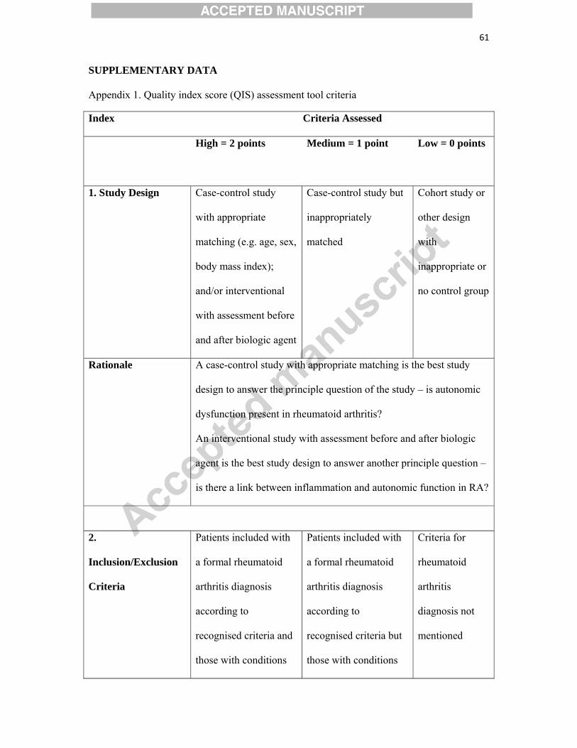
61
SUPPLEMENTARY DATA
Appendix 1. Quality index score (QIS) assessment tool criteria
Index Criteria Assessed
High = 2 points Medium = 1 point Low = 0 points
1. Study Design Case-control study
with appropriate
matching (e.g. age, sex,
body mass index);
and/or interventional
with assessment before
and after biologic agent
Case-control study but
inappropriately
matched
Cohort study or
other design
with
inappropriate or
no control group
Rationale A case-control study with appropriate matching is the best study
design to answer the principle question of the study – is autonomic
dysfunction present in rheumatoid arthritis?
An interventional study with assessment before and after biologic
agent is the best study design to answer another principle question –
is there a link between inflammation and autonomic function in RA?
2.
Inclusion/Exclusion
Criteria
Patients included with
a formal rheumatoid
arthritis diagnosis
according to
recognised criteria and
those with conditions
Patients included with
a formal rheumatoid
arthritis diagnosis
according to
recognised criteria but
those with conditions
Criteria for
rheumatoid
arthritis
diagnosis not
mentioned
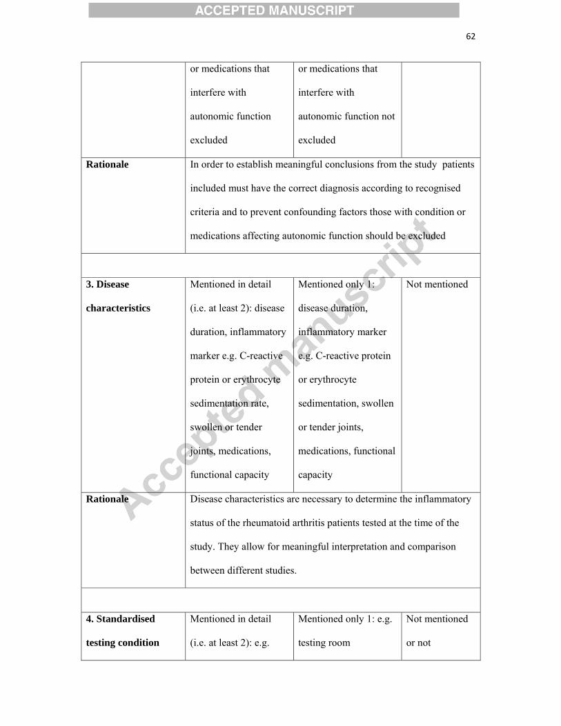
62
or medications that
interfere with
autonomic function
excluded
or medications that
interfere with
autonomic function not
excluded
Rationale In order to establish meaningful conclusions from the study patients
included must have the correct diagnosis according to recognised
criteria and to prevent confounding factors those with condition or
medications affecting autonomic function should be excluded
3. Disease
characteristics
Mentioned in detail
(i.e. at least 2): disease
duration, inflammatory
marker e.g. C-reactive
protein or erythrocyte
sedimentation rate,
swollen or tender
joints, medications,
functional capacity
Mentioned only 1:
disease duration,
inflammatory marker
e.g. C-reactive protein
or erythrocyte
sedimentation, swollen
or tender joints,
medications, functional
capacity
Not mentioned
Rationale Disease characteristics are necessary to determine the inflammatory
status of the rheumatoid arthritis patients tested at the time of the
study. They allow for meaningful interpretation and comparison
between different studies.
4. Standardised
testing condition
Mentioned in detail
(i.e. at least 2): e.g.
Mentioned only 1: e.g.
testing room
Not mentioned
or not
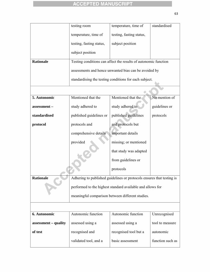
63
testing room
temperature, time of
testing, fasting status,
subject position
temperature, time of
testing, fasting status,
subject position
standardised
Rationale Testing conditions can affect the results of autonomic function
assessments and hence unwanted bias can be avoided by
standardising the testing conditions for each subject.
5. Autonomic
assessment –
standardised
protocol
Mentioned that the
study adhered to
published guidelines or
protocols and
comprehensive details
provided
Mentioned that the
study adhered to
published guidelines
and protocols but
important details
missing; or mentioned
that study was adapted
from guidelines or
protocols
No mention of
guidelines or
protocols
Rationale Adhering to published guidelines or protocols ensures that testing is
performed to the highest standard available and allows for
meaningful comparison between different studies.
6. Autonomic
assessment – quality
of test
Autonomic function
assessed using a
recognised and
validated tool, and a
Autonomic function
assessed using a
recognised tool but a
basic assessment
Unrecognised
tool to measure
autonomic
function such as
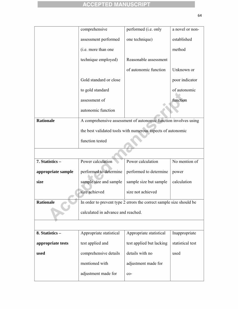
64
comprehensive
assessment performed
(i.e. more than one
technique employed)
Gold standard or close
to gold standard
assessment of
autonomic function
performed (i.e. only
one technique)
Reasonable assessment
of autonomic function
a novel or non-
established
method
Unknown or
poor indicator
of autonomic
function
Rationale A comprehensive assessment of autonomic function involves using
the best validated tools with numerous aspects of autonomic
function tested
7. Statistics –
appropriate sample
size
Power calculation
performed to determine
sample size and sample
size achieved
Power calculation
performed to determine
sample size but sample
size not achieved
No mention of
power
calculation
Rationale In order to prevent type 2 errors the correct sample size should be
calculated in advance and reached.
8. Statistics –
appropriate tests
used
Appropriate statistical
test applied and
comprehensive details
mentioned with
adjustment made for
Appropriate statistical
test applied but lacking
details with no
adjustment made for
co-
Inappropriate
statistical test
used
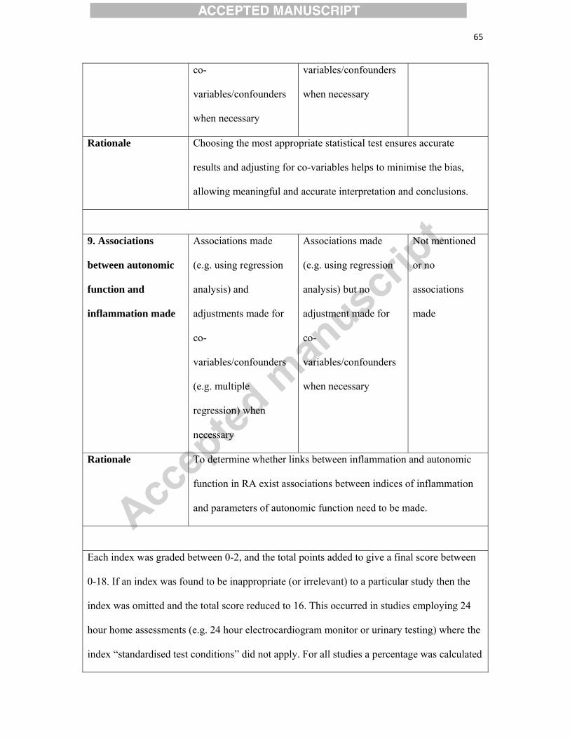
65
co-
variables/confounders
when necessary
variables/confounders
when necessary
Rationale Choosing the most appropriate statistical test ensures accurate
results and adjusting for co-variables helps to minimise the bias,
allowing meaningful and accurate interpretation and conclusions.
9. Associations
between autonomic
function and
inflammation made
Associations made
(e.g. using regression
analysis) and
adjustments made for
co-
variables/confounders
(e.g. multiple
regression) when
necessary
Associations made
(e.g. using regression
analysis) but no
adjustment made for
co-
variables/confounders
when necessary
Not mentioned
or no
associations
made
Rationale To determine whether links between inflammation and autonomic
function in RA exist associations between indices of inflammation
and parameters of autonomic function need to be made.
Each index was graded between 0-2, and the total points added to give a final score between
0-18. If an index was found to be inappropriate (or irrelevant) to a particular study then the
index was omitted and the total score reduced to 16. This occurred in studies employing 24
hour home assessments (e.g. 24 hour electrocardiogram monitor or urinary testing) where the
index “standardised test conditions” did not apply. For all studies a percentage was calculated
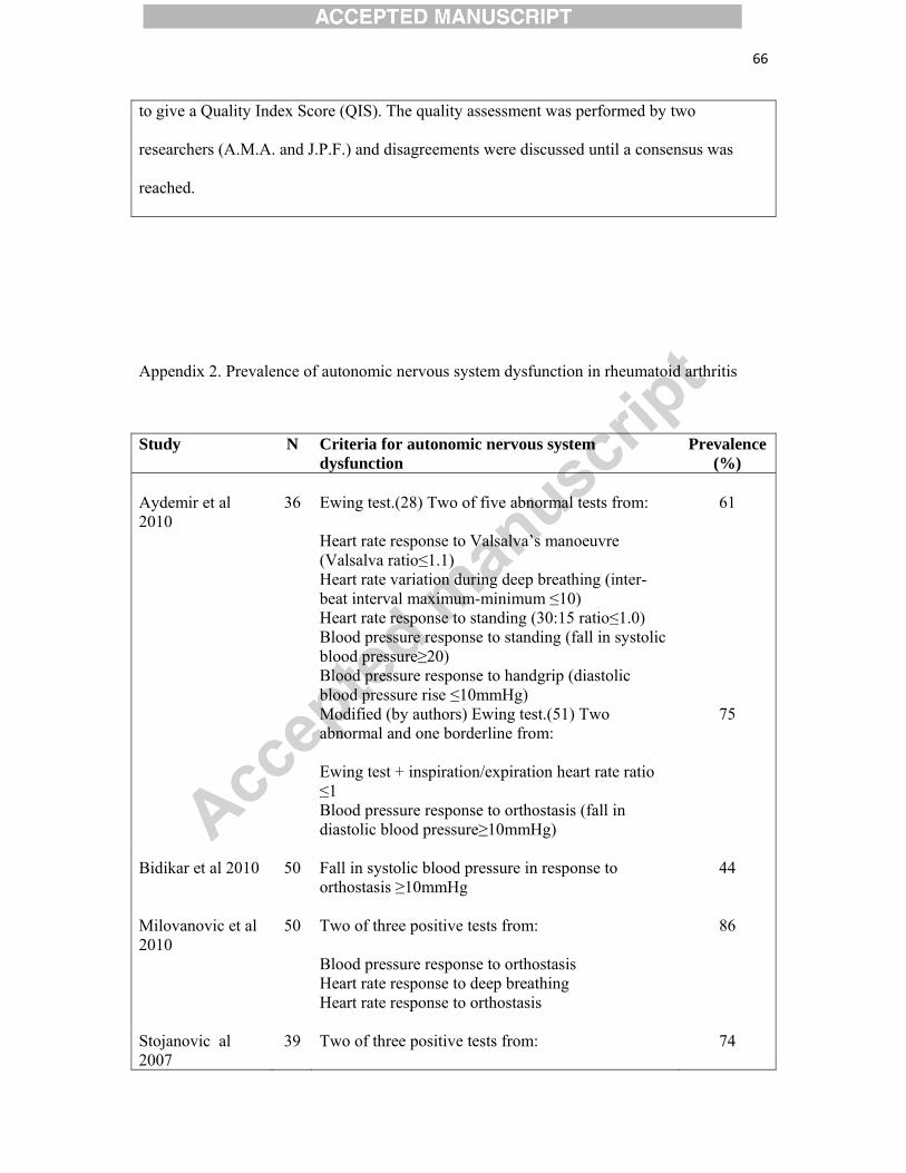
66
to give a Quality Index Score (QIS). The quality assessment was performed by two
researchers (A.M.A. and J.P.F.) and disagreements were discussed until a consensus was
reached.
Appendix 2. Prevalence of autonomic nervous system dysfunction in rheumatoid arthritis
Study N Criteria for autonomic nervous system dysfunction
Prevalence (%)
Aydemir et al 2010
36 Ewing test.(28) Two of five abnormal tests from: Heart rate response to Valsalva’s manoeuvre (Valsalva ratio≤1.1) Heart rate variation during deep breathing (inter-beat interval maximum-minimum ≤10) Heart rate response to standing (30:15 ratio≤1.0) Blood pressure response to standing (fall in systolic blood pressure≥20) Blood pressure response to handgrip (diastolic blood pressure rise ≤10mmHg)
61
Modified (by authors) Ewing test.(51) Two abnormal and one borderline from: Ewing test + inspiration/expiration heart rate ratio ≤1 Blood pressure response to orthostasis (fall in diastolic blood pressure≥10mmHg)
75
Bidikar et al 2010 50 Fall in systolic blood pressure in response to
orthostasis ≥10mmHg 44
Milovanovic et al 2010
50 Two of three positive tests from: Blood pressure response to orthostasis Heart rate response to deep breathing Heart rate response to orthostasis
86
Stojanovic al 2007
39 Two of three positive tests from:
74
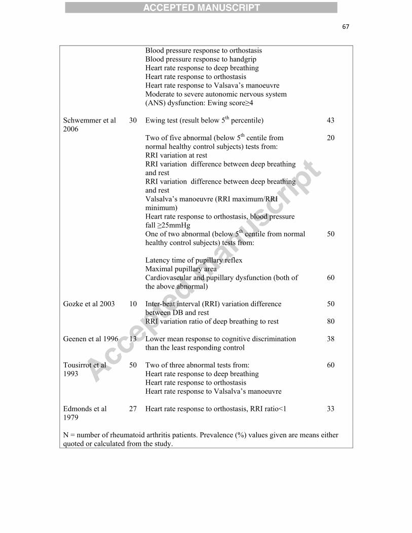
67
Blood pressure response to orthostasis Blood pressure response to handgrip Heart rate response to deep breathing Heart rate response to orthostasis Heart rate response to Valsava’s manoeuvre Moderate to severe autonomic nervous system (ANS) dysfunction: Ewing score≥4
Schwemmer et al 2006
30 Ewing test (result below 5th percentile) 43
Two of five abnormal (below 5th centile from normal healthy control subjects) tests from: RRI variation at rest RRI variation difference between deep breathing and rest RRI variation difference between deep breathing and rest Valsalva’s manoeuvre (RRI maximum/RRI minimum) Heart rate response to orthostasis, blood pressure fall ≥25mmHg
20
One of two abnormal (below 5th centile from normal healthy control subjects) tests from: Latency time of pupillary reflex Maximal pupillary area
50
Cardiovascular and pupillary dysfunction (both of the above abnormal)
60
Gozke et al 2003 10 Inter-beat interval (RRI) variation difference
between DB and rest 50
RRI variation ratio of deep breathing to rest 80 Geenen et al 1996 13 Lower mean response to cognitive discrimination
than the least responding control 38
Tousirrot et al 1993
50 Two of three abnormal tests from: Heart rate response to deep breathing Heart rate response to orthostasis Heart rate response to Valsalva’s manoeuvre
60
Edmonds et al 1979
27 Heart rate response to orthostasis, RRI ratio<1 33
N = number of rheumatoid arthritis patients. Prevalence (%) values given are means either quoted or calculated from the study.
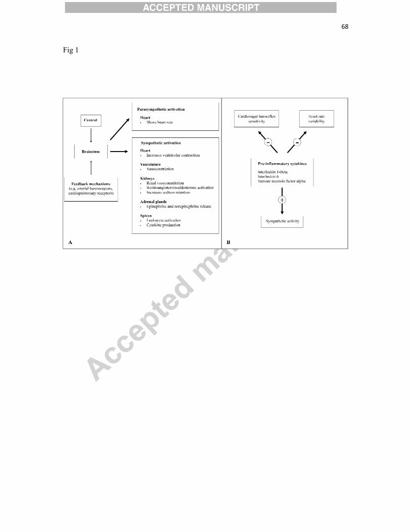
68
Fig 1
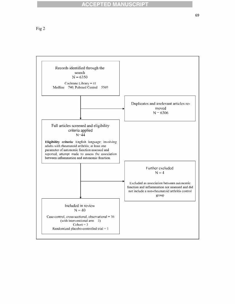
69
Fig 2