Embed Size (px)
Citation preview
Autonomic Nervous System IPhysiological Anatomy
Physiology lecture 28
Dr. Waleed R. Ezzat
Lecture objectives:
Def. the Autonomic nervous system (ANS).
Discuss the physiological anatomy.
Describe the autonomic nervous system efferent pathways from the CNS to effector organs and explain how these differ from the pathway of a motor neuron.
Explain the difference between the parasympathetic and sympathetic nervous systems in terms of preganglionic neuronal location, ganglia location.
Introduction:
The autonomic nervous system (ANS) is the portion of the nervous system that controls most visceral functions of the body.
The ANS also includes the enteric nervous system that functions within the gastrointestinal tract and influences the pancreas, liver, and gallbladder, thereby controlling gastrointestinal motility, secretion, and blood flow.
The ANS is characterized by its rapid and intense control of visceral functions.
Physiological Anatomy- General Description:
ANS is activated mainly by centers located in the spinal cord, brain stem, and hypothalamus.
These central regulators of the ANS also adjust the secretion of hormones that influence blood volume and total peripheral resistance.
The central regulators of the ANS also coordinate the stress response (e.g., fight-or-flight response), reproduction, and thermoregulation.
At the conscious level, the limbic cortex transmit signals to the lower centers and can, as such, influence autonomic control.
The ANS operates through subconscious sensory signals and subconscious reflex responses to control visceral activities.
Physiological Anatomy- General Description (cont.):
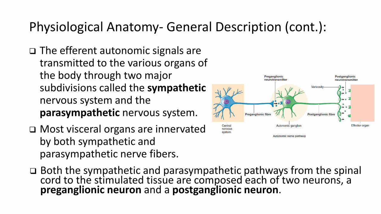
The efferent autonomic signals are transmitted to the various organs of the body through two major subdivisions called the sympatheticnervous system and the parasympathetic nervous system.
Most visceral organs are innervated by both sympathetic and parasympathetic nerve fibers.
Both the sympathetic and parasympathetic pathways from the spinal cord to the stimulated tissue are composed each of two neurons, a preganglionic neuron and a postganglionic neuron.
A.
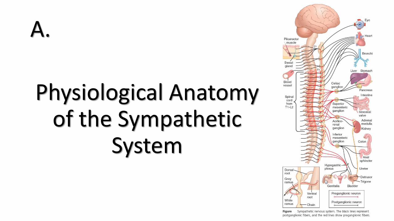
Physiological Anatomy of the Sympathetic
System

Physiological Anatomy of the Sympathetic System:
The peripheral portion of the Sympathetic System include:
1. Two paravertebral sympathetic chains of ganglia (also called the sympathetic trunk), that are interconnected with the spinal nerves on the side of the vertebral column.
2. Prevertebral ganglia or collateral ganglia (the celiac, superior mesenteric, aortico-renal, inferior mesenteric, and hypogastric).
3. Nerves extending from the ganglia to the different internal organs.
The sympathetic nerve fibers originate in the spinal cord along with spinal nerves between cord segments T1 and L2 and pass first into the sympathetic chain and then to the tissues and organs that are stimulated by the sympathetic nerves.
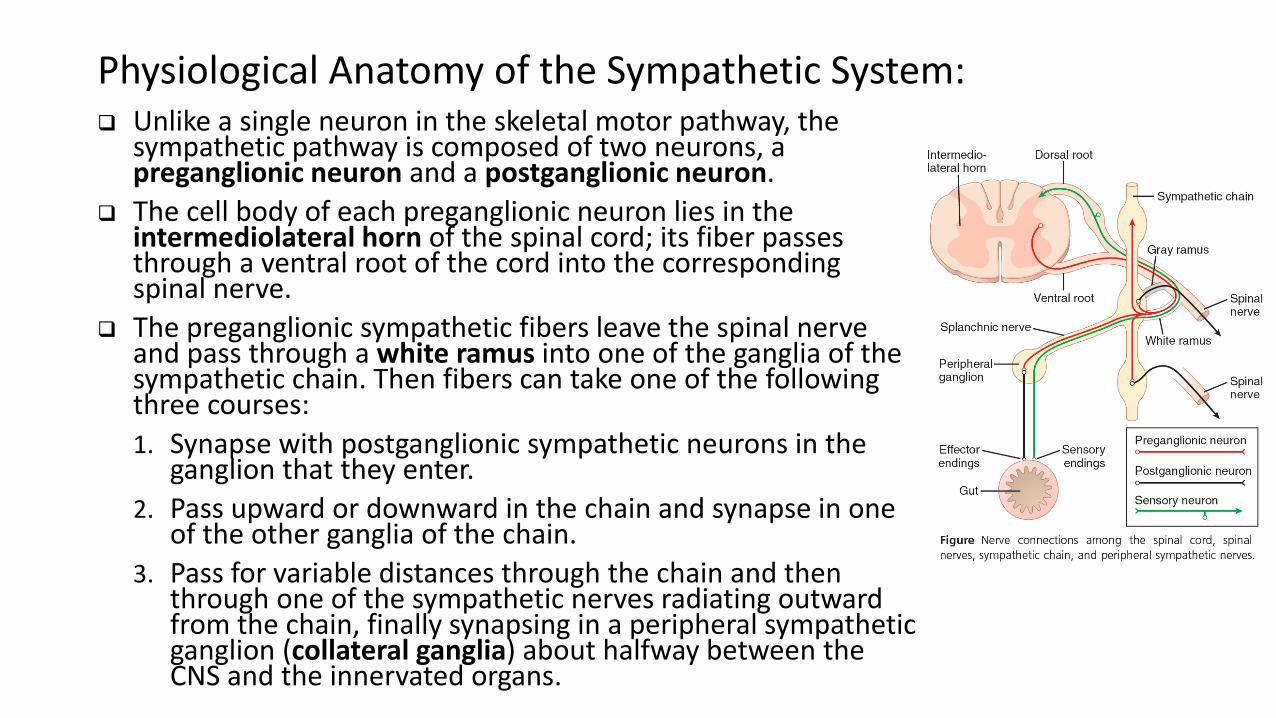
Physiological Anatomy of the Sympathetic System: Unlike a single neuron in the skeletal motor pathway, the
sympathetic pathway is composed of two neurons, a preganglionic neuron and a postganglionic neuron.
The cell body of each preganglionic neuron lies in the intermediolateral horn of the spinal cord; its fiber passes through a ventral root of the cord into the corresponding spinal nerve.
The preganglionic sympathetic fibers leave the spinal nerve and pass through a white ramus into one of the ganglia of the sympathetic chain. Then fibers can take one of the following three courses:
1. Synapse with postganglionic sympathetic neurons in the ganglion that they enter.
2. Pass upward or downward in the chain and synapse in one of the other ganglia of the chain.
3. Pass for variable distances through the chain and then through one of the sympathetic nerves radiating outward from the chain, finally synapsing in a peripheral sympathetic ganglion (collateral ganglia) about halfway between the CNS and the innervated organs.
Physiological Anatomy of the Sympathetic System:
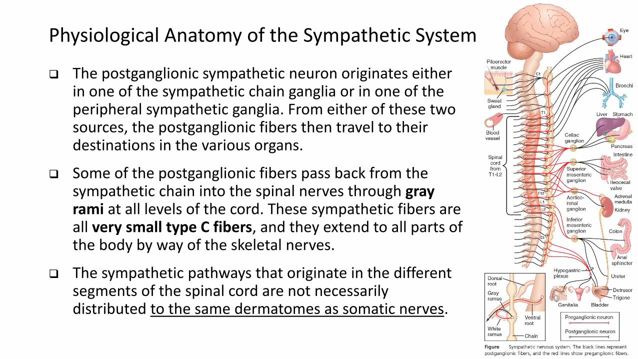
The postganglionic sympathetic neuron originates either in one of the sympathetic chain ganglia or in one of the peripheral sympathetic ganglia. From either of these two sources, the postganglionic fibers then travel to their destinations in the various organs.
Some of the postganglionic fibers pass back from the sympathetic chain into the spinal nerves through gray rami at all levels of the cord. These sympathetic fibers are all very small type C fibers, and they extend to all parts of the body by way of the skeletal nerves.
The sympathetic pathways that originate in the different segments of the spinal cord are not necessarily distributed to the same dermatomes as somatic nerves.
Physiological Anatomy of the Sympathetic System:
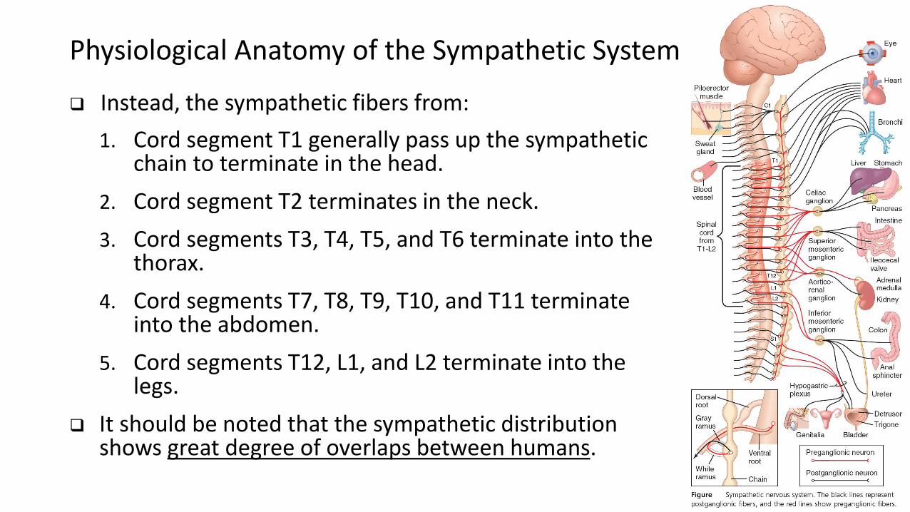
Instead, the sympathetic fibers from:
1. Cord segment T1 generally pass up the sympathetic chain to terminate in the head.
2. Cord segment T2 terminates in the neck.
3. Cord segments T3, T4, T5, and T6 terminate into the thorax.
4. Cord segments T7, T8, T9, T10, and T11 terminate into the abdomen.
5. Cord segments T12, L1, and L2 terminate into the legs.
It should be noted that the sympathetic distribution shows great degree of overlaps between humans.
Physiological Anatomy of the Sympathetic System:
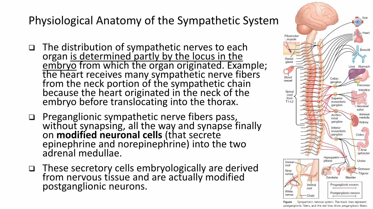
The distribution of sympathetic nerves to each organ is determined partly by the locus in the embryo from which the organ originated. Example; the heart receives many sympathetic nerve fibers from the neck portion of the sympathetic chain because the heart originated in the neck of the embryo before translocating into the thorax.
Preganglionic sympathetic nerve fibers pass, without synapsing, all the way and synapse finally on modified neuronal cells (that secrete epinephrine and norepinephrine) into the two adrenal medullae.
These secretory cells embryologically are derived from nervous tissue and are actually modified postganglionic neurons.
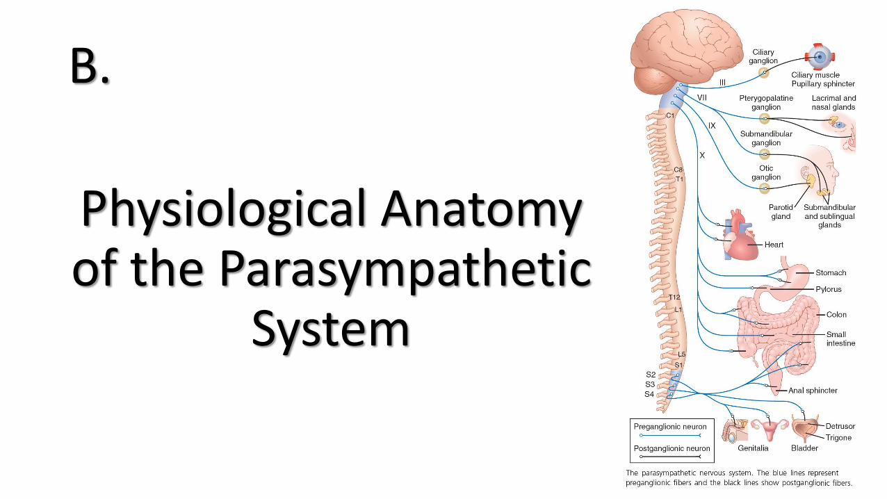
B.
Physiological Anatomy of the Parasympathetic
System
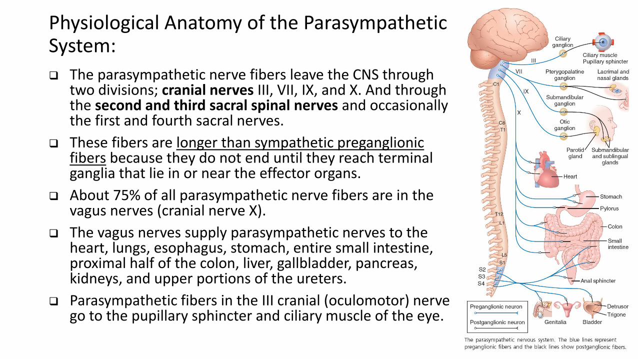
Physiological Anatomy of the Parasympathetic System:
The parasympathetic nerve fibers leave the CNS through two divisions; cranial nerves III, VII, IX, and X. And through the second and third sacral spinal nerves and occasionally the first and fourth sacral nerves.
These fibers are longer than sympathetic preganglionic fibers because they do not end until they reach terminal ganglia that lie in or near the effector organs.
About 75% of all parasympathetic nerve fibers are in the vagus nerves (cranial nerve X).
The vagus nerves supply parasympathetic nerves to the heart, lungs, esophagus, stomach, entire small intestine, proximal half of the colon, liver, gallbladder, pancreas, kidneys, and upper portions of the ureters.
Parasympathetic fibers in the III cranial (oculomotor) nerve go to the pupillary sphincter and ciliary muscle of the eye.
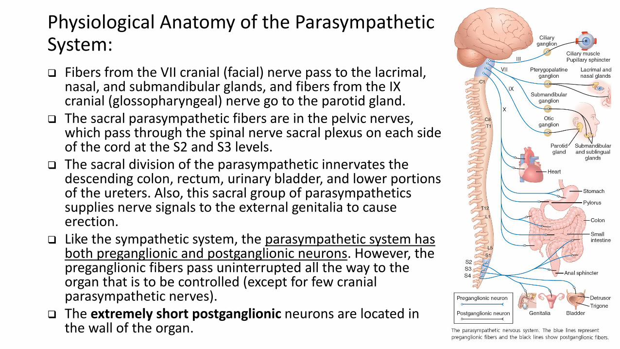
Physiological Anatomy of the Parasympathetic System:
Fibers from the VII cranial (facial) nerve pass to the lacrimal, nasal, and submandibular glands, and fibers from the IX cranial (glossopharyngeal) nerve go to the parotid gland.
The sacral parasympathetic fibers are in the pelvic nerves, which pass through the spinal nerve sacral plexus on each side of the cord at the S2 and S3 levels.
The sacral division of the parasympathetic innervates the descending colon, rectum, urinary bladder, and lower portions of the ureters. Also, this sacral group of parasympatheticssupplies nerve signals to the external genitalia to cause erection.
Like the sympathetic system, the parasympathetic system has both preganglionic and postganglionic neurons. However, the preganglionic fibers pass uninterrupted all the way to the organ that is to be controlled (except for few cranial parasympathetic nerves).
The extremely short postganglionic neurons are located in the wall of the organ.
Test Question:
Q. The autonomic ganglia:A. Are 5 typesB. Are the sites of relay of afferent neuronsC. Function as distributing centersD. Are located inside the CNSE. Are relay stations for all preganglionic fibers
passing through them