Embed Size (px)
Citation preview
BritishJ7ournal ofOphthalmology 1993; 77:473-479
ORIGINAL ARTICLES - Clinical science
Autosomal dominant retinitis pigmentosa withapparent incomplete penetrance: a clinical,electrophysiological, psychophysical, and moleculargenetic study
A T Moore, F Fitzke,M Jay, G B Arden, C F Inglehearn, T J Keen, S S Bhattacharya, A C Bird
AbstractTwenty five symptomatic individuals and sixasymptomatic obligate gene carriers from fourfamilies with autosomal dominant retinitis pig-mentosa (adRP) showing apparent incompletepenetrance have been studied. Symptomaticindividuals from three families showed earlyonset of night blindness, non-recordable rodelectroretinograms, and marked elevation ofboth rod and cone thresholds in all subjectstested. In the fourth family, there was morevariation in the age of onset of night blindnessand some symptomatic individuals showedwell preserved rod and cone function in someretinal areas. All asymptomatic individualstested had evidence of mild abnormalities ofrod and cone function, indicating that thesefamilies show marked variation in expressivityrather than true non-penetrance of the adRP-gene. No mutations of the rhodopsin or RDSgenes were found in these families and theprecise genetic mutation(s) remain to be identi-fied.(Br_' Ophthalmol 1993; 77: 473-479)
Institute ofOphthalmology, andMoorfields Eye Hospital,LondonA T MooreF FitzkeM JayG B ArdenC F IngleheamT J KeenS S BhattacharyaA C Bird
Addenbrooke's Hospital,CambridgeAT MooreCorrespondence to:Mr A T Moore,Addenbrooke's Hospital, HillsRoad, Cambridge CB2 2QQ.Accepted for publication31 May 1993
Clinical and psychophysical studies in autosomaldominant retinitis pigmentosa (adRP) have sug-gested that there may be genetic heterogeneitywithin the disorder.`'5 This has been confirmedby the finding ofmutations in the rhodopsin geneon chromosome 367 and the retinal degenerationslow (RDS) gene on chromosome 6"1o in somefamilies with adRP. Some forms of adRP do notshow linkage to or mutations of either therhodopsin gene or the RDS gene and additionalgenetic mutations remain to be identified.Most families, including those with known
mutations, show complete penetrance of theadRP gene. In a few families incomplete pene-trance is a notable feature"-'5 and it has beensuggested on the basis ofthe electroretinographicfindings that this may be a distinct form ofadRP." 12We have studied both symptomatic indi-
viduals and asymptomatic obligate gene carriersfrom four families showing apparent incompletepenetrance in order to characterise the pattern of
retinal disease in affected family members and toinvestigate rod and cone function in thoseobligate gene carriers who remain asymptomaticin the fourth or fifth decade of life. We have alsoinvestigated whether any of the families showmutations of the rhodopsin or RDS genes, orlinkage to chromosomes 3 or 6.
Patients and methodsTwenty five symptomatic individuals (mean age32 years; range 13-58 years) and six asympto-matic obligate gene carriers (mean age 46 years;range 39-60 years) from four families withapparent incomplete penetrance took part in thestudy. In each family there was transmission ofthe disease through at least three generations (Fig1), and in three there was evidence of male tomale transmission, In the fourth, although therewas no male to male transmission, all the affectedfemales had severe disease, suggesting autosomaldominant inheritance.Each subject underwent a full clinical evalua-
tion. A variety of investigations including anelectroretinography (ERG), electro-oculography(EOG), Goldmann perimetry, and detailed darkadapted static perimetry were performed in themajority of subjects (subjects with advanced RPwith extensive field loss did not undergo psycho-physical testing). In addition, photopic flickertesting was performed on each of the six asymp-tomatic obligate gene carriers. Detailed Gold-mann perimetry was performed on each eyeusing the IV 4e and I 4e targets. The ERG was
Family 1
IV
Figure 1
473
on Decem
ber 26, 2019 by guest. Protected by copyright.
http://bjo.bmj.com
/B
r J Ophthalm
ol: first published as 10.1136/bjo.77.8.473 on 1 August 1993. D
ownloaded from
Moore, Fitzke, Jay, Arden, Inglehearn, Keen, Bhattacharya, Bird
Xl
IIIA
IIA
IA
AAl
III
Family 2
11111
IV
V
VI
VIIIVillVil
Family 3
11
III
IV
V
Family 4
III 5 i 27 29 tIVI
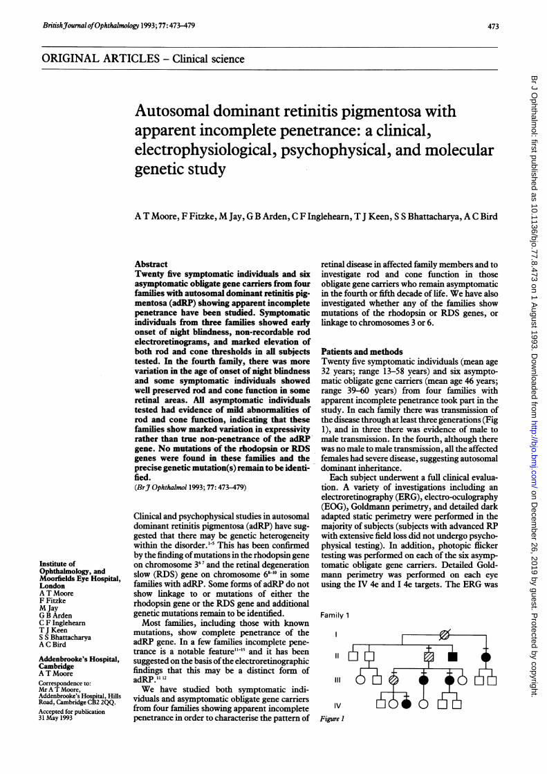
Figure 1 Pedigrees offamilies 1, 2, 3, and 4. O :symptomatic individual; 3 0: asymptomatic obligate gene carrier; -: propositus; +: examinedby the authors.
474
:ll
on Decem
ber 26, 2019 by guest. Protected by copyright.
http://bjo.bmj.com
/B
r J Ophthalm
ol: first published as 10.1136/bjo.77.8.473 on 1 August 1993. D
ownloaded from
Autosomal dominant retinitis pigmnentosa with apparent incomplete penetrance
Table I Age ofonset ofnight blindness
Onset ofnight blindnessNo ofpatients
Family <10years 11-20years >21 years examined
1 4 0 0 42 2 3 5 103 2 0 0 24 4 4 0 9*
*One patient denied any night blindness.
performed in accordance with the protocol des-cribed in Arden et al'6 and the details of themethod used for photopic fficker are given inTyler et al."7 Dark adapted perimetry was per-formed on one eye (which showed least field losson Goldmann perimetry) of each subject usingred (dominant wavelength 660 mm, subtending0.9) and green (dominant wavelength 530 nm,subtending 0.90) targets. The pupil was dilatedwith 1% cyclopentolate and the eye dark adaptedfor 40 minutes before starting the test. At least 17points at different retinal locations in both upperand lower fields were tested in each case. Theapparatus and method for the dark adapted staticperimetry have been described previously. 8Symptomatic members from each family were
screened for mutations in the rhodopsin andRDS genes. Exon sequences were amplified bythe polymerase chain reaction using primersflanking the exons.9192' Amplified fragmentswere run on hydrolink gels in order to detectmutations as heteroduplexed fragments ofnormal and mutated DNA sequences.2' Thismethod has successfully detected 13 differentmutations in the rhodopsin and RDS genes in 17different families with retinal dystrophiesl'2' andappears to detect the same proportion of muta-tions in screening of RP families as single strandconformation polymorphism (SSCP)22 anddenaturing gradient gel electrophoresis(DGGE).7
Linkage analysis was performed on markers
90
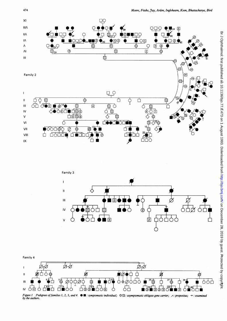
Figure 2 Goldmann field performed on the right eye ofsubject IV-34 fromfamily 4.
within the rhodopsin and RDS genes in thefamilies. The rhodopsin poly-CA tract23 and theRDS gene poly-T tract24 were amplified by thepolymerase chain reaction in symptomatic familymembers. One primer in each amplification waskinase end labelled with 32P-y ATP, then sampleswere run on denaturing acrylamide gels. The gelswere dried and exposed to autoradiographic filmovernight. Inheritance of alleles relative to thedisease was analysed using the program LINK-AGE, from the LINKAGE package version5.10.25Informed consent was obtained after the
nature of the procedures had been fullyexplained.
Table 2 Affected patients: clinicalfindings
Acuity Goldmann fieldsFamily Subject Age (R;L) Cataract Maculae (IV4e target) Electroretinogram
1 III-S 34 6/18;6/18 Yes MO 10 degrees Non-recordable1 IV-3 18 6/18;6/12 No MH 20 degrees Non-recordable1 III-4 30 6/36;6/36 Yes MA 10 degrees Non-recordable1 II-5 42 6/18;6/24 Yes MO 10 degrees Non-recordable2 VIII-12 39 6/12;6/12 Yes MO 10 degrees Not performed2 VIII-47 13 615;6/5 No Normal 10 degrees Absent rod, minimal cone responses2 VII-25 58 6/36;NPL Yes MA 5 degrees Not performed2 VIII-30 19 6/9;6/9 No Normal 40 degrees Rod and cone responses2 VIII-31 22 616;6/6 No Normal 40 degrees Rod and cone responses2 VII-28 33 6/18;6/12 Yes MO 5 degrees Non-recordable2 VII-26 42 6/18;6/18 Yes MA 5 degrees Not performed2 VII-14 46 6/18;6/18 Yes MO 40 degrees Absent rod, reduced cone responses2 VII-13 56 6/12;6/12 No MO 5 degrees Non-recordable2 VII-17 24 615;6/5 No Normal Full* Non-recordable3 IV-14 53 6/36;6/36 Yes MA 15 degrees Non-recordable3 V-2 22 6/6;6/5 No Normal 60 degrees Non-recordable4 IV-il 37 6/18;6/18 Yes MO 10 degrees Non-recordable4 III-15 45 6/9;6/9 Yes Normal 5 degrees Not performed4 IV-48 19 6/9;6/6 No Normal Full* Absent rod, reduced cone responses4 IV-34 16 6/6;6/6 No Normal Full* Absent rod, reduced cone responses4 IV-8 20 6/12;6/12 Yes MO 10 degrees Non-recordable4 111-3 45 6/18;6/9 Yes MO 10 degrees Non-recordable4 III-29 40 LP;LP Yes MA <5 degrees Not performed4 III-19 18 LPt;6/5 No Normal Full* Absent rod, reduced cone responses4 IV-43 21 6/6;616 No Normal Full* Non-recordable
MO=macular oedema.MA=macular atrophy.MH=macular hole.*Field loss evident with smaller targets.tRight optic nerve hypoplasia.
I -IV4/1-41 IV4 notseen
475 on D
ecember 26, 2019 by guest. P
rotected by copyright.http://bjo.bm
j.com/
Br J O
phthalmol: first published as 10.1136/bjo.77.8.473 on 1 A
ugust 1993. Dow
nloaded from
Moore, Fitzke, Jay, Arden, Inglehearn, Keen, Bhatacharya, Bird
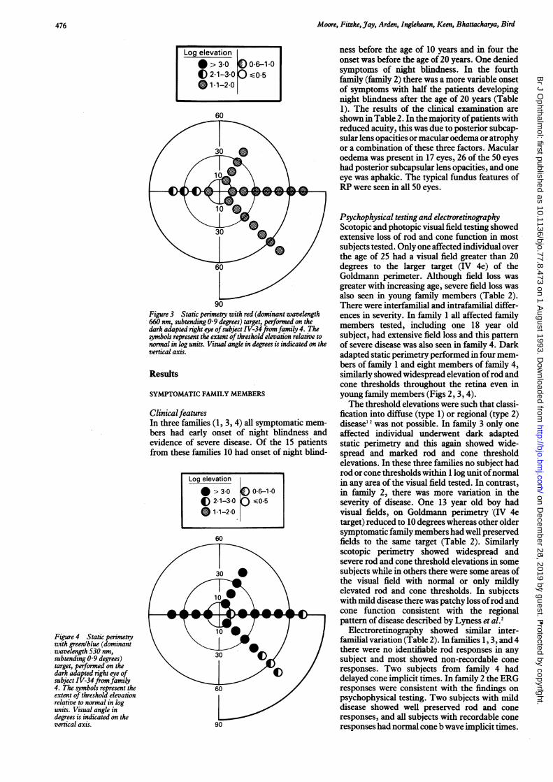
r Log elevation* > 3.0Q 0-1 00 2-1-300 Q0.5o 11-2-0
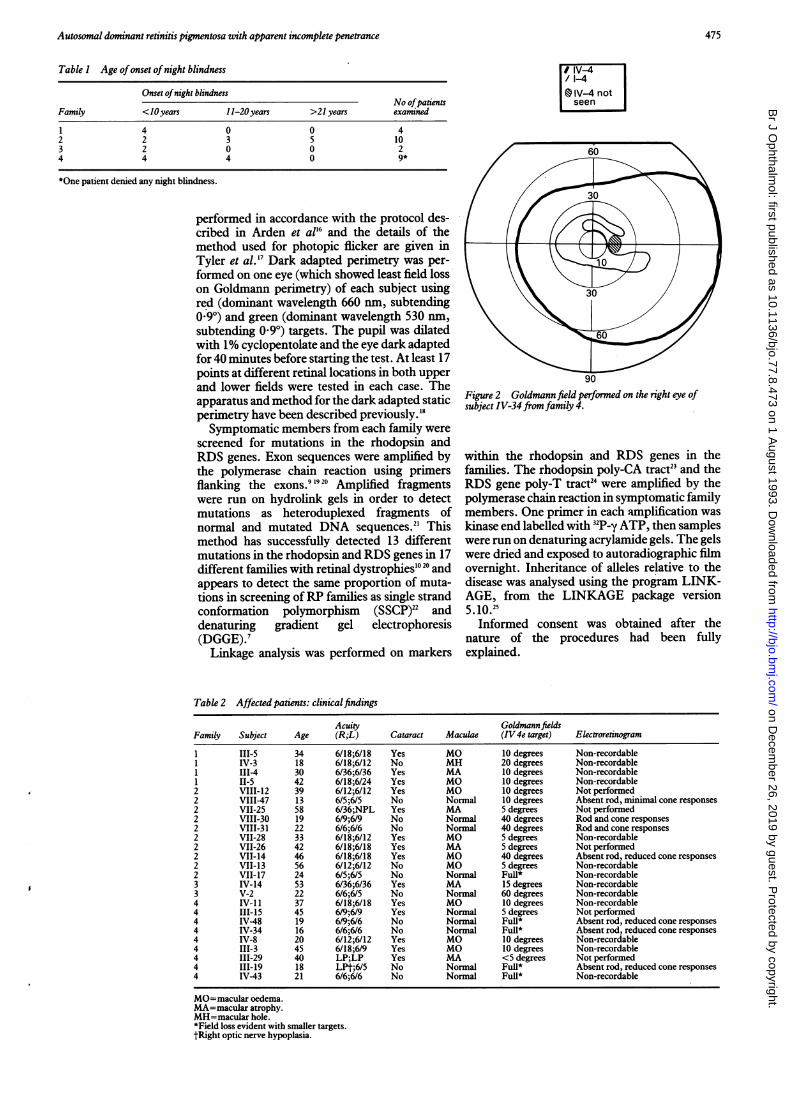
90Figure 3 Static perimetry with red (dominant wavelength660 nm, subtending 0 9 degrees) target, performed on thedark adapted right eye ofsubject IV-34 fromfamily 4. Thesymbols represent the extent ofthreshold elevation relative tonormal in log units. Visual angle in degrees is indicated on thevertical axis.
Results
SYMPTOMATIC FAMILY MEMBERS
ClinicalfeaturesIn three families (1, 3, 4) all symptomatic mem-bers had early onset of night blindness andevidence of severe disease. Of the 15 patientsfrom these families 10 had onset of night blind-
60
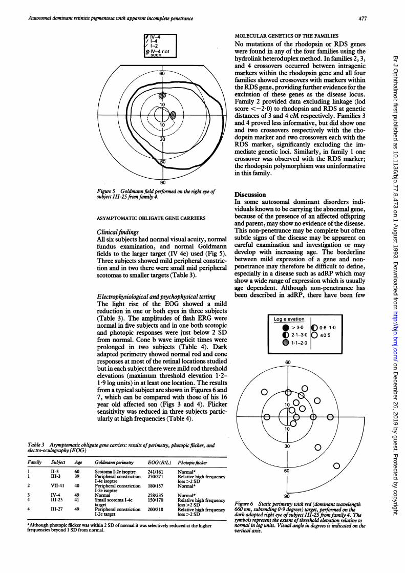
Figure 4 Static perimetrywith greenlblue (dominantwavelength 530 nm,subtending 0-9 degrees)target, performed on thedark adapted right eye ofsubject IV-34 from family4. The symbols represent theextent of threshold elevationrelative to normal in logunits. Visual angle indegrees is indicated on thevertical axis. 90
ness before the age of 10 years and in four theonset was before the age of 20 years. One deniedsymptoms of night blindness. In the fourthfamily (family 2) there was a more variable onsetof symptoms with half the patients developingnight blindness after the age of 20 years (Table1). The results of the clinical examination areshown in Table 2. In the majority ofpatients withreduced acuity, this was due to posterior subcap-sular lens opacities or macular oedema or atrophyor a combination of these three factors. Macularoedema was present in 17 eyes, 26 of the 50 eyeshad posterior subcapsular lens opacities, and oneeye was aphakic. The typical fundus features ofRP were seen in all 50 eyes.
Psychophysical testing and electroretinographyScotopic and photopic visual field testing showedextensive loss of rod and cone function in mostsubjects tested. Only one affected individual overthe age of 25 had a visual field greater than 20degrees to the larger target (IV 4e) of theGoldmann perimeter. Although field loss wasgreater with increasing age, severe field loss wasalso seen in young family members (Table 2).There were interfamilial and intrafamilial differ-ences in severity. In family 1 all affected familymembers tested, including one 18 year oldsubject, had extensive field loss and this patternof severe disease was also seen in family 4. Darkadapted static perimetry performed in four mem-bers of family 1 and eight members of family 4,similarly showed widespread elevation ofrod andcone thresholds throughout the retina even inyoung family members (Figs 2, 3, 4).The threshold elevations were such that classi-
fication into diffuse (type 1) or regional (type 2)disease'2 was not possible. In family 3 only oneaffected individual underwent dark adaptedstatic perimetry and this again showed wide-spread and marked rod and cone thresholdelevations. In these three families no subject hadrod or cone thresholds within 1 log unit ofnormalin any area of the visual field tested. In contrast,in family 2, there was more variation in theseverity of disease. One 13 year old boy hadvisual fields, on Goldmann perimetry (IV 4etarget) reduced to 10 degrees whereas other oldersymptomatic family members had well preservedfields to the same target (Table 2). Similarlyscotopic perimetry showed widespread andsevere rod and cone threshold elevations in somesubjects while in others there were some areas ofthe visual field with normal or only mildlyelevated rod and cone thresholds. In subjectswith mild disease there was patchy loss ofrod andcone function consistent with the regionalpattern of disease described by Lyness et al.2
Electroretinography showed similar inter-familial variation (Table 2). In families 1, 3, and 4there were no identifiable rod responses in anysubject and most showed non-recordable coneresponses. Two subjects from family 4 haddelayed cone implicit times. In family 2 the ERGresponses were consistent with-the findings onpsychophysical testing. Two subjects with milddisease showed well preserved rod and coneresponses, and all subjects with recordable coneresponses had normal cone b wave implicit times.
Log elevation
* >3.0 Aoo0-i-0
( 2.1-3.0 0 o0.5* 1*1-2-0
476
on Decem
ber 26, 2019 by guest. Protected by copyright.
http://bjo.bmj.com
/B
r J Ophthalm
ol: first published as 10.1136/bjo.77.8.473 on 1 August 1993. D
ownloaded from
Autosomal dominant retinitis pigmentosa with apparent incomplete penetrance
I IV-4/ 1-4fl1-2IV-4notseen
-,~~~~~~~~~~~~~~~60
10I7 h
90Figure 5 Goldmannfield performed on the right eye ofsubject III-25fromfamily 4.
ASYMPTOMATIC OBLIGATE GENE CARRIERS
ClinicalfindingsAll six subjects had normal visual acuity, normalfundus examination, and normal Goldmannfields to the larger target (IV 4e) used (Fig 5).Three subjects showed mild peripheral constric-tion and in two there were small mid peripheralscotomas to smaller targets (Table 3).
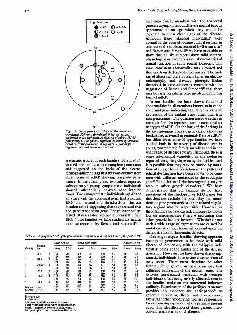
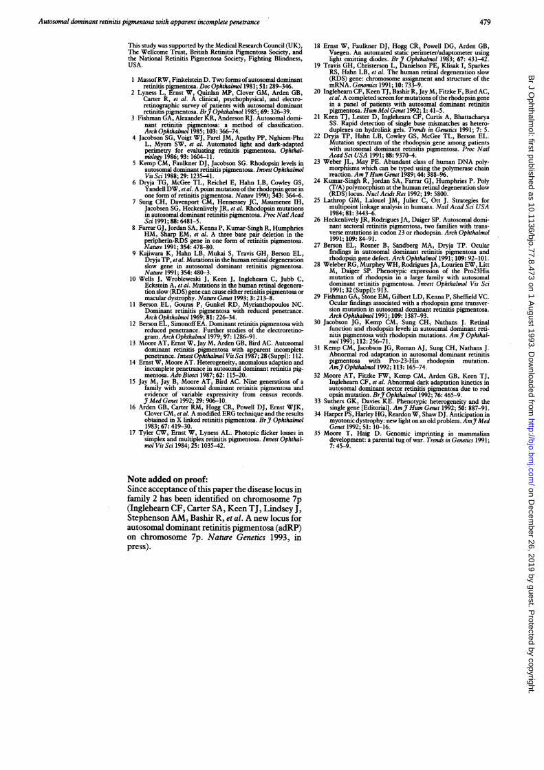
Electrophysiological andpsychophysical testingThe light rise of the EOG showed a mildreduction in one or both eyes in three subjects(Table 3). The amplitudes of flash ERG werenormal in five subjects and in one both scotopicand photopic responses were just below 2 SDfrom normal. Cone b wave implicit times wereprolonged in two subjects (Table 4). Darkadapted perimetry showed normal rod and coneresponses at most of the retinal locations studiedbut in each subject there were mild rod thresholdelevations (maximum threshold elevation 1-2-1-9 log units) in at least one location. The resultsfrom a typical subject are shown in Figures 6 and7, which can be compared with those of his 16year old affected son (Figs 3 and 4). Flickersensitivity was reduced in three subjects partic-ularly at high frequencies (Table 4).
Table 3 Asymptomatic obligate gene cariers: results ofperimetry, photopicflicker, andelectro-oculography (EOG)
Family Subject Age Goldmann perimetry EOG(R/L) Photopicflicker
1 11-3 60 Scotoma I-2e isoptre 241/161 Normal*1 111-3 39 Peripheral constriction 250/271 Relative high frequency
I-4e isoptre loss >2 SD2 VII-41 40 Peripheral constriction 180/157 Normal*
I-2e isoptre3 IV-4 49 Normal 258/235 Normal*4 111-25 41 Small scotoma I-4e 150/170 Relative high frequency
target loss >2 SD4 III-27 49 Peripheral constriction 200/218 Relative high frequency
I-2e target loss >2 SD
*Although photopic flicker was within 2 SD ofnormal it was selectively reduced at the higherfrequencies beyond 1 SD from normal.
MOLECULAR GENETICS OF THE FAMILIES
No mutations of the rhodopsin or RDS geneswere found in any of the four families using thehydrolink heteroduplex method. In families 2, 3,and 4 crossovers occurred between intragenicmarkers within the rhodopsin gene and all fourfamilies showed crossovers with markers withintheRDS gene, providing further evidence for theexclusion of these genes as the disease locus.Family 2 provided data excluding linkage (lodscore <-2-0) to rhodopsin and RDS at geneticdistances of 3 and 4 cM respectively. Families 3and 4 proved less informative, but did show oneand two crossovers respectively with the rho-*dopsin marker and two crossovers each with theRDS marker, significantly excluding the im-mediate genetic loci. Similarly, in family 1 onecrossover was observed with the RDS marker;the rhodopsin polymorphism was uninformativein this family.
DiscussionIn some autosomal dominant disorders indi-viduals known to be carrying the abnormal gene,because of the presence of an affected offspringand parent, may show no evidence ofthe disease.This non-penetrance may be complete but oftensubtle signs of the disease may be apparent oncareful examination and investigation or maydevelop with increasing age. The borderlinebetween mild expression of a gene and non-penetrance may therefore be difficult to define,especially in a disease such as adRP which mayshow a wide range of expression which is usuallyage dependent. Although non-penetrance hasbeen described in adRP, there have been few
Log elevation
* > 3-0 A 06-1-0
0 2.1-3-0 <051-1-2-0
90
Figure 6 Static perimetry with red (dominant wavelength660 nm, subtending 0 9 degrees) target, performed on thedark adapted right eye ofsubject III-25fromfamily 4. Thesymbols represent the extent ofthreshold elevation relative tonormal in log units. Visual angle in degrees is indicated on thevertical axis.
477
on Decem
ber 26, 2019 by guest. Protected by copyright.
http://bjo.bmj.com
/B
r J Ophthalm
ol: first published as 10.1136/bjo.77.8.473 on 1 August 1993. D
ownloaded from
Moore, Fitzke, Jay, Arden, Inglehearn, Keen, Bhattacharya, Bird
90Figure 7 Static perimetry with green/blue (dominantwavelength 530 nm, subtending 0 9 degrees) target,performed on the dark adapted right eye ofsubject III-25fromfamily 4. The symbols represent the extent ofthresholdelevation relative to normal in log units. Visual angle indegrees is indicated on the vertical axis.
systematic studies of such families. Berson et al"studied one family with incomplete penetranceand suggested on the basis of the electro-retinographic findings that this was distinct fromother forms of adRP showing complete pene-trance. In their family and two others reportedsubsequently'2 young symptomatic individualsshowed substantially delayed cone implicittimes. Two asymptomatic individuals age 42 and75 years with the abnormal gene had a normalERG and normal rod thresholds at the onelocation tested suggesting that they showed truenon-penetrance ofthe gene. The younger patienttested 10 years later retained a normal full fieldERG.12 The families we have studied are similarto those reported by Berson and Simonoff"2 in
Table 4 Asymptomatic obligate gene carriers: amplitude and implicit times oftheflash ERG
Scotopic blue Brightflash white Flicker (30 Hz)Subject
Family eye b amp b imp a amp a imp b amp b imp b amp b imp
1 II-3 R 190 70 180 15 200 55 38 33L 160 80 160 15 200 55 16 33
1 III-3 R 270 60 160 15 260 55 32 31L 250 60 120 15 360 55 32 31
2 VII-41 R 60 60 120 16 80 50 30 35L 50 60 120 16 80 50 30 35
3 IV-4 R 260 70 140 15 330 50 35 31L 225 68 140 16 360 52 40 31
4 III-25 R 260 66 200 15 240 50 25 32L 230 66 200 14 360 48 30 30
4 III-27 R 180 60 240 15 240 50 25 32L 140 60 200 15 360 48 30 30
Normal mean 319 57 262 15 404 50 43 29Normal-2 SD 141 69 133 20 227 56 10 32
R=right eye.L=left eye.a amp=amplitude a wave in microvolts.a imp=implicit time a wave in milliseconds.b amp=amplitude b wave in microvolts.b imp=implicit time b wave in milliseconds.
that some family members with the abnormalgene are asymptomatic and have a normal fundusappearance at an age when they would beexpected to show clear signs of the disease.Although these 'skipped individuals' werenormal on the basis of routine clinical testing, incontrast to the subjects reported by Berson et al"and Berson and Simonoff" we have been able toshow that all six subjects show mild electro-physiological or psychophysical abnormalities ofretinal function in some retinal locations. Themost consistent abnormality was elevated rodthresholds on dark adapted perimetry. The find-ing of abnormal cone implicit times on electro-retinography and elevated photopic ffickerthresholds in some subjects is consistent with thesuggestion of Berson and Simonoff" that theremay be early peripheral cone involvement in thisform of adRP.
In our families we have shown functionalabnormalities in all members known to have theabnormal gene indicating that there is variableexpression of the mutant gene rather than truenon-penetrance. The question arises whether ornot such families represent one or more distinctsubtypes ofadRP. On the basis of the findings inthe asymptomatic obligate gene carriers they canbe classified as type II or regional (R) type adRP'2but differ from other R type families we havestudied both in the severity of disease seen inyoung symptomatic family members and in thewide range of disease severity. Although there issome interfamilial variability in the pedigreesreported here, they share many similarities, andit is possible that they may be caused by muta-tions at a single locus. Variations in the pattern ofretinal dysfunction have been shown to be com-mon with different mutations in the rhodopsingene2"32 and similar allelic heterogeneity may beseen in other genetic disorders.33 We havedemonstrated that our families do not havemutations of the rhodopsin or RDS genes butthis does not exclude the possibility that muta-tions of gene promoters or other related regula-tory regions may be responsible. However, inthree families we have excluded linkage to knownloci on chromosomes 3 and 6 indicating thatother genetic loci are involved. Whether or notsuch a wide range of expressions are caused bymutations at a single locus will depend upon thedemonstration of the genetic defects.One might expect families showing apparent
incomplete penetrance to be those with milddisease of late onset, with the 'skipped indi-viduals' being at the milder end of the diseasespectrum. However, we have shown that symp-tomatic individuals have severe disease often ofearly onset. There must therefore be otherfactors, either genetic or environmental, thatinfluence expression of the mutant gene. Theextreme intrafamilial variation, with youngerindividuals often being severly affected, seen inour families make an environmental influenceunlikely. Examination of the pedigree structureprovides no evidence for anticipation' orgenomic imprinting effects35 and it seems morelikely that other 'modifying' loci are responsiblefor influencing expression of the primary mutantgene. The identification of these genetic inter-actions remains a major challenge.
Log elevation
* > 3.0 A 06-10
( 2 1-3-0 Q s0.5
o 11-2-0
478 on D
ecember 26, 2019 by guest. P
rotected by copyright.http://bjo.bm
j.com/
Br J O
phthalmol: first published as 10.1136/bjo.77.8.473 on 1 A
ugust 1993. Dow
nloaded from
Autosomal dominant retinitis pigmentosa with apparent incomplete penetrance
This study was supported by the Medical Research Council (UK),The Weilcome Trust, British Retinitis Pigmentosa Society, andthe National Retinitis Pigmentosa Society, Fighting Blindness,USA.
1 MassofRW, Finkelstein D. Two forms ofautosomal dominantretinitis pigmentosa. Doc Ophthalmol 1981; 51: 289-346.
2 Lyness L, Ernst W, Quinlan MP, Clover GM, Arden GB,Carter R, et al. A clinical, psychophysical, and electro-retinographic survey of patients with autosomal dominantretinitis pigmentosa. BrJ Ophthalmol 1985; 69: 326-39.
3 Fishman GA, Alexander KR, Anderson RJ. Autosomal domi-nant retinitis pigmentosa: a method of classification.Arch Ophthalmol 1985; 103: 366-74.
4 Jacobson SG, Voigt WJ, Parel JM, Apathy PP, Nghiem-PhuL, Myers SW, et al. Automated light and dark-adaptedperimetry for evaluating retinitis pigmentosa. Ophthal-mology 1986; 93: 1604-11.
5 Kemp CM, Faulkner DJ, Jacobson SG. Rhodopsin levels inautosomal dominant retinitis pigmentosa. Invest OphthalmolVis Sci 1988; 29: 1235-41.
6 Dryja TG, McGee TL, Reichel E, Hahn LB, Cowley GS,Yandell DW, etal. A point mutation ofthe rhodopsin gene inone form of retinitis pigmentosa. Nature 1990; 343: 364-6.
7 Sung CH, Davenport CM, Hennessey JC, Maumenee IH,Jacobsen SG, Heckenlively JR, et al. Rhodopsin mutationsin autosomal dominant retinitis pigmentosa. Proc NatlAcadSCi 1991; 88: 6481-5.
8 Farrar GJ, Jordan SA, Kenna P, Kumar-Singh R, HumphriesHM, Sharp EM, et al. A three base pair deletion in theperipherin-RDS gene in one form of retinitis pigmentosa.Nature 1991; 354: 478-80.
9 Kajiwara K, Hahn LB, Mukai S, Travis GH, Berson EL,Dryja TP, etal. Mutations in the human retinal degenerationslow gene in autosomal dominant retinitis pigmentosa.Nature 1991; 354: 480-3.
10 Wells J, Wrobleweski J, Keen J, Inglehearn C, Jubb C,Eckstein A, et al. Mutations in the human retinal degenera-tion slow (RDS) gene can cause either retinitis pigmentosa ormacular dystrophy. Nature Genet 1993; 3: 213-8.
11 Berson EL, Gouras P, Gunkel RD, Myrianthopoulos NC.Dominant retinitis pigmentosa with reduced penetrance.Arch Ophthalmol 1969; 81: 226-34.
12 Berson EL, SimonoffEA. Dominant retinitis pigmentosa withreduced penetrance. Further studies of the electroretino-gram. Arch Ophthalmol 1979; 97: 1286-91.
13 Moore AT, Ernst W, Jay M, Arden GB, Bird AC. Autosomaldominant retinitis pigmentosa with apparent incompletepenetrance. Invest Ophthalmol Vis Sci 1987; 28 (Suppl): 112.
14 Ernst W, Moore AT. Heterogeneity, anomalous adaption andincomplete penetrance in autosomal dominant retinitis pig-mentosa. Adv Biosci 1987; 62: 115-20.
15 jay M, Jay B, Moore AT, Bird AC. Nine generations of afamily with autosomal dominant retinitis pigmentosa andevidence of variable expressivity from census records.J Med Genet 1992; 29: 906-10.
16 Arden GB, Carter RM, Hogg CR, Powell DJ, Ernst WJK,Clover CM, etal. A modified ERG technique and the resultsobtained in X linked retinitis pigmentosa. BrJ Ophthalmol1983; 67: 419-30.
17 Tyler CW, Ernst W, Lyness AL. Photopic fficker losses insimplex and multiplex retinitis pigmentosa. Invest Ophthal-mol Vis Sci 1984; 25: 1035-42.
18 Ernst W, Faulkner DJ, Hogg CR, Powell DG, Arden GB,Vaegen. An automated static perimeter/adaptometer usinglight emitting diodes. BrJ Ophthalmol 1983; 67: 431-42.
19 Travis GH, Christerson L, Danielson PE, Klisak I, SparkesRS, Hahn LB, et al. The human retinal degeneration slow(RDS) gene: chromosome assignment and structure of themRNA. Genomics 1991; 10: 733-9.
20 Inglehearn CF, Keen TJ, Bashir R, Jay M, Fitzke F, Bird AC,et al. A completed screen for mutations ofthe rhodopsin genein a panel of patients with autosomal dominant retinitispigmentosa. HumMol Genet 1992; 1: 41-5.
21 Keen TJ, Lester D, Inglehearn CF, Curtis A, BhattacharyaSS. Rapid detection of single base mismatches as hetero-duplexes on hydrolink gels. Trends in Genetics 1991; 7: 5.
22 Dryja TP, Hahn LB, Cowley GS, McGee TL, Berson EL.Mutation spectrum of the rhodopsin gene among patientswith autosomal dominant retinitis pigmentosa. Proc Nat!Acad Sci USA 1991; 88: 9370-4.
23 Weber JL, May PE. Abundant class of human DNA poly-morphisms which can be typed using the polymerase chainreaction. AmJ Hum Genet 1989; 44: 388-96.
24 Kumar-Singh R, Jordan SA, Farrar GJ, Humphries P. Poly(T/A) polymorphism at the human retinal degeneration slow(RDS) locus. Nucl Acids Res 1992; 19: 5800.
25 Lathrop GM, Lalouel JM, Julier C, Ott J. Strategies formultipoint linkage analysis in humans. Nat! Acad Sci USA1984; 81: 3443-6.
26 Heckenlively JR, Rodrigues JA, Daiger SP. Autosomal domi-nant sectoral retinitis pigmentosa, two families with trans-verse mutations in codon 23 or rhodopsin. Arch Ophthalmol1991; 109: 84-91.
27 Berson EL, Rosner B, Sandberg MA, Dryja TP. Ocularfindings in autosomal dominant retinitis pigmentosa andrhodopsin gene defect. Arch Ophthalmol 1991; 109: 92-101.
28 Weleber RG, Murphey WH, Rodrigues JA, Lourien EW, LittM, Daiger SP. Phenotypic expression of the Pro23Hismutation of rhodopsin in a large family with autosomaldominant retinitis pigmentosa. Invest Ophthalmol Vis Sci1991; 32 (Suppl): 913.
29 Fishman GA, Stone EM, Gilbert LD, Kenna P, Sheffield VC.Ocular findings associated with a rhodopsin gene transver-sion mutation in autosomal dominant retinitis pigmentosa.Arch Ophthalmol 1991; 109: 1387-93.
30 Jacobson JG, Kemp CM, Sung CH, Nathans J. Retinalfunction and rhodopsin levels in autosomal dominant reti-nitis pigmentosa with rhodopsin mutations. AmJI Ophthal-mo! 1991; 112:256-71.
31 Kemp CM, Jacobson JG, Roman AJ, Sung CH, Nathans J.Abnormal rod adaptation in autosomal dominant retinitispigmentosa with Pro-23-His rhodopsin mutation.AmJ Ophthalmol 1992; 113: 165-74.
32 Moore AT, Fitzke FW, Kemp CM, Arden GB, Keen TJ,Inglehearn CF, et al. Abnormal dark adaptation kinetics inautosomal dominant sector retinitis pigmentosa due to rodopsin mutation. BrJ Ophthalmol 1992; 76: 465-9.
33 Suthers GK, Davies KE. Phenotypic heterogeneity and thesingle gene [Editorial]. AmJr Hum Genet 1992; 50: 887-91.
34 Harper PS, Harley HG, Reardon W, Shaw DJ. Anticipation inmyotonic dystrophy: new light on an old problem. AmJ3MedGenet 1992; 51: 10-16.
35 Moore T, Haig D. Genomic imprinting in mammaliandevelopment: a parental tug of war. Trends in Genetics 1991;7:45-9.
Note added on proof:Since acceptance ofthis paper the disease locus infamily 2 has been identified on chromosome 7p(Inglehearn CF, Carter SA, Keen TJ, Lindsey J,Stephenson AM, Bashir R, et al. A new locus forautosomal dominant retinitis pigmentosa (adRP)on chromosome 7p. Nature Genetics 1993, inpress).
479 on D
ecember 26, 2019 by guest. P
rotected by copyright.http://bjo.bm
j.com/
Br J O
phthalmol: first published as 10.1136/bjo.77.8.473 on 1 A
ugust 1993. Dow
nloaded from