Embed Size (px)
Citation preview
Patil V N et al / IJRAP 2011, 2 (4) 1297-1302
International Journal of Research in Ayurveda & Pharmacy, 2(4), 2011 1297-1302
Research Article Available online through www.ijrap.net ISSN 2229-3566
PHARMACOGNOSTIC AND PHYTOCHEMICAL EVALUATION OF
CHLOROPHYTUM GLAUCUM DALZ. – A MEDICINALLY IMPORTANT PLANT Patil V. N.*1, Abyari M. 2 and Deokule S. S.1
1Department of Botany, University of Pune, Pune, MS, India 2Department of Biology, Urmia University, Urmia, West Azarbayjan, Iran
Received on: 13/06/2011 Revised on: 21/07/2011 Accepted on: 09/08/2011
ABSTRACT Chlorophytum glaucum Dalz. belongs to family Liliaceae and is being used in the indigenous systems of medicine as a galactogogue and aphrodisiac. It is being sold in the market under the common name “safed musali”. The white tuberous roots of this plant are the medicinally useful parts. The tuberous roots of other species of Chlorophytum, Asparagus, Bombax and Orchids are also sometimes called safed musali leading to confusion. In order to ensure correct botanical standardization, the detailed pharmacognostic study on tuberous roots of Chlorophytum glaucum has been carried out in this study. KEY WORDS: Chlorophytum glaucum, Pharmacognostic evaluation, phytochemistry. *Author for Correspondence Vishal N. Patil, Student, Department of Botany, Ph. D. Scholar, University of Pune, Pune-411 007 (MS) India. Email: [email protected] INTRODUCTION Chlorophytum glaucum Dalz. Baker belongs to family Liliaceae. In India, it is found in rain fed areas. The plant generally grows along the forest margins, grassy slopes and rocky places along valleys (between 1300-2800 m)1,2. This is an erect plant growing up to a height of 1 – 1.5 ft with sheathing leaf base acute to acuminate with entire margin. Tuberous root are cylindrical and are measuring 10-14 cm long and 1-1.4 cm diameter3. The tuberous roots are medicinally important and are known commonly as safed musali. Safed musali is used as an aphrodisiac and galactogogue4-6 as well as for its nutritive, health promoting properties and immunoenhancing, hepatoprotective and antioxidants activities7-11. The tubers are also used in fever, leucorrhoea and also as an aphrodisiac12. The species Asparagus, Bombax and Orchids are also known as safed musali4, 5 in literature. It is therefore important to define specifications that will allow correct identification of the plant that is being sold as “safed musali”. In addition there are 17 species of Chlorophytum recorded in India out of these 11 species of Chlorophytum are found to be growing in Maharashtra13. We chose Chlorophytum glaucum for the present investigation as it is being sold widely in the market
under the common name safed musali because of its white tuberous roots. MATERIAL AND METHODS Collection and Identification of Plant Materials The plant materials were collected from in and around Pune district of Maharashtra during the rainy season for correct botanical identification. Efforts were made to collect the plant in flowering and fruiting condition for the correct botanical identification. It was identified with help of Flora of The Presidency of Bombay3. Herbarium specimen were prepared and authenticated from Botanical Survey of India, Western Circle, Pune (India). The voucher specimen number is PAVICH4/ 2009. Microscopic and Macroscopic evaluation Thin (25µ) hand cut sections were taken from the fresh tuberous roots, permanently double stained and finally mounted in Canada balsam as per the plant microtechnique method of Johansen14. The macroscopic evaluation was studied by the method of Trease and Evans15 and Wallis16. Histochemical study The thin transverse sections of fresh root were taken (about 25µ). It was treated with respective reagent for the detection and localization of chemicals in the tissues as per the method of Krishnamurthy17.
Patil V N et al / IJRAP 2011, 2 (4) 1297-1302
International Journal of Research in Ayurveda & Pharmacy, 2(4), 2011 1297-1302
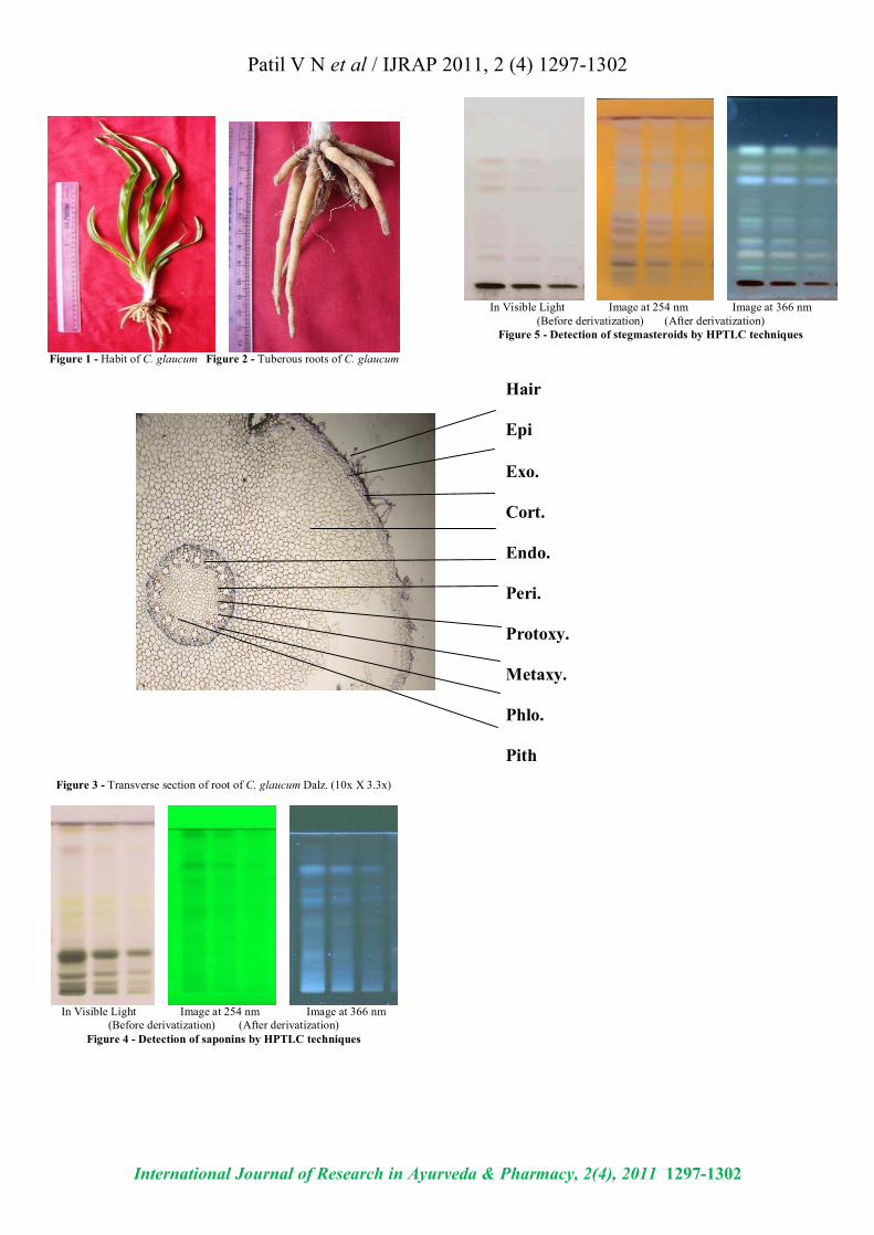
Phytochemical evaluation Some roots were dried under the shade so as to avoid the decomposition of chemical constituents, powdered in a blender and finally stored in dry air tied containers for phytochemical screening. Ash and percentage extractive content was measured by following standard pharmacopoeal techniques18. Fluorescence analysis was carried out as per Chase and Pratt19. Qualitative phytochemical tests were carried out by standard methods of Harborne20 and Trease and Evans15. Quantitative phytochemical analysis was determined for proteins, carbohydrates and saponins by the methods of Lowry et al.21; Nelson22 and Obadoni and Ochuko23 respectively. The phytochemical screening was also done by High Performance-Thin Layer Chromatography (HPTLC). HPTLC study was carried out on Linomat 5 for application using Densitometer- TLC Scanner 3 with “WINCATS” software (Camag, Switzerland). These studies were carried out on pre-coated aluminum fluorescent plates (E. Merck). For HPTLC studies, an extract of methanol (25% GR) solvent system was used and after development, plate was scanned at 254 and 366 nm24,25,26. RESULTS AND DISCUSSIONS Macroscopic evaluation (Figure 1 and 2) The details of the macroscopic examination are mentioned in Table 1 and illustrated in Figures 1 and 2. Microscopic characters The transverse section of the root had a circular outline. The outermost layer is the epidermis consisting of uniseriate trichomes followed by a very large zone of the cortex. The outermost layer of the cortex just below the epidermis consists of cells which are mostly rectangular, appearing longer than wide. The rest of the cortex are rounded to polygonal parenchymatous cells and have no intercellular spaces. The innermost layer of the cortex is a single layered endodermis. The stellar structure shows that the endodermis is followed by the pericycle layer. The xylem is exarch variety and the phloem is in between the xylem along with the parenchyma. The central region is occupied by large pith mostly polygonal in shape (Figure 3). Histochemical Screening Histochemical screening showed the presence of starch, protein, fat, saponins, tannin, sugars and alkaloids (Table2). Phytochemical Studies The tuber had a total ash content of 10.7%, the acid insoluble ash being 3.5% (Table 3). The values of percentage extractives were higher in chloroform and lower in benzene solvent (Table 4). Fluorescence analysis was carried out to check the purity of the drug.
The powder drug was observed in visible light as yellowish white in color. The powder was treated with nitrocellulose, 1 N sodium hydroxide, 1 N sodium hydroxide in nitrocellulose and dried for 30 minutes. After this it was observed under ultraviolet light and it emits the color as shown in (Table 5). Qualitative analysis of the root indicated the presence of proteins, reducing and non-reducing sugars, saponins, fats, tannin, glycoside and alkaloids (Table 6). The quantity of proteins is higher than saponins and carbohydrates (Table 7). Saponins are the important chemical and justify the use of tubers of this plant and are used as a well known health tonic, aphrodisiac and galactogogue4-6,27,28. In HPTLC study, the methanolic extract is ultrasonic for 15 minutes and filtered. The filtrate is used as an application for saponins and stegmasteroids. For each application 20 µl, 10µ and 5µl extracts were used and loaded on instrument comprising of Linomat 5 for application using Densitometer- TLC Scanner 3 with “WINCATS” software (Camag, Switzerland). These studies were carried out on pre-coated aluminum fluorescent plates (E. Merck). The plates were scanned at 254 and at 366 nm24-27. Analytical studies (Saponins) The HPTLC analysis showed that, the saponins from the C. glaucum root samples gave light yellow bands in visible light and blue bands after derivatization in fluorescence light. The plates were scanned at 254 and 366 nm. When images were compared with the graph and table values, it showed maximum area 31.19 % at 366nm after derivatization. The table also indicates the starting Rf values and end Rf values (Figure 4; Graph 1-3; Table 8-10). Analytical studies (Stegmasteroids) In HPTLC analysis, stegmasteroids revealed white bands in visible light. After derivatization in fluorescence light it showed the dark blue bands. The plates were scanned at 254 and 366 nm. It was covered the area 25.16% at 254 nm. The tables also indicate the Rf values for all the peaks scanned by “WINCATS” software (Figure 5; Graph 4-6; Table 11-13). CONCLUSION The plant C. glaucum showed the correct taxonomy which is helpful for the standardization of drug. The morphological characters and histochemical study with double staining of the root, percentage extractives, fluorescence and ash analysis and the phytochemical screening of the plant. As in case of saponins and stegmasteroids, the peaks are denoted by the Rf values. These investigations will be useful for the correct botanical identification and authentication of the drug.

Patil V N et al / IJRAP 2011, 2 (4) 1297-1302
International Journal of Research in Ayurveda & Pharmacy, 2(4), 2011 1297-1302
After getting the overall results of C. glaucum and if data is comparable with the above mention species of safed musali, it can be used as a substitute for them. ACKNOWLEDGEMENT Both the authors would like to express a sincere thank to Head, Department of Botany, University of Pune, Pune-411 007 for encouragement and necessary laboratory facilities and The first author is grateful to authorities of Pune University for providing financial support in the form of research stipend. REFERENCES 1. Hara H. The Flora of Eastern Himalaya. Tokyo University Press,
Japan; 1966. p. 407. 2. Patil VN, Deokule SS. Pharmacognostic standardization of
Chlorophytum bharuchae Ans., Ragh. & Hem. International Journal of Current Research 2010; 3: 27- 32.
3. Cooke T. Flora of Presidency of Bombay. BSI Calcutta; 1958; 3: 280-289.
4. Nadkarni AK. KM Nadkarni’s Indian Materia Medica. Popular Book Depot., Lamington Road, Bombay, 3rd Edtn. 1927; 1: 208-209.
5. Chopra RN, Nayer SL, Chopra IC. Glossary of Indian medicinal Plants CSIR New Delhi; 1956. p. 218.
6. Govindarajan R, Vijayakumar M, Pushpangadan P. Antioxidant approach to disease management and the role of ‘Rasayana’ herbs of Ayurveda. Journal of Ethnopharmacology 2005; 99: 165-178.
7. Anonymous. Medicinal Plants more on Safed Musali. Agriculture and Industry Survey; 2001. p. 38-39.
8. Dhuley JN. Effect of some Indian herbs on macrophase functions in Ochratoxin A treated mice. Journal of Ethnopharmacology 1997; 58: 15-20.
9. Nergard CS, Diallo D, Michaelsen TE, Malterud KE, Kiyohara H, Matsumoto T. Isolation, Partial characterization and immunostimulation activity of polysaccharides from Verninia kotschyana Sch. Bip. Ex. Walp. Journal of Ethnopharmacology 2004; 91: 141-152.
10. Kirtikar KR, Basu BD: Indian Medicinal Plants. Liliaceae: Chlorophytum. In Kirtikar KR; Basu BD (Eds.) L.M. Basu Publishers. Allahabad, India; 1975. p. 2508-2509.
11. Sreevidya N, Kumar V, Kumar S, Sikarwar RLS. Utilization, depletion and conservation of Safed Musali (Chlorophytum spp.). Journal of non- Timber Forest Products 2003; 10: 155-157.
12. The Wealth of India- A dictionary of Indian raw materials and industrial products, Revised Edn. Publication and Information Directorate, CSIR. Dr. R.S. Krishnan Marg, New Delhi. 1992; 3: 482-483.
13. Marais W, Reilly J. Chlorophytum and its related Genera (Liliaceae). Kew Bulletin 1978; 32 (3): 653-663.
14. Johansen DA. Plant Microtechnique, McGraw-Hill Book Co. Inc. New York; 1940. p. 151-154, 182-203.
15. Trease GE, Evans WC. Trease and Evans Pharmacognosy, 15th Ed. W. B. Saunders Edinburgh London, New York, Philadelphia St. Louis Sydney Toronto; 2002. p. 3-4, 528-533, 538-547.
16. Wallis TE. A Text Book of Pharmacognosy, reprinted edition, Churchill, Livingstone. London; 1967. p. 578 – 617.
17. Krishnamurthy KV. Methods in the Plant Histochemistry, Viswanadhan Pvt. Limited, Madras; 1988. p. 1 – 77.
18. Anonymous. Pharmacopoeia of India, Government of India, Ministry of Health Manager, Publications Delhi, 1st Edn.; 1955. p. 370 & 864.
19. Chase CR, Pratt R. Fluorescence of powdered vegetable drugs with particular reference to development of a system of identification. Journal of Amer. Phar. Asso. (Sci. ed.) 1949; 38: 324-330.
20. Harborne JB. Phytochemical Methods, Chapman and Hall International Edition, London. 2nd edition; 1973. p. 5-8.
21. Lowry OH, Rosebrough NJ, Farr AL, Randall RJ. Protein measurement with the Folin-Phenol reagent. Jour. of Biol. Chem. 1951; 193: 265-275.
22. Nelson N. A photometric adaptation of the Somogyi method for the determination of Glucose. Jour. of Biol. Chem. 1944; 153: 375-380.
23. Obadoni BO, Ochuko PO. Phytochemical studies and comparative efficacy of crude extracts of some homeostatic plants in Edo and Delta state of Nigeria. Global Jour. Pure Appl. Sci. 2001; 8: 203-208.
24. Patil VN, Deokule SS. Pharmacognostic Evaluation of Chlorophytum orchidastrum Lindl. Annals of Biological Research 2010; 1 (4): 126-137.
25. Wagner H, Baldt S. Plant Drug Analysis: A Thin Layer Chromatography Atlas. Springer – Verlag, Berlin; 1996. p. 129, 144, 155, 157, 176, 178, 206.
26. Reich E, Schibii A. High Performance- Thin Layer Chromatography for the analysis of medicinal plants, Thieme medical publishers. Inc; 2007. p. 129-160, 206-210, 224-240.
27. Patil VN, Deokule SS. Pharmacognostic evaluation of Chlorophytum tuberosum Baker, Govt. of India (AYUSH) International Journal of Ayurveda Research 2010; 1 (4): 175 – 180.
28. Oudhia P. Problem perceived by Safed Musali (Chlorophytum borivilianum) growers of Chhattisgarh (India) region: A study. Journal of Medicinal and Aromatic Plant Sciences 2001; 22/4A & 23/ 1A: 396-399.
Table 1 - Macroscopic examination of C. glaucum Dalz.
Herb 1 - 1.5 ft. in height. Roots Tuberous root are fibers cylindrical and are measuring
10-14 cm long, 1-1.4 cm diameter. Leaves
Green, 6 – 9 in number, oblanceolate, acute, glaucous, glabrous, 20 – 35 ´ 2.8 – 4.5 cm, short broad petiole.
Scape Densely clothed with sheaths, erect, 3 – 12 ft. long. Flower White, dense raceme, lanceolate, acuminate.
Bract Persistent forming a terminal coma before flowering, the lower 0.5 cm long, upper 0.5 – 0.8 cm long.
Pedicels Ascending, 0.5 – 0.8 cm long, slender, jointed at or below the middle.
Perianth
White, naked, segments less than 0.8 cm long by 0.5 cm filaments minutely papillose, 5 nerved.
Stamen. 0.5 – 0.8 cm long, anther 0.5 cm long. Style 0.8 cm long, stigma minute.
Capsule Globose, emarginate, 3 winged. Seeds Black, orbicular, 2- 4, 0.3 – 0.5 cm.

Patil V N et al / IJRAP 2011, 2 (4) 1297-1302
International Journal of Research in Ayurveda & Pharmacy, 2(4), 2011 1297-1302
Table 2 - Histochemical study of C. glaucum Dalz.
Test Reagents Color Tissue Starch I2KI Blue Cort., Xy. Phlo.
pith Protein Potassium
Ferrocyanide + water + acetic acid + 60%
alcohol + FeCl3
Blue Epi., Cort., Peri., Phlo., xy.
Tannin Acidic FeCl3 Light brown
Epi., Cort., Peri., Phlo., xy.
Saponin Conc. H2SO4 Yellow Epi., Peri., Phlo., xy.
Fat Sudan III Pink Epi., endo., phlo., xy. pith
Sugar 20% aq. NaOH Yellow Epi., Cort., Xy. Phlo.
Glycosides Guignard’s Test Brown Hair, Epi. Alkaloids Mayer’s Reagent Colorless Hair, Epi., Cort
xy. Phlo..
Wagner’s Reagent Orange Cort. Endo., peri., phlo., xy.
Dragendorff’s Reagent
Orange to dark
brown
Epi., Cort., Phlo., xy. pith
Tannic acid Buff color
Cort., Phlo., xy. pith
Hager’s Reagent Yellow Cort.
Abbreviations: I2KI: Potassium iodide, FeCl3: Ferric chloride, Conc. H2SO4: Concentrated sulphuric acid, NaOH: Sodium hydroxide.
Epi: Epidermis, Endo: Endodermis, Peri: Pericycle, Cort: Cortex, Xy: Xylem, Phlo: Phloem
+ Sign indicates the addition of Potassium Ferrocyanide in water, then acetic acid , 60% alcohol and lastly FeCl3
Table 3 - Ash and Acid Insoluble Ash of C. glaucum Dalz.
Parameter Results Total Ash 10.7 % dry wt.
Acid Insoluble Ash 3.5 % dry wt.
Table 4 - Percentage extractives of C. glaucum Dalz.
Solvent Used Extract (%) Distilled Water 4.70 %
Absolute Alcohol 0.28 % Petroleum ether 0.24 %
Benzene 0.19 % Chloroform 9.31 %
Diethyl ether 0. 32 % Acetone 0.35 %
Table 5 - Fluorescence analysis of C. glaucum Dalz. at 230 nm
Treatments Color Emits
Powder as such Yellowish white
Powder as such in UV-light Pale white Powder + Nitrocellulose Grayish white
Powder + 1 N NaOH in Methanol Whitish gray Powder + 1 N NaOH in Methanol dry for 30 min. + Nitrocellulose
Grayish white
Table 6 - Phytochemical study of C. glaucum Dalz.
Compound Reagents Results
Water Extracts Starch I2KI + ve
Protein Millon’s reagent + ve
Tannins Acidic FeCl3 + ve
Saponin Distilled water + ve
Steroids Liebermann – Burchard’s Test + ve
Salkowski Test + ve
Anthroquinone’s Benzene + 10% NH4OH - ve
Sugars Benedict’s reagent + ve
Fats Sudan III + ve
Alcoholic extracts
Alkaloids
a Mayer’s Reagent + ve
b Wagner’s Reagent + ve
c Dragendorff’s Reagent + ve
d Tannic acid + ve
e Hager’s Reagent + ve
f Folin-Phenol Reagent + ve
Glycosides Benzene + ve
Table 7 - Quantitative estimation of C. glaucum Dalz.
Quantitative estimation (mg /g )
Protein 1.69740 Reducing Sugar 0.03951
Non - Reducing Sugar 0.06829 Starch 0.04946
saponins 1.1024
Table 8 - showing the peak values for saponins for 20 µl plant extract
Table 9 - showing the peak values for saponins for 10 µl plant extract
Patil V N et al / IJRAP 2011, 2 (4) 1297-1302
International Journal of Research in Ayurveda & Pharmacy, 2(4), 2011 1297-1302
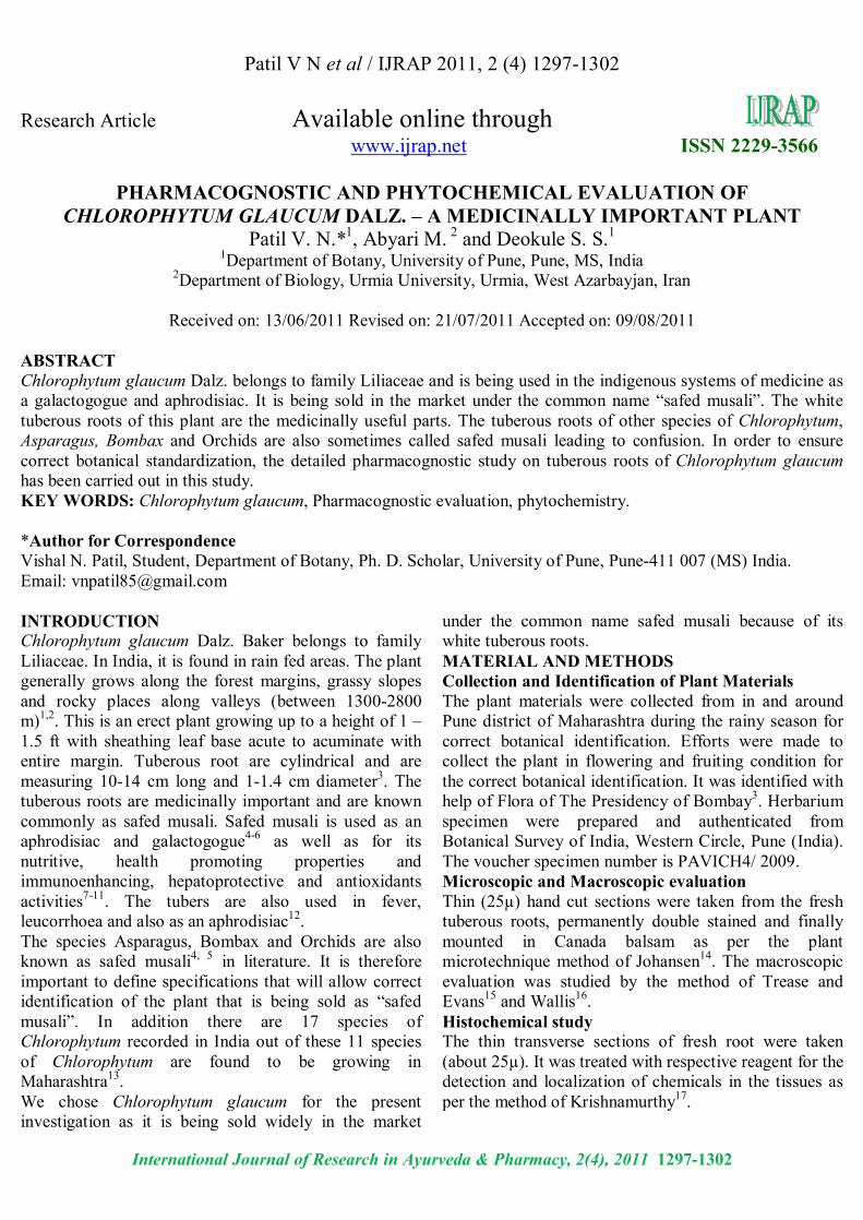
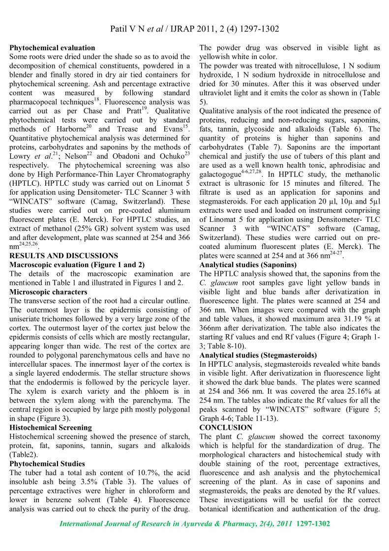
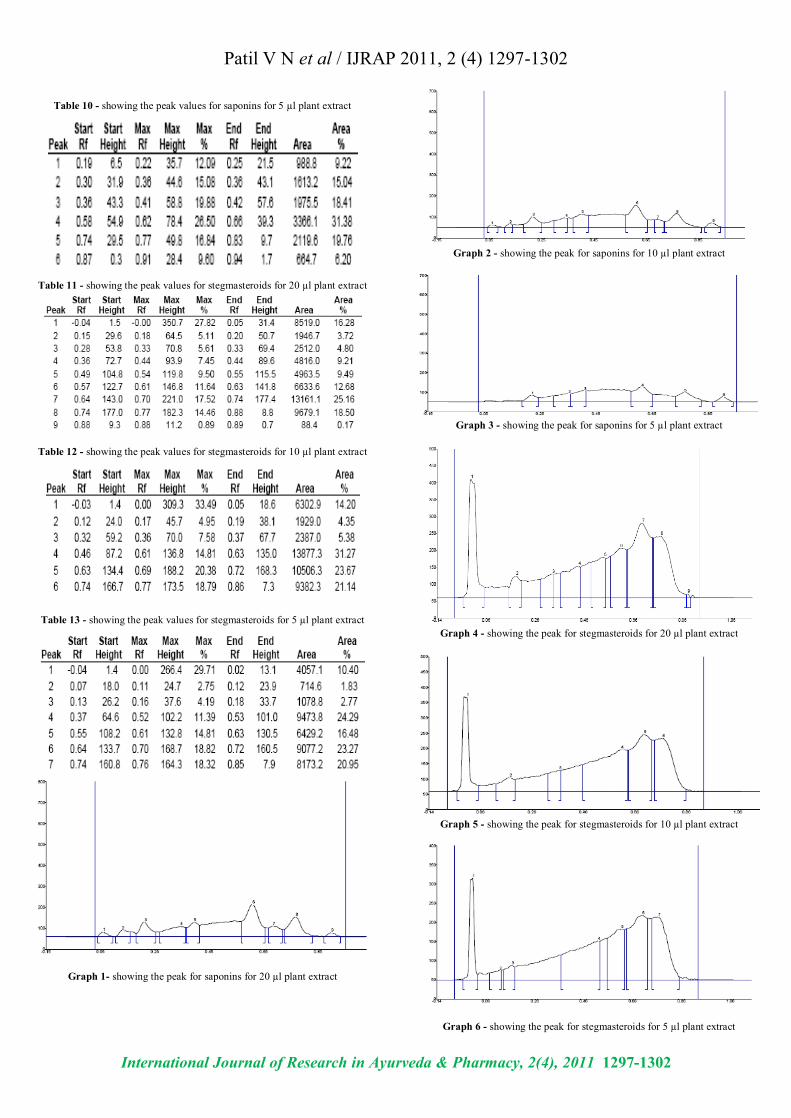
Table 10 - showing the peak values for saponins for 5 µl plant extract
Table 11 - showing the peak values for stegmasteroids for 20 µl plant extract
Table 12 - showing the peak values for stegmasteroids for 10 µl plant extract
Table 13 - showing the peak values for stegmasteroids for 5 µl plant extract
Graph 1- showing the peak for saponins for 20 µl plant extract
Graph 2 - showing the peak for saponins for 10 µl plant extract
Graph 3 - showing the peak for saponins for 5 µl plant extract
Graph 4 - showing the peak for stegmasteroids for 20 µl plant extract
Graph 5 - showing the peak for stegmasteroids for 10 µl plant extract
Graph 6 - showing the peak for stegmasteroids for 5 µl plant extract
Patil V N et al / IJRAP 2011, 2 (4) 1297-1302
International Journal of Research in Ayurveda & Pharmacy, 2(4), 2011 1297-1302
Figure 1 - Habit of C. glaucum Figure 2 - Tuberous roots of C. glaucum
Figure 3 - Transverse section of root of C. glaucum Dalz. (10x X 3.3x)
In Visible Light Image at 254 nm Image at 366 nm
(Before derivatization) (After derivatization) Figure 4 - Detection of saponins by HPTLC techniques
In Visible Light Image at 254 nm Image at 366 nm
(Before derivatization) (After derivatization) Figure 5 - Detection of stegmasteroids by HPTLC techniques
Hair Epi Exo. Cort. Endo. Peri. Protoxy. Metaxy. Phlo. Pith