Embed Size (px)
Citation preview
Mulay Shashikant K et al / IJRAP 2011, 2 (3) 715-722
International Journal of Research in Ayurveda & Pharmacy, 2(3), 2011 715-722
Research Article Available online through www.ijrap.net ISSN 2229-3566
STUDY OF JANU MARMA IN RELATION TO ITS VAIKALYAKARA EFFECTS WHEN
INJURED Mulay Shashikant K. 1*, Paranjape M. H. 2, Bhingare S. D. 3
1Associate professor and Head of department, Dept. of Sharir Rachana, Govt. Ayurved College, Vazirabad road, Nanded, Maharashtra, India
2Ex-Professor & Head of department, Dept. of Sharir Rachana, Ashtanga Ayurved College, Pune, Maharashtra, India 3P.G. Scholar (Final Year), Dept. of Sharir Rachana, Govt. Ayurved College, Vazirabad road, Nanded, Maharashtra,
India
Received on: 20/04/2011 Revised on: 21/05/2011 Accepted on: 09/06/2011 ABSTRACT This study was undertaken to investigate “Vaikalyakara Marma” with special reference to injuries occur at Janu Marma (knee joint). Location of Janu Marma was done with help of description of Marma in Ayurvedic texts, dissection of Knee joint & with help of X-ray documentation. 160 patients having trauma to knee joints due to various causes such as fall, road traffic accident, direct trauma on knee joint, h/o trauma year back, known c/o knee surgery & injury due to burn were included in this study. Major symptoms like Khanjata (lameness), pain, swelling, loss of power, restricted movements, muscle wasting & associated symptoms like tingling sensation, heaviness, syncope, sweating, dizziness, vomiting were reviewed. At end of study it was found that Janu (knee joint) is definitely a Vaikalyakara Marma. Disabilities like restriction of movements, swelling & atrophy were remains of injured knee joint inspite of best surgical treatment. KEYWORDS: Ayurveda, Marma, Vaikalyakara Marma, Janu-Marma (Knee joint). *Corresponding Author Dr. Muley Shashikant Krishnanath, Associate Professor and Head of Department, Dept. of Sharir Rachana, Govt. Ayurved College, Vazirabad road, Nanded, Maharashtra. Email: [email protected] INTRODUCTION Ayurveda possesses some basic principles which are most exceptional & significant in all life sciences, such as Panchabhautik Siddhant1, Tridosha Siddhant2, Dosha-Dhatu- Mala Siddhant3, Atma Vijnana4, Mano Vijnana5, Garbha Vijnana6 & last but not least “Marma Vijnana”7. Marma, defined as an anatomical vulnerable area where arteries, tendons, flesh, veins, bones and joints congregate to form the spot of life or Prana8. Marma, Definitions of Marma, Types of Marma, symptoms produced after injurious & their management is described by nearly all Ayurvedic texts, especially in “Trimarmiya Siddhi”9, “Trimarmiya Chikitsa”10 Chapters in Charaka Samhita, “Marmavibhaga” Chapter in Ashtanga Sangraha11, “Shariravichaya Sharir’ chapter in Kashyapa Samhita12. Little injuries to these Marma points or anatomical areas can be fatal comparing with major injuries at anywhere else in body so, detailed knowledge of these Marma points is crucial for Ayurvedic physician. Sushruta mentioned that Marma
points should be avoided during surgical procedures due to its fatality13. Deep knowledge of these fatal points is compulsory for surgeon. In ancient era, even after best treatment available at that time, some sort of disability remains at end of therapy. Anatomically knee joint is most important joint as it is weight bearing joint and more prone to get diseased or injured while doing routine activities. According to Ayurvedacharya Janu Marma (three digit area of knee joint) is vaikalyakara marma. So injury to this marma may cause disabilities as described in Sushrut Sharirsthana 6th chapter which may interrupt daily activities. Nowadays in spite of super specialty modalities available, does modern science is capable of curing these disabilities? To rule out above question study of Janu marma in relation to its Vaikalyakara effect when injured is carried out.
Mulay Shashikant K et al / IJRAP 2011, 2 (3) 715-722
International Journal of Research in Ayurveda & Pharmacy, 2(3), 2011 715-722
AIM & OBJECTIVE 1. To locate appropriate position of Janu Marma
manually in healthy volunteer as per Ayurvedic text. 2. To locate appropriate position of Janu Marma with the
help of X-Ray finding. 3. Study of structure included in Janu marma by doing
dissection of knee joint in cadaver. 4. Survey of Janu Marmaghat (injury at 3 digit parimana
area of knee joint) patients to study its signs & symptoms & their Vaikalyakara effect (deformities) formed after injuries or surgical treatment.
MATERIAL & METHODS Materials For Structural Study of Janu Marma 1. X -ray instrument 2. Knee joint of cadaver 3. Scalpel 4. Surgical scissor 5. Plain & tooth forceps 6. Retractors 7. Camera For Survey of Janu Marma (Study of Marmaghat Symptoms) 1. CRF for the survey of patients of Janu Marmaghata
(Injuries to Janu Marma) 2. X ray film & their reports 3. Goniometer (For assessment of restriction of
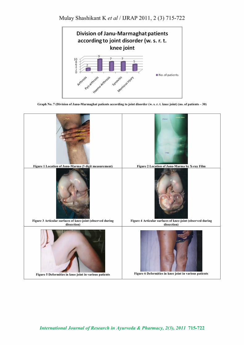
movement) Methods For Location of Janu Marma in Healthy Volunteer To the study of the structures which is included in site of Janu Marma, it is essential to determine the appropriate location of Janu Marma, for the conveniences in the dissection as well as during survey. In the Ayurvedic text book the Janu marma is located at meeting point leg and thigh14 i.e. Knee joint and the measurement of Janu Marma is about three Angula (three digits)15. To decide this three Angula area on knee joint, healthy adult volunteer having all proportionate body parts was selected. Fully extended Index, Middle & Ring finger of left hand, put parallel on the right knee-joint, from medial to lateral on anterior side, where the long axis of middle finger touches to apex of the Patella [Photo-1]. Anterio-posterior Radiograph in the above situation shows, ‘Three digit area’ is the location of Janu - Marma [Photo-2]. For the convenience of study anterior aspect is preferred and structures situated beneath this three finger area were studied. Study of Structures Involved In Janu Marma Steps of Dissection of Knee Joint Include Dissection of knee joint is always started from dorsal aspect of knee. Left knee-joint of cadaver was selected
for dissection, step by step dissection procedure was done, and observations about superficial fascia, Boundaries, Contents, Floor and Vessels of the fossa were noted. Also observation about Ligaments, Synovial membrane, then Menisci, Cruciate ligaments and articular surfaces of knee-joint were noted [Photo-3]. Hence procedure done was described below16 1. Horizontal incision on back of thigh (at upper 2/3rd &
lower 1/3rd junction) 2. Another horizontal incision on back of leg (at upper
1/3rd & lower 2/3rd junction) 3. Vertical incision joining midpoint of above two
horizontal incisions 4. Reflection of skin and superficial fascia 5. Observations of cutaneous vessels, identification of
boundaries 6. Contents of Popliteal fossa were noted. 7. The semitendinosus and biceps femoris were striped
from the posterior side of the knee joint. 8. The semimembranosus was traced to its insertion and
the aponeurotic fibers which extend to the capsule of the knee joint and form the oblique popliteal ligament were noted.
9. The popliteal tendon was traced to its femoral attachment, and the fibular (lateral) collateral ligament was located and isolated.
10. Isolate and expose the tibial (medial) collateral ligament to the medial side of the joint and. Its deep portion extending posteriorly and attaching to the margin of the medial condyle was noted.
11. All elements of the quadriceps femoris was made free from the femur and reflected downward, cutting the capsule of the joint as it is seen.
12. The capsule of the joint was cut along the anterior border of the tibial and fibular collateral ligaments.
13. The quadriceps with the contained patella was pulled downward. The combined insertions of the quadriceps femoris, the patella, the patellar retinacula and fascia lata combine to provide anterior reinforcement for the joint.
14. Variation in size of its different articular surfaces was noted. The infrapatellar synovial fat pad, its alar folds and its attachment at the intercondylar fossa were seen. The joint cavity, the communication between right and left halves beneath the infrapatellar synovial fold, the suprapatellar bursa and the deep infrapatellar bursa (deep to the patellar tendon) was defined.
15. The attachment of the infrapatellar synovial fold was cut. The anterior cruciate ligament, the posterior cruciate ligament, the medial and lateral menisci, and the transverse genicular ligament was identified.
Mulay Shashikant K et al / IJRAP 2011, 2 (3) 715-722
International Journal of Research in Ayurveda & Pharmacy, 2(3), 2011 715-722
16. The posterior and anterior cruciate ligaments were identified from the dorsal side.
17. The joint was opened and the attachments of the menisci to the intercondylar eminence and (medially) to the capsule were noted.
18. The attachments of cruciate ligaments were examined. The joint was extended and flexed to determine the varying axes of rotation of the femoral condyles and collateral ligaments, the rotation of the femur in flexion and extension, and the freedom of motion of the menisci.
For Survey (Study of Marmaghat Symptoms of Janu Marma) Following Points Were Taken Into Consideration – Inclusion Criteria
a. H/o Trauma at either knee-joint, b. H/o Trauma in either sex, c. Age group of 06 to 80 yrs, d. Registered patients at Government Medical College, Exclusion Criteria a. Patients having below age of 06yrs & above 80 yrs, b. Unconscious patients, c. Deaf, mute & dump patients, d. Psychic patients, e. Poliomyelitis patients Since the patient having age below 6 years and above 80 years, Unconscious, deaf mute & dump patients, psychic patients, cannot cooperate for study so these patients were not included in study. Muscle paresis and paralysis can sometimes result in skeletal deformities, tightening of the joints and movement disability in poliomyelitis so patients of poliomyelitis were excluded from study. Criteria For Diagnosis Criteria for diagnosis were done on the basis of sign and symptom available in the Ayurvedic and Modern texts as well as with the help of following parameters. a) Presence of pain, Tenderness, Swelling and loss of
muscle power, muscle atrophy and restricted movement at knee joint.
b) Foot flexion test. c) Foot extension test. d) Radiological assessment, X-ray, knee joint AP &
lateral view was carried out in all patients to ascertain the diagnosis as well as the differential diagnosis.
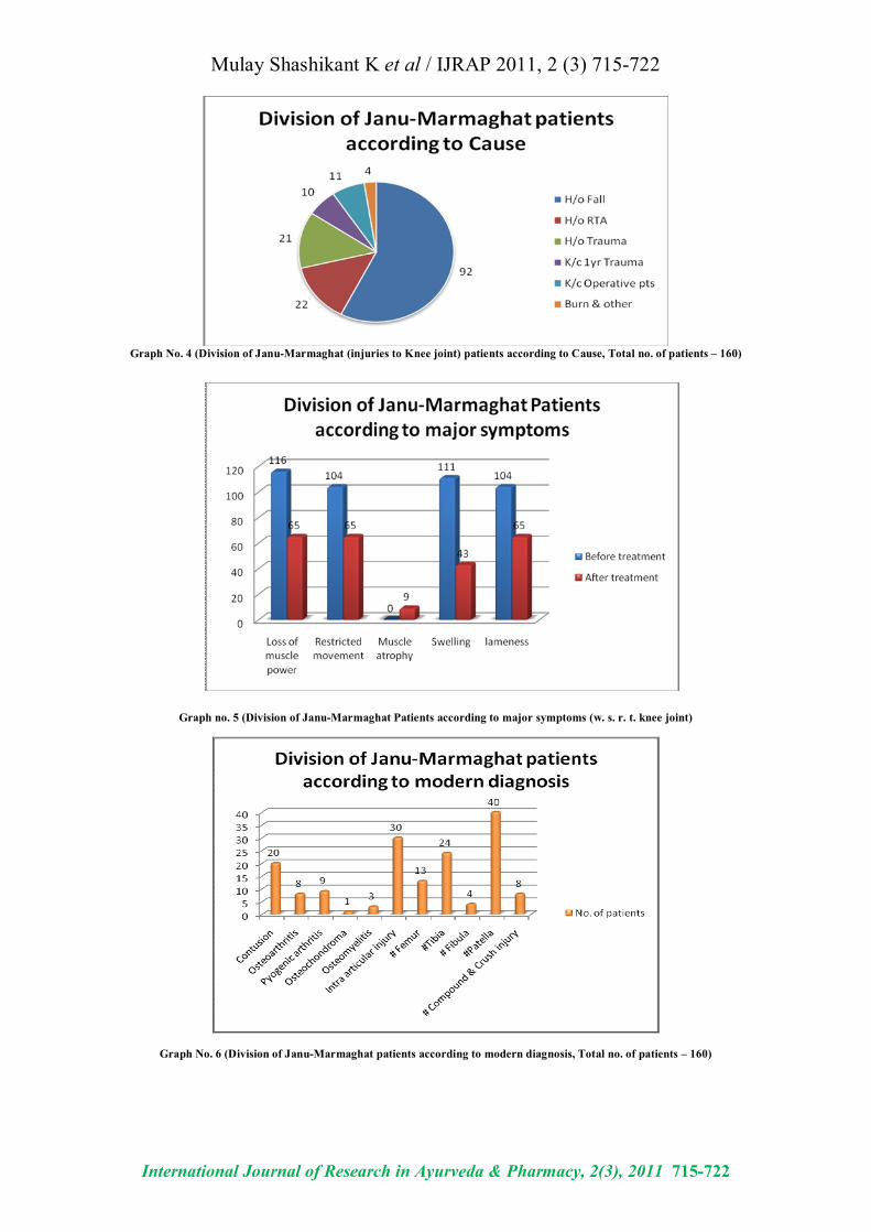
Assessment Criteria For Symptoms of Janu Marmaghat (Injuries To Knee Joint) a. For Pain, Tenderness, Swelling (Mild - +, Moderate - ++, Severe +++) RESULTS Total no. of 160 patients, registered at OPD/IPD of government Medical College were assessed on symptoms like Pain, Tenderness, Swelling, Loss of
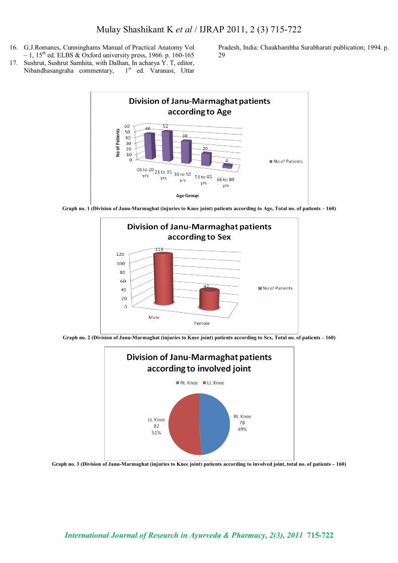
activities, Muscle wasting, walking inability (restricted movements) & other associated symptoms. Observations were made & noted as following. Patients were divided into 5 groups. The larger group belonged to the age group of 21 – 35 yrs. i.e. 52 patients (32.50%), 46pts (28.75%) belonged to the age group 06 to 20 yrs, 38 pts (23.75 %) of age group 36 to 50 yrs, 20 pts (12.50 %) belonged to the age group 51 – 65 yrs and remaining 04 pts (02.50%) belonged to the age 66 to 80 yrs. Out of 160 patients, large no. of samples i.e. 118 were males (73.75%) & remaining 42 were females (26.25%). Out of 160 patients, Left knee joint was more involved in trauma or injury than right knee joint. According to external cause of injury, in majority of patients Janu Marmaghat was consequence of fall from height (92 patients). The other causes included were road traffic accident, trauma (direct / indirect); trauma caused before one year, post operative, burns etc. After reviewing major & associated symptoms after knee injuries, it was found that majority of patients had symptoms like loss of muscle power, restricted movements, swelling and lameness after injury to knee joint, but no one had muscle atrophy. Those patients observed post allopathic treatment, were found with unchanged symptoms even after treatment. These patients when diagnosed and categorized on modern diagnosis, it was found that large samples were have a medical condition like # Patella, intra articular injury, # tibia and contusion before developing major symptoms. Further, these samples were reviewed to find internal cause of joint injury as joint disorder & it was found that large samples were have a medical condition like Pyo-arthrosis -30%, then Haemo-Arthrosis and Synovitis- each 23.33%, meniscus injury-16.666% and Arthrosis -6.666%. DISCUSSION After analyzing x-ray films of knee joint of healthy volunteer, it was explicable that Janu Marma is formed by lateral intercondylar tibio-femoral joint, medial intercondylar tibio-femoral joint, superior tibio-fibular joint, patelo-femoral joint. In Text, Sushruta Samhita sharirsthana ch. 6 mentioned that Mansa (muscle), Sira (vessels), Snayu (tendons) are present nearby in small fraction and asthi (bones), sandhi (joints) are present in higher percentage. After dissection of cadaveric knee joint, same was found which was stated earlier by Acharya Sushruta as Janu Marma is also a Vaikalyakara Marma. In Clinical study only those patients were included in study in which injuries to knee joint occurs in areas of
Mulay Shashikant K et al / IJRAP 2011, 2 (3) 715-722
International Journal of Research in Ayurveda & Pharmacy, 2(3), 2011 715-722
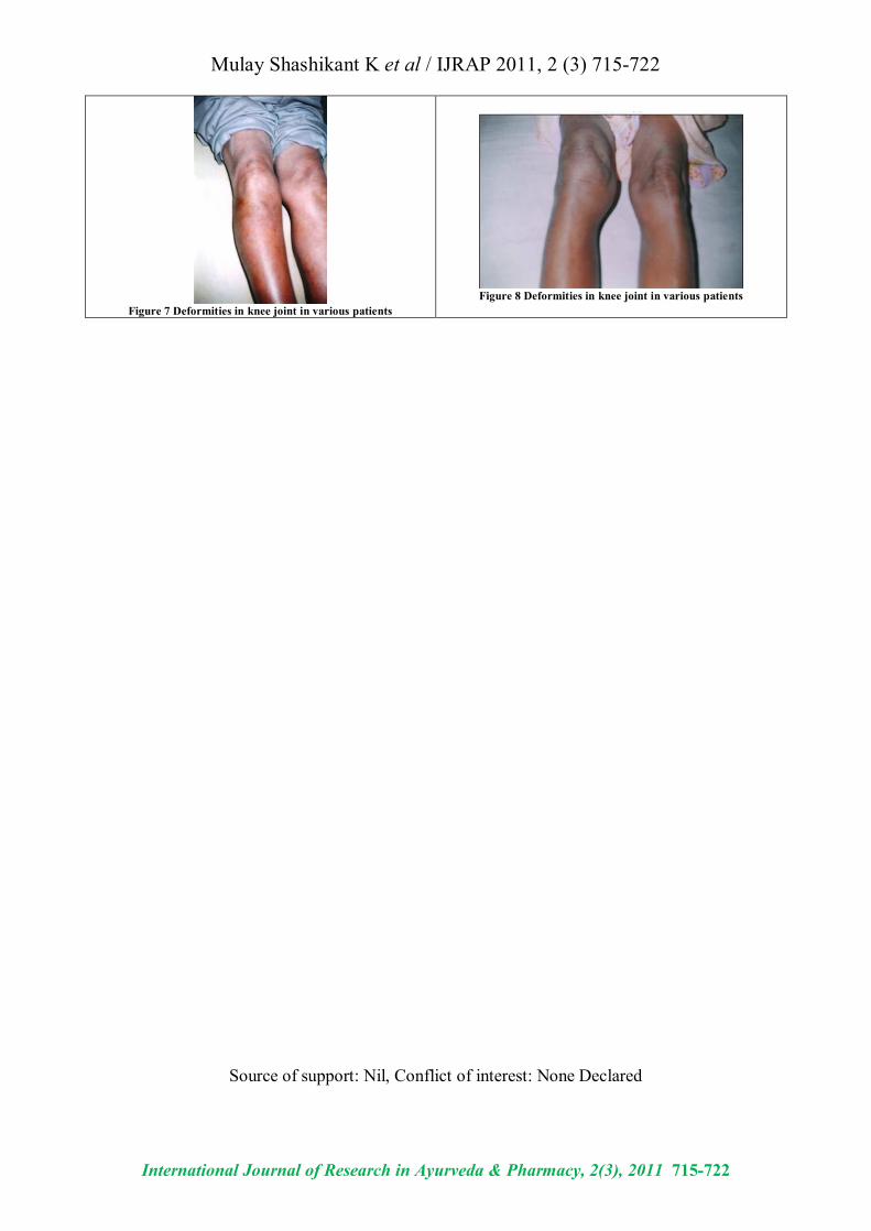
three Angula Parimana (length of 3 digits). As per modern concept, # Medial condyle femur, # Lateral condyle femur, # Supra-condyle femur, # Inter-condyle femur, # Medial condyle tibia, # Lateral condyle tibia, # Bi-condyle tibia, # Displaced patella, # Un-displaced Patella, Synovitis, Arthrosis, Haemo-Arthrosis, Pyo-arthrosis Bursitis, Meniscus injury are includes in Janu marmaghat. After the survey of Janu- Marmaghat patients, Swelling, Loss of power, Restricted movements (flexion & extension), deformity e.g. Difficulty in climbing, squatting posture is observed in 65 (40.62 %) patients’ inspite of best surgical treatment. Cause of Khanjata (lameness) or deformity was changing the normal structure of articular surface in knee joint, which was confirmed through study of post operative x-rays. CONCLUSION With reference to Sharir – Sthana of Sushruta (Ch. 6) Vaikalyakara Marma are Somaguna bhuyistha i.e. it posses Soma Mahabhuta means lunar properties, which have its own characters like steadiness & coolness. Although Injuries to this site are not life threatening but definitely it produces deformity so the normal movements of that part gets affected. Janu Marma is congregation point of Uru (Thigh) & Jangha (Leg).17 Its parimana (dimension) is three angula (three digits) & effect wise it’s Vaikalyakara Marma. Structurally it is a Sandhi Marma. In the current study it was observed that in the region of three digits of Knee joint (shown in pic – 3) contents like Mansa (muscle), Sira (vessels), Snayu (tendons) are present nearby in small fraction and Asthi (bones), Sandhi (joints) are present in higher percentage. After studying three digit area of knee joint with help of X-ray study it was concluded that knee joint is congregation of medial tibio-femoral intercondylar joint, lateral tibio-femoral intercondylar joint, patelo-femoral joint, superior tibio-fibular joint and related articular part of involved bone. Hence it is proved that Janu Marma (knee joint) is Sandhi Marma. According to Ayurvedic texts injuries to Knee joint produces symptoms like loss of muscle power, restricted movements, atrophy of knee joint, swelling, lameness. After clinical evaluation of patients of knee joint injuries it was concluded that all above symptoms related to knee joint which ultimately produces Khanjata (lameness) are present in most of the patient. 65 patients out of 160 (i.e. 40.625%) were found to affected with flexion deformity, edema of knee joint, restricted movements, painful movements, atrophic knee. So we can definitely state that Janu (knee joint) is Vaikalyakara Marma.
Kalari / Varman and marma may vary in number i.e. In Tamil traditions they are 108 Marma points, and in Kalari tradition there are 365 Marma points. But the concept behind this is one and the same i.e. these are the vital point where Prana is located. Along with their ability to kill, however, comes an ability to heal. REFERENCES 1. Charaka, Charaka Samhita, In Acharya V. S., editor, Vaidya-
manorama hindi commentary, 1st ed. Delhi, India: Chaukhambha Sanskrit publication; 2003. p. 356
2. Charaka, Charaka Samhita, In Acharya V. S., editor, Vaidya-manorama hindi commentary, 1st ed. Delhi, India: Chaukhambha Sanskrit publication; 2003. p. 30
3. Sushrut, Sushrut Samhita, with Dalhan, In acharya Y. T, editor, Nibandhasangraha commentary, 1st ed. Varanasi, Uttar Pradesh, India: Chaukhambha Surabharati publication; 1994. p. 58
4. Charaka, Charaka Samhita, In Acharya V. S., editor, Vaidya-manorama hindi commentary, 1st ed. Delhi, India: Chaukhambha Sanskrit publication; 2003. p. 17
5. Charaka, Charaka Samhita, In Acharya V. S., editor, Vaidya-manorama hindi commentary, 1st ed. Delhi, India: Chaukhambha Sanskrit publication; 2003. p. 17
6. Sushrut, Sushrut Samhita, with Dalhan, In acharya Y. T, editor, Nibandhasangraha commentary, 1st ed. Varanasi, Uttar Pradesh, India: Chaukhambha Surabharati publication; 1994. p. 282
7. Sushrut, Sushrut Samhita, with Dalhan, In acharya Y. T, editor, Nibandhasangraha commentary, 1st ed. Varanasi, Uttar Pradesh, India: Chaukhambha Surabharati publication; 1994. p. 288
8. Sushrut, Sushrut Samhita, with Dalhan, In acharya Y. T, editor, Nibandhasangraha commentary, 1st ed. Varanasi, Uttar Pradesh, India: Chaukhambha Surabharati publication; 1994. p. 288
9. Charaka, Charaka Samhita, In Acharya V. S., editor, Vaidya-manorama hindi commentary, 1st ed. Delhi, India: Chaukhambha Sanskrit publication; 2003. p. 945-962
10. Charaka, Charaka Samhita, In Acharya V. S., editor, Vaidya-manorama hindi commentary, 1st ed. Delhi, India: Chaukhambha Sanskrit publication; 2003. p. 622-667
11. Vagbhat, Ashtanga Sangraha, in Acharya S. S., editor, Shashilekha Sanskrit commentary, 1st ed. Varanasi, Uttar Pradesh, India: Chaukhambha Sanskrit Series office publication; 2006. p. 319-326
12. Kashyapa, Kashyap Samhita, Vidyothini Hindi commentary, Bhishagacharya Shri. Satyapal Sharma, 2nd ed. Chaukhambha Sanskrit Sansthan 1964, p. 66
13. Sushrut, Sushrut Samhita, with Dalhan, In acharya Y. T, editor, Nibandhasangraha commentary, 1st ed. Varanasi, Uttar Pradesh, India: Chaukhambha Surabharati publication; 1994. p. 292
14. Sushrut, Sushrut Samhita, with Dalhan, In acharya Y. T, editor, Nibandhasangraha commentary, 1st ed. Varanasi, Uttar Pradesh, India: Chaukhambha Surabharati publication; 1994. p. 290
15. Sushrut, Sushrut Samhita, with Dalhan, In acharya Y. T, editor, Nibandhasangraha commentary, 1st ed. Varanasi, Uttar Pradesh, India: Chaukhambha Surabharati publication; 1994. p. 292
Mulay Shashikant K et al / IJRAP 2011, 2 (3) 715-722
International Journal of Research in Ayurveda & Pharmacy, 2(3), 2011 715-722
16. G.J.Romanes, Cunninghams Manual of Practical Anatomy Vol – 1, 15th ed. ELBS & Oxford university press, 1966. p. 160-165
17. Sushrut, Sushrut Samhita, with Dalhan, In acharya Y. T, editor, Nibandhasangraha commentary, 1st ed. Varanasi, Uttar
Pradesh, India: Chaukhambha Surabharati publication; 1994. p. 29
Graph no. 1 (Division of Janu-Marmaghat (injuries to Knee joint) patients according to Age, Total no. of patients – 160)
Graph no. 2 (Division of Janu-Marmaghat (injuries to Knee joint) patients according to Sex, Total no. of patients – 160)
Graph no. 3 (Division of Janu-Marmaghat (injuries to Knee joint) patients according to involved joint, total no. of patients – 160)
Mulay Shashikant K et al / IJRAP 2011, 2 (3) 715-722
International Journal of Research in Ayurveda & Pharmacy, 2(3), 2011 715-722
Graph No. 4 (Division of Janu-Marmaghat (injuries to Knee joint) patients according to Cause, Total no. of patients – 160)
Graph no. 5 (Division of Janu-Marmaghat Patients according to major symptoms (w. s. r. t. knee joint)
Graph No. 6 (Division of Janu-Marmaghat patients according to modern diagnosis, Total no. of patients – 160)
Mulay Shashikant K et al / IJRAP 2011, 2 (3) 715-722
International Journal of Research in Ayurveda & Pharmacy, 2(3), 2011 715-722
Graph No. 7 (Division of Janu-Marmaghat patients according to joint disorder (w. s. r. t. knee joint) (no. of patients – 30)
Figure 1 Location of Janu-Marma (3 digit measurement)
Figure 2 Location of Janu-Marma by X-ray Film
Figure 3 Articular surfaces of knee-joint (observed during
dissection)
Figure 4 Articular surfaces of knee-joint (observed during
dissection)
Figure 5 Deformities in knee joint in various patients
Figure 6 Deformities in knee joint in various patients
Mulay Shashikant K et al / IJRAP 2011, 2 (3) 715-722
International Journal of Research in Ayurveda & Pharmacy, 2(3), 2011 715-722
Figure 7 Deformities in knee joint in various patients
Figure 8 Deformities in knee joint in various patients
Source of support: Nil, Conflict of interest: None Declared