Embed Size (px)
Citation preview
AD______________ Award Number: W81XWH-11-1-0106 TITLE: Molecular Mechanisms Underlying Genomic Instability in Brca-Deficient Cells PRINCIPAL INVESTIGATOR: Dr. Andre Nussenzweig CONTRACTING ORGANIZATION: The Geneva Foundation Lakewood, WA 98499 REPORT DATE: March 2013 TYPE OF REPORT: Annual PREPARED FOR: U.S. Army Medical Research and Materiel Command Fort Detrick, Maryland 21702-5012 DISTRIBUTION STATEMENT: Approved for public release; distribution unlimited The views, opinions and/or findings contained in this report are those of the author(s) and should not be construed as an official Department of the Army position, policy or decision unless so designated by other documentation.
REPORT DOCUMENTATION PAGE Form Approved
OMB No. 0704-0188 Public reporting burden for this collection of information is estimated to average 1 hour per response, including the time for reviewing instructions, searching existing data sources, gathering and maintaining the data needed, and completing and reviewing this collection of information. Send comments regarding this burden estimate or any other aspect of this collection of information, including suggestions for reducing this burden to Department of Defense, Washington Headquarters Services, Directorate for Information Operations and Reports (0704-0188), 1215 Jefferson Davis Highway, Suite 1204, Arlington, VA 22202-4302. Respondents should be aware that notwithstanding any other provision of law, no person shall be subject to any penalty for failing to comply with a collection of information if it does not display a currently valid OMB control number. PLEASE DO NOT RETURN YOUR FORM TO THE ABOVE ADDRESS. 1. REPORT DATE (DD-MM-YYYY) 2. REPORT TYPE 3. DATES COVERED (From - To)
4. TITLE AND SUBTITLE 5a. CONTRACT NUMBER
5b. GRANT NUMBER
5c. PROGRAM ELEMENT NUMBER
6. AUTHOR(S) 5d. PROJECT NUMBER
5e. TASK NUMBER
E-Mail: 5f. WORK UNIT NUMBER 7. PERFORMING ORGANIZATION NAME(S) AND ADDRESS(ES) 8. PERFORMING ORGANIZATION REPORT NUMBER
9. SPONSORING / MONITORING AGENCY NAME(S) AND ADDRESS(ES) 10. SPONSOR/MONITOR’S ACRONYM(S) U.S. Army Medical Research and Materiel Command
Fort Detrick, Maryland 21702-5012 11. SPONSOR/MONITOR’S REPORT NUMBER(S) 12. DISTRIBUTION / AVAILABILITY STATEMENT Approved for Public Release; Distribution Unlimited
13. SUPPLEMENTARY NOTES 14. ABSTRACT
15. SUBJECT TERMS
16. SECURITY CLASSIFICATION OF:
17. LIMITATION OF ABSTRACT
18. NUMBER OF PAGES
19a. NAME OF RESPONSIBLE PERSON USAMRMC
a. REPORT U
b. ABSTRACT U
c. THIS PAGE U
UU
19b. TELEPHONE NUMBER (include area code)
Standard Form 298 (Rev. 8-98) Prescribed by ANSI Std. Z39.18
W81XWH-11-1-0106
Molecular Mechanisms Underlying Genomic Instability in Brca-Deficient Cells
Dr. Andre Nussenzweig
The Geneva Foundation Lakewood, WA 98499
Our proposal is to explore the novel notion that it may be possible to restore near normal HR activity in Brca1 cells and tissues. We believe that this phenomenon will lead to targeted therapies to reduce lifetime risk of tumor formation in BRCA1 and potentially BRCA2 carriers.
BRCA1, 53BP1, cancer biology, DNA repair, tumorigenesis
56
1 March 2012 - 28 February 2013AnnualMarch 2013
3
Page
Introduction……………………………………………..…………………………...4
Body…………………………………………………………………………………..4
Key Research Accomplishments………………………………………….………...7
Reportable Outcomes………………………………………………………………..8
Conclusion……………………………………………………………………………8
References…………………………………………………………………………….8
Appendices……………………………………………………………………………8
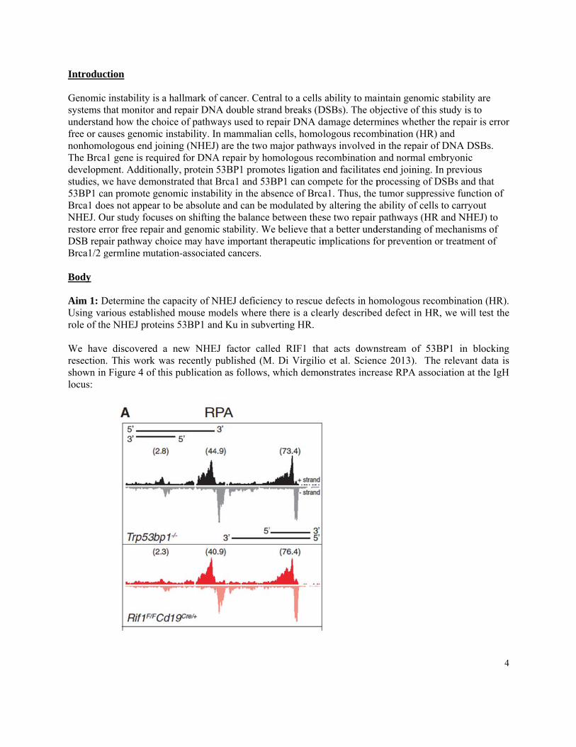
Introduct Genomic systems thunderstanfree or caunonhomolThe Brca1developmstudies, w53BP1 caBrca1 doeNHEJ. Ourestore errDSB repaBrca1/2 g Body Aim 1: DUsing varrole of the We have resection.shown in locus:
tion
instability is hat monitor an
nd how the chuses genomiclogous end jo1 gene is requ
ment. Additionwe have demoan promote gees not appear ur study focusror free repair
air pathway chgermline muta
Determine the rious establishe NHEJ prote
discovered This work wFigure 4 of th
a hallmark ofnd repair DNoice of pathw
c instability. Ioining (NHEJuired for DNAnally, protein nstrated that B
enomic instabto be absolutses on shiftingr and genomihoice may havation-associat
capacity of Nhed mouse meins 53BP1 an
a new NHEwas recently phis publicatio
f cancer. CentNA double straways used to r
n mammalian) are the two
A repair by ho53BP1 promoBrca1 and 53ility in the ab
te and can be g the balancec stability. Wve important ted cancers.
NHEJ deficienmodels where nd Ku in subv
EJ factor callpublished (M
on as follows,
tral to a cells and breaks (Drepair DNA dn cells, homolmajor pathwa
omologous reotes ligation a
3BP1 can combsence of Brcamodulated by between thes
We believe thattherapeutic im
ncy to rescue there is a clea
verting HR.
led RIF1 thaM. Di Virgilio
which demo
ability to maDSBs). The obdamage determlogous recomays involved
ecombination and facilitates
mpete for the pa1. Thus, the y altering the se two repair
at a better undmplications fo
defects in hoarly describe
at acts downo et al. Scienonstrates incre
aintain genombjective of thimines whethe
mbination (HRin the repair oand normal es end joining.processing oftumor suppreability of celpathways (H
derstanding offor prevention
omologous red defect in H
nstream of 5nce 2013). Thease RPA ass
mic stability aris study is to
er the repair isR) and of DNA DSBembryonic . In previous f DSBs and thessive functiolls to carryout
HR and NHEJ)f mechanismsn or treatment
combination HR, we will te
3BP1 in blohe relevant dociation at th
4
re
s error
Bs.
hat on of t ) to s of t of
(HR). est the
ocking data is he IgH
WBasR Twsh
Tin
Aim 2: combinedregulate th We are usGiven thasensitivityactivity. Sinclude fuphospho-mphosphoryyet behavrelevant d
We are currenBrca1f/fRIF1+/+
ssaying for thRPA phosphor
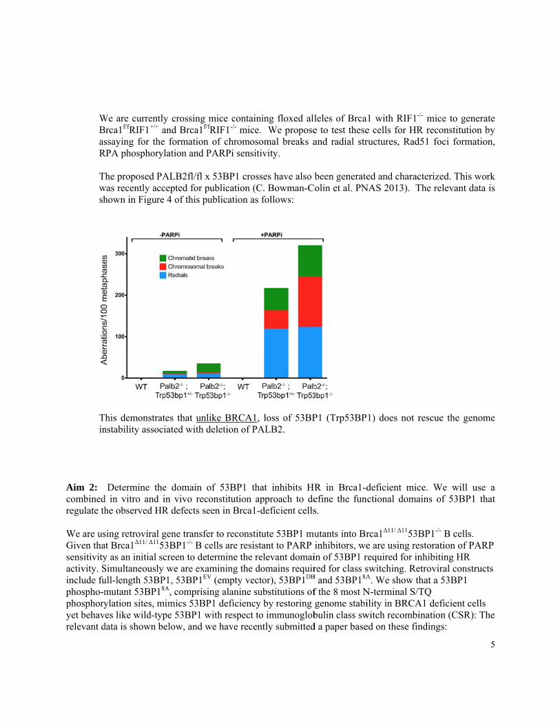
The proposed Pwas recently ahown in Figur
This demonstrnstability asso
Determine td in vitro andhe observed H
sing retroviralat Brca1Δ11/ Δ11
y as an initial Simultaneouslull-length 53Bmutant 53BPylation sites, mes like wild-t
data is shown
ntly crossing m+ and Brca1f/f
he formation rylation and P
PALB2fl/fl xccepted for pre 4 of this pu
rates that unliociated with d
the domain od in vivo recoHR defects se
l gene transfe153BP1-/- B cescreen to det
ly we are examBP1, 53BP1EV
18A, comprisinmimics 53BPtype 53BP1 wbelow, and w
mice containifRIF1-/- mice. of chromoso
PARPi sensiti
x 53BP1 crossublication (Cublication as f
ike BRCA1, deletion of PA
of 53BP1 thaonstitution apeen in Brca1-d
er to reconstituells are resistatermine the remining the do
V (empty vectng alanine su
P1 deficiency with respect towe have recen
ing floxed all We propose
omal breaks aivity.
ses have also bC. Bowman-C
follows:
loss of 53BPALB2.
at inhibits HRpproach to dedeficient cells
ute 53BP1 muant to PARP
elevant domaiomains requiror), 53BP1DB
ubstitutions ofby restoring
o immunoglobntly submitted
leles of Brcae to test theseand radial str
been generateolin et al. PN
P1 (Trp53BP
R in Brca1-define the funs.
utants into Brinhibitors, wein of 53BP1 rred for class s
B and 53BP18A
f the 8 most Ngenome stabibulin class swd a paper base
a1 with RIF1-
e cells for HRructures, Rad
ed and characNAS 2013). T
1) does not r
deficient micnctional doma
rca1Δ11/ Δ1153Be are using rerequired for inswitching. ReA. We show th
N-terminal S/Tility in BRCA
witch recombied on these fin
-/- mice to genR reconstitutid51 foci form
cterized. ThisThe relevant d
rescue the ge
ce. We willains of 53BP
BP1-/- B cellsestoration of Pnhibiting HR etroviral consthat a 53BP1 TQ
A1 deficient cination (CSR)ndings:
5
nerate ion by
mation,
s work data is
enome
use a 1 that
. PARP
tructs
cells ): The
Tde
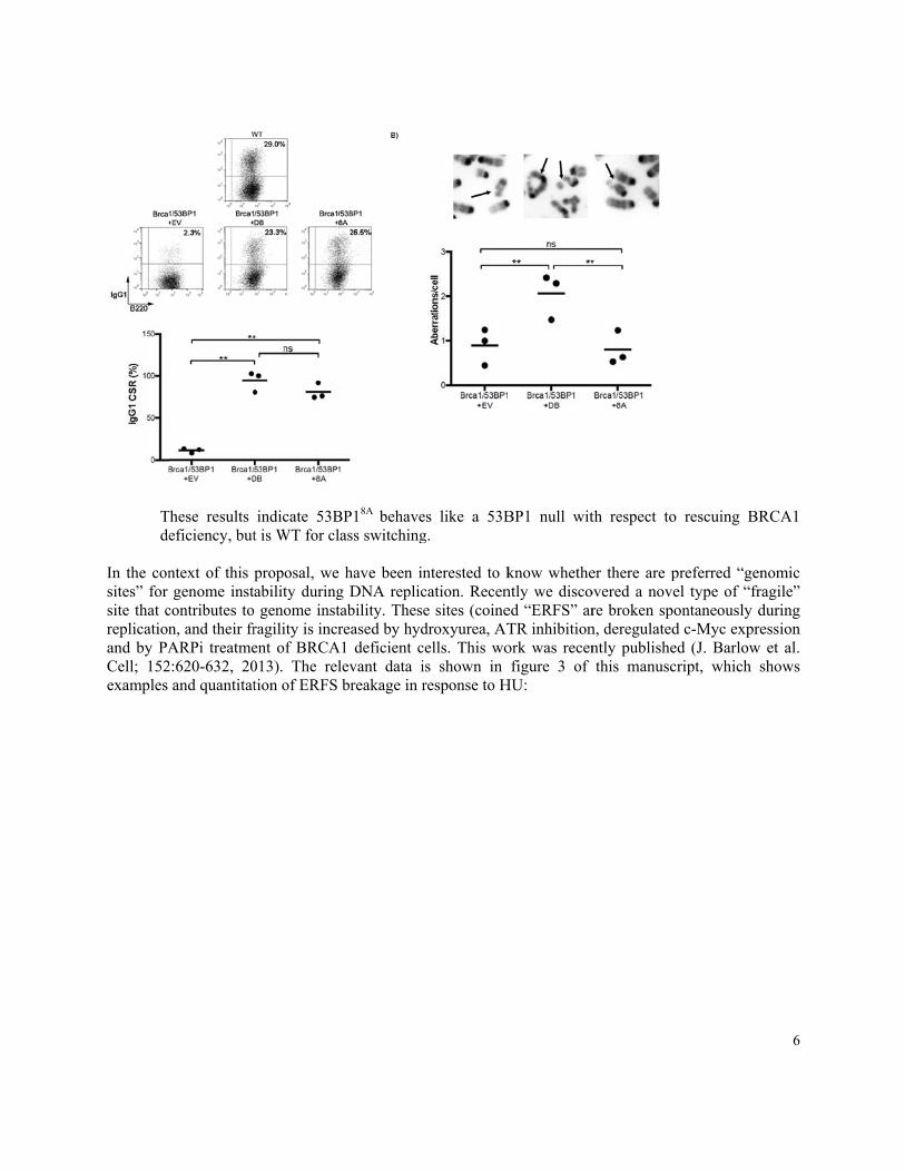
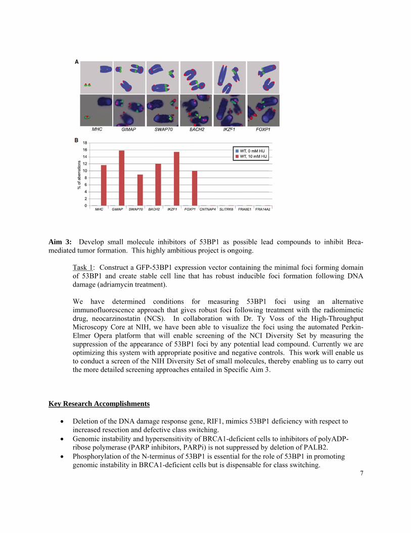
In the consites” for site that creplicationand by PACell; 152examples
These results eficiency, but
ntext of this genome inst
contributes ton, and their frARPi treatme
2:620-632, 20and quantitat
indicate 53Bt is WT for cl
proposal, weability during genome instragility is incrent of BRCA013). The reltion of ERFS
BP18A behavelass switching
have been ing DNA replictability. Thesreased by hyd
A1 deficient clevant data ibreakage in r
es like a 53Bg.
nterested to kcation. Recene sites (coinedroxyurea, ATcells. This wois shown in response to H
BP1 null wit
know whethently we discoed “ERFS” arTR inhibitionork was recenfigure 3 of
HU:
th respect to
er there are povered a novere broken spon, deregulatedntly publishethis manuscr
rescuing BR
preferred “genel type of “fraontaneously dd c-Myc expred (J. Barlow ript, which s
6
RCA1
nomic agile” during ession et al.
shows
Aim 3: mediated
Tofda WimdrMEsuoptoth
Key Rese
Din
Gri
Phge
Develop smtumor format
Task 1: Constf 53BP1 andamage (adriam
We have dmmunofluoresrug, neocarzi
Microscopy CElmer Opera uppression ofptimizing thiso conduct a sche more detail
earch Accom
Deletion of thencreased resec
Genomic instaibose polymerhosphorylatioenomic instab
mall moleculetion. This hig
truct a GFP-5d create stablmycin treatm
determined cscence approinostatin (NCore at NIH, wplatform thatf the appearans system withcreen of the Nled screening
mplishments
e DNA damagction and defe
ability and hyprase (PARP inon of the N-tebility in BRCA
e inhibitors oghly ambitiou
53BP1 exprese cell line thent).
conditions fach that give
CS). In colwe have beent will enablence of 53BP1h appropriate NIH Diversityg approaches e
ge response gective class swpersensitivitynhibitors, PAerminus of 53A1-deficient
of 53BP1 as us project is o
ssion vector chat has robus
for measuries robust focilaboration wn able to visu
e screening o1 foci by anypositive and
y Set of smallentailed in Sp
gene, RIF1, mwitching.
y of BRCA1-dARPi) is not su
BP1 is essentcells but is di
possible leaongoing.
containing thest inducible f
ing 53BP1 i following tr
with Dr. Ty ualize the fo
of the NCI Dy potential lea
negative conl molecules, t
pecific Aim 3
mimics 53BP1
deficient cellsuppressed by tial for the roispensable for
ad compound
e minimal fofoci formatio
foci usingreatment withVoss of the ci using the
Diversity Set ad compoundntrols. This wthereby enabl.
deficiency w
s to inhibitorsdeletion of Ple of 53BP1 ir class switch
ds to inhibit
ci forming doon following
g an alternh the radiomi
High-Throuautomated Peby measurin
d. Currently wwork will enabling us to carr
with respect to
s of polyADPPALB2. in promoting hing.
7
Brca-
omain DNA
native imetic
ughput erkin-
ng the we are ble us ry out
o
-
8
PARPi treatment leads to genome instability at preferred genomic sites.
Reportable Outcomes
1. Three high-impact papers have been published or are in press based on this work with the awardee, Dr. Andre Nussenzweig (M. Di Virgilio et al. Science 2013. C. Bowman-Colin et al. PNAS 2013, J. Barlow et al. Cell; 152:620-632, 2013). One additional paper has been submitted for publication.
Conclusion This work has revealed for the first time that deletion of 53BP1 prevents genomic instability in cells lacking the tumor suppressor, BRCA1. 53BP1 therefore plays a key role in cellular changes leading to cancer in individuals with BRCA1 mutations. This validates the targeting of 53BP1 as a chemopreventive measure to avert the appearance of cancer in women with mutations in BRCA1, which accounts for ~5% of all annual cases of breast cancer and a higher proportion of ovarian cancers. This work further suggests that 53BP1 inactivation or deregulation of downstream pathways (eg. RIF1) is a potential mechanism leading to resistance of BRCA1-deficient tumors to chemotherapeutic regimens involving PARP inhibitors. References Di Virginio M, Callen E, Yamane A, Jankovic M, Gitlin AD, Feldhahn N, Resch W, Chait BT, Nussenzweig A, Casellas R, Robbiani DF, Nussenweig MC. Rif1 prevents resection of DNA breaks and promotes immunoglobulin class switching, Science, 339(6120): 711-5, 2013 Feb 8. Bowman-Colin C, Xia B, Bunting SF, Klijn C, Drost R, Bouwman P, Fineman L, Chen X, Culhane AC, Bronson RT, Jonkers J, Nussenzweig A, Kanellopoulou C, Livingston, DM. Palb2 synergizes with Trp53 to suppress mammary tumor formation in a model of inherited breast cancer, PNAS in press 2013. Barlow J, Faryabi RB, Callen E, Wong N, Malhowski A, Chen HT, Gutierez-Cruz G, Sun HW, McKinnon P, Wright G, Casellas R, Robbiani DF, Staudt L, Fernandez-Capetillo O, Nussenzweig A. Identification of early replicating fragile sites that contribute to genome instability. Cell, 152(3):620-32, 2013 Jan 31
Appendices 1. Di Virginio M, Callen E, Yamane A, Jankovic M, Gitlin AD, Feldhahn N, Resch W, Chait BT,
Nussenzweig A, Casellas R, Robbiani DF, Nussenweig MC. Rif1 prevents resection of DNA breaks and promotes immunoglobulin class switching, Science, 339(6120): 711-5, 2013 Feb 8.

9
2. Barlow J, Faryabi RB, Callen E, Wong N, Malhowski A, Chen HT, Gutierez-Cruz G, Sun HW, McKinnon P, Wright G, Casellas R, Robbiani DF, Staudt L, Fernandez-Capetillo O, Nussenzweig A. Identification of early replicating fragile sites that contribute to genome instability. Cell, 152(3):620-32, 2013 Jan 31
3. Bowman-Colin C, Xia B, Bunting SF, Klijn C, Drost R, Bouwman P, Fineman L, Chen X, Culhane AC, Bronson RT, Jonkers J, Nussenzweig A, Kanellopoulou C, Livingston, DM. Palb2 synergizes with Trp53 to suppress mammary tumor formation in a model of inherited breast cancer, PNAS in press 2013.
DOI: 10.1126/science.1230624, 711 (2013);339 Science
et al.Michela Di VirgilioImmunoglobulin Class SwitchingRif1 Prevents Resection of DNA Breaks and Promotes
This copy is for your personal, non-commercial use only.
clicking here.colleagues, clients, or customers by , you can order high-quality copies for yourIf you wish to distribute this article to others
here.following the guidelines
can be obtained byPermission to republish or repurpose articles or portions of articles
): April 22, 2013 www.sciencemag.org (this information is current as of
The following resources related to this article are available online at
http://www.sciencemag.org/content/339/6120/711.full.htmlversion of this article at:
including high-resolution figures, can be found in the onlineUpdated information and services,
http://www.sciencemag.org/content/suppl/2013/01/09/science.1230624.DC1.html can be found at: Supporting Online Material
http://www.sciencemag.org/content/339/6120/711.full.html#relatedfound at:
can berelated to this article A list of selected additional articles on the Science Web sites
http://www.sciencemag.org/content/339/6120/711.full.html#ref-list-1, 15 of which can be accessed free:cites 48 articlesThis article
http://www.sciencemag.org/content/339/6120/711.full.html#related-urls2 articles hosted by HighWire Press; see:cited by This article has been
http://www.sciencemag.org/cgi/collection/immunologyImmunology
subject collections:This article appears in the following
registered trademark of AAAS. is aScience2013 by the American Association for the Advancement of Science; all rights reserved. The title
CopyrightAmerican Association for the Advancement of Science, 1200 New York Avenue NW, Washington, DC 20005. (print ISSN 0036-8075; online ISSN 1095-9203) is published weekly, except the last week in December, by theScience
on
Apr
il 22
, 201
3w
ww
.sci
ence
mag
.org
Dow
nloa
ded
from
higher numbers from colon contents than wasthe nitrate respiration–deficient mutant (Fig. 3Hand fig. S8B). Collectively, these data suggestedthat nitrate respiration conferred a marked growthadvantage on commensal E. coli in the lumenof the inflamed gut.
The picture emerging from this study is thatnitrate generated as a by-product of the host in-flammatory response can be used by E. coli, andlikely by other commensal Enterobacteriaceae,to edge out competing microbes that rely on fer-mentation to generate energy for growth. Obli-gate anaerobic microbes in the intestine competefor nutrients that are available for fermentationbut cannot use nonfermentable nutrients (suchas fermentation end products). The ability todegrade nonfermentable substrates probablyenables E. coli to sidestep this competition, whichexplains the fitness advantage conferred by ni-trate respiration in the inflamed gut. Throughthis mechanism, inflammation contributes to abloom of nitrate-respiration–proficient Entero-bacteriaceae, providing a plausible explanationfor the dysbiosis associated with intestinal in-flammation (3–12). This general principle mightalso influence the dynamics of host-associated
bacterial communities outside the large bowel,as nitrate respiration confers a fitness advantagein the oxygen-poor and nitrate-rich environmentof the cystic fibrosis airway (21).
References and Notes1. P. B. Eckburg et al., Science 308, 1635 (2005).2. R. E. Ley et al., Proc. Natl. Acad. Sci. U.S.A. 102, 11070
(2005).3. A. Krook, B. Lindström, J. Kjellander, G. Järnerot, L. Bodin,
J. Clin. Pathol. 34, 645 (1981).4. M. H. Giaffer, C. D. Holdsworth, B. I. Duerden, J. Med.
Microbiol. 35, 238 (1991).5. P. Seksik et al., Gut 52, 237 (2003).6. U. Gophna, K. Sommerfeld, S. Gophna, W. F. Doolittle,
S. J. Veldhuyzen van Zanten, J. Clin. Microbiol. 44, 4136(2006).
7. D. N. Frank et al., Proc. Natl. Acad. Sci. U.S.A. 104,13780 (2007).
8. M. M. Heimesaat et al., PLoS ONE 2, e662 (2007).9. C. Lupp et al., Cell Host Microbe 2, 119 (2007).10. B. Stecher et al., PLoS Biol. 5, e244 (2007).11. M. Barman et al., Infect. Immun. 76, 907 (2008).12. W. S. Garrett et al., Cell Host Microbe 8, 292 (2010).13. J. O. N. Lundberg, J. M. Lundberg, K. Alving,
P. M. Hellström, Lancet 344, 1673 (1994).14. I. I. Singer et al., Gastroenterology 111, 871 (1996).15. A. Enocksson, J. Lundberg, E. Weitzberg, A. Norrby-Teglund,
B. Svenungsson, Clin. Diagn. Lab. Immunol. 11, 250(2004).
16. C. Szabó, H. Ischiropoulos, R. Radi, Nat. Rev. Drug Discov.6, 662 (2007).
17. C. Schöneich, Biochim. Biophys. Acta 1703, 111(2005).
18. B. Balagam, D. E. Richardson, Inorg. Chem. 47, 1173(2008).
19. R. B. Gennis, V. Stewart, in Escherichia coli andSalmonella. Cellular and Molecular Biology,F. C. Neidhardt et al., Eds. (ASM Press, Washington,DC, 1996), vol. 1, pp. 217–261.
20. M. C. Pils et al., Inflamm. Bowel Dis. 17, 2038 (2011).21. L. R. Hoffman et al., PLoS Pathog. 6, e1000712 (2010).
Acknowledgments: We thank W. Müller for providingIl10flox/flox Cd4-cre mice and E. Romao for technical assistance.The data reported in the manuscript are tabulated in themain paper and in the supplementary materials. This workwas supported by the California Agricultural Experiment Station(I.E.P. and S.J.P.) and Public Health Service grants AI090387(R.M.T), AI076246 (L.G.A and A.J.B.), and AI088122 (A.J.B.).P.T. was supported by a scholarship from the Faculty of Medicine,Chiang Mai University, Thailand.
Supplementary Materialswww.sciencemag.org/cgi/content/full/339/6120/708/DC1Materials and MethodsFigs. S1 to S11Tables S1 and S2References (22–39)
7 November 2012; accepted 5 December 201210.1126/science.1232467
Rif1 Prevents Resection ofDNA Breaks and PromotesImmunoglobulin Class SwitchingMichela Di Virgilio,1 Elsa Callen,3* Arito Yamane,4* Wenzhu Zhang,5* Mila Jankovic,1
Alexander D. Gitlin,1 Niklas Feldhahn,1 Wolfgang Resch,4 Thiago Y. Oliveira,1,6,7 Brian T. Chait,5
André Nussenzweig,3 Rafael Casellas,4 Davide F. Robbiani,1 Michel C. Nussenzweig1,2†
DNA double-strand breaks (DSBs) represent a threat to the genome because they can lead tothe loss of genetic information and chromosome rearrangements. The DNA repair proteinp53 binding protein 1 (53BP1) protects the genome by limiting nucleolytic processing of DSBsby a mechanism that requires its phosphorylation, but whether 53BP1 does so directly isnot known. Here, we identify Rap1-interacting factor 1 (Rif1) as an ATM (ataxia-telangiectasiamutated) phosphorylation-dependent interactor of 53BP1 and show that absence of Rif1 resultsin 5′-3′ DNA-end resection in mice. Consistent with enhanced DNA resection, Rif1 deficiencyimpairs DNA repair in the G1 and S phases of the cell cycle, interferes with class switchrecombination in B lymphocytes, and leads to accumulation of chromosome DSBs.
The DNA damage response factor p53 bind-ing protein 1 (53BP1) is a multidomain pro-tein containing a chromatin-binding tudor
domain, an oligomerization domain, tandem breastcancer 1 (BRCA1) C-terminal (BRCT) domains,and an N-terminal domain with 28 SQ/TQ poten-tial phosphorylation sites for phosphatidylinositol3-kinase–related kinases [PIKKs, ataxia-telangiectasiamutated (ATM)/ATM and Rad3-related/DNA-dependent protein kinase catalytic subunit (DNA-PKcs)] (1–3). 53BP1 contributes to DNA repairin several ways: This protein facilitates joiningbetween intrachromosomal double-strand breaks(DSBs) at a distance (synapsis) (4–7), it enablesheterochromatic DNA repair through relaxa-
tion of nucleosome compaction (2, 3), and itprotects DNA ends from resection and therebyfavors repair of DSBs that occur in G1 phase bynonhomologous end joining (NHEJ) (4, 5, 8).Consistent with its role in DNA-end protection,53BP1 is essential for class switch recombina-tion (CSR) in B lymphocytes (9, 10).
Structure-function studies indicate that, be-sides the recruitment of 53BP1 to DNA ends,protection requires 53BP1 phosphorylation (4),but how this protective effect is mediated is un-known. To identify phosphorylation-dependentinteractors of 53BP1, we applied stable isotopelabeling by amino acids in cell culture (SILAC).Trp53bp1−/− (Trp53bp1 encodes 53BP1)B cellswere
infected with retroviruses encoding a C-terminaldeleted version of 53BP1 (53BP1DB) or a phospho-mutant in which all 28 N-terminal potential PIKKphosphorylation sites were mutated to alanine(53BP1DB28A) (4), inmedia containing isotopicallyheavy (53BP1DB) or light (53BP1DB28A) lysine andarginine (fig. S1, A to C) (11).
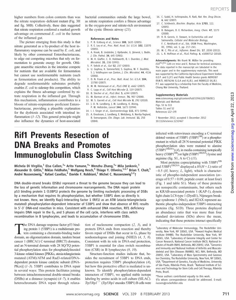
Most proteins coprecipitating with 53BP1DB
and 53BP1DB28A displayed aH/(H + L) ratio of~0.5 (H, heavy; L, light), which is character-istic of phospho-independent association (av-erage of 0.57 T 0.09, peptide count: at least four)(Fig. 1 and table S1). Many of these proteinsare nonspecific contaminants, but others suchas KRAB-associated protein 1 (KAP-1), dyneinlight chain LC8-type 1 (Dynll1), Nijmegen break-age syndrome 1 (Nbs1), and H2AX represent au-thentic phospho-independent 53BP1-interactingproteins (fig. S1D). Three proteins displayedan abundance ratio that was more than fourstandard deviations (SDs) above the mean,indicating that these proteins interact specifically
1Laboratory of Molecular Immunology, The Rockefeller Uni-versity, New York, NY 10065, USA. 2Howard Hughes MedicalInstitute (HHMI), The Rockefeller University, New York, NY10065, USA. 3Laboratory of Genome Integrity and Center forCancer Research, National Cancer Institute (NCI), National In-stitutes of Health (NIH), Bethesda, MD 20892, USA. 4Genomicsand Immunity and National Institute of Arthritis and Musculo-skeletal and Skin Diseases (NIAMS), NCI, NIH, Bethesda, MD20892, USA. 5Laboratory of Mass Spectrometry and GaseousIon Chemistry, The Rockefeller University, New York, NY 10065,USA. 6Department of Genetics, Faculty of Medicine, Universityof São Paulo, Ribeirão Preto, Brazil. 7National Institute of Sci-ence and Technology for Stem Cells and Cell Therapy, RibeirãoPreto, Brazil.
*These authors contributed equally to this work.†To whom correspondence should be addressed. E-mail:[email protected]
www.sciencemag.org SCIENCE VOL 339 8 FEBRUARY 2013 711
REPORTS
on
Apr
il 22
, 201
3w
ww
.sci
ence
mag
.org
Dow
nloa
ded
from
with phosphorylated 53BP1: Pax interactionwith transcription-activation domain protein-1(Paxip1, or PTIP; 0.95), PTIP-associated protein1 (Pa1; 0.97), and Rap1-interacting factor 1 (Rif1)(0.96) (Fig. 1 and figs. S1D and S2). PTIP wasknown to interact with 53BP1 in a phospho-dependent manner (12), whereas Pa1 and Rif1were not.
Rif1 was originally identified in buddingyeast as a protein with a key role in telomerelength maintenance (13). However, in mam-malian cells, Rif1 is not essential for telomerehomeostasis, but has been assigned a number ofdifferent roles in maintaining genome stability,including participation in the DNA damage re-sponse (14–16), repair of S-phase DNA damage(17, 18), and regulation of origin firing duringDNA replication (19, 20). However, the mech-anism by which Rif1 might contribute to DNArepair and maintenance of genome stability is notknown.
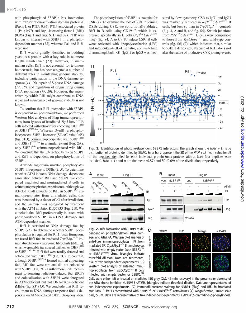
To confirm that Rif1 interaction with 53BP1is dependent on phosphorylation, we performedWestern blot analysis of Flag immunoprecipi-tates from lysates of irradiated Trp53bp1−/− Bcells infectedwith retroviruses encoding 53BP1DB
or 53BP1DB28A. Whereas Dynll1, a phospho-independent 53BP1 interactor (SILAC ratio: 0.55)(fig. S1D), coimmunoprecipitated with 53BP1DB
and 53BP1DB28A to a similar extent (Fig. 2A),only 53BP1DB coimmunoprecipitated with Rif1.We conclude that the interaction between 53BP1and Rif1 is dependent on phosphorylation of53BP1.
Ataxia-telangiectasia mutated phosphorylates53BP1 in response to DSBs (1, 3). To determinewhether ATM induces DNA damage–dependentassociation between Rif1 and 53BP1, we com-pared irradiated and nonirradiated B cells incoimmunoprecipitation experiments. Although wedetected small amounts of Rif1 in 53BP1DB im-munoprecipitates from unirradiated cells, thiswas increased by a factor of >3 after irradiation,and the increase was abrogated by treatmentwith the ATM inhibitor KU55933 (Fig. 2B). Weconclude that Rif1 preferentially interacts withphosphorylated 53BP1 in a DNA damage- andATM-dependent manner.
Rif1 is recruited to DNA damage foci by53BP1 (15). To determine whether 53BP1 phos-phorylation is required for Rif1 focus formation,we tested Rif1 foci in irradiated Trp53bp1−/− im-mortalizedmouse embryonic fibroblasts (iMEFs),whichwere stably transducedwith either 53BP1DB
or 53BP1DB28A. Rif1 fociwere readily detected andcolocalized with 53BP1DB (Fig. 2C). In contrast,although 53BP1DB28A formed normal-appearingfoci, Rif1 foci were rare and did not colocalizewith 53BP1 (Fig. 2C). Furthermore, Rif1 recruit-ment to ionizing radiation–induced foci (IRIF)and colocalization with 53BP1 were abrogatedin ATM-deficient but not DNA-PKcs–deficientiMEFs (fig. S3) (15). We conclude that Rif1 re-cruitment to DNA damage response foci is de-pendent on ATM-mediated 53BP1 phosphorylation.
The phosphorylation of 53BP1 is essential forCSR (4). To examine the role of Rif1 in joiningDSBs during CSR, we conditionally ablatedRif1 in B cells using CD19Cre, which is ex-pressed specifically in B cells (Rif1F/FCd19Cre/+
mice) (fig. S4, A to C). To induce CSR, B cellswere activated with lipopolysaccharide (LPS)and interleukin-4 (IL-4) in vitro, and switchingto immunoglobulin G1 (IgG1) or IgG3 was mea-
sured by flow cytometry. CSR to IgG1 and IgG3was markedly reduced in Rif1F/FCd19Cre/+ Bcells, but less so than in Trp53bp1−/− controls(Fig. 3, A and B, and fig. S5). Switch junctionsfrom Rif1F/FCd19Cre/+ B cells were comparableto those from Trp53bp1−/− and wild-type con-trols (fig. S6) (7), which indicates that, similarto 53BP1 deficiency, absence of Rif1 does notalter the nature of productive CSR joining events.
H
L
H/(
H+
L)
0
0.2
0.4
0.6
0.8
1 Pa1 Rif1 Paxip1
H/(H+L)
σ
2σ
3σ
4σ
- σ
- 2σ
- 3σ
- 4σ
Fig. 1. Identification of phospho-dependent 53BP1 interactors. The graph shows the H/(H + L) ratiodistribution of proteins identified by SILAC. Error bars represent the SD of the H/(H + L) mean value for allof the peptides identified for each individual protein (only proteins with at least four peptides wereincluded).
—H/(H + L) and s are the mean (0.57) and SD (0.09) of the distribution, respectively.
A
C
vec
53BP1 DB
53BP1 DB28A
Input
Dynll1
Flag
Rif1
Flag IP
+ + IR++ ++
-+
vec
γH2AX
Input
- - - + ATMi
+ - + + IR
- - +- + +
vec53BP1DB
Flag IP
Flag
Rif1
B
53BP1 Rif1 Merge + DAPI
vec53BP1 DB
53BP1 DB28A
53BP1DB
53B
P1D
B53
BP
1DB
28A
Fig. 2. Rif1 interaction with 53BP1 is de-pendent on phosphorylation, DNA dam-age, and ATM. (A) Western blot analysis ofanti-Flag immunoprecipitates (IP) fromirradiated (IR) Trp53bp1−/− B lymphocytesinfected with empty vector (vec), 53BP1DB,or 53BP1DB28A virus. Triangles indicatethreefold dilution. Data are representa-tive of two independent experiments. (B)Western blot analysis of anti-Flag immu-noprecipitates from Trp53bp1−/− B cellsinfected with empty vector or 53BP1DB.Cells were either left untreated or irradiated [50 gray (Gy), 45-min recovery] in the presence or absence ofthe ATM kinase inhibitor KU55933 (ATMi). Triangles indicate threefold dilution. Data are representative oftwo independent experiments. (C) Immunofluorescent staining for 53BP1 (Flag) and Rif1 in irradiatedTrp53bp1−/− iMEFs reconstituted with 53BP1DB or 53BP1DB28A retroviruses (4). Magnification, 100×; scalebars, 5 mm. Data are representative of two independent experiments. DAPI, 4′,6-diamidino-2-phenylindole.
8 FEBRUARY 2013 VOL 339 SCIENCE www.sciencemag.org712
REPORTS
on
Apr
il 22
, 201
3w
ww
.sci
ence
mag
.org
Dow
nloa
ded
from
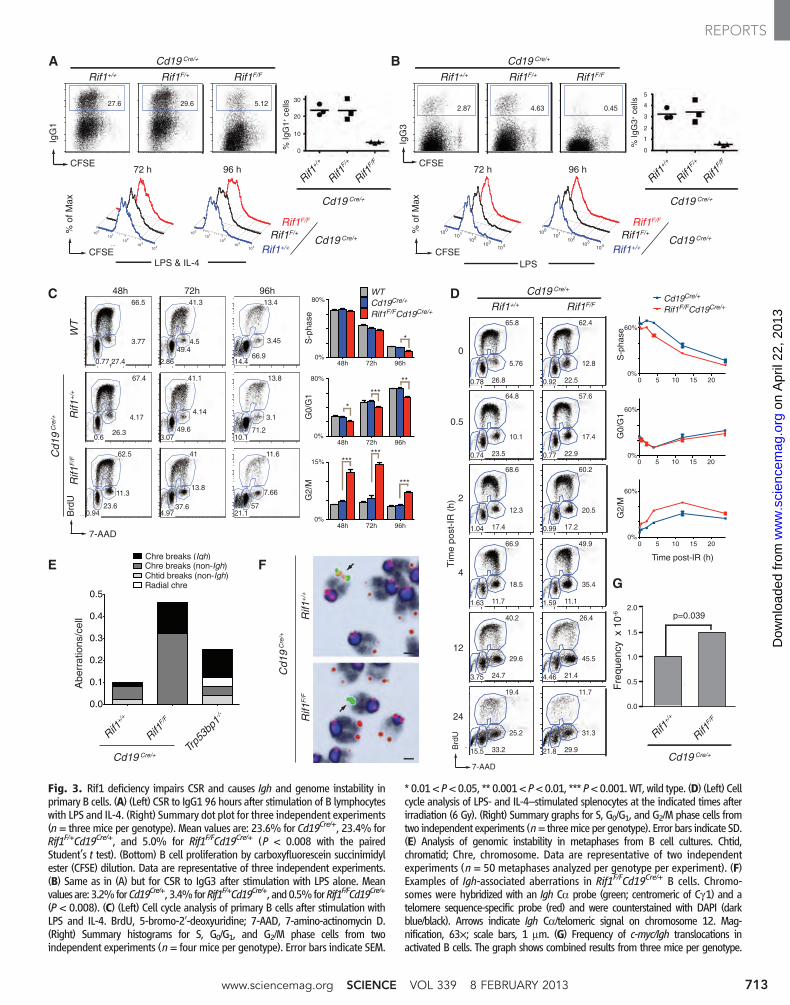
Fig. 3. Rif1 deficiency impairs CSR and causes Igh and genome instability inprimary B cells. (A) (Left) CSR to IgG1 96 hours after stimulation of B lymphocyteswith LPS and IL-4. (Right) Summary dot plot for three independent experiments(n = three mice per genotype). Mean values are: 23.6% for Cd19Cre/+, 23.4% forRif1F/+Cd19Cre/+, and 5.0% for Rif1F/FCd19Cre/+ (P < 0.008 with the pairedStudent’s t test). (Bottom) B cell proliferation by carboxyfluorescein succinimidylester (CFSE) dilution. Data are representative of three independent experiments.(B) Same as in (A) but for CSR to IgG3 after stimulation with LPS alone. Meanvaluesare: 3.2%forCd19Cre/+, 3.4%forRif1F/+Cd19Cre/+, and0.5%forRif1F/FCd19Cre/+
(P < 0.008). (C) (Left) Cell cycle analysis of primary B cells after stimulation withLPS and IL-4. BrdU, 5-bromo-2′-deoxyuridine; 7-AAD, 7-amino-actinomycin D.(Right) Summary histograms for S, G0/G1, and G2/M phase cells from twoindependent experiments (n = four mice per genotype). Error bars indicate SEM.
* 0.01< P<0.05, ** 0.001< P<0.01, *** P<0.001.WT, wild type. (D) (Left) Cellcycle analysis of LPS- and IL-4–stimulated splenocytes at the indicated times afterirradiation (6 Gy). (Right) Summary graphs for S, G0/G1, and G2/M phase cells fromtwo independent experiments (n= threemice per genotype). Error bars indicate SD.(E) Analysis of genomic instability in metaphases from B cell cultures. Chtid,chromatid; Chre, chromosome. Data are representative of two independentexperiments (n = 50 metaphases analyzed per genotype per experiment). (F)Examples of Igh-associated aberrations in Rif1F/FCd19Cre/+ B cells. Chromo-somes were hybridized with an Igh Ca probe (green; centromeric of Cg1) and atelomere sequence-specific probe (red) and were counterstained with DAPI (darkblue/black). Arrows indicate Igh Ca/telomeric signal on chromosome 12. Mag-nification, 63×; scale bars, 1 mm. (G) Frequency of c-myc/Igh translocations inactivated B cells. The graph shows combined results from three mice per genotype.
IgG
1
CFSE
IgG
3
CFSE
A B
% Ig
G1+
cel
ls
20
10
0
30
Rif1F/+
Rif1F/F
Rif1+/
+
Cd19 Cre/+
% Ig
G3+
cel
ls
5
4
3
2
1
0
Rif1F/+
Rif1F/F
Rif1+/
+
Cd19 Cre/+
C
E
48h 72h 96h
Rif1
F/F
Cd1
9C
re/+
Brd
U
0
0.5
2
4
12
24
Tim
e po
st-I
R (
h)
Brd
U
7-AAD
WT
F
7-AAD
Cd19 Cre/+
Rif1F/+ Rif1F/FRif1+/+
Cd19 Cre/+
Rif1F/+ Rif1F/FRif1+/+%
of M
ax
CFSE
100
101
102
103
104
100
101
102
103
104
72 h 96 h
Cd19 Cre/+Rif1F/+
Rif1F/F
Rif1+/+
100101
102
103104
100101
102
103104
72 h 96 h
Cd19 Cre/+Rif1F/+
Rif1F/F
Rif1+/+
% o
f Max
CFSE
21.157
7.66
11.6
4.9737.6
13.8
41
0.9423.6
11.3
62.5
10.171.2
3.1
13.8
3.0749.6
4.14
41.1
0.626.3
4.17
67.4
14.466.9
3.45
13.4
2.86
49.44.5
41.3
0.77 27.4
3.77
66.5
Fre
quen
cy x
10-6 p=0.039
0.0
0.5
1.0
1.5
2.0
Rif1F/F
Rif1+/
+
Cd19 Cre/+
Cd19 Cre/+
Rif1F/FRif1+/+
0.78 26.8
5.76
65.8
1.04 17.4
12.3
68.6
1.63 11.7
18.5
66.9
3.75 24.7
29.6
40.2
15.5 33.2
25.2
19.4
0.74 23.5
10.1
64.8
0.92 22.5
12.8
62.4
0.99 17.2
20.5
60.2
1.59 11.1
35.4
49.9
4.46 21.4
45.5
26.4
21.8 29.9
31.3
11.7
0.77 22.9
17.4
57.6
5.1229.627.62.87 4.63 0.45
LPS & IL-4 LPS
Abe
rrat
ions
/cel
l
G
Cd1
9C
re/+
Rif1
F/F
Rif1
+/+
0.0
0.1
0.2
0.3
0.4
0.5
Chtid breaks (non-Igh)Chre breaks (non-Igh)
Radial chre
Chre breaks (Igh)
Rif1F/F
Rif1+/
+
Cd19 Cre/+Tr
p53b
p1-/-
D
48h 72h 96h0%
20%
40%
60%
80%WTCd19Cre/+
Rif1F/FCd19Cre/+S
-pha
se
48h 72h 96h0%
20%
40%
60%
80%
G0/
G1
48h 72h 96h0%
5%
10%
15%
G2/
M
*
*
*****
******
***
Rif1
+/+
0 5 10 15 200%
20%
40%
60%
G0/
G1
0 5 10 15 200%
20%
40%
60%
Time post-IR (h)
G2/
M
0 5 10 15 200%
20%
40%
60%
Cd19Cre/+
Rif1F/FCd19Cre/+
S-p
hase
www.sciencemag.org SCIENCE VOL 339 8 FEBRUARY 2013 713
REPORTS
on
Apr
il 22
, 201
3w
ww
.sci
ence
mag
.org
Dow
nloa
ded
from
A similar CSR defect was also obtained byconditionally deleting Rif1 with 4-hydroxy-tamoxifen (4HT) in Rif1F/FROSA26Cre-ERT2/+
B cells (fig. S7). Finally, short hairpin RNA–mediated partial down-regulation of CtBP-interacting protein (CtIP), which interacts withRif1 (fig. S8C) and has been implicated in pro-cessing of DNA ends (21, 22), resulted in a verysmall but reproducible increase in CSR (fig. S8,A and B). Thus, Rif1 is essential for normal CSR,and CtIP may not be the only factor that contrib-utes to end processing in Rif1-deficient B cells.
Class switch recombination requires cell di-vision, activation-induced cytidine deaminase(AID) expression, and Igh germline transcription(23). There are conflicting reports that Rif1 isrequired for proliferation in MEFs, but not inDT40 B cells (17, 18). We found that cell divi-sion profiles of Rif1F/FCd19Cre/+ and 4HT-treatedRif1F/FROSA26Cre-ERT2/+ B cells were indistin-guishable from controls (Fig. 3, A and B; and fig.S7, A, C, E, and G), indicating that Rif1 is dis-pensable for B cell proliferation in vitro. Finally,AIDmRNAand protein expression and Igh germ-
line transcription were not affected by Rif1 de-letion (fig. S4, B and D).
We next examined the role of Rif1 in cellcycle progression in primary B cells. We foundno major differences in the percentage of cellsin G0/G1 and S phases (Fig. 3C). However, thenumber of cells in G2/M phase was increasedapproximately twofold in the absence of Rif1(2.64-, 2.56-, and 1.91-fold at 48, 72, and 96 hours,respectively) (Fig. 3C). We obtained similarresults with the use of Rif1F/FROSA26Cre-ERT2/+
B cells treated with 4HT (fig. S7, H and I).
53BP1P
ATM53BP1 Rif1
DSBs RPA
Rad51
Rif1
5’5’
CSR (Igh locus)
A B
Cd19Cre/+
Rif1F/FCd19Cre/+
Rad51
chr12:114,470,000-114,691,762
C
D
E(47.6)
(742.3)
(78.7)
(361.1)
(2.4)
(12.7)
RPA
chr7:132,660,000-132,780,000
Il21rIl4ra 10 Kb
Cd19Cre/+
Rif1F/FCd19Cre/+
(47.0)
(9.9)
(20.5)
(14.3)
Rad51
chr7:132,660,000-132,780,000
Il21rIl4ra 10 Kb
Cd19Cre/+
Rif1F/FCd19Cre/+
(106.0)
(15.9)
(30.7)
(17.5)
RPA
Trp53bp1-/-
Cd19Cre/+
Rif1F/FCd19Cre/+
5’5’3’
3’
5’5’3’3’
+ strand
- strand
chr12:114,470,000-114,691,762
(7.2)
(76.4)
(11.8)
(73.4)
(40.9)
(44.9)
(1.2)
(2.3)
(2.8)
10 KbIgµIgγ1Igε
10 KbIgµIgγ1Igε
chr17:29,590,000-29,670,000
Pim1 10 Kb
(12.7)
(3.8)
Cd19Cre/+
Rif1F/FCd19Cre/+
Resection
chr17:29,590,000-29,670,000
Pim1 10 Kb
(27.7)
(6.5)
Cd19Cre/+
Rif1F/FCd19Cre/+
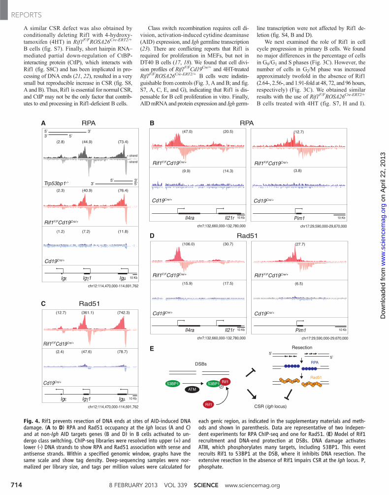
Fig. 4. Rif1 prevents resection of DNA ends at sites of AID-induced DNAdamage. (A to D) RPA and Rad51 occupancy at the Igh locus (A and C)and at non-Igh AID targets genes (B and D) in B cells activated to un-dergo class switching. ChIP-seq libraries were resolved into upper (+) andlower (-) DNA strands to show RPA and Rad51 association with sense andantisense strands. Within a specified genomic window, graphs have thesame scale and show tag density. Deep-sequencing samples were nor-malized per library size, and tags per million values were calculated for
each genic region, as indicated in the supplementary materials and meth-ods and shown in parenthesis. Data are representative of two indepen-dent experiments for RPA ChIP-seq and one for Rad51. (E) Model of Rif1recruitment and DNA-end protection at DSBs. DNA damage activatesATM, which phosphorylates many targets, including 53BP1. This eventrecruits Rif1 to 53BP1 at the DSB, where it inhibits DNA resection. Theextensive resection in the absence of Rif1 impairs CSR at the Igh locus. P,phosphate.
8 FEBRUARY 2013 VOL 339 SCIENCE www.sciencemag.org714
REPORTS
on
Apr
il 22
, 201
3w
ww
.sci
ence
mag
.org
Dow
nloa
ded
from
Furthermore, irradiation increases the accumulationof Rif1F/FCd19Cre/+ B cells in G2/M phase (Fig.3D). In addition, Trp53bp1−/− iMEFs expressing53BP1DB28A, which did not recruit Rif1 to IRIF(Fig. 2C), exhibited delayed progression throughS phase following DNA damage with accumula-tion of cells in G2 phase after irradiation (fig. S9).
Accumulation of cells in G2/M phase mayreflect the persistence of unrepaired DNA dam-age in a fraction of Rif1-deficient cells. To investi-gate this possibility, we analyzedmetaphase spreadsfrom B cells dividing in response to LPS andIL-4 in vitro. These cells express AID, whichproduces DSBs in Igh and, less frequently at off-target sites throughout the genome, in the G1
phase of the cell cycle (24–26). Chromosomalaberrations were increased in Rif1F/FCd19Cre/+
B cells compared to controls (Fig. 3E), with manylocalized to the Igh locus (Fig. 3E). Consistentwith the observation that Igh is targeted by AIDin the G1 phase of the cell cycle, all of the Ighbreaks were chromosome breaks (Fig. 3, E and F).Interestingly, the frequency of c-myc/Igh translo-cations ismoderately increased inRif1F/FCd19Cre/+
B cells; however, the breakpoint distribution wassimilar to the Cd19Cre/+ control (1.5 × 10⁻⁶ ver-sus 1.0 × 10⁻⁶ in the control; P = 0.039) (Fig. 3Gand fig. S10). We conclude that in the absence ofRif1, DSBs fail to be resolved efficiently in theG1, S, or G2 phases, which leads to increasedlevels of genomic instability, including chromo-some breaks at Igh and translocations in dividingB cells.
In the absence of 53BP1, DSBs producedby AID at the Igh locus accumulate the single-stranded DNA-binding replication protein A com-plex (RPA) as a result of increased DNA-endresection (24). To determine if Rif1 is requiredfor DNA-end protection by 53BP1, we performedRPA–chromatin immunoprecipitation followedby massive parallel sequencing (ChIP-seq) ex-periments onRif1F/FCd19Cre/+ and control B cells.Ablation of Rif1 was indistinguishable fromthe loss of 53BP1 in that in its absence, RPA dec-orates the Igh locus asymmetrically, in a mannerconsistent with 5′-3′ resection (Fig. 4A) (27). Inaddition, absence of Rif1 also results in RPAaccumulation at non-Igh genes, such as Il4ra andPim1, that are damaged by AID in G1 phase (Fig.4B) (24, 25). Rad51 is the recombinase thatmediates repair of DSBs by homologous recom-bination in S/G2/M phase (22). To confirm thatRif1 prevents resection that takes place in Sphase, we monitored Rad51 accumulation in ac-tivated B cells by ChIP-seq. Loss of Rif1 was
indistinguishable from the loss of 53BP1 (27), inthat it led to asymmetric Rad51 accumulation atsites of AID-inflicted DNA damage (Fig. 4, Cand D). We conclude that in the absence of Rif1,AID-induced DSBs incurred in G1 phase persistand undergo extensive 5′-3′ DNA-end resectionin S/G2/M phase, as measured by RPA and Rad51accumulation.
A role for Rif1 in maintenance of genomestability and protection of DNA ends against re-section is consistent with its phosphorylation-dependent recruitment to the N-terminal domainof 53BP1 (4). 53BP1 facilitates DNA repair andprevents DNA-end resection during CSR. In theabsence of 53BP1, AID-induced DSBs are re-solved inefficiently in G1 phase, leading to chro-mosome breaks, Igh instability, and resolution byalternative NHEJ or homologous recombinationinstead of classical NHEJ (4, 8, 27). Our exper-iments show that in the absence of Rif1, 53BP1 isinsufficient to promote genomic stability or me-diate efficient Igh repair, DNA-end protection, orCSR. Thus, these 53BP1 activities require Rif1recruitment to the phosphorylated N terminus of53BP1. Rif1 is likely to have additional functionsbeyond 53BP1, CSR, and DNA-end protectionbecause although Trp53bp1−/− mice are viable,Rif1 deletion is lethal (17). Indeed, Rif1 is be-lieved to play a role in the repair of S-phase DNAdamage (17, 18), as well as in the regulation ofreplication timing (19, 20, 28). Analogously, ad-ditional CSR factor(s) may exist downstream of53BP1, as class switching in Rif1-deficienct Bcells is significantly higher than in Trp53bp1−/−.
In summary, our data are consistent with amodel in which ATM-mediated phosphorylationof 53BP1 recruits Rif1 to sites of DNA damage,where it facilitates DNA repair in part by pro-tecting DNA ends from resection (Fig. 4E). In theabsence of Rif1, DNA breaks incurred in G1
phase fail to be repaired by NHEJ and undergoextensive 5′-3′ end resection, resulting in the ac-cumulation of chromosome breaks and genomeinstability.
References and Notes1. M. M. Adams, P. B. Carpenter, Cell Div. 1, 19 (2006).2. J. Lukas, C. Lukas, J. Bartek, Nat. Cell Biol. 13, 1161
(2011).3. A. T. Noon, A. A. Goodarzi, DNA Repair 10, 1071
(2011).4. A. Bothmer et al., Mol. Cell 42, 319 (2011).5. S. Difilippantonio et al., Nature 456, 529 (2008).6. N. Dimitrova, Y. C. Chen, D. L. Spector, T. de Lange,
Nature 456, 524 (2008).7. B. Reina-San-Martin, J. Chen, A. Nussenzweig,
M. C. Nussenzweig, Eur. J. Immunol. 37, 235 (2007).
8. A. Bothmer et al., J. Exp. Med. 207, 855 (2010).9. J. P. Manis et al., Nat. Immunol. 5, 481 (2004).
10. I. M. Ward et al., J. Cell Biol. 165, 459 (2004).11. Materials and methods are available as supplementary
materials on Science Online.12. I. A. Manke, D. M. Lowery, A. Nguyen, M. B. Yaffe,
Science 302, 636 (2003).13. C. F. Hardy, L. Sussel, D. Shore, Genes Dev. 6, 801
(1992).14. S. Kumar et al., Cell Cycle 11, 1183 (2012).15. J. Silverman, H. Takai, S. B. Buonomo, F. Eisenhaber,
T. de Lange, Genes Dev. 18, 2108 (2004).16. L. Xu, E. H. Blackburn, J. Cell Biol. 167, 819 (2004).17. S. B. Buonomo, Y. Wu, D. Ferguson, T. de Lange,
J. Cell Biol. 187, 385 (2009).18. D. Xu et al., EMBO J. 29, 3140 (2010).19. D. Cornacchia et al., EMBO J. 31, 3678 (2012).20. S. Yamazaki et al., EMBO J. 31, 3667 (2012).21. A. A. Sartori et al., Nature 450, 509 (2007).22. L. S. Symington, J. Gautier, Annu. Rev. Genet. 45, 247
(2011).23. R. Pavri, M. C. Nussenzweig, Adv. Immunol. 110,
1 (2011).24. O. Hakim et al., Nature 484, 69 (2012).25. S. Petersen et al., Nature 414, 660 (2001).26. A. Yamane et al., Nat. Immunol. 12, 62 (2011).27. A. Yamane et al., Cell Rep. 10.1016/j.celrep.2012.12.006
(2013).28. M. Hayano et al., Genes Dev. 26, 137 (2012).
Acknowledgments: We thank all members of the Nussenzweiglaboratory for discussion, D. Bosque and T. Eisenreich forhelp in managing mouse colonies, A. Gazumyan for assistancewith Igh germline and AID transcript levels analysis, andK. Yao for help with genotyping. We thank T. de Lange(The Rockefeller University, New York) for Rif1F/F mice;S. Buonomo (European Molecular Biology Laboratory MouseBiology Unit, Monterotondo, Italy) for the anti-mouse Rif1serum #1240; G. Gutierrez (NIAMS, NIH, Bethesda, MD) forIllumina sequencing; N. Zampieri (Columbia University,New York) for assistance with immunofluorescence imageprocessing, and M. P. Rout, J. LaCava, S. Obado, and L. Hough(The Rockefeller University) for invaluable help, discussions,and protocols for cryolysis and magnetic bead-mediatedimmunoisolation. The data presented in the manuscript aretabulated in the main text and in the supplementary materials.Sequence data shown in Fig. 4 have been deposited in theGene Expression Omnibus database (accession numberGSE42298) at www.ncbi.nlm.nih.gov/geo/. M.D.V. was a Fellowof the American Italian Cancer Foundation, and A.D.G. wassupported by NIH Medical Scientist Training Programgrant GM007739. This work was supported in part by NIHgrants AI037526 (M.C.N.), RR022220 (B.T.C.), RR00862(B.T.C.), and GM103314 (B.T.C.); and by the intramuralprogram of NIAMS at the NIH (R.C.); and the intramuralresearch program of NCI at the NIH and Center for CancerResearch (A.N. and E.C.). M.C.N. is an HHMI Investigator.
Supplementary Materialswww.sciencemag.org/cgi/content/full/science.1230624/DC1Materials and MethodsFigs. S1 to S10Table S1References (29–49)
24 September 2012; accepted 16 November 2012Published online 10 January 2013;10.1126/science.1230624
www.sciencemag.org SCIENCE VOL 339 8 FEBRUARY 2013 715
REPORTS
on
Apr
il 22
, 201
3w
ww
.sci
ence
mag
.org
Dow
nloa
ded
from
Identification of Early ReplicatingFragile Sites that Contributeto Genome InstabilityJacqueline H. Barlow,1,9 Robert B. Faryabi,1,9 Elsa Callen,1 Nancy Wong,1 Amy Malhowski,1 Hua Tang Chen,1
Gustavo Gutierrez-Cruz,3 Hong-Wei Sun,4 Peter McKinnon,6 George Wright,2 Rafael Casellas,5 Davide F. Robbiani,7
Louis Staudt,2 Oscar Fernandez-Capetillo,8 and Andre Nussenzweig1,*1Laboratory of Genome Integrity2Metabolism Branch Center for Cancer ResearchNational Cancer Institute, NIH, Bethesda, Maryland 20892, USA3Laboratory of Muscle Stem Cells and Gene Regulation4Biodata Mining and Discovery Section, Office of Science and Technology5Laboratory of Immunogenetics
National Institute of Arthritis and Musculoskeletal and Skin Diseases, NIH, Bethesda, MD 20892, USA6Department of Genetics, St. Jude Children’s Research Hospital, Memphis, TN 38105, USA7Laboratory of Molecular Immunology, The Rockefeller University, New York, NY 10065, USA8Genomic Instability Group, Spanish National Cancer Research Centre (CNIO), E-28029 Madrid, Spain9These authors contributed equally to this work
*Correspondence: [email protected]
http://dx.doi.org/10.1016/j.cell.2013.01.006
SUMMARY
DNA double-strand breaks (DSBs) in B lymphocytesarise stochastically during replication or as a result oftargeted DNA damage by activation-induced cyti-dine deaminase (AID). Here we identify recurrent,early replicating, and AID-independent DNA lesions,termed early replication fragile sites (ERFSs), bygenome-wide localization of DNA repair proteinsin B cells subjected to replication stress. ERFSscolocalize with highly expressed gene clusters andare enriched for repetitive elements and CpGdinucleotides. Although distinct from late-replicatingcommon fragile sites (CFS), the stability of ERFSsand CFSs is similarly dependent on the replication-stress response kinase ATR. ERFSs break spon-taneously during replication, but their fragility isincreased by hydroxyurea, ATR inhibition, or deregu-lated c-Myc expression. Moreover, greater than50% of recurrent amplifications/deletions in humandiffuse large B cell lymphoma map to ERFSs. Insummary, we have identified a source of sponta-neous DNA lesions that drives instability at preferredgenomic sites.
INTRODUCTION
Double-strand breaks (DSBs) arise spontaneously during DNA
replication, as a result of oncogenic stress, and as a part of the
gene diversification programs in lymphocytes (Bartek et al.,
620 Cell 152, 620–632, January 31, 2013 ª2013 Elsevier Inc.
2007; Callen et al., 2007; Gostissa et al., 2011; Halazonetis
et al., 2008). When B lymphocytes are activated, they undergo
rapid proliferation and simultaneously initiate two-genome re-
modeling reactions, termed somatic hypermutation (SHM) and
class switch recombination (CSR). The coupling of rapid cycling
and programmed DNA damage poses the B cell genome at high
risk for destabilization.
SHM introduces point mutations in the variable region of
immunoglobulin (Ig) genes, which can increase antibody affinity,
whereas CSR is a DNA deletion event that replaces one Ig
constant region gene for another. Both of these reactions are
initiated by the enzyme activation-induced cytidine deaminase
(AID), which deaminates cytosine residues in single-stranded
DNA exposed during Ig gene transcription (Chaudhuri and Alt,
2004). In addition to Ig genes, AID causes a considerable amount
of collateral genomic damage (Chiarle et al., 2011; Kato et al.,
2012; Klein et al., 2011; Liu et al., 2008), including oncogenic
targets such as c-Myc (Robbiani et al., 2008). Nevertheless,
many recurrent mutations in B cell lymphoma are not associated
with AID activity, and the mechanisms of rearrangements at
these sites remain unclear.
The DNA damage response (DDR) is activated during pro-
grammed rearrangements in lymphocytes to ensure faithful
DNA repair and prevent chromosomal translocation (Chen
et al., 2000; Petersen et al., 2001). The DDR is also triggered
by aberrant oncogene expression that induces precocious entry
into S phase and perturbs replication fork progression (Bartek
et al., 2007; Bester et al., 2011; Halazonetis et al., 2008). Replica-
tion fork instability can also be triggered by exogenous agents
such as hydroxyurea (HU), which depletes deoxynucleotide
pools, or by deficiencies in homologous recombination path-
ways that are needed to complete DNA replication after fork
stalling or collapse (Schlacher et al., 2012).
Oncogenic stress has been shown to preferentially target
genomic regions called common fragile sites (CFSs) (Bartek
et al., 2007; Halazonetis et al., 2008). Historically, CFSs have
been mapped in lymphocytes but are induced in all cell types
under conditions that obstruct replication, such as treatment
with low doses of the DNA polymerase inhibitor aphidicolin.
DNA breakage within CFSs spans megabase regions. Neverthe-
less, CFSs share characteristic features including association
with very large genes, enrichment of long stretches of AT dinu-
cleotide-rich repeats, and incomplete DNA replication (Durkin
and Glover, 2007).
Replication-stress-induced DNA damage is also observed in
yeast. Similar to CFSs, sites located in ‘‘replication slow zones’’
(RSZs) are late replicating and breakage prone (Cha and Kleck-
ner, 2002). In addition to late replicating areas, irreversible repli-
cation fork collapse in response to acute doses of hydroxyurea
has been observed preferentially around a subset of early firing
replication origins in yeast (Raveendranathan et al., 2006), which
do not overlap with RSZs (Cha and Kleckner, 2002; Hashash
et al., 2011). Although the molecular mechanisms governing
replication initiation in yeast and mammalian cells are distinct,
we wondered if fragility at early firing origins is also a feature of
mammalian cells. Here, we identify highly unstable regions of
the B cell genome designated as ‘‘early replicating fragile sites’’
(ERFSs). We propose that ERFSs are a new class of fragile sites
in mammalian cells that contribute to recurrent rearrangements
during lymphomagenesis.
RESULTS
Genome-wide Mapping of Replication-Induced DNADamageSingle-strand DNA (ssDNA) mapping has been used to localize
origins of replication in yeast (Feng et al., 2006). To identify
potential sites of fork collapse, we first profiled the location
and extent of ssDNA genome-wide using chromatin immunopre-
cipitation (ChIP) with an anti-replication protein A (RPA) antibody
(Figure 1). RPA associates with ssDNA at stalled forks near early
firing origins when fork movement is inhibited by HU (Tanaka and
Nasmyth, 1998).
Freshly isolated mouse B cells are arrested in the G0 phase of
the cell cycle (Figure 1A). Upon stimulation with LPS/IL4, cells
synchronously enter into the cell cycle so that by 22 hr, approx-
imately 8% of cells have entered S phase, whereas at 28 hr over
30% are in S/G2 phases (Figure 1A). To profile early replication
origins, we treated cells at 22 hr with 10 mM HU for 6 hr to fully
arrest cells at G1/S (Figures 1A and 1B). We then performed
ChIP-seq of RPA in both untreated and HU-treated cells at
28 hr (Figures 1A and 1B). Two independent experiments
showed reproducibility of genome-wide RPA association in
HU-treated cells (Figure S1A available online). We generated
profiles of RPA in untreated and treated samples, centered on
individual RPA-bound sites (Figure S1B), and observed amarked
increase in the intensity of RPA in HU-treated B cells relative to
untreated cells where 5,939 out of 11,942 genomic regions
(49.7%) displayed more than a 4-fold increase in RPA recruit-
ment. In addition to the 53% overlap of RPA-associated regions
between HU-untreated versus -treated cells, we also observed
that 1,441 regions were present only in HU-treated samples (Fig-
ure S1B). These HU-dependent ssDNA regions may correspond
to the firing of new replication origins to compensate for ineffi-
cient replication.
To confirm that RPA recruitment maps early replication zones,
we used the Repli-Seq approach (Hansen et al., 2010) to identify
replication origins in B cells during HU arrest. Approximately
12,000 early activating replication origins across the murine B
cell genomewere identified (FigureS1C).Bycomparing thedistri-
bution of BrdU incorporation relative to the individual RPA-occu-
pied genomic regions,we observed association of BrdU incorpo-
ration with nearly 80% of RPA-bound regions (Figure S1C).
Moreover, more than 86% of RPA/BrdU enriched genomic sites
coincided with previously mapped early replicating regions in
the mouse B cell line CH12 (Stamatoyannopoulos et al., 2012)
(p(permutation) < 1 3 10�5, Figure S1D). Thus, HU-arrested B
cells exhibited an enrichment of RPA at early replicating zones,
consistent with an early S phase cell-cycle arrest (Figure 1A).
Early replicating regions are associated with accessible chro-
matin configuration (MacAlpine et al., 2004). In agreement with
this, we found that more than 67% of RPA-bound regions in
HU-arrested cells reside within intragenic sequences (Figures
S1E and S1I), a frequency significantly higher than expected
(p(permutation) < 1 3 10�5). Moreover, RPA preferentially asso-
ciated with DNaseI hypersensitive sites (DHS) and euchromatic
promoters marked by H3K4me3 (Figure S1F). Finally, we
measured transcriptional activity in HU-treated B cells directly
by genome-wide RNA sequencing. We observed high transcrip-
tion activity within the RPA-occupied genomic regions as shown
by the aggregated pattern of RNA-Seq centered on those
regions (Figure S1G). Moreover, 6,100 RPA-bound RefSeq
genes exhibited significantly higher average mRNA abundance
than those that did not show RPA binding (p < 1 3 10�16, Fig-
ure S1H). Thus, HU-induced RPA recruitment in early S phase
maps to actively transcribed genes that show the hallmarks of
euchromatin.
Replisome stalling in response to HU triggers the activation of
the ATR kinase (Ward and Chen, 2001), which protects forks
from collapse (Cimprich and Cortez, 2008), and leads to phos-
phorylation of H2AX (g-H2AX) (Ward and Chen, 2001), which
colocalizes with RPA (Petermann et al., 2010). To examine the
relative distribution of g-H2AX and RPA genome-wide, we
carried out ChIP-seq with an antibody that recognizes g-H2AX
(Figure S1A) and examined their profiles with respect to the
center of RPA-bound sites. g-H2AX-associated genomic
regionsweremuch broader than RPA, but these regions overlap-
ped with 93% of RPA-bound sites marking ssDNA in HU-treated
cells (Figure 1C), consistent with the finding that g-H2AX marks
stalled forks even prior to DSB formation (Petermann et al.,
2010). g-H2AX/RPA enriched loci may therefore correspond to
a combination of stalled and broken replisomes.
Cells deficient in homologous recombination (HR) pathway
components, such as XRCC2, often accumulate spontaneous
chromosome breaks and exhibit hypersensitivity to HU (Sonoda
et al., 1998). Consistent with increased spontaneous DNA
damage at replication forks, untreated XRCC2�/� cells exhibited
accumulation of g-H2AX at similar genomic regions and at
almost similar levels observed in HU-treated wild-type (WT)
Cell 152, 620–632, January 31, 2013 ª2013 Elsevier Inc. 621
G0 G1/S
stimulate 22h grow
6h
G1/S arrest+HU
G1/S/G2+0
B
SMC5 (10 mM HU)
BRCA1 (10 mM HU)RPA (10 mM HU)
56112538
2829
2498
1298276
2204
E
S/G26.24%
28h, 10mM HUDNA content
F
0.0
0.5
1.0
1.5
DNA Transposon
LINE(L1)
LINE(L2)LTR
SINE
tRNA/rRNA/sn
RNA
scRNA/srp
RNA
Simple Repeat
rela
tive
frequ
ency
G
D4022
RPA SMC5 BRCA110 mM HU
0
>20
C RPA γ-H2AX
0
>400
H
10 mM HU
20kb
S/G230.7%
28h, 0mM HUDNA content
S/G27.9%
22h, 0 mM HUDNA content
S/G20.42%
0h, 0mM HUDNA content
cell
coun
t
20kb 20kb200kb 2Mbcentered on RPA-bound regions centered on RPA-bound regions
I
*
* * *
RP
A-b
ound
regi
ons
RP
A-b
ound
regi
ons
1.0
1.5
2.0
2.5
3.0
3.5
4.0
20 0 20distance from ERFS center (kb)
150
200
250
300
350
400
tota
l CpG
isla
nd s
eque
nces
(kb)
ERFS
trans
crip
t pai
r (co
unt)
20
40
60
80
100
120
ERFS ERFS
convergent divergent
norm
aliz
ed re
ad c
ount
A
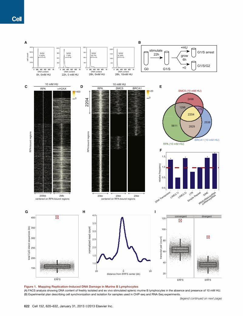
Figure 1. Mapping Replication-Induced DNA Damage in Murine B Lymphocytes
(A) FACS analysis showing DNA content of freshly isolated and ex vivo stimulated splenic murine B lymphocytes in the absence and presence of 10 mM HU.
(B) Experimental plan describing cell synchronization and isolation for samples used in ChIP-seq and RNA-Seq experiments.
(legend continued on next page)
622 Cell 152, 620–632, January 31, 2013 ª2013 Elsevier Inc.

B cells (Figures S2A–S2C). 90% of g-H2AX-associated genomic
regions in untreated XRCC2�/� cells correlate with the regions
enriched for this protein in HU-treated WT B cells (Figure S2B),
and nearly 80% of the regions with enriched g-H2AX observed
in HU-treated WT B cells overlapped with those seen in HU-
treated XRCC2�/� cells (Figure S2C). These data indicate that
XRCC2 deficiency leads to increased endogenous levels of
replication stress mostly at the same loci where HU induces
replication fork stalling and/or breakage in WT cells.
RPA, BRCA1, and SMC5 Colocalization Marks the Sitesof Replication Stress in Early Replicating ZonesLike XRCC2, BRCA1 and members of the structural mainte-
nance of chromosome (SMC) family have been implicated
in promoting replication fork restart (Schlacher et al., 2012;
Stephan et al., 2011). To determine whether HR proteins bind
to a subset of stalled forks marked by RPA and g-H2AX, we
also defined the genome-wide profile of BRCA1 and SMC5.
We confirmed BRCA1 and SMC5 ChIP-seq efficacy by
observing their association at both Sm and Sg1 in 53BP1�/�
cells, where the breaks in IgH persist unrepaired and undergo
extensive resection (Figure S3A) (Bothmer et al., 2010; Bunting
et al., 2010, 2012; Yamane et al., 2011, 2013).
We then determined the localization of BRCA1 and SMC5 in
HU-arrested B cells. Two independent experiments showed
reproducibility of genome-wide BRCA1 and SMC5 association
(Figures S3B and S3C). To identify the RPA genomic sites co-
occupied by the HR proteins BRCA1 and SMC5, we plotted
the distribution of their binding with respect to the center of
individual RPA-bound regions. Overall, 2,204 regions spanning
10 kbp on average showed RPA/BRCA1/SMC5 triple colocaliza-
tion (Figures 1D and 1E). We found that RPA was recruited to
more than 88% of genomic sites exhibiting BRCA1 and SMC5
association (Figure 1E). Furthermore, genome-wide analysis of
RPA/BRCA1/SMC5 profiles in untreated cells revealed more
than a 21% increase in the number of genomic regions occupied
by these three proteins after HU treatment (Figure S4A). Never-
theless, 48% of RPA/BRCA1/SMC5 triple colocalizations were
(C) For each RPA-bound site in response to 10mMHU (y axis), each column depic
RPA-bound sites. Colormap corresponds to binding intensitieswhere ‘‘black’’ rep
bound sites.
(D) RPA, SMC5, and BRCA1 co-occupy 2,204 genomic regions in response to 1
RPA, SMC5, and BRCA1 genomic occupancy in response to HU centered on RPA
sites.
(E) The Venn diagram shows the overlap of sites bound by RPA, SMC5, and BRC
each shared and unique area.
(F) Relative frequency of ERFSs in classes of repetitive sequences is shown. Da
(*, enriched repetitive element classes; p < 1 3 10�3).
(G) ERFSs are enriched in CpG islands. Total CpG island sequences in all the 2,20
model as indicated by the gray points. Each gray point corresponds to the total C
plot depicts the quantiles of total CpG sequences based on the permutation mo
(H) ERFS genomic regions are transcriptionally active. The line plot represents th
center of the ERFSs.
(I) ERFSs are enriched in transcriptionally active convergent and divergent gen
indicated by the crossed red point is compared to the permutation model as ind
divergent/convergent gene pairs observed in an iteration of the permutation mode
pair count based on the permutation model (p < 13 10�5). For definition of conve
S3, S4.
common between the unperturbed and HU-arrested B cells
(Figure S4A). Therefore, we hypothesized that chromatin
with concomitant RPA, BRCA1, and SMC5 binding might corre-
spond to regions undergoing replication fork collapse both in
response to replication stress and during normal DNA replica-
tion. Given that our analysis focused on early replicating sites,
which contrasts with late replicating CFSs, we designated these
regions as ERFSs.
We then characterized ERFSs to determine whether they
share common underlying primary sequence characteristics.
Indeed, these loci were enriched at known repetitive elements,
including LINE L2, SINE, DNA transposons, and tRNA elements
(p(permutation) < 13 10�3, Figure 1F), which are known replica-
tion fork barriers (Mirkin and Mirkin, 2007). Furthermore, ERFSs
showed significantly higher G and C nucleotide content
compared to the whole mouse genome, in contrast to CFSs
that are enriched in A+T sequences (p(Wilcoxon) < 1 3 10�16,
Figure S4B). Twenty-six percent of the ERFSs regions overlap-
ped with CpG islands, which are highly enriched at translocation
breakpoints in B cell lymphoma (Tsai et al., 2008). Conversely,
CpG islands covered approximately 400,000 nucleotides
within these regions (p(permutation) < 1 3 10�5, Figure 1G). As
anticipated, ERFSs clustered at early replication origins (Fig-
ure S4C), and over 66% of the loci overlapped with intragenic
or promoter sequences of RefSeq annotated protein coding
genes (p(permutation) < 1 3 10�3, Figures S4D and S4E).
Moreover, ERFSs are more transcriptionally active relative to
flanking genomic regions shown by relative mRNA enrichment
by RNA-Seq (Figure 1H). Indeed, more than 86% of the RefSeq
annotated genes with ERFSs are among the highest transcribed
genes (p(binomial) < 13 10�16, Figure S4F). Finally, ERFSs were
significantly enriched in gene pairs that are transcribed in
converging or diverging directions (see Experimental Proce-
dures), such as the convergent transcription pair of IKZF1 and
FIGNL1 shown in Figure 2A. Compared to expected values,
ERFSs were at least two times more likely to localize in regions
containing gene pairs exhibiting convergent and/or divergent
gene pairs (p(permutation) < 1 3 10�5, Figure 1I).
ts the presence of RPA (left) and g-H2AX (right) within a window centered on the
resents no binding. K-mean clustering algorithmwas used to group the protein-
0 mM HU. The plot in each column, from left to right, represents the pattern of
-bound sites. K-mean clustering algorithm is used to group the protein-bound
A1 in response to 10 mM HU. The total number of bound sites is indicated for
shed line indicates the expected frequency based on the permutation model
4 ERFSs as indicated by the crossed red point is compared to the permutation
pG island sequences covered in an iteration of the permutation model. The box
del (p < 1 3 10�5).
e average RNA tag count (loess smoothed) in a genomic window around the
e pairs. Count of divergent/convergent gene pairs coinciding with ERFSs as
icated by the gray points. Each gray point corresponds to the total number of
l. The box plot depicts the quantiles of the total convergent/divergent transcript
rgent/divergent gene pairs see Experimental Procedures. See also Figures S1,
Cell 152, 620–632, January 31, 2013 ª2013 Elsevier Inc. 623
A
CERFS Name
1 MHCII2 GIMAP3 SWAP704 BACH25 IKZF16 FOXP17 PTPRC8 SLA9 PVT1
10 ETS111 IRF412 BCL213 USP2214 NFκB115 PRKCB
Convergent and/orDivergent Transcripts
YesYesYesYesYesNoYesYesYesYesYesYesYesNoNo
MHC II
GIMAP
SWAP70
BACH2
IKZF1
FOXP1
PTPRC
SLAPVT1
ETS1 IRF4
BCL2
UPS22
NFKB1 PRKCB
chr1
chr2
chr3
chr4
chr5
chr6
chr7
chr8
chr9
chr10
chr11
chr12
chr13
chr14
chr15
chr16
chr17
chr18
chr19 ch
rXch
rY
B
Rearranged in B Cell Cancer
Yes
Yes
Yes
Yes
Yes
Yes
Yes
Yes
No
No
NoNo
No
No
No
chr11:11000000 chr11:12000000500 kbp
RPA (10 mM HU)
BRCA1 (10 mM HU)
SMC5 (10 mM HU)
γ−H2AX (10 mM HU)
BrdU (10 mM HU)
ERFS hotspot ERFS
5
5
5
1
5
Ikzf1 Fignl1 Grb101700042O10Rik Ddc
2930415F15RikVwc2Zpbp
AID Target
Yes
YesYesNoNoYesNoNoNo
YesYesNoNoYesNo
17674
1161
1515
913
111
37
Chromosome
1121.6498.8
333128
314.6419.4281.4431.8
669.8520283
656.860.2117
170.6
Length (Kb)YesYesYesNoYesNoYesYesNoYesYesYesNoNoNo
Gene ClusterYesYesYesYesYesYesYesYesYesYesYesYesYesYesYes
Transcribed
RP
M
coordinate
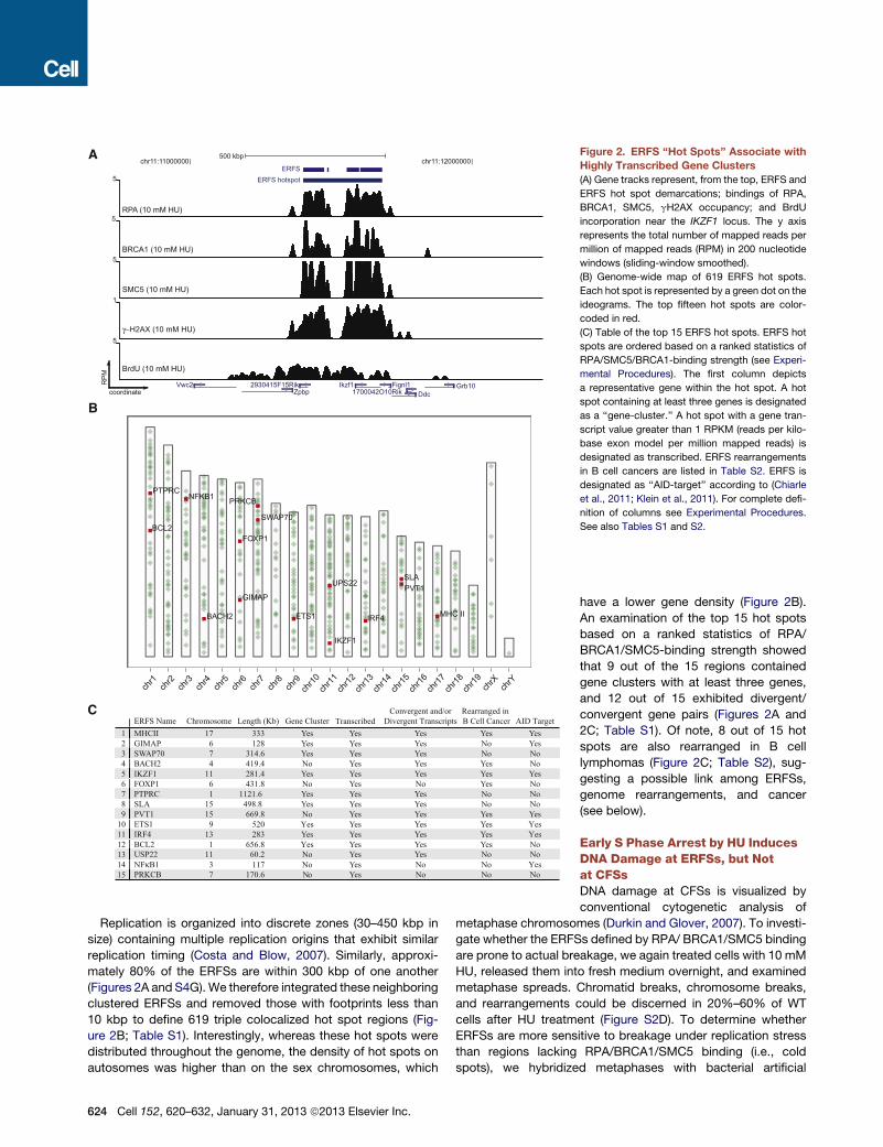
Figure 2. ERFS ‘‘Hot Spots’’ Associate with
Highly Transcribed Gene Clusters
(A) Gene tracks represent, from the top, ERFS and
ERFS hot spot demarcations; bindings of RPA,
BRCA1, SMC5, gH2AX occupancy; and BrdU
incorporation near the IKZF1 locus. The y axis
represents the total number of mapped reads per
million of mapped reads (RPM) in 200 nucleotide
windows (sliding-window smoothed).
(B) Genome-wide map of 619 ERFS hot spots.
Each hot spot is represented by a green dot on the
ideograms. The top fifteen hot spots are color-
coded in red.
(C) Table of the top 15 ERFS hot spots. ERFS hot
spots are ordered based on a ranked statistics of
RPA/SMC5/BRCA1-binding strength (see Experi-
mental Procedures). The first column depicts
a representative gene within the hot spot. A hot
spot containing at least three genes is designated
as a ‘‘gene-cluster.’’ A hot spot with a gene tran-
script value greater than 1 RPKM (reads per kilo-
base exon model per million mapped reads) is
designated as transcribed. ERFS rearrangements
in B cell cancers are listed in Table S2. ERFS is
designated as ‘‘AID-target’’ according to (Chiarle
et al., 2011; Klein et al., 2011). For complete defi-
nition of columns see Experimental Procedures.
See also Tables S1 and S2.
Replication is organized into discrete zones (30–450 kbp in
size) containing multiple replication origins that exhibit similar
replication timing (Costa and Blow, 2007). Similarly, approxi-
mately 80% of the ERFSs are within 300 kbp of one another
(Figures 2A and S4G). We therefore integrated these neighboring
clustered ERFSs and removed those with footprints less than
10 kbp to define 619 triple colocalized hot spot regions (Fig-
ure 2B; Table S1). Interestingly, whereas these hot spots were
distributed throughout the genome, the density of hot spots on
autosomes was higher than on the sex chromosomes, which
624 Cell 152, 620–632, January 31, 2013 ª2013 Elsevier Inc.
have a lower gene density (Figure 2B).
An examination of the top 15 hot spots
based on a ranked statistics of RPA/
BRCA1/SMC5-binding strength showed
that 9 out of the 15 regions contained
gene clusters with at least three genes,
and 12 out of 15 exhibited divergent/
convergent gene pairs (Figures 2A and
2C; Table S1). Of note, 8 out of 15 hot
spots are also rearranged in B cell
lymphomas (Figure 2C; Table S2), sug-
gesting a possible link among ERFSs,
genome rearrangements, and cancer
(see below).
Early S Phase Arrest by HU InducesDNA Damage at ERFSs, but Notat CFSsDNA damage at CFSs is visualized by
conventional cytogenetic analysis of
metaphase chromosomes (Durkin and Glover, 2007). To investi-
gate whether the ERFSs defined by RPA/ BRCA1/SMC5 binding
are prone to actual breakage, we again treated cells with 10 mM
HU, released them into fresh medium overnight, and examined
metaphase spreads. Chromatid breaks, chromosome breaks,
and rearrangements could be discerned in 20%–60% of WT
cells after HU treatment (Figure S2D). To determine whether
ERFSs are more sensitive to breakage under replication stress
than regions lacking RPA/BRCA1/SMC5 binding (i.e., cold
spots), we hybridized metaphases with bacterial artificial
A
B
C D
FE
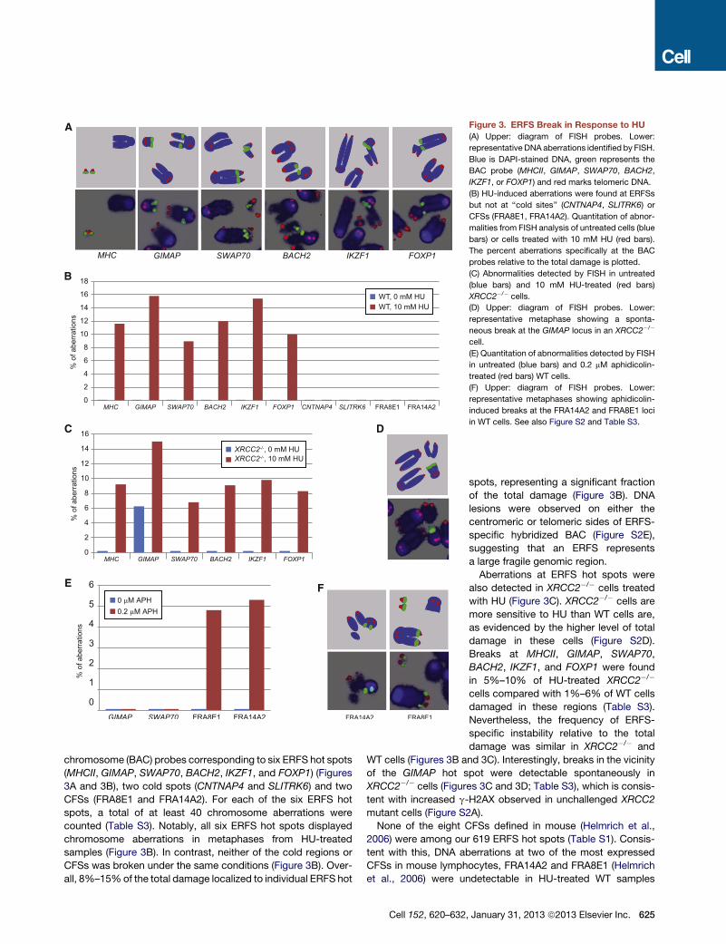
Figure 3. ERFS Break in Response to HU
(A) Upper: diagram of FISH probes. Lower:
representative DNA aberrations identified by FISH.
Blue is DAPI-stained DNA, green represents the
BAC probe (MHCII, GIMAP, SWAP70, BACH2,
IKZF1, or FOXP1) and red marks telomeric DNA.
(B) HU-induced aberrations were found at ERFSs
but not at ‘‘cold sites’’ (CNTNAP4, SLITRK6) or
CFSs (FRA8E1, FRA14A2). Quantitation of abnor-
malities from FISH analysis of untreated cells (blue
bars) or cells treated with 10 mM HU (red bars).
The percent aberrations specifically at the BAC
probes relative to the total damage is plotted.
(C) Abnormalities detected by FISH in untreated
(blue bars) and 10 mM HU-treated (red bars)
XRCC2�/� cells.
(D) Upper: diagram of FISH probes. Lower:
representative metaphase showing a sponta-
neous break at the GIMAP locus in an XRCC2�/�
cell.
(E) Quantitation of abnormalities detected by FISH
in untreated (blue bars) and 0.2 mM aphidicolin-
treated (red bars) WT cells.
(F) Upper: diagram of FISH probes. Lower:
representative metaphases showing aphidicolin-
induced breaks at the FRA14A2 and FRA8E1 loci
in WT cells. See also Figure S2 and Table S3.
chromosome (BAC) probes corresponding to six ERFS hot spots
(MHCII, GIMAP, SWAP70, BACH2, IKZF1, and FOXP1) (Figures
3A and 3B), two cold spots (CNTNAP4 and SLITRK6) and two
CFSs (FRA8E1 and FRA14A2). For each of the six ERFS hot
spots, a total of at least 40 chromosome aberrations were
counted (Table S3). Notably, all six ERFS hot spots displayed
chromosome aberrations in metaphases from HU-treated
samples (Figure 3B). In contrast, neither of the cold regions or
CFSs was broken under the same conditions (Figure 3B). Over-
all, 8%–15%of the total damage localized to individual ERFS hot
Cell 152, 620–632
spots, representing a significant fraction
of the total damage (Figure 3B). DNA
lesions were observed on either the
centromeric or telomeric sides of ERFS-
specific hybridized BAC (Figure S2E),
suggesting that an ERFS represents
a large fragile genomic region.
Aberrations at ERFS hot spots were
also detected in XRCC2�/� cells treated
with HU (Figure 3C). XRCC2�/� cells are
more sensitive to HU than WT cells are,
as evidenced by the higher level of total
damage in these cells (Figure S2D).
Breaks at MHCII, GIMAP, SWAP70,
BACH2, IKZF1, and FOXP1 were found
in 5%–10% of HU-treated XRCC2�/�
cells compared with 1%–6% of WT cells
damaged in these regions (Table S3).
Nevertheless, the frequency of ERFS-
specific instability relative to the total
damage was similar in XRCC2�/� and
WT cells (Figures 3B and 3C). Interestingly, breaks in the vicinity
of the GIMAP hot spot were detectable spontaneously in
XRCC2�/� cells (Figures 3C and 3D; Table S3), which is consis-
tent with increased g-H2AX observed in unchallenged XRCC2
mutant cells (Figure S2A).
None of the eight CFSs defined in mouse (Helmrich et al.,
2006) were among our 619 ERFS hot spots (Table S1). Consis-
tent with this, DNA aberrations at two of the most expressed
CFSs in mouse lymphocytes, FRA14A2 and FRA8E1 (Helmrich
et al., 2006) were undetectable in HU-treated WT samples
, January 31, 2013 ª2013 Elsevier Inc. 625
A
B
C D E
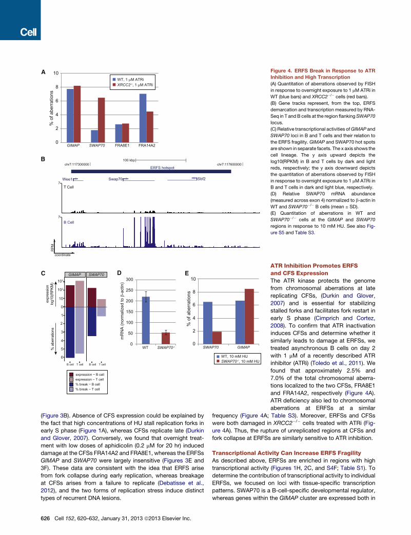
Figure 4. ERFS Break in Response to ATR
Inhibition and High Transcription
(A) Quantitation of aberrations observed by FISH
in response to overnight exposure to 1 mM ATRi in
WT (blue bars) and XRCC2�/� cells (red bars).
(B) Gene tracks represent, from the top, ERFS
demarcation and transcription measured by RNA-
Seq in T and B cells at the region flanking SWAP70
locus.
(C) Relative transcriptional activities ofGIMAP and
SWAP70 loci in B and T cells and their relation to
the ERFS fragility. GIMAP and SWAP70 hot spots
are shown in separate facets. The x axis shows the
cell lineage. The y axis upward depicts the
log10(RPKM) in B and T cells by dark and light
reds, respectively; the y axis downward depicts
the quantitation of aberrations observed by FISH
in response to overnight exposure to 1 mM ATRi in
B and T cells in dark and light blue, respectively.
(D) Relative SWAP70 mRNA abundance
(measured across exon 4) normalized to b-actin in
WT and SWAP70�/� B cells (mean ± SD).
(E) Quantitation of aberrations in WT and
SWAP70�/� cells at the GIMAP and SWAP70
regions in response to 10 mM HU. See also Fig-
ure S5 and Table S3.
(Figure 3B). Absence of CFS expression could be explained by
the fact that high concentrations of HU stall replication forks in
early S phase (Figure 1A), whereas CFSs replicate late (Durkin
and Glover, 2007). Conversely, we found that overnight treat-
ment with low doses of aphidicolin (0.2 mM for 20 hr) induced
damage at the CFSs FRA14A2 and FRA8E1, whereas the ERFSs
GIMAP and SWAP70 were largely insensitive (Figures 3E and
3F). These data are consistent with the idea that ERFS arise
from fork collapse during early replication, whereas breakage
at CFSs arises from a failure to replicate (Debatisse et al.,
2012), and the two forms of replication stress induce distinct
types of recurrent DNA lesions.
626 Cell 152, 620–632, January 31, 2013 ª2013 Elsevier Inc.
ATR Inhibition Promotes ERFSand CFS ExpressionThe ATR kinase protects the genome
from chromosomal aberrations at late
replicating CFSs, (Durkin and Glover,
2007) and is essential for stabilizing
stalled forks and facilitates fork restart in
early S phase (Cimprich and Cortez,
2008). To confirm that ATR inactivation
induces CFSs and determine whether it
similarly leads to damage at ERFSs, we
treated asynchronous B cells on day 2
with 1 mM of a recently described ATR
inhibitor (ATRi) (Toledo et al., 2011). We
found that approximately 2.5% and
7.0% of the total chromosomal aberra-
tions localized to the two CFSs, FRA8E1
and FRA14A2, respectively (Figure 4A).
ATR deficiency also led to chromosomal
aberrations at ERFSs at a similar
frequency (Figure 4A; Table S3). Moreover, ERFSs and CFSs
were both damaged in XRCC2�/� cells treated with ATRi (Fig-
ure 4A). Thus, the rupture of unreplicated regions at CFSs and
fork collapse at ERFSs are similarly sensitive to ATR inhibition.
Transcriptional Activity Can Increase ERFS FragilityAs described above, ERFSs are enriched in regions with high
transcriptional activity (Figures 1H, 2C, and S4F; Table S1). To
determine the contribution of transcriptional activity to individual
ERFSs, we focused on loci with tissue-specific transcription
patterns. SWAP70 is a B-cell-specific developmental regulator,
whereas genes within the GIMAP cluster are expressed both in
B and in T cells (Figures 4B and S5A). Treatment with ATRi led to
a similar frequency of damage atGIMAP in B and T cells, consis-
tent with insignificant changes in gene expression between the
two cell types (Figure 4C). In contrast, damage near SWAP70
was 3-fold lower in T than in B cells (Figure 4C; Table S3), which
correlated with the decreased transcription of SWAP70 in T cells
(Figure 4B). Nevertheless, the replication timing near SWAP70
was similar in both cell types (Figure S5B). To further delineate
the role of transcription on ERFS breakage, we used
SWAP70�/� mice in which 2.7 kbp, including the first exon and
part of the 50 untranslated region, is removed (Borggrefe et al.,
2001), allowing us to compare the fragility of ERFSs in the
same genomic region in knockout B cells. We determined that
SWAP70 mRNA in SWAP70�/� B cells was reduced by approx-
imately 4-fold relative to levels in WT (Figure 4D). Moreover, DNA
damage near SWAP70 was approximately 2.5-fold lower in
SWAP70�/� relative to levels in WT B cells (Figure 4E). In
contrast, DNA damage near GIMAP remained at a similar level
both in WT and SWAP70�/� cells (Figure 4E). Although our
data indicate that high level of transcription contributes to the
breakage of some ERFSs, other molecular features, including
repetitive elements (Figure 1F), covalently bound protein
complexes, and RNA:DNA hybrids, might also be sources of
ERFS fragility.
Oncogenic Stress Can Trigger ERFS and CFS FragilityOncogene deregulation is thought to compromise genome
integrity preferentially at CFSs (Bartek et al., 2007; Halazonetis
et al., 2008), and CFS deletion has been associated with various
cancers (Bignell et al., 2010). To determine whether oncogenic
stress similarly induces DNA damage at ERFSs, we overex-
pressed c-myc in B cells because it has been implicated in regu-
lating replication initiation and origin firing (Dominguez-Sola
et al., 2007).XRCC2�/� cells were utilized to increase the amount
of replicative stress and DNA damage as a result of decreased
HR (Figure S2D). c-myc overexpression led to induction of
p53 (Figure 5A), which correlated with an approximately 1.6-
fold increase in overall DNA damage in XRCC2�/� cells overex-
pressing c-myc compared to empty vector (EV)-infected cells
(Table S3). Moreover, 7.3% of the total breaks generated in
c-myc overexpressing cells were found near SWAP70,
compared to 2.4% of total breaks at this ERFS in EV-infected
B cells (Figure 5B). Similarly, out of 43 breaks observed in
c-myc-infected cells, 3 (7%) were found at the GIMAP cluster,
and 3 (6.7%) were found near BACH2. c-myc overexpression
also induced breaks at FRA8E1, showing a 2-fold relative
increase in breaks relative to EV-infected cells (Figure 5B).
Thus, DNA damage induced by c-myc overexpression can occur
at ERFSs and CFSs.
ERFS Fragility Is AID IndependentMutations and DSBs at various oncogenes, including c-myc, are
due to AID off-target activity (Robbiani et al., 2008). Recently,
a number of genome-wide studies in primary B cells mapped
AID-induced DNA translocation events, and identified several
novel hot spots for AID-dependent translocations at non-Ig
genes (Chiarle et al., 2011; Kato et al., 2012; Klein et al., 2011).
Among these translocation hot spots, MHCII, GIMAP, IKZF1,
PVT1, ETS1, IRF4, and NFkB1 were located within the top 15
ERFS hot spots in this study, whereas the IgH locus (the physio-
logic target of AID) was not ranked high on the list (Figure 2C;
Table S1). To determine whether AID contributes to ERFS
fragility, we stimulated WT and AID knockout B cells with LPS/
IL4 for 2 days, and then treated them with ATRi overnight. These
conditions induce robust AID-dependent DNA damage simulta-
neously with replication stress. We probed metaphases with
BACs spanning the IgH locus, the GIMAP cluster, and IKZF1-
all AID translocation hot spots—as well as BACH2, SWAP70,
FOXP1, and BCL2 (Figure S2E)—ERFSs that are frequently rear-
ranged in B cell lymphoma (Figure 2C; Tables S1 and S2). In WT,
the IgH locus was damaged in 3.8% of cells, but the frequency of
IgH-specific instability did not increase with ATRi (Figure S2F),
despite the fact that ATRi greatly increased overall damage
(Table S3). Upon ATRi treatment, the frequency of breaks at
the ERFSs GIMAP, IKZF1, BACH2, SWAP70, and FOXP1, and
BCL2 were elevated to the levels similar to those observed at
the IgH in activated B cells (Figure S2F). Breaks at some ERFSs
were even spontaneously detected (FOXP1 and GIMAP,
Figure S2F).
To determine whether AID expression contributes to aberra-
tions observed at ERFSs, we next analyzed their breakage
frequency in AID�/� cells. Unlike WT cells, IgH breaks were
absent in AID�/� cells. In contrast, all ERFSs exhibited similar
levels of breakage both in WT and AID�/� cells (Figure 5C; Table
S3). Therefore, whereas IgH breaks in B cells are entirely AID
dependent, the breakage of ERFSs is AID independent. Alto-
gether, these data suggest that some recurrent rearrangements
in B cell lymphoma are due to AID-independent replicative stress
at ERFSs.
Genome Instability at ERFSs Is Observed in MouseModels and Human CancerAmong the top 15 ERFS hot spot that break in response to AID-
independent replication stress, we have identified three partners
that recurrently translocate to IgH in lymphomas: BACH2,
FOXP1, and BCL2 (Table S2). We hypothesized that if AID-
dependent DSBs in G1 persisted into early S phase, transloca-
tions between AID-dependent breaks and ERFS might be
detectable. To test this, we examined cells transgenically over-
expressing AID and simultaneously deficient for 53BP1
(IgkAID/53BP1�/�), thus allowing the persistence of G1 IgH
breaks into S phase where they could be joined to ERFSs.
Indeed, 26% and 7% of IgkAID/53BP1�/� B cells carried IgH
locus andBACH2 breaks, respectively (Figure 5D). These breaks
are fusogenic because IgH- and BACH2-associated transloca-
tions to unidentified partner chromosomes were found in 7.3%
and 1.2% of the metaphases, respectively (Figure 5D). Impor-
tantly, we also detected one IgH/BACH2 translocation among
750 cells (Figure 5E), reminiscent of the IgH/BACH2 transloca-
tions observed in human B cell lymphoma (Kobayashi et al.,
2011). Thus, AID-dependent breaks generated in G1 (Petersen
et al., 2001) can join to ERFS breaks triggered in early S phase.
A hallmark of cancer genomes is widespread copy-number
changes, insertions, and deletions. To determine whether dele-
tions and/or amplifications at ERFSs are a general feature of
the B cell lymphoma genome, we compared our ERFSs with
Cell 152, 620–632, January 31, 2013 ª2013 Elsevier Inc. 627
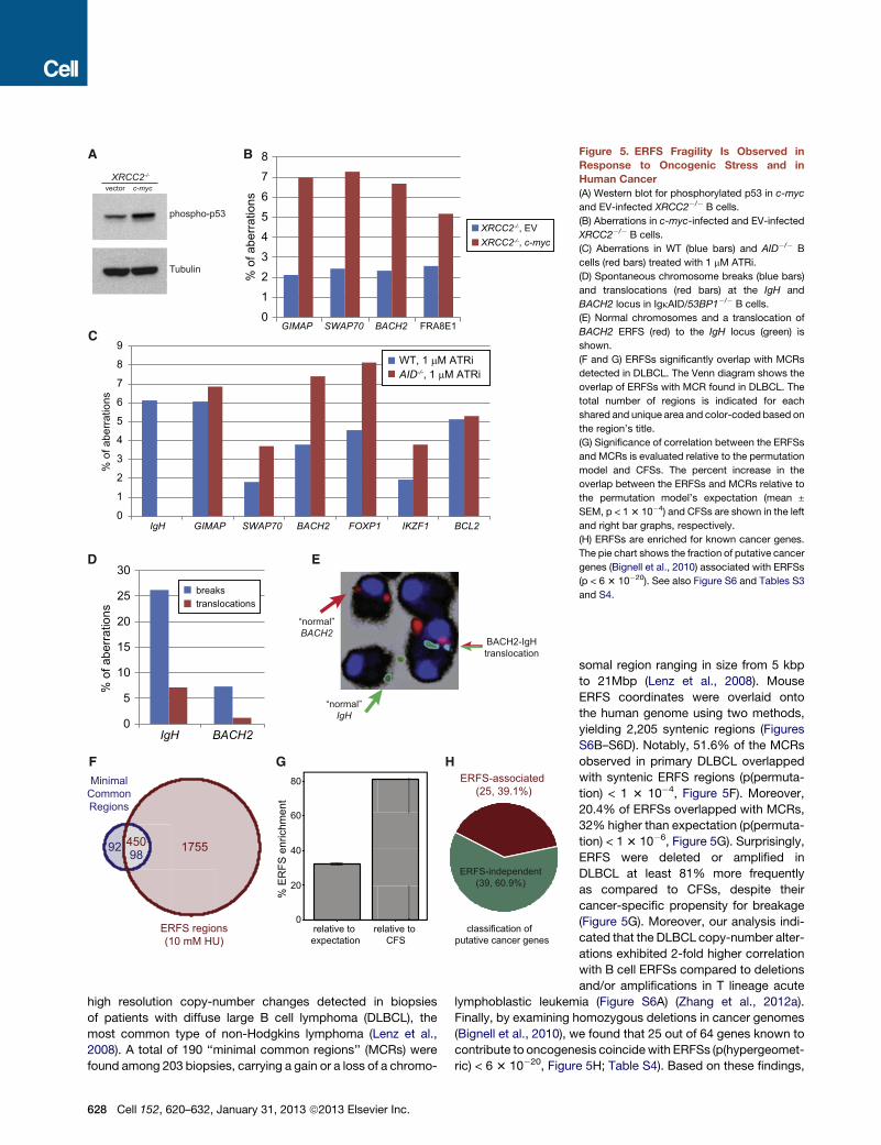
A B
C
D E
F G H
Figure 5. ERFS Fragility Is Observed in
Response to Oncogenic Stress and in
Human Cancer
(A) Western blot for phosphorylated p53 in c-myc
and EV-infected XRCC2�/� B cells.
(B) Aberrations in c-myc-infected and EV-infected
XRCC2�/� B cells.
(C) Aberrations in WT (blue bars) and AID�/� B
cells (red bars) treated with 1 mM ATRi.
(D) Spontaneous chromosome breaks (blue bars)
and translocations (red bars) at the IgH and
BACH2 locus in IgkAID/53BP1�/� B cells.
(E) Normal chromosomes and a translocation of
BACH2 ERFS (red) to the IgH locus (green) is
shown.
(F and G) ERFSs significantly overlap with MCRs
detected in DLBCL. The Venn diagram shows the
overlap of ERFSs with MCR found in DLBCL. The
total number of regions is indicated for each
shared and unique area and color-coded based on
the region’s title.
(G) Significance of correlation between the ERFSs
and MCRs is evaluated relative to the permutation
model and CFSs. The percent increase in the
overlap between the ERFSs and MCRs relative to
the permutation model’s expectation (mean ±
SEM, p < 13 10�4) and CFSs are shown in the left
and right bar graphs, respectively.
(H) ERFSs are enriched for known cancer genes.
The pie chart shows the fraction of putative cancer
genes (Bignell et al., 2010) associated with ERFSs
(p < 6 3 10�20). See also Figure S6 and Tables S3
and S4.
high resolution copy-number changes detected in biopsies
of patients with diffuse large B cell lymphoma (DLBCL), the
most common type of non-Hodgkins lymphoma (Lenz et al.,
2008). A total of 190 ‘‘minimal common regions’’ (MCRs) were
found among 203 biopsies, carrying a gain or a loss of a chromo-
628 Cell 152, 620–632, January 31, 2013 ª2013 Elsevier Inc.
somal region ranging in size from 5 kbp
to 21Mbp (Lenz et al., 2008). Mouse
ERFS coordinates were overlaid onto
the human genome using two methods,
yielding 2,205 syntenic regions (Figures
S6B–S6D). Notably, 51.6% of the MCRs
observed in primary DLBCL overlapped
with syntenic ERFS regions (p(permuta-
tion) < 1 3 10�4, Figure 5F). Moreover,
20.4% of ERFSs overlapped with MCRs,
32% higher than expectation (p(permuta-
tion) < 13 10�6, Figure 5G). Surprisingly,
ERFS were deleted or amplified in
DLBCL at least 81% more frequently
as compared to CFSs, despite their
cancer-specific propensity for breakage
(Figure 5G). Moreover, our analysis indi-
cated that the DLBCL copy-number alter-
ations exhibited 2-fold higher correlation
with B cell ERFSs compared to deletions
and/or amplifications in T lineage acute
lymphoblastic leukemia (Figure S6A) (Zhang et al., 2012a).
Finally, by examining homozygous deletions in cancer genomes
(Bignell et al., 2010), we found that 25 out of 64 genes known to
contribute to oncogenesis coincidewith ERFSs (p(hypergeomet-
ric) < 6 3 10�20, Figure 5H; Table S4). Based on these findings,
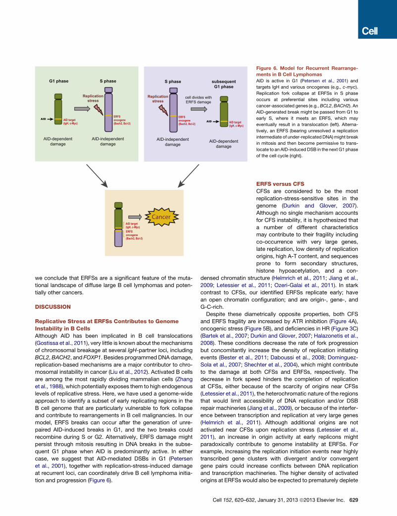
Figure 6. Model for Recurrent Rearrange-
ments in B Cell Lymphomas
AID is active in G1 (Petersen et al., 2001) and
targets IgH and various oncogenes (e.g., c-myc).
Replication fork collapse at ERFSs in S phase
occurs at preferential sites including various
cancer-associated genes (e.g.,BCL2,BACH2). An
AID-generated break might be passed from G1 to
early S, where it meets an ERFS, which may
eventually result in a translocation (left). Alterna-
tively, an ERFS (bearing unresolved a replication
intermediate of under-replicated DNA)might break
in mitosis and then become permissive to trans-
locate to an AID-induced DSB in the next G1 phase
of the cell cycle (right).
we conclude that ERFSs are a significant feature of the muta-
tional landscape of diffuse large B cell lymphomas and poten-
tially other cancers.
DISCUSSION
Replicative Stress at ERFSs Contributes to GenomeInstability in B CellsAlthough AID has been implicated in B cell translocations
(Gostissa et al., 2011), very little is known about the mechanisms
of chromosomal breakage at several IgH-partner loci, including
BCL2,BACH2, and FOXP1. Besides programmedDNAdamage,
replication-based mechanisms are a major contributor to chro-
mosomal instability in cancer (Liu et al., 2012). Activated B cells
are among the most rapidly dividing mammalian cells (Zhang
et al., 1988), which potentially exposes them to high endogenous
levels of replicative stress. Here, we have used a genome-wide
approach to identify a subset of early replicating regions in the
B cell genome that are particularly vulnerable to fork collapse
and contribute to rearrangements in B cell malignancies. In our
model, ERFS breaks can occur after the generation of unre-
paired AID-induced breaks in G1, and the two breaks could
recombine during S or G2. Alternatively, ERFS damage might
persist through mitosis resulting in DNA breaks in the subse-
quent G1 phase when AID is predominantly active. In either
case, we suggest that AID-mediated DSBs in G1 (Petersen
et al., 2001), together with replication-stress-induced damage
at recurrent loci, can coordinately drive B cell lymphoma initia-
tion and progression (Figure 6).
Cell 152, 620–632
ERFS versus CFSCFSs are considered to be the most
replication-stress-sensitive sites in the
genome (Durkin and Glover, 2007).
Although no single mechanism accounts
for CFS instability, it is hypothesized that
a number of different characteristics
may contribute to their fragility including
co-occurrence with very large genes,
late replication, low density of replication
origins, high A-T content, and sequences
prone to form secondary structures,
histone hypoacetylation, and a con-
densed chromatin structure (Helmrich et al., 2011; Jiang et al.,
2009; Letessier et al., 2011; Ozeri-Galai et al., 2011). In stark
contrast to CFSs, our identified ERFSs replicate early; have
an open chromatin configuration; and are origin-, gene-, and
G-C-rich.
Despite these diametrically opposite properties, both CFS
and ERFS fragility are increased by ATR inhibition (Figure 4A),
oncogenic stress (Figure 5B), and deficiencies in HR (Figure 3C)
(Bartek et al., 2007; Durkin and Glover, 2007; Halazonetis et al.,
2008). These conditions decrease the rate of fork progression
but concomitantly increase the density of replication initiating
events (Bester et al., 2011; Daboussi et al., 2008; Dominguez-
Sola et al., 2007; Shechter et al., 2004), which might contribute
to the damage at both CFSs and ERFSs, respectively. The
decrease in fork speed hinders the completion of replication
at CFSs, either because of the scarcity of origins near CFSs
(Letessier et al., 2011), the heterochromatic nature of the regions
that would limit accessibility of DNA replication and/or DSB
repair machineries (Jiang et al., 2009), or because of the interfer-
ence between transcription and replication at very large genes
(Helmrich et al., 2011). Although additional origins are not
activated near CFSs upon replication stress (Letessier et al.,
2011), an increase in origin activity at early replicons might
paradoxically contribute to genome instability at ERFSs. For
example, increasing the replication initiation events near highly
transcribed gene clusters with divergent and/or convergent
gene pairs could increase conflicts between DNA replication
and transcription machineries. The higher density of activated
origins at ERFSs would also be expected to prematurely deplete
, January 31, 2013 ª2013 Elsevier Inc. 629
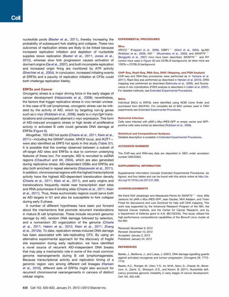
nucleotide pools (Bester et al., 2011), thereby increasing the
probability of subsequent fork stalling and collapse. These two
outcomes of replication stress are likely to be linked because
increased replication initiation and depletion of nucleotide
supplies slows replication (Bester et al., 2011; Jones et al.,
2012), whereas slow fork progression causes activation of
dormant origins (Ge et al., 2007), and both incomplete replication
and increased origin firing are monitored by ATR activity
(Shechter et al., 2004). In conclusion, increased initiating events
at ERFSs and a paucity of replication initiation at CFSs could
both challenge replication fidelity.
ERFSs and CancerOncogenic stress is a major driving force in the early stages of
cancer development (Halazonetis et al., 2008); nevertheless,
the factors that trigger replicative stress in vivo remain unclear.
In the case of B cell lymphomas, oncogenic stress can be initi-
ated by the activity of AID, which by targeting non-Ig genes
such as c-myc (Robbiani et al., 2008), leads to c-myc/IgH trans-
locations and consequent aberrant c-myc expression. This form
of AID-induced oncogenic stress or high levels of proliferative
activity in activated B cells could generate DNA damage at
ERFSs (Figure 6).
Altogether, 103 AID hot spots (Chiarle et al., 2011; Klein et al.,
2011)—including the GIMAP cluster, MHCII locus, and IKZF1—
were also identified as ERFS hot spots in this study (Table S1).
It is possible that the overlap observed between a subset of
off-target AID sites and ERFSs is due to common underlying
features of these loci. For example, AID is recruited to ssDNA
regions (Chaudhuri and Alt, 2004), which are also generated
during replicative stress; AID-dependent DSBs and ERFSs are
also both enriched in repeat elements (Staszewski et al., 2011).
In addition, chromosomal regionswith the highest transcriptional
activity have the highest AID-dependent translocation density
(Chiarle et al., 2011; Klein et al., 2011), and early origins and
translocations frequently reside near transcription start sites
and RNA polymerase-II-binding sites (Chiarle et al., 2011; Klein
et al., 2011). Thus, these euchromatic regions could serve both
as AID targets in G1 and also be susceptible to fork collapse
during early S phase.
A number of different hypotheses have been put forward
about the mechanisms that promote recurrent translocations
in mature B cell lymphomas. These include recurrent genomic
damage by AID, random DNA damage followed by selection,
and a nonrandom 3D organization of the genome (Chiarle
et al., 2011; Hakim et al., 2012; Klein et al., 2011; Zhang
et al., 2012b). To date, replication-stress-induced DNA damage
has been associated with late-replicating CFS. By using an
alternative experimental approach for the discovery of fragile
site expression during early replication, we have identified
a novel source of recurrent AID-independent DNA breaks
that may play a mechanistic role in some of the most common
genome rearrangements during B cell lymphomagenesis.
Because transcriptional activity and replication timing of a
genomic region vary among different cell lineages (Hansen
et al., 2010), different sets of ERFSs might also account for
recurrent chromosomal rearrangements in cancers of distinct
cellular origins.
630 Cell 152, 620–632, January 31, 2013 ª2013 Elsevier Inc.
EXPERIMENTAL PROCEDURES
Mice
XRCC2�/�(Frappart et al., 2009), 53BP1�/� (Ward et al., 2004), IgkAID
(Robbiani et al., 2009), AID�/� (Muramatsu et al., 2000), and SWAP70�/�
(Borggrefe et al., 2001) mice have been described. SWAP70�/� and WT
control mice used in Figure 4D are C57BL/6 background; all other mice are
129/Sv x C57BL/6 background.
ChIP-Seq, Repli-Seq, RNA-Seq, DHS I Mapping, and FISH Analysis
ChIP-seq and RNA-Seq procedures were performed as in Yamane et al.
(2011), Repli-Seq was performed as described in Hansen et al. (2010). DHSI
mapping was performed as described (Sekimata et al., 2009), and fluores-
cence in situ hybridization (FISH) analysis is described in Callen et al. (2007).
For detailed methods, see Extended Experimental Procedures.
BACs
Individual BACs to ERFSs were identified using NCBI clone finder and
purchased from BACPAC. For complete list of BAC probes used in FISH
experiments see Extended Experimental Procedures.
Retroviral Infection
Cells were infected with pMX-c-Myc-IRES-GFP or empty vector and GFP-
positive cells were sorted as described (Robbiani et al., 2008).
Statistical and Computational Analyses
Detailed description is available in Extended Experimental Procedures.
ACCESSION NUMBERS
The ChIP-seq and RNA-seq data are deposited in GEO under accession
number GSE43504.
SUPPLEMENTAL INFORMATION
Supplemental Information includes Extended Experimental Procedures, six
figures, and four tables and can be found with this article online at http://dx.
doi.org/10.1016/j.cell.2013.01.006.
ACKNOWLEDGMENTS
We thank Rolf Jessberger and Alessandra Pernis for SWAP70�/� mice, Mila
Jankovic for pMX-c-Myc-IRES-GFP, Jean Gautier, Mirit Aladjem, and Toren
Finkel for discussions and Lars Grontved for help with DHS mapping. This
work was supported by the Intramural Research Program of the NIH, the
National Cancer Institute, and the Center for Cancer Research, and by
a Department of Defense grant to A.N. (BC102335). This study utilized the
high-performance computational capabilities of the Biowulf Linux cluster at
the NIH.
Received: November 9, 2012
Revised: December 10, 2012
Accepted: January 2, 2013
Published: January 24, 2013
REFERENCES
Bartek, J., Bartkova, J., and Lukas, J. (2007). DNA damage signalling guards
against activated oncogenes and tumour progression. Oncogene 26, 7773–
7779.
Bester, A.C., Roniger, M., Oren, Y.S., Im, M.M., Sarni, D., Chaoat, M., Bensi-
mon, A., Zamir, G., Shewach, D.S., and Kerem, B. (2011). Nucleotide defi-
ciency promotes genomic instability in early stages of cancer development.
Cell 145, 435–446.
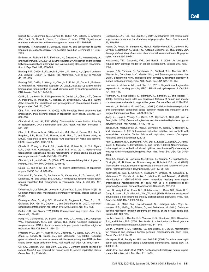
Bignell, G.R., Greenman, C.D., Davies, H., Butler, A.P., Edkins, S., Andrews,
J.M., Buck, G., Chen, L., Beare, D., Latimer, C., et al. (2010). Signatures of
mutation and selection in the cancer genome. Nature 463, 893–898.
Borggrefe, T., Keshavarzi, S., Gross, B., Wabl, M., and Jessberger, R. (2001).
Impaired IgE response in SWAP-70-deficient mice. Eur. J. Immunol. 31, 2467–
2475.
Bothmer, A., Robbiani, D.F., Feldhahn, N., Gazumyan, A., Nussenzweig, A.,
andNussenzweig,M.C. (2010). 53BP1 regulates DNA resection and the choice
between classical and alternative end joining during class switch recombina-
tion. J. Exp. Med. 207, 855–865.
Bunting, S.F., Callen, E., Kozak, M.L., Kim, J.M., Wong, N., Lopez-Contreras,
A.J., Ludwig, T., Baer, R., Faryabi, R.B., Malhowski, A., et al. (2012). Mol. Cell
46, 125–135.
Bunting, S.F., Callen, E., Wong, N., Chen, H.T., Polato, F., Gunn, A., Bothmer,
A., Feldhahn, N., Fernandez-Capetillo, O., Cao, L., et al. (2010). 53BP1 inhibits
homologous recombination in Brca1-deficient cells by blocking resection of
DNA breaks. Cell 141, 243–254.
Callen, E., Jankovic, M., Difilippantonio, S., Daniel, J.A., Chen, H.T., Celeste,
A., Pellegrini, M., McBride, K., Wangsa, D., Bredemeyer, A.L., et al. (2007).
ATM prevents the persistence and propagation of chromosome breaks in
lymphocytes. Cell 130, 63–75.
Cha, R.S., and Kleckner, N. (2002). ATR homolog Mec1 promotes fork
progression, thus averting breaks in replication slow zones. Science 297,
602–606.
Chaudhuri, J., and Alt, F.W. (2004). Class-switch recombination: interplay
of transcription, DNA deamination and DNA repair. Nat. Rev. Immunol. 4,
541–552.
Chen, H.T., Bhandoola, A., Difilippantonio, M.J., Zhu, J., Brown, M.J., Tai, X.,
Rogakou, E.P., Brotz, T.M., Bonner, W.M., Ried, T., and Nussenzweig, A.
(2000). Response to RAG-mediated VDJ cleavage by NBS1 and gamma-
H2AX. Science 290, 1962–1965.
Chiarle, R., Zhang, Y., Frock, R.L., Lewis, S.M., Molinie, B., Ho, Y.J., Myers,
D.R., Choi, V.W., Compagno, M., Malkin, D.J., et al. (2011). Genome-wide
translocation sequencing reveals mechanisms of chromosome breaks and
rearrangements in B cells. Cell 147, 107–119.
Cimprich, K.A., and Cortez, D. (2008). ATR: an essential regulator of genome
integrity. Nat. Rev. Mol. Cell Biol. 9, 616–627.
Costa, S., and Blow, J.J. (2007). The elusive determinants of replication
origins. EMBO Rep. 8, 332–334.
Daboussi, F., Courbet, S., Benhamou, S., Kannouche, P., Zdzienicka, M.Z.,
Debatisse, M., and Lopez, B.S. (2008). A homologous recombination defect
affects replication-fork progression in mammalian cells. J. Cell Sci. 121,
162–166.
Debatisse, M., Le Tallec, B., Letessier, A., Dutrillaux, B., and Brison, O. (2012).
Common fragile sites: mechanisms of instability revisited. Trends Genet. 28,
22–32.
Dominguez-Sola, D., Ying, C.Y., Grandori, C., Ruggiero, L., Chen, B., Li, M.,
Galloway, D.A., Gu, W., Gautier, J., and Dalla-Favera, R. (2007). Non-tran-
scriptional control of DNA replication by c-Myc. Nature 448, 445–451.
Durkin, S.G., and Glover, T.W. (2007). Chromosome fragile sites. Annu. Rev.
Genet. 41, 169–192.
Feng, W., Collingwood, D., Boeck, M.E., Fox, L.A., Alvino, G.M., Fangman,
W.L., Raghuraman, M.K., and Brewer, B.J. (2006). Genomic mapping of
single-stranded DNA in hydroxyurea-challenged yeasts identifies origins of
replication. Nat. Cell Biol. 8, 148–155.
Frappart, P.O., Lee, Y., Russell, H.R., Chalhoub, N., Wang, Y.D., Orii, K.E.,
Zhao, J., Kondo, N., Baker, S.J., and McKinnon, P.J. (2009). Recurrent
genomic alterations characterize medulloblastoma arising from DNA double-
strand break repair deficiency. Proc. Natl. Acad. Sci. USA 106, 1880–1885.
Ge, X.Q., Jackson, D.A., and Blow, J.J. (2007). Dormant origins licensed by
excess Mcm2-7 are required for human cells to survive replicative stress.
Genes Dev. 21, 3331–3341.
Gostissa, M., Alt, F.W., and Chiarle, R. (2011). Mechanisms that promote and
suppress chromosomal translocations in lymphocytes. Annu. Rev. Immunol.
29, 319–350.
Hakim, O., Resch, W., Yamane, A., Klein, I., Kieffer-Kwon, K.R., Jankovic, M.,
Oliveira, T., Bothmer, A., Voss, T.C., Ansarah-Sobrinho, C., et al. (2012). DNA
damage defines sites of recurrent chromosomal translocations in B lympho-
cytes. Nature 484, 69–74.
Halazonetis, T.D., Gorgoulis, V.G., and Bartek, J. (2008). An oncogene-
induced DNA damage model for cancer development. Science 319, 1352–
1355.
Hansen, R.S., Thomas, S., Sandstrom, R., Canfield, T.K., Thurman, R.E.,
Weaver, M., Dorschner, M.O., Gartler, S.M., and Stamatoyannopoulos, J.A.
(2010). Sequencing newly replicated DNA reveals widespread plasticity in
human replication timing. Proc. Natl. Acad. Sci. USA 107, 139–144.
Hashash, N., Johnson, A.L., and Cha, R.S. (2011). Regulation of fragile sites
expression in budding yeast by MEC1, RRM3 and hydroxyurea. J. Cell Sci.
124, 181–185.
Helmrich, A., Stout-Weider, K., Hermann, K., Schrock, E., and Heiden, T.
(2006). Common fragile sites are conserved features of human and mouse
chromosomes and relate to large active genes. Genome Res. 16, 1222–1230.
Helmrich, A., Ballarino, M., and Tora, L. (2011). Collisions between replication
and transcription complexes cause common fragile site instability at the
longest human genes. Mol. Cell 44, 966–977.
Jiang, Y., Lucas, I., Young, D.J., Davis, E.M., Karrison, T., Rest, J.S., and Le
Beau, M.M. (2009). Common fragile sites are characterized by histone hypoa-
cetylation. Hum. Mol. Genet. 18, 4501–4512.
Jones, R.M., Mortusewicz, O., Afzal, I., Lorvellec, M., Garcıa, P., Helleday, T.,
and Petermann, E. (2012). Increased replication initiation and conflicts with
transcription underlie Cyclin E-induced replication stress. Oncogene.
Published online September 3, 2012.
Kato, L., Begum, N.A., Burroughs, A.M., Doi, T., Kawai, J., Daub, C.O., Kawa-
guchi, T., Matsuda, F., Hayashizaki, Y., and Honjo, T. (2012). Nonimmunoglo-
bulin target loci of activation-induced cytidine deaminase (AID) share unique
features with immunoglobulin genes. Proc. Natl. Acad. Sci. USA 109, 2479–
2484.
Klein, I.A., Resch, W., Jankovic, M., Oliveira, T., Yamane, A., Nakahashi, H.,
Di Virgilio, M., Bothmer, A., Nussenzweig, A., Robbiani, D.F., et al. (2011).
Translocation-capture sequencing reveals the extent and nature of chromo-
somal rearrangements in B lymphocytes. Cell 147, 95–106.
Kobayashi, S., Taki, T., Chinen, Y., Tsutsumi, Y., Ohshiro, M., Kobayashi, T.,
Matsumoto, Y., Kuroda, J., Horiike, S., Nishida, K., and Taniwaki, M. (2011).
Identification of IGHCd-BACH2 fusion transcripts resulting from cryptic
chromosomal rearrangements of 14q32 with 6q15 in aggressive B-cell
lymphoma/leukemia. Genes Chromosomes Cancer 50, 207–216.
Lenz, G., Wright, G.W., Emre, N.C., Kohlhammer, H., Dave, S.S., Davis, R.E.,
Carty, S., Lam, L.T., Shaffer, A.L., Xiao, W., et al. (2008). Molecular subtypes of
diffuse large B-cell lymphoma arise by distinct genetic pathways. Proc. Natl.
Acad. Sci. USA 105, 13520–13525.
Letessier, A., Millot, G.A., Koundrioukoff, S., Lachages, A.M., Vogt, N.,
Hansen, R.S., Malfoy, B., Brison, O., and Debatisse, M. (2011). Cell-type-
specific replication initiation programs set fragility of the FRA3B fragile site.
Nature 470, 120–123.
Liu, M., Duke, J.L., Richter, D.J., Vinuesa, C.G., Goodnow, C.C., Kleinstein,
S.H., and Schatz, D.G. (2008). Two levels of protection for the B cell genome
during somatic hypermutation. Nature 451, 841–845.
Liu, P., Carvalho, C.M., Hastings, P.J., and Lupski, J.R. (2012). Mechanisms
for recurrent and complex human genomic rearrangements. Curr. Opin.
Genet. Dev. 22, 211–220.
MacAlpine, D.M., Rodrıguez, H.K., and Bell, S.P. (2004). Coordination of repli-
cation and transcription along a Drosophila chromosome. Genes Dev. 18,
3094–3105.
Mirkin, E.V., and Mirkin, S.M. (2007). Replication fork stalling at natural imped-
iments. Microbiol. Mol. Biol. Rev. 71, 13–35.
Cell 152, 620–632, January 31, 2013 ª2013 Elsevier Inc. 631
Muramatsu, M., Kinoshita, K., Fagarasan, S., Yamada, S., Shinkai, Y., and
Honjo, T. (2000). Class switch recombination and hypermutation require acti-
vation-induced cytidine deaminase (AID), a potential RNA editing enzyme. Cell
102, 553–563.
Ozeri-Galai, E., Lebofsky, R., Rahat, A., Bester, A.C., Bensimon, A., and
Kerem, B. (2011). Failure of origin activation in response to fork stalling leads
to chromosomal instability at fragile sites. Mol. Cell 43, 122–131.
Petermann, E., Orta, M.L., Issaeva, N., Schultz, N., and Helleday, T. (2010).
Hydroxyurea-stalled replication forks become progressively inactivated and
require two different RAD51-mediated pathways for restart and repair. Mol.
Cell 37, 492–502.
Petersen, S., Casellas, R., Reina-San-Martin, B., Chen, H.T., Difilippantonio,
M.J., Wilson, P.C., Hanitsch, L., Celeste, A., Muramatsu, M., Pilch, D.R.,
et al. (2001). AID is required to initiate Nbs1/gamma-H2AX focus formation
and mutations at sites of class switching. Nature 414, 660–665.
Raveendranathan, M., Chattopadhyay, S., Bolon, Y.T., Haworth, J., Clarke,
D.J., and Bielinsky, A.K. (2006). Genome-wide replication profiles of S-phase
checkpoint mutants reveal fragile sites in yeast. EMBO J. 25, 3627–3639.
Robbiani, D.F., Bothmer, A., Callen, E., Reina-San-Martin, B., Dorsett, Y.,
Difilippantonio, S., Bolland, D.J., Chen, H.T., Corcoran, A.E., Nussenzweig,
A., and Nussenzweig, M.C. (2008). AID is required for the chromosomal breaks
in c-myc that lead to c-myc/IgH translocations. Cell 135, 1028–1038.
Robbiani, D.F., Bunting, S., Feldhahn, N., Bothmer, A., Camps, J., Deroubaix,
S., McBride, K.M., Klein, I.A., Stone, G., Eisenreich, T.R., et al. (2009).
AID produces DNA double-strand breaks in non-Ig genes and mature B cell
lymphomas with reciprocal chromosome translocations. Mol. Cell 36,
631–641.
Schlacher, K., Wu, H., and Jasin, M. (2012). A distinct replication fork protec-
tion pathway connects Fanconi anemia tumor suppressors to RAD51-BRCA1/
2. Cancer Cell 22, 106–116.
Sekimata, M., Perez-Melgosa, M., Miller, S.A., Weinmann, A.S., Sabo, P.J.,
Sandstrom, R., Dorschner, M.O., Stamatoyannopoulos, J.A., and Wilson,
C.B. (2009). CCCTC-binding factor and the transcription factor T-bet orches-
trate T helper 1 cell-specific structure and function at the interferon-gamma
locus. Immunity 31, 551–564.
Shechter, D., Costanzo, V., and Gautier, J. (2004). ATR and ATM regulate the
timing of DNA replication origin firing. Nat. Cell Biol. 6, 648–655.
Sonoda, E., Sasaki, M.S., Buerstedde, J.M., Bezzubova, O., Shinohara, A.,
Ogawa, H., Takata, M., Yamaguchi-Iwai, Y., and Takeda, S. (1998). Rad51-
deficient vertebrate cells accumulate chromosomal breaks prior to cell death.
EMBO J. 17, 598–608.
Stamatoyannopoulos, J.A., Snyder, M., Hardison, R., Ren, B., Gingeras, T.,
Gilbert, D.M., Groudine, M., Bender, M., Kaul, R., Canfield, T., et al.; Mouse
ENCODE Consortium. (2012). An encyclopedia of mouse DNA elements
(Mouse ENCODE). Genome Biol. 13, 418.
632 Cell 152, 620–632, January 31, 2013 ª2013 Elsevier Inc.
Staszewski, O., Baker, R.E., Ucher, A.J., Martier, R., Stavnezer, J., and
Guikema, J.E. (2011). Activation-induced cytidine deaminase induces repro-
ducible DNA breaks at many non-Ig Loci in activated B cells. Mol. Cell 41,
232–242.
Stephan, A.K., Kliszczak, M., and Morrison, C.G. (2011). The Nse2/Mms21
SUMO ligase of the Smc5/6 complex in the maintenance of genome stability.
FEBS Lett. 585, 2907–2913.
Tanaka, T., and Nasmyth, K. (1998). Association of RPA with chromosomal
replication origins requires an Mcm protein, and is regulated by Rad53, and
cyclin- and Dbf4-dependent kinases. EMBO J. 17, 5182–5191.
Toledo, L.I., Murga, M., Zur, R., Soria, R., Rodriguez, A., Martinez, S., Oyarza-
bal, J., Pastor, J., Bischoff, J.R., and Fernandez-Capetillo, O. (2011). A cell-
based screen identifies ATR inhibitors with synthetic lethal properties for
cancer-associated mutations. Nat. Struct. Mol. Biol. 18, 721–727.
Tsai, A.G., Lu, H., Raghavan, S.C., Muschen, M., Hsieh, C.L., and Lieber, M.R.
(2008). Human chromosomal translocations at CpG sites and a theoretical
basis for their lineage and stage specificity. Cell 135, 1130–1142.
Ward, I.M., and Chen, J. (2001). Histone H2AX is phosphorylated in an ATR-
dependent manner in response to replicational stress. J. Biol. Chem. 276,
47759–47762.
Ward, I.M., Reina-San-Martin, B., Olaru, A., Minn, K., Tamada, K., Lau, J.S.,
Cascalho, M., Chen, L., Nussenzweig, A., Livak, F., et al. (2004). 53BP1 is
required for class switch recombination. J. Cell Biol. 165, 459–464.
Yamane, A., Resch, W., Kuo, N., Kuchen, S., Li, Z., Sun, H.W., Robbiani, D.F.,
McBride, K., Nussenzweig, M.C., and Casellas, R. (2011). Deep-sequencing
identification of the genomic targets of the cytidine deaminase AID and its
cofactor RPA in B lymphocytes. Nat. Immunol. 12, 62–69.
Yamane, A., Robbiani, D.F., Resch, W., Bothmer, A., Nakahashi, H., Oliveira,
T., Rommel, P.C., Brown, E.J., Nussenzweig, A., Nussenzweig, M.C., et al.
(2013). RPA accumulation during class switch recombination represents
50-30 DNA end resection during S-G2/M phase of the cell cycle. Cell Rep.
Published January 02, 2013. http://dx.doi.org/10.1016/j.celrep.2012.12.006.
Zhang, J., MacLennan, I.C., Liu, Y.J., and Lane, P.J. (1988). Is rapid prolifera-
tion in B centroblasts linked to somatic mutation in memory B cell clones?
Immunol. Lett. 18, 297–299.
Zhang, J., Ding, L., Holmfeldt, L., Wu, G., Heatley, S.L., Payne-Turner, D.,
Easton, J., Chen, X., Wang, J., Rusch, M., et al. (2012a). The genetic basis
of early T-cell precursor acute lymphoblastic leukaemia. Nature 481, 157–163.
Zhang, Y., McCord, R.P., Ho, Y.J., Lajoie, B.R., Hildebrand, D.G., Simon, A.C.,
Becker, M.S., Alt, F.W., and Dekker, J. (2012b). Spatial organization of the
mouse genome and its role in recurrent chromosomal translocations. Cell
148, 908–921.
Palb2 synergizes with Trp53 to suppress mammary tumor formation in
a novel model of familial breast cancer
Christian Bowman-Colin1, Bing Xia2, Samuel Bunting4, Christiaan Klijn3,6, Rinske
Drost3, Peter Bouwman3, Laura Fineman1, Xixi Chen1, Hong Cai2, Jos Jonkers3, Andre
Nussenzweig5, Chryssa Kanellopoulou*1,7 and David M. Livingston*1.
*corresponding authors
1Department of Cancer Biology, The Dana-Farber Cancer Institute, 450 Brookline Ave –
SM870, Boston, MA 02215, USA.
2The Cancer Institute of New Jersey, 195 Little Albany Street, New Brunswick, NJ
08901, USA.
3Division of Molecular Pathology, The Netherlands Cancer Institute, Plesmanlaan 121,
1066 CX Amsterdam, The Netherlands.
4Rutgers University, Department of Molecular Biology and Biochemistry, 679 Hoes
Lane, Piscataway, NJ 08854, USA.
5Laboratory of Genome Integrity, National Cancer Institute, National Institutes of Health,
37 Convent Drive, Bethesda, MD 20892, USA.
6Genentech, 1 DNA Way, South San Francisco, CA 94080, USA. 7Current address: Laboratory of Immunology, National Institute of Allergy and Infectious
Diseases, National Institutes of Health, Bethesda, MD 20892, USA.
ABSTRACT
Germline mutations in PALB2 lead to a familial predisposition to breast and
pancreatic cancer or to Fanconi Anemia subtype N. PALB2 performs its tumor
suppressor role, at least in part, by supporting homologous recombination through
interactions with BRCA1, BRCA2 and RAD51. To further understand the biology of
PALB2-mediated tumor suppression, we targeted Palb2 in the mouse. Palb2-deficient
murine cells recapitulated DNA damage and checkpoint defects caused by PALB2
depletion in human cells. Similar to what happens after Brca1 and Brca2 ablation,
germline Palb2 deficiency in mice led to embryonic lethality, which could be delayed by
ablation of checkpoint regulator genes such as Cdkn1a (p21) and Cdkn2a
(p16INK4A/p19ARF). In contrast, somatic deletion of Palb2 driven by K14-Cre led to tumor
formation in the mammary glands, skin, and oral cavity with long latency. Mutation or
loss of heterozygosity (LOH) of Trp53 was frequently observed in these tumors. Palb2
loss synergized with somatic Trp53 loss to markedly accelerate tumor formation.
Comparative genomic hybridization (CGH) analysis of Palb2/Trp53 compound mutant
breast tumors revealed distinct chromosomal aberrations that only partially overlapped
with Brca1/Trp53 and Brca2/Trp53 compound mutant tumors, implying that the Palb2
tumor suppression process is, in part, distinct from that executed by Brca1 and Brca2.
These observations underscore the usefulness of this new mouse model to investigate the
molecular mechanisms underlying the breast tumor suppression functions of Palb2.
INTRODUCTION
PALB2 (Partner and Localizer of BRCA2) was recently identified as a major
interacting protein of the breast cancer susceptibility gene product BRCA21. This
interaction is required for the repair of DNA double strand breaks by homologous
recombination (HR), since PALB2 is necessary for the chromatin association of BRCA2
and its partner, RAD511. RAD51 is the central recombinase in HR, involved in D-loop
formation and strand displacement (PMID: 12778123). PALB2 also plays a BRCA2-
independent role in the HR process by enhancing RAD51 function2,(PMID: 20871615).
PALB2 was also found to interact with BRCA1 and, as such, supports the long
known interaction between BRCA1 and BRCA23, 4. Loss of PALB2 does not affect
BRCA1 recruitment to irradiation-induced foci (IRIF), but abrogates localization of
BRCA2 and RAD51 at these structures. Genetic analyses have shown that, like BRCA2,
a member of Fanconi anemia complementation group D1, PALB2 is also a Fanconi
anemia protein (FANCN)5, 6 and a breast tumor suppressor in its own right7-10. Unlike the
case of BRCA1 and BRCA2-mutant tumors, only some PALB2-associated breast cancers
have undergone loss of PALB2 heterozygosity (LOH)7, 8. This implies that a reduction of
PALB2 gene copy number by half is sufficient to allow breast cancer development in
some, but not all settings. Why this difference exists is an open question, and an
experimental to address this question, among others, is in order.
Breast cancer in PALB2-mutated families appears to be of intermediate
penetrance, unlike that in BRCA1/2 families8, 10. Although PALB2 mutations are rarer
than BRCA1/2 mutations, available clinical data indicate that heterozygous, germline
PALB2 mutations do not phenocopy precisely either BRCA1 or BRCA2 cancer
predisposition syndromes7, 8, consistent with the notion that PALB2 biological functions
extend beyond solely bridging BRCA1-BRCA2 complex formation. Moreover, PALB2
also interacts with MRG15 (also known as MORF4L1)11 a subunit of HAT/HDAC
complexes and with KEAP1, a major regulator of the antioxidant transcription factor
NRF2 (also known as NFE2L2)12. In addition, a highly conserved chromatin-associated
domain (ChAM) has been described in a region of PALB2 for which there are no known
binding partners13. The contribution of these PALB2 binding partners and of the ChAM
domain to the BRCA1-PALB2-BRCA2 HR machinery and/or to hitherto unknown
functions of PALB2, unrelated to homologous recombination is so far unclear.
In an effort to study how PALB2 contributes to breast cancer suppression, we
generated the first model of high penetrance Palb2 breast cancer in the mouse and
assessed a number of its most salient properties. These insights are reported below.
RESULTS AND DISCUSSION
Targeting the mouse Palb2 gene and the generation of Palb2-deficient ES cells
In order to generate a Palb2 allele that could be conditionally inactivated upon
Cre recombinase expression, we inserted loxP sites flanking exons 2 and 3 of the Palb2
gene (Fig. S1B). These exons encode a putative nuclear localization signal (NLS)
sequence and the PALB2 coiled-coil domain (Fig. 1A). The latter was previously shown
to mediate the PALB2 interaction with BRCA13, 4. Deletion of these exons resulted in
out-of-frame reading of exon 4, leading to premature termination of the protein and
exclusion of the BRCA2-interacting seven-bladed WD40-type β-propeller domain
encoded by downstream exons. Due to the premature truncation of the Palb2 open
reading frame (ORF), the resulting transcript is also a candidate target for degradation by
the nonsense-mediated decay (NMD) pathway.
Targeting of the Palb2 locus and integration of both loxP sites was confirmed by
Southern blot analysis (Fig. S1C). Heterozygous ES cells (Palb2neo/+) were injected into
blastocysts and the resulting chimeras from 2 individual clones were bred to either Flp-
deleter mice (to eliminate the Frt-flanked Neor cassette) or Cre-deleter mice (to generate
a conventional Palb2 knockout allele). Germline transmission occurred from nearly all
chimeras, and mice were PCR-genotyped for the Flp- or Cre-recombined alleles (Palb2fl
or Palb2-, respectively; Fig S1B).
Palb2-/- ES cells were derived from blastocysts of heterozygous Palb2fl/- crosses
according to standard protocols. Three independent ES cell lines (1.1, 1.3 and 1.4) were
generated from one such timed mating. Expression analysis of Palb2 mRNA by
quantitative real-time RT-PCR (qRT-PCR) confirmed that one of them was Palb2fl/fl and
the other two were Palb2-/- (Fig. S1D), demonstrating that Palb2 loss does not prevent ES
cell derivation and subsequent survival. The three ES cell lines that were derived were
morphologically comparable, proliferated at a similar rate to wild-type (WT) ES cells and
could undergo differentiation into embryoid bodies (data not shown). The loss of full
length Palb2 expression in these lines was confirmed by qRT-PCR for Palb2 mRNA
(Fig. S1D) and by Western blotting, using an affinity-purified rabbit polyclonal anti-
mouse PALB2 antibody (raised against the first 200 residues of the mouse PALB2
protein) (Fig. 1B).
ES cells that are deficient for Brca1 or Brca2 have been notoriously difficult to
isolate and are severely compromised in their proliferation (PMID: 8698242, PMID:
9126738). Since PALB2 acts immediately upstream of BRCA2 and is required for
BRCA2 localization at double strand breaks (DSBs), it is possible that the viability and
robustness of the Palb2-/- cells was due to residual expression of a truncated Palb2
species resulting from a downstream translation initiation that we could not detect with
our antibody. In keeping with this possibility, Palb2-/- ES cells could not be derived from
embryos carrying a conventional PALB2 gene-trap allele14.
To test whether the conditional gene targeting approach that was employed had
generated a functionally null allele, the response of Palb2fl/fl and Palb2-/- ES cells to DNA
damaging agents that cause double strand breaks was analyzed. Normally, exposure of
PALB2-proficient cells to ionizing radiation (IR) leads to the formation of γH2AX
nuclear foci and subsequent recruitment to these structures of BRCA1, BRCA2 and
RAD51 to these structures. As expected, γH2AX and BRCA1 foci formation was
unaffected in Palb2-/- cells (Fig. 1C, Fig. S2A). However, the recruitment of RAD51 to
IRIF was severely compromised, and no RAD51 colocalization with γH2AX foci could
be detected after exposure of cells to IR (Fig. 1C). This defect was also evident at the
biochemical level, since no increase in chromatin loading of RAD51 after IR was
detected in Palb2-/- cells (Fig. 1D). Since biallelic PALB2 mutations in humans cause
Fanconi anemia (FA), a hallmark of which is increased sensitivity to DNA cross-linking
agents such as Mitomycin C (MMC), the sensitivity of Palb2-/- ES cells to MMC as well
other DNA damaging agents was assayed. Both Palb2-deficient lines displayed increased
sensitivity to MMC and IR (Fig. 1E, Fig. S2B-C). These findings reinforce the notion that
these Palb2-/- cells are functional knock-outs for Palb2, as they appear compromised in
all known Palb2-associated functions. Thus, upon Cre-mediated recombination in vivo,
the aforementioned conditional Palb2 allele should be converted to a Palb2-null allele.
Loss of Palb2 in the germline results in early embryonic lethality
Germline deletion of Brca1 or Brca2 results in early embryonic lethality15-17.
Although Palb2-/- ES cells displayed no significant growth defects in the absence of
exogenous DNA damage when compared with Palb2fl/fl controls, Palb2 loss could be
deleterious in differentiated progeny cells, and thereby negatively affect mouse
development. Indeed, we were unable to obtain Palb2-/- mice from heterozygous crosses
either at the time of weaning or at birth, consistent to previous reports14, 18. Dissection of
embryos from timed pregnancies revealed that Palb2-null embryos could only be
recovered up to E11.5, but only at sub-Mendelian ratios, and always presenting severe
malformations. At earlier time points, morphological aberrations of Palb2-/- embryos
were less obvious, although these mutant embryos were evidently smaller than WT or
heterozygous littermates (Fig. 2A), some displaying exencephaly as well as
malformations of the labyrinth and yolk sac-associated hematopoietic blood islets (Fig.
2C-F). The finding that Palb2 deficiency results in embryonic lethality detectable at
E8.5-E10.5 is largely consistent with earlier reports that homozygous Palb2-deficient
mice, generated using a randomly-inserted gene-trap vector, also die during
embryogenesis ~E8.514, 18.
Embryonic lethality due to loss of Brca1 or Brca2 can be delayed by concomitant
loss of P53 and/or p2119,20. Likewise, p53 loss delayed the lethality of Palb2 KO
embryos18, which also display increased levels of p2118. We therefore tested whether p21
deficiency would also have an effect on the embryonic lethality associated with Palb2
nullizygosity. As expected, p21 deficiency did delay embryonic lethality of Palb2 KO
embryos by 2-3 days (Fig. 2B). A similar effect was observed when Palb2-deficient
embryos were bred to a p16INK4A-null allele (Fig. 2B); however, all Palb2/p21 or
Palb2/p16INK4A double knockout embryos still displayed multiple malformations and
impaired growth as compared with Palb2 heterozygous or WT littermates (not shown),
and were eventually resorbed.
Because the establishment of the placenta and onset of embryonic hematopoiesis
are critical developmental steps that take place around the time of lethality of Palb2
embryos, we set out to verify whether the lethality of Palb2 KO embryos could be by-
passed by a WT placenta. To this end, we used the Palb2fl/fl and Palb2-/- ES cells to
perform tetraploid complementation assays. We found that embryos derived from the
Palb2-/- ES cells were underdeveloped and malformed already at E9.5 when compared to
their Palb2fl/fl counterparts (Fig. 2I-J). At E12.5, embryos derived from Palb2fl/fl ES
looked normal, whereas embryos from Palb2-/- ES cells were completely degenerated and
resorbed (Fig. S3). Likewise, breeding of the Palb2 conditional allele to Meox2-Cre KI
mice (in which Cre is expressed from the endogenous Meox2 locus only in the embryo
proper21) only yielded mice in which Palb2 deletion was incomplete (data not shown).
Collectively, these findings indicate that Palb2 is a developmentally essential gene, and
its deficiency leads to multiple defects. These findings are also in accord with previous
studies in which the lethality of Brca1-/- embryos was not rescued by tetraploid
complementation assay15.
Palb2 is a breast tumor suppressor in mice
In order to assess the effect of Palb2 loss-of-function in mammary tumorigenesis,
we crossed Palb2fl/fl mice with K14-Cre transgenic mice22. K14-Cre transgenic mice
preferentially express Cre recombinase in the basal epithelium of the mammary ducts, as
well as in other epithelial compartments such as skin and oral mucosa. K14-Cre has
previously been used to model Brca1 and Brca2 loss-mediated mammary tumorigenesis
in the mouse23, 25.
In humans and mice, mammary tumor formation initiated by BRCA1 or BRCA2
loss requires concomitant loss of functional P53 (encoded by Trp53 in mice)23, 24.
Therefore, we set out to generate cohorts of Palb2/Trp53 double conditional mice by
crossing Palb2fl/fl;K14-Cre transgenic mice with Trp53-floxed mice in order to assess the
contribution of each gene alone and in combination to suppressing mammary tumor
formation23. All mice that harbored the K14-Cre transgene or conditional alleles for
Palb2 and/or Trp53 were phenotypically normal, fertile, and capable of nursing their
litters.
During the period of tumor monitoring (up to 600 days after birth), Trp53fl/fl;K14-
Cre female mice developed spontaneous tumors with a frequency of ~80% and a mean
tumor-free interval (T50) of 320 days. In contrast, Palb2fl/fl;Trp53fl/fl;K14-Cre double
conditional mice displayed significantly decreased latency in tumor formation (T50 = 192
days, P = 2.4×10⁻⁵), indicating that Palb2 loss significantly accelerates tumor formation
on a Trp53 conditional null background (Fig. 3A). Most tumors in this double conditional
KO group appeared between 130-200 days.
Somatic loss of one Trp53 allele displayed, as expected, a haploinsufficient tumor
suppressor phenotype, given that Palb2fl/fl;Trp53fl/+;K14-Cre mice developed tumors
significantly faster than Palb2fl/fl;Trp53+/+;K14-Cre mice (T50 = 225 and 420 days,
respectively, P = 2.5×10⁻¹², Fig. S4C). Palb2 loss of function also accelerated tumor
formation on a Trp53 heterozygous (fl/+) background, reiterating the synergistic
interaction of these two genes (Fig 3B).
K14-Cre-mediated conditional loss of Palb2 and Trp53 predominantly led to
tumor formation in breast, skin and oral mucosa (Fig. 3E), as previously reported for
Brca1 and Brca223, 25, 26. While most of the tumors found in Palb2+/+;Trp53fl/fl;K14-Cre
mice were breast carcinomas, Palb2/Trp53 compound conditional KO mice displayed an
expanded spectrum of tissues affected by tumors (Fig. 3E), suggesting that PALB2 tumor
suppressor activities are not restricted to the mammary gland. Mice harboring conditional
alleles for Palb2 and Trp53, but no K14-Cre transgene, and Palb2+/+;Trp53+/+;K14-Cre
mice did not display increased tumor susceptibility during the observation period, despite
the intrinsic mutagenic activity of Cre in mammalian cells27, 28. Taken together, our
results imply that the short latency and high penetrance of tumor formation observed in
Palb2fl/fl;Trp53fl/fl;K14-Cre mice is primarily a result of synergy of the combined loss of
Palb2 and Trp53.
In order to assess the contribution of the loss of Palb2 and/or Trp53 alleles as
driver mutations in the tumors that arose in K14-Cre mice, we determined the gene copy
number for Palb2 and Trp53 by qPCR on tumor genomic DNA. All analyzed tumors
from Palb2fl/fl;Trp53fl/fl;K14-Cre mice (12 out of 12) lost both floxed copies of Palb2 and
Trp53 (Fig. 3D). Similarly, in all tumors from Palb2fl/fl;Trp53fl/+;K14-Cre mice (15 of 15)
only residual signals could be detected for Palb2 and Trp53 allele copy number,
suggesting that the conditional Palb2 and Trp53 alleles were fully recombined in most
cells, whereas the WT copy of Trp53 was lost through LOH. On the other hand, tumors
that arose in Palb2fl/+ mice displayed a heterogeneous pattern of loss of the conditional
and WT Palb2 or Trp53 allele. Early reports describing lack of PALB2 LOH in clinical
tumor samples from heterozygous patients suggested that PALB2 could be a
haploinsufficient tumor suppressor in humans. Other reports showed that multiple PALB2
tumors revealed PALB2 LOH, implying that the PALB2 tumor formation process is not
rigidly uniform7, 29, 30. In our experimental setting, no haploinsufficiency was observed
for Palb2, as indicated by the comparable latency in Palb2fl/+;Trp53fl/fl;K14-Cre and
Palb2+/+;Trp53fl/fl;K14-Cre tumor development (P = 0.46, Fig. S4A). The same held true
when similar cohorts of Palb2+/+ and Palb2fl/+ mice were compared on a Trp53fl/+;K14-
Cre background (P = 0.96, Fig S4B). Likewise, Palb2 heterozygous mouse breast tumor
lines displayed proper RAD51 localization at IRIF, consistent with preserved HR
function. Tumor formation in this setting was not accelerated when compared with
Trp53-/- tumor formation (Fig. 4C). As expected, Palb2-deficient breast tumor cell lines
displayed the same defect in RAD51 accumulation observed in Palb2-deficient primary
cells (Fig. 4C). This again points to a role for HR deficiency in the genesis of these
tumors. Finally, all analyzed breast tumors from K14-Cre mice were negative for
estrogen receptor (ER), progesterone receptor (PR) and ErbB2 (HER2), irrespective of
the germline genotype (Fig 3F-H). This is consistent with what has been previously
described for mouse breast tumors arising from K14-Cre-mediated conditional deletion of
tumor suppressor genes23, 25, 26.
Long latency tumors were also observed in Palb2fl/fl;K14-Cre mice on a Trp53
WT background (T50 = 420 days), but the tumor formation is still highly significant
when compared with Palb2+/+;Trp53+/+;K14-Cre control mice (P = 5.4×10⁻¹⁰) (Fig. 3C).
The majority of these tumors were small lesions in the head/neck cavities, whereas a
smaller subset of animals also developed mammary tumors. The finding that loss of
Palb2 alone is sufficient to induce tumor formation contrasts with most Brca1 and Brca2
mouse models in which short-medium latency breast tumor formation due to somatic loss
of Brca1/2 cannot be detected, unless Trp53 is also co-deleted23, 25, 31, 32. One possible
explanation for this finding is that somatic Palb2 loss might be better tolerated than
somatic Brca1/2 loss on a Trp53 WT background, which would be in keeping with the
observation that Palb2 deficiency leads to a less severe lethality phenotype in ES cells
compared to Brca1/Brca2 deficiency in the same setting. These Palb2-null and HR-
deficient cells would survive and accumulate additional mutations that would eventually
lead to tumor formation. The majority of the macroscopically dissectible tumors that
arose in Palb2fl/fl;Trp53+/+;K14-Cre mice had recombined both copies of Palb2. Two out
of 7 mammary tumors gave rise to tumor cell lines. These tumors harbored inactivating
Trp53 mutations (not shown), again suggesting strongly that loss of p53 is ultimately
required for Palb2 loss-mediated tumor formation.
Genomic features of Palb2/Trp53-deficient mammary tumors
Genomic instability is a hallmark of human cancer, and it is believed to promote
tumor initiation and progression. Experimental mouse tumor models have recapitulated
this aspect of human tumorigenesis. In addition, aberrant tumor genomic profiles in such
models have been shown to correlate with clinico-pathological features of the relevant
tumor as well as with the results of certain signal transduction events. In order to gain
insight into the genomic nature of the tumors that arose due to the loss of Palb2, we
performed high-resolution comparative genomic hybridization (CGH) analysis of
Palb2/Trp53, Brca1/Trp53, Brca2/Trp53 and Trp53 only-deficient mammary tumors.
Segmentation analysis of the CGH data was performed for each tumor in order to
assess the number of genomic segments with deviating copy number changes as a readout
of genomic instability33. The mammary tumors arising in Palb2fl/fl;Trp53fl/fl;K14-Cre
mice (n=8) displayed higher numbers of amplified segments of the genome (log2 dose ≥
0.5) than Brca2fl/fl;Trp53fl/fl;K14-Cre tumors (n=5; p = 0.046, Fig. S5). When compared to
Brca1/Trp53 tumors (n=5), which are known to display high levels of genomic
instability, the CGH profiles of Palb2/Trp53 tumors were equally fragmented (p = 0.94),
indicating that Palb2 loss also leads to formation of tumors that display a high degree of
genomic instability. On the other hand, the number of homozygous deletions (log2 dose ≤
0.5) detected in Palb2/Trp53 tumors was significantly lower than that observed in
Brca1/Trp53 tumors (p = 0.035; Fig. S5), whereas Palb2/Trp53 tumors were
indistinguishable from Brca2/Trp53 tumors in that regard (p = 0.45). Based on these
analyses, Palb2/Trp53 tumors seem to be phenotypically closer to Brca1/Trp53 driven
tumors in terms of genomic instability, but still distinct from tumors generated by either
Brca1/Trp53 or Brca2/Trp53 loss. The implication for the trends observed is that PALB2
might possess biological roles that are, at least in part, not overlapping with its role in the
regulation of BRCA2 function. Notably, we were unable to identify chromosomal regions
of imbalance that were unique to Palb2/Trp53 tumors, but it is possible that a more
comprehensive analysis with a larger collection of tumor samples might allow the
identification of such regions.
53BP1 loss fails to rescue the HR defect caused by PALB2 deficiency
Loss of 53BP1 can rescue an HR defect observed in BRCA1-null cells and the
lethality of Brca1-/- mice34, 35 (REF 36, PMID: 22445484). Similarly, decreased
expression of 53BP1 expression was linked to triple negative breast cancers as well as to
human BRCA1 tumors36. We then asked whether Trp53bp1 is absent from Palb2/Trp53
KO tumors, and whether its absence would rescue the HR defect associated with Palb2
loss. Quantitative RT-PCR analysis of Trp53bp1 mRNA in freshly isolated Palb2 tumor
samples showed that Trp53bp1 mRNA levels were not significantly downregulated in
Palb2-deficient breast tumors in comparison with Palb2 WT tumor controls, although
some tumors displayed reduced Trp53bp1 mRNA levels (Fig. 4A).
To determine whether HR deficiency due to Palb2 loss could be complemented
by Trp53bp1 loss in primary cells, we generated Palb2fl/fl;CD19-Cre mice that were or
were not deficient in Trp53bp1. Cultured primary splenocytes from these mice were then
assayed for their HR competence upon treatment with PARP1 inhibitors (PARPi), which
selectively induces DNA damage and chromosomal aberrations in HR-deficient cells37.
Treatment with Olaparib (PARPi) induced accumulation of chromosomal and chromatid
breaks, and radial structures in chromosomal spreads from cultured Palb2fl/fl; Trp53bp1+/-
; CD19-Cre primary splenocytes. The number of chromosomal aberrations was
comparable in Palb2fl/fl; Trp53bp1-/-; CD19-Cre splenocytes, implying that Trp53bp1
deletion did not complement the HR defect caused by Palb2 deficiency (Fig. S6).
Trp53bp1 deletion also failed to rescue the chromosomal aberrations found in spreads
from PARPi-treated Brca2fl/fl; Trp53bp1-/-; CD19-Cre splenocytes. In the same
experiment, complete rescue of the DNA repair deficiency in Brca1fl/fl; Trp53bp1-/-;
CD19-Cre splenocytes was observed as previously described35, 36. These observations
suggest that the contributions of PALB2 and BRCA2 to HR-based DSB repair are
distinct from those of BRCA1, and cannot be complemented by 53BP1 loss. In keeping
with existing evidence, PALB2 and BRCA2 may well be de facto downstream HR
effectors, which cannot be replaced or bypassed, except by artificially forcing the loading
of RAD51 onto chromatin at/near DSB (REF 36, 38-40) . These observations, along with
earlier results (PMID: 20871615), also suggest that PARP1 inhibition is a potential
therapeutic target in PALB2-deficient tumors, to the same extent that it is in BRCA1- and
BRCA2-associated tumors37.
CONCLUSIONS
In summary, we have shown that Palb2 is a breast tumor suppressor gene in mice
as it is in humans, and synergizes with Trp53 to suppress tumor formation. However,
tumorigenesis driven by Palb2 loss in the mouse is not entirely suppressed on a Trp53
WT germline background, unlike most Brca1 and Brca2 mouse models of breast
tumorigenesis (PMID: 16998503). Despite some similarities to Brca2;Trp53;K14-Cre
breast tumors, Palb2 tumors still display certain divergent features that, together, support
the notion that PALB2 possesses additional biological functions that might not be related
to HR and/or its interaction with BRCA2, as judged by the divergent chromosomal
aberration pattern observed in between Palb2- and Brca2-derived tumors and for the fact
that Palb2 fl/fl;K14-Cre conditional mice which are Trp53 WT still develop tumors with
long latency, unlike Brca2fl/fl;K14-Cre mice. Finally, haploinsufficiency for Palb2 tumor
suppression was not detected in this model, although one cannot rule out that it would be
manifest in a different model system and/or with enlarged cohorts of experimental mice.
For example, the tumors in this mouse model driven by K14-Cre were uniformly of the
triple negative phenotype. This difference might well contribute to the absence of
haploinsufficiency in our system, in the same fashion that distinct cell populations in the
human mammary gland display preferential patterns of consecutive LOH events along
the tumorigenesis pathway associated with BRCA1 loss41. We believe that this mouse
model will be useful in further studies aimed at unraveling the molecular pathways in
which PALB2 plays a role.
Figure legends
Figure 1. Conditional gene targeting of mouse Palb2. A) Schematic representation of
Palb2 domains and the exons from which they are encoded. The shaded area corresponds
to the truncated open reading frame encoded upon recombination of the inserted loxP
recombination sites. B) Western blot analysis for PALB2 in chromatin fraction extracts
(S420) of three independent ES cell lines. The full length mouse PALB2 protein is ~120
kDa. An unspecific background band is indicated by an asterisk, and can be used as an
internal loading control. C) Recruitment of RAD51 to DSBs marked by γH2AX IRIF 2h
after exposure to 5 Gy of ionizing radiation in Palb2 fl/fl and Palb2−/− ES cells. D) Western
blot analysis of chromatin-bound (S420) RAD51 in Palb2 fl/fl and Palb2−/− ES cells that
received 10 Gy of IR and their respective unirradiated control. Histone H3 was used as a
loading control. E) Dose-response curves of Palb2fl/fl and Palb2−/− ES cells after exposure
to increasing concentrations of the radiomimetic neocarzinostatin.
Figure 2. Early lethality of Palb2−/− embryos. A) Bright-field images of Palb2+/+ and
Palb2-/- embryos at E10.5. Arrow indicates brain exencephaly. B) Graph indicating the p-
values of Hardy-Weinberg disequilibrium for Palb2 KO embryos retrieved from Palb2+/-
matings at various embryonic ages. Both Cdkn1a (p21)- or Cdkn2a (p16INK4A)-deficient
backgrounds delay the lethality of Palb2-deficient embryos and the time point at which
the counterselection of Palb2 KO embryos become significant (p <0.05). C-H)
Hematoxylin-eosin staining of placenta (C, F) and yolk sac blood islets of WT (C-E) or
Palb2-/- embryos (F-H). Pictures E and H display higher magnification of the areas
delimited on D and G. I-J) Hematoxylin-eosin staining of in utero embryo sections from
tetraploid complementation assay at E9.5, utilizing Palb2fl/fl ES cells (I) or Palb2-/- ES
cells (J).
Figure 3. Tumor formation in Palb2fl/fl;Trp53fl/fl;K14-Cre mice. A-C) Kaplan-Meier
curves displaying that Palb2 loss accelerates tumor formation both under a Trp53
conditional null background (A) as well under a Trp53 conditional heterozygous
background (B) and finally on a Trp53 WT background. D) Palb2 (top panels) and Trp53
(lower panels) gene dosages of mammary tumors derived from Palb2/Trp53 double
conditional mouse cohorts. The germline Palb2 and Trp53 genotypes of the mice are
indicated in red below the graphs. E) Spectrum of tumors arising in mouse cohorts with
different combinations of Palb2 and Trp53 alleles. The genotypes of the mice are shown
below the graphs. F-H) Mammary tumors arising in Palb2/Trp53 double conditional
mice are triple-negative. Representative IHC stainings for ER (F), PR (G) and HER2 (H).
The arrows on the top left corner of F) and G) indicates an in situ ductal carcinoma
structures that displays the typical nuclear positivity for ER and PR, respectively.
Figure 4. The HR defect in Palb2-deficient cells and tumors. A) RT-qPCR for Trp53bp1
mRNA in freshly isolated tumor samples that are either Palb2-proficient (+/+ and +/-) or
Palb2-deficient (-/-). B) Distinct defects observed in chromosome spreads of acute
chromosomal damage and genome instability following PARPi treatment are not rescued
by Trp53bp1 deletion in Palb2fl/fl;CD19-Cre B-lymphocytes. C) Established
Palb2/Trp53-deficient breast tumor cell lines have not reversed the defect of recruitment
of RAD51 to IRIF, whereas Palb2 heterozygosity does not impair the proper IRIF
localization of RAD51 in breast tumor lines, compared with Palb2 WT control breast
tumor lines.
Figure S1. A) Schematic representation of the targeting strategy. The wild-type allele
(WT), targeting construct and targeted allele are depicted. Dashed lines indicate regions
of homology. Black triangles represent loxP sites, and arrows represents the primers used
for RT-PCR. B) Southern blot of genomic DNA from targeted ES cell clones digested
with indicated restriction enzyme (BamHI or BsaBI), and hybridized with 5' and 3' probe.
Note that only some of the targeted clones (3' probe) that show cointegration of the
second loxP site with the 5' probe were used. C) Real-time RT-PCR analysis of Palb2
mRNA levels in three ES cell lines of the indicated genotypes derived from heterozygous
Palb2fl/- crosses.
Figure S2. Response of Palb2-/- cells to DNA damage. A) Recruitment of BRCA1 to
DSBs marked by γH2AX foci 2h after exposure to 5 Gy of ionizing radiation both in
Palb2fl/fl and Palb2-/- cells. B-C) Survival curves of Palb2fl/fl and Palb2-/- ES cell lines to
DNA damaging agents such as mitomycin C (B) and IR (C).
Figure S3. Palb2 WT placentas do not rescue the lethality of Palb2-/- embryos.
Hematoxylin-eosin section staining of embryos from tetraploid complementation assay.
E12.5 derived from WT tetraploid embryo aggregation with either Palb2fl/fl ES cells (left)
or Palb2-/- ES cells (right). By this stage (E12.5), all Palb2-/- embryos have been
reabsorbed (arrows).
Figure S4. Effects of Palb2 and Trp53 heterozygosity in K14-Cre mice. Kaplan-Meier
curves of tumor-free survival indicating that Palb2 is not haploinsufficient for tumor
formation either on a Trp53 conditional null (A) or conditional heterozygous background
(B). On the other hand, Trp53 displays a strong haploinsufficient tumor phenotype on a
Palb2fl/fl background (C).
Figure S5. CGH analysis of Palb2 tumors. A) Control (spleen) and tumor DNA was
hybridized to whole genome arrays to determine regions of loss or gains in mouse breast
tumor samples. Representative rainbow graphs for each tumor genotype showing log2
DNA copy number ratio (tumor/spleen) across the entire genome. (B-C) Dot plots of the
number and dose ratio of high-level amplification (log2 > 0.5, B) or homozygous deletion
events (log2 < -0.5, C). Each dot represents an amplification/deletion event, with its
corresponding relative dose. Asterisks indicate differences that were statistically
significant (p < 0.05).
Figure S6. Rescue of the PARPi-induced acute chromosomal damage in Brca1/2-
deficient B-lymphocytes by Trp53bp1 deletion. Trp53bp1 deletion is able to rescue
defects observed in chromosome spreads from PARPi-treated Brca1-deficient B-
lymphocytes (Brca1fl/fl;CD19-Cre) (A), but cannot rescue the damage in Brca2-deficient
B-lymphocytes (B).
REFERENCES
1. Xia, B. et al. Control of BRCA2 cellular and clinical functions by a nuclear partner, PALB2. Mol Cell 22, 719-29 (2006).
2. Dray, E. et al. Enhancement of RAD51 recombinase activity by the tumor suppressor PALB2. Nat Struct Mol Biol 17, 1255-9 (2010).
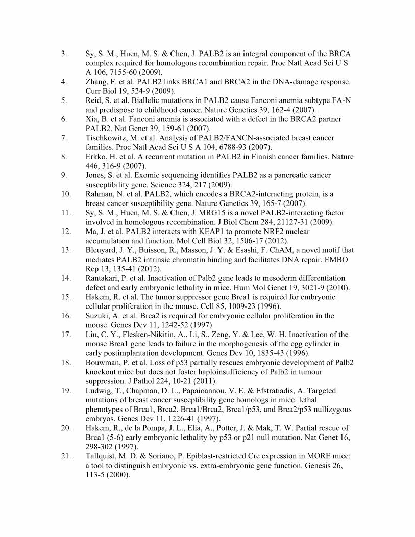
3. Sy, S. M., Huen, M. S. & Chen, J. PALB2 is an integral component of the BRCA complex required for homologous recombination repair. Proc Natl Acad Sci U S A 106, 7155-60 (2009).
4. Zhang, F. et al. PALB2 links BRCA1 and BRCA2 in the DNA-damage response. Curr Biol 19, 524-9 (2009).
5. Reid, S. et al. Biallelic mutations in PALB2 cause Fanconi anemia subtype FA-N and predispose to childhood cancer. Nature Genetics 39, 162-4 (2007).
6. Xia, B. et al. Fanconi anemia is associated with a defect in the BRCA2 partner PALB2. Nat Genet 39, 159-61 (2007).
7. Tischkowitz, M. et al. Analysis of PALB2/FANCN-associated breast cancer families. Proc Natl Acad Sci U S A 104, 6788-93 (2007).
8. Erkko, H. et al. A recurrent mutation in PALB2 in Finnish cancer families. Nature 446, 316-9 (2007).
9. Jones, S. et al. Exomic sequencing identifies PALB2 as a pancreatic cancer susceptibility gene. Science 324, 217 (2009).
10. Rahman, N. et al. PALB2, which encodes a BRCA2-interacting protein, is a breast cancer susceptibility gene. Nature Genetics 39, 165-7 (2007).
11. Sy, S. M., Huen, M. S. & Chen, J. MRG15 is a novel PALB2-interacting factor involved in homologous recombination. J Biol Chem 284, 21127-31 (2009).
12. Ma, J. et al. PALB2 interacts with KEAP1 to promote NRF2 nuclear accumulation and function. Mol Cell Biol 32, 1506-17 (2012).
13. Bleuyard, J. Y., Buisson, R., Masson, J. Y. & Esashi, F. ChAM, a novel motif that mediates PALB2 intrinsic chromatin binding and facilitates DNA repair. EMBO Rep 13, 135-41 (2012).
14. Rantakari, P. et al. Inactivation of Palb2 gene leads to mesoderm differentiation defect and early embryonic lethality in mice. Hum Mol Genet 19, 3021-9 (2010).
15. Hakem, R. et al. The tumor suppressor gene Brca1 is required for embryonic cellular proliferation in the mouse. Cell 85, 1009-23 (1996).
16. Suzuki, A. et al. Brca2 is required for embryonic cellular proliferation in the mouse. Genes Dev 11, 1242-52 (1997).
17. Liu, C. Y., Flesken-Nikitin, A., Li, S., Zeng, Y. & Lee, W. H. Inactivation of the mouse Brca1 gene leads to failure in the morphogenesis of the egg cylinder in early postimplantation development. Genes Dev 10, 1835-43 (1996).
18. Bouwman, P. et al. Loss of p53 partially rescues embryonic development of Palb2 knockout mice but does not foster haploinsufficiency of Palb2 in tumour suppression. J Pathol 224, 10-21 (2011).
19. Ludwig, T., Chapman, D. L., Papaioannou, V. E. & Efstratiadis, A. Targeted mutations of breast cancer susceptibility gene homologs in mice: lethal phenotypes of Brca1, Brca2, Brca1/Brca2, Brca1/p53, and Brca2/p53 nullizygous embryos. Genes Dev 11, 1226-41 (1997).
20. Hakem, R., de la Pompa, J. L., Elia, A., Potter, J. & Mak, T. W. Partial rescue of Brca1 (5-6) early embryonic lethality by p53 or p21 null mutation. Nat Genet 16, 298-302 (1997).
21. Tallquist, M. D. & Soriano, P. Epiblast-restricted Cre expression in MORE mice: a tool to distinguish embryonic vs. extra-embryonic gene function. Genesis 26, 113-5 (2000).
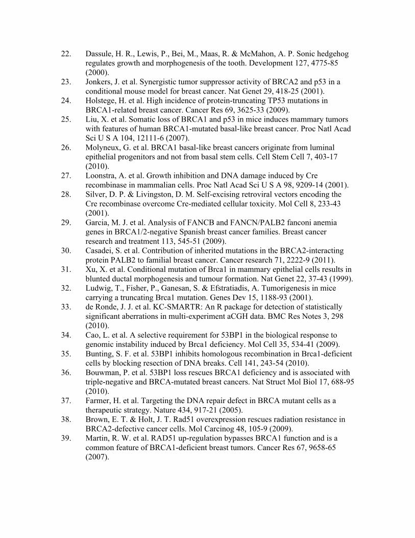
22. Dassule, H. R., Lewis, P., Bei, M., Maas, R. & McMahon, A. P. Sonic hedgehog regulates growth and morphogenesis of the tooth. Development 127, 4775-85 (2000).
23. Jonkers, J. et al. Synergistic tumor suppressor activity of BRCA2 and p53 in a conditional mouse model for breast cancer. Nat Genet 29, 418-25 (2001).
24. Holstege, H. et al. High incidence of protein-truncating TP53 mutations in BRCA1-related breast cancer. Cancer Res 69, 3625-33 (2009).
25. Liu, X. et al. Somatic loss of BRCA1 and p53 in mice induces mammary tumors with features of human BRCA1-mutated basal-like breast cancer. Proc Natl Acad Sci U S A 104, 12111-6 (2007).
26. Molyneux, G. et al. BRCA1 basal-like breast cancers originate from luminal epithelial progenitors and not from basal stem cells. Cell Stem Cell 7, 403-17 (2010).
27. Loonstra, A. et al. Growth inhibition and DNA damage induced by Cre recombinase in mammalian cells. Proc Natl Acad Sci U S A 98, 9209-14 (2001).
28. Silver, D. P. & Livingston, D. M. Self-excising retroviral vectors encoding the Cre recombinase overcome Cre-mediated cellular toxicity. Mol Cell 8, 233-43 (2001).
29. Garcia, M. J. et al. Analysis of FANCB and FANCN/PALB2 fanconi anemia genes in BRCA1/2-negative Spanish breast cancer families. Breast cancer research and treatment 113, 545-51 (2009).
30. Casadei, S. et al. Contribution of inherited mutations in the BRCA2-interacting protein PALB2 to familial breast cancer. Cancer research 71, 2222-9 (2011).
31. Xu, X. et al. Conditional mutation of Brca1 in mammary epithelial cells results in blunted ductal morphogenesis and tumour formation. Nat Genet 22, 37-43 (1999).
32. Ludwig, T., Fisher, P., Ganesan, S. & Efstratiadis, A. Tumorigenesis in mice carrying a truncating Brca1 mutation. Genes Dev 15, 1188-93 (2001).
33. de Ronde, J. J. et al. KC-SMARTR: An R package for detection of statistically significant aberrations in multi-experiment aCGH data. BMC Res Notes 3, 298 (2010).
34. Cao, L. et al. A selective requirement for 53BP1 in the biological response to genomic instability induced by Brca1 deficiency. Mol Cell 35, 534-41 (2009).
35. Bunting, S. F. et al. 53BP1 inhibits homologous recombination in Brca1-deficient cells by blocking resection of DNA breaks. Cell 141, 243-54 (2010).
36. Bouwman, P. et al. 53BP1 loss rescues BRCA1 deficiency and is associated with triple-negative and BRCA-mutated breast cancers. Nat Struct Mol Biol 17, 688-95 (2010).
37. Farmer, H. et al. Targeting the DNA repair defect in BRCA mutant cells as a therapeutic strategy. Nature 434, 917-21 (2005).
38. Brown, E. T. & Holt, J. T. Rad51 overexpression rescues radiation resistance in BRCA2-defective cancer cells. Mol Carcinog 48, 105-9 (2009).
39. Martin, R. W. et al. RAD51 up-regulation bypasses BRCA1 function and is a common feature of BRCA1-deficient breast tumors. Cancer Res 67, 9658-65 (2007).

40. Schild, D. & Wiese, C. Overexpression of RAD51 suppresses recombination defects: a possible mechanism to reverse genomic instability. Nucleic Acids Res 38, 1061-70 (2010).
41. Martins FC et al. Evolutionary pathways in BRCA1-associated breast tumors. Cancer Discov 6, 503-11 (2012).
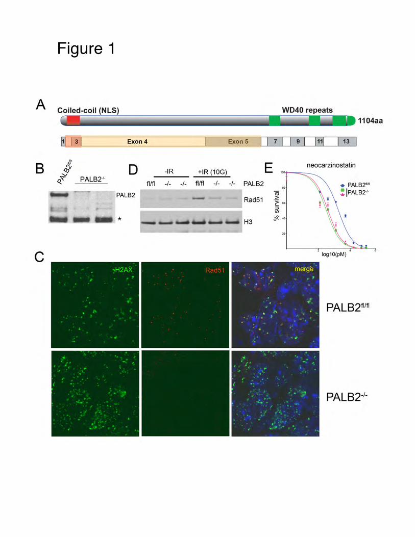
Figure 1!
A
Exon 4
B ~
tE' D -IR +IR (10G) ~ PALB2·'· ~ fl/fl -1- -1- fl/fl -1-- PALB2 -
*
c
1104aa
Exon 5 I I 7 I I 9 I 1111 13
E 100
-1- PALB2 80
(ij
Rad51 > -~ 60
:J (/)
H3 'Cf?. 40
20
neocarzinostatin
+ PALB2MI ! IPALB2-1-
log10(pM)
PALB2flttl
PALB2-t-
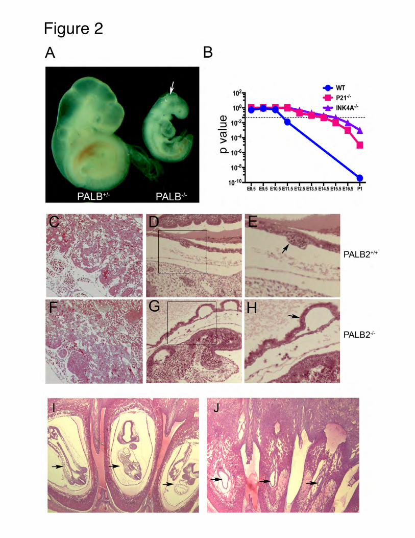
Figure 2!A B
102 + wr • P21·1•
10° + INK4A·1•
a> 10"2
::::J -ro 10"4 > 0..1o·6
E8.5 E9.5 E10.5 E11.5 E12.5 E13.5 E14.5 E15.5 E16.5 P1
PALB2+'+
PALB2 '
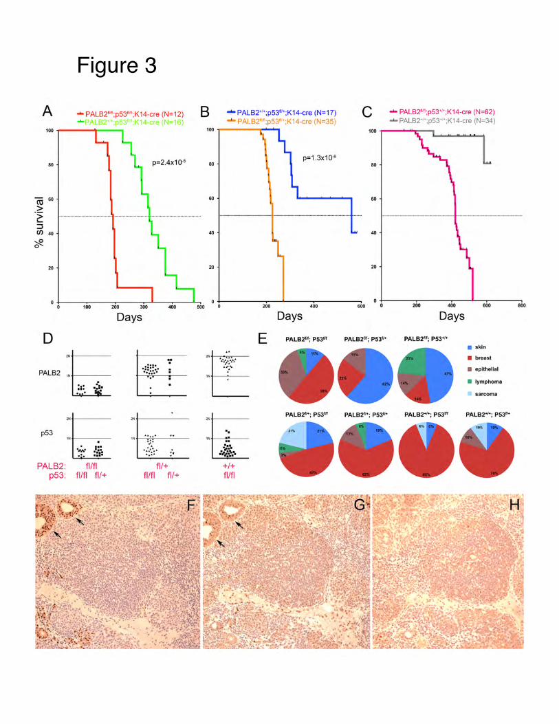
Figure 3!
A .._ PALB211111;p5311111;K14-cre (N=12)
100
eo
ro 60
>
... . I
p=2.4x1 0-5
"2: ······································ ........................ ···································· ::J C/)4l)
~
20
D
PALB2 :EI. ~ ... . . . .
... p53
IN
.,.. ••• .;,;. ... . ... PALB2: fllfl
p53: fl/fl fl/+
Days
. :::::. ······ .. .
:E ·:.: ·:":•:":• :··
fl/+ fl/fl fl/+
B .._ PALB<~'1' ; p531v'; K14-cre (N=17) c ... PALB2111";p53'1' ;K14-cre (N=62} ..._ PALS:!' ";p53' K14 ere (N=35) _.. PALB2·-;p53''';K14-cre (N=34)
100 100
p=1.3x1o.a 80 80
60 60
40 40
20 20
200 400 600 0 +-----,200,...----400-r-- --'-- 600-r----..... 800
Days Days
E PALB2'n; PS311f PALB211f; P53"' PALB2'n; P53'' ' • skin
• breast
• epithelial
• lymphoma
sarcoma
"' PALB2U+; P5311f PALB2U•; P53U• PALB2'~+; P53"' PAL.B2'"; P53U•
.. 1N ..•
·:!:. :,.;.:~.\
+I+ fllfl
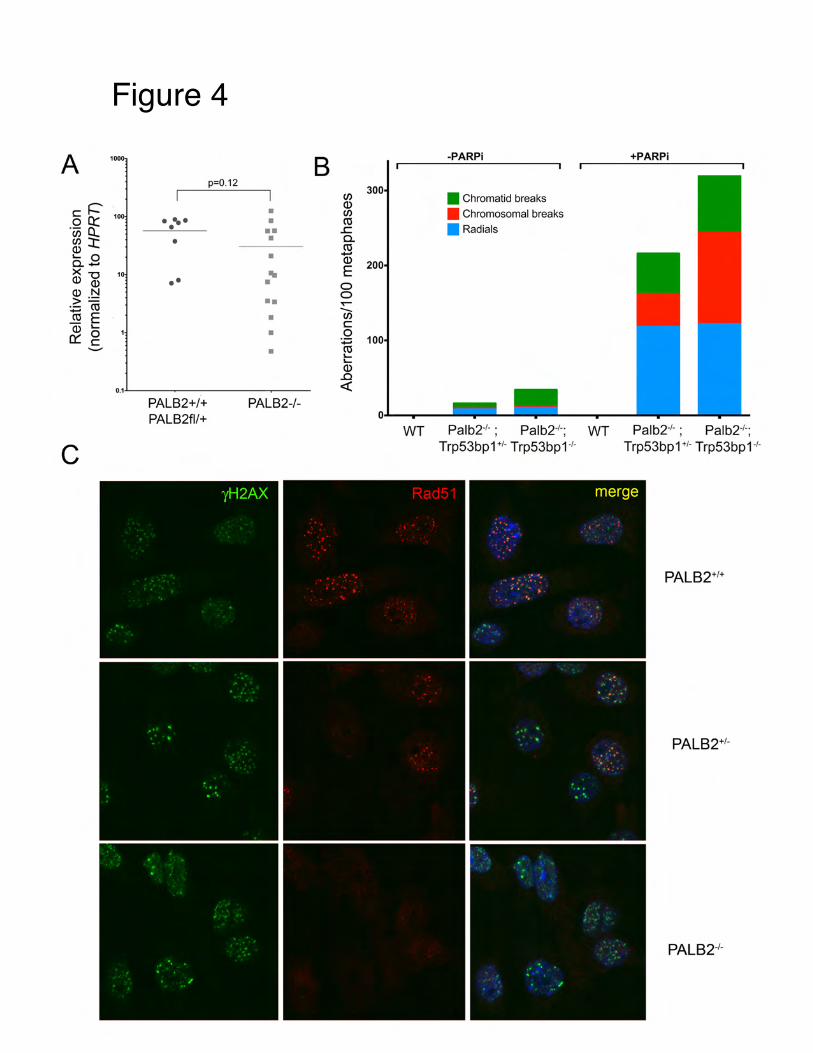
Figure 4!
A
c: ~ 10 oa:: ·u; Q.. CJ):t Q) ..... 0 o..X-o Q) Q)
Q) N >= :o:; ro ro E Q) .....
0:: g
..... •
••
p=0.12
• • •• • •
"' • •• • • •
0.1.1...._ __________ _
c
PALB2+/+ PALB2fl/+
PALB2-/-
B fJ) Q) fJ) ro .c 0.. ro -Q)
E 0 0 T""" -fJ) c: 0
:o:; ro ..... ..... Q) .0 <(
-PARPi
• Chromatid breaks • Chromosomal breaks
• Radials
WT Palb2·'· ; Palb2+; WT Trp53bp1 +'· Trp53bp1 ·'·
Palb2·'· ; Palb2+; Trp53bp1 +'· Trp53bp1 ·'·
PALB2+t+
PALB2+'-
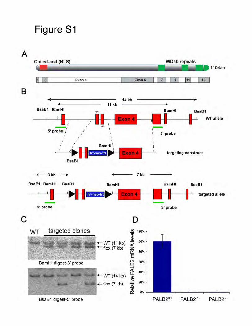
Figure S1!
A
B
c
Exon 4
BsaB1 BamHI
- I 5' probe 1
I
I I
I
I
BsaB1
+-- 3kb ~
BsaB1 BamHI BsaB1
-5' probe
I
I I
I
WT targeted clones
BamHI digest-3' probe
BsaB1 digest-S' probe
1104aa
Exon 5 I I 7 I I 9 I 1111 13
14 kb 11 kb
-
\
I I
Bam HI
~ WT (11 kb) ~ flox (7 kb)
7 kb
D U) 120% Q) > Q)
100% <l: z 0::: 80%
E N 60% co _J
0: 40%
Q) > 20% ~ ro Q) 0::: 0%
I ,-
Bam HI
1 3' probe I I I
BsaB1
WT allele
targeting construct
Bam HI BsaB1
targeted allele
- 3' probe
PALB211m PALB2·1· PALB2·'-
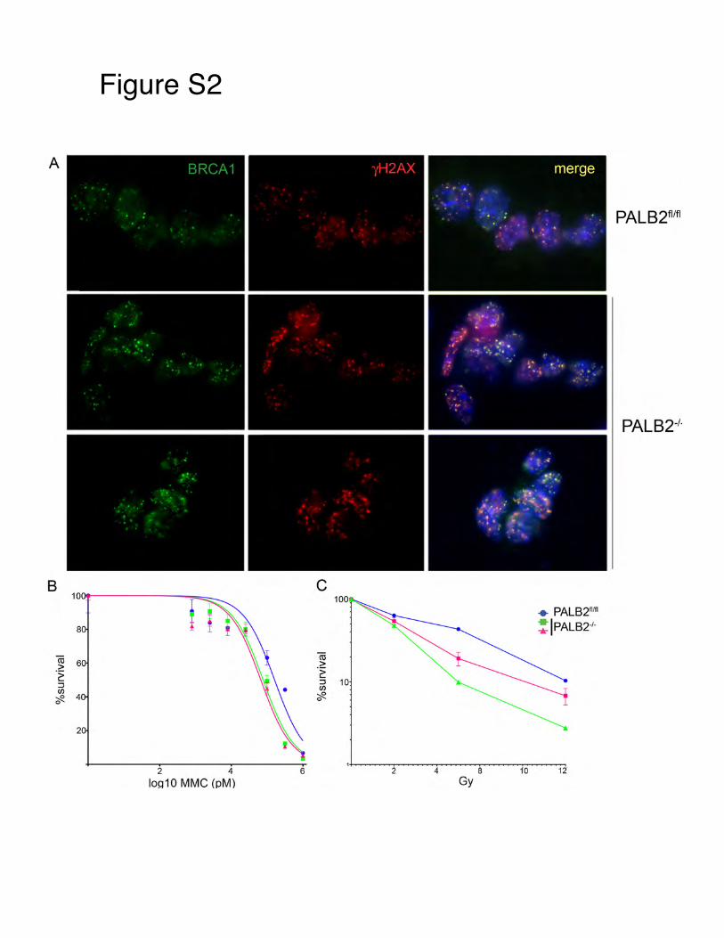
Figure S2!
A
PALB2flm
PALB2·'·
B c
i + PALB2fllfl ! IPALB2"'·
ro > ·~ ::J (/)
10
~ 0
2 4 6 2 4 8 12
log10 MMC (pM) Gy
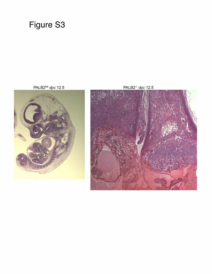
Figure S3!
PALB2fl'fl dpc 12.5
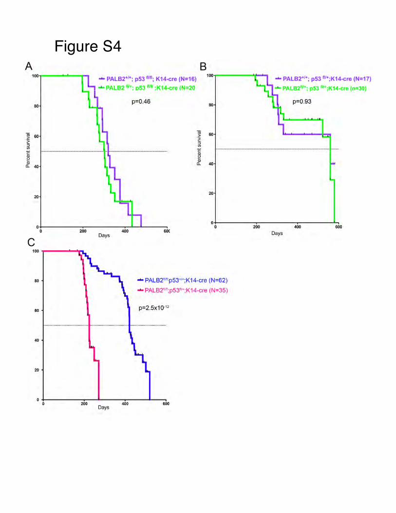
Figure S4!A
60
~ 60 -~ :l
"' c ~ Q)
Q.. 40
20
..._ PALB2+i+; p53 filii ; K14·cre (N=16)
-..PALB2 tlh; D53 lflfl ·Kt4-<:re (N=20
p=0.46
B
80
20
..._ PALB2•1• ; p53 111• ;K14-cre (N=17)
oo~.. PAl.B2111• ; pS3 nr-;K14-efe (n==30)
p=0.93
0+---------~~--------~--------~~ 0+-----------r---------~~~L------, 0
0 200
c Days
80
60
40
20
400 60(
..._ PALB2nlf1;p53•";K14-cre (N=62)
..._ PALB2"111;p53111•;K14-cre (N=35)
p=2.5x10⁻¹²
0 +---------~~~~----~------~--~ 0 200
Days 400 600
200 400 600
Days
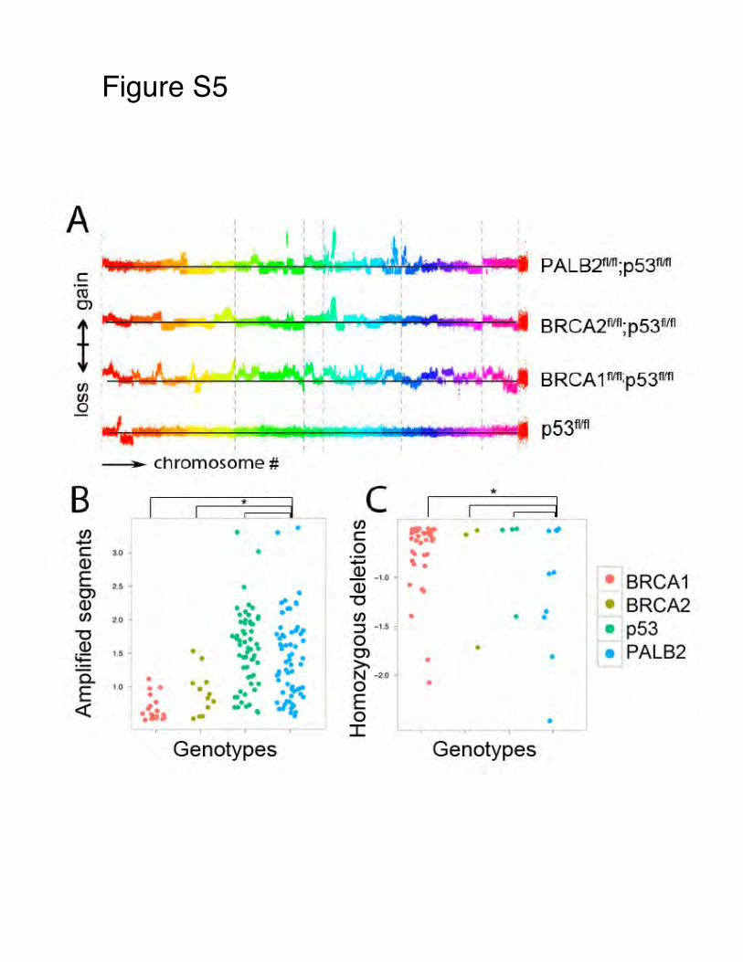
Figure S5!
A I I
'\s~~t t...-t '""'.Jot~ ....... ...,, •• • PALB2tvn;p53wn c:: I ' ro I
I
0> I
rtuww1! I "'..,. ... BRCA2Wfl;p53fltfl
i $ ;« ,.-n?_r I
' ' . ~ BRCA 1rvn:p53r:m
(/) en 0
t4xr~ I
::' ,1;;;; • I . 12 7
' :)lilfU1 p53fllfl
chromosome #
B c * • • C/)
It'! • • ... • • (/) • • c ....... 0 c 3.0 • ...
a.> ....... ' . E a.> ... to .. • BRCA1 ~· ' • a.> ... 0> • a.> ·.:- ,.. "0 • BRCA2 •• • (/) 2.0 :..• C/) • • • . ;. .,, :::J -1.5 - • p53 "0 ...... a.> .. , ..... 0 • PALB2 • ~ 0> • ~ '. - • -1.: • • >. • • f'" a. • ** N ... 0 • • •• • 0 • E 1,0 • • • • ••• •K • '· E <( ~· • -·#. ~ · . .;. •• 0 • I
Genotypes Genotypes
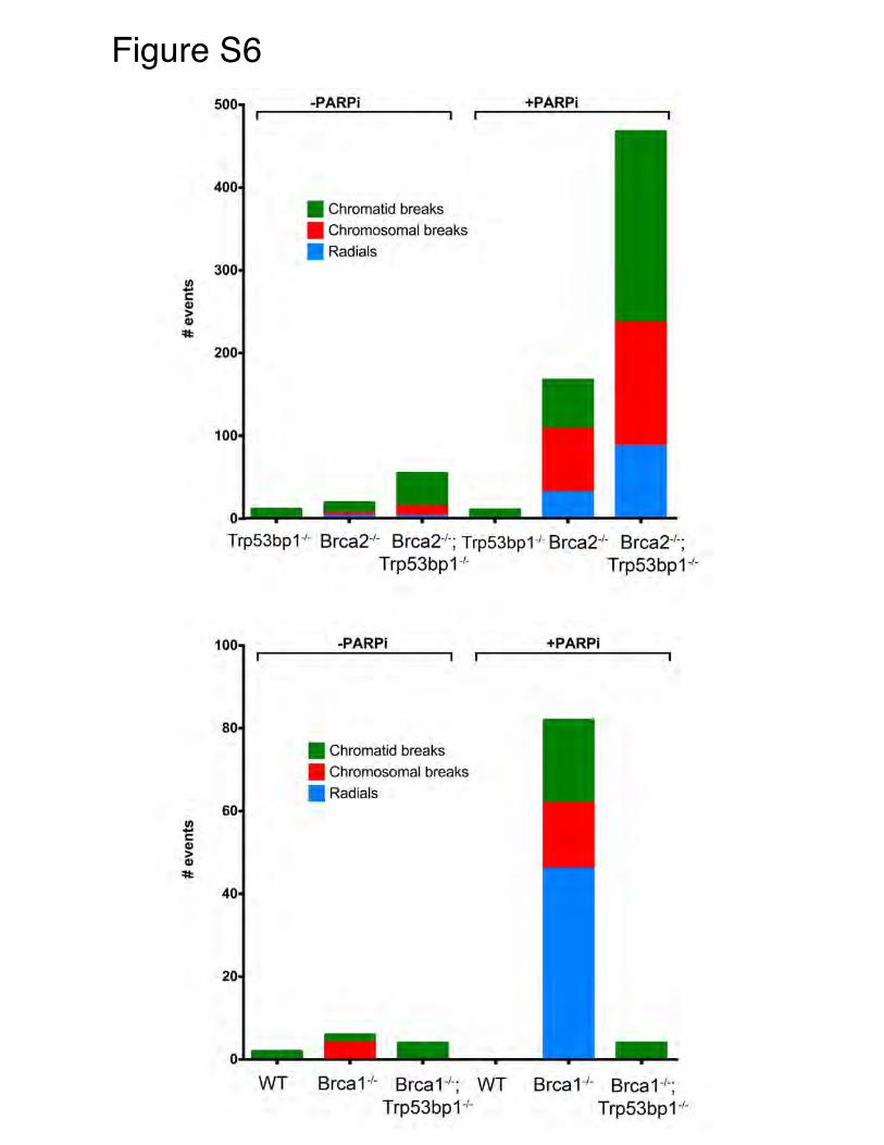
Figure S6!
1/) -c (I)
> (I)
~
1
-PAR Pi +PARPi
• Chromatid breaks
• Chromosomal breaks
• Radials
Trp53bp1 ·'- Brca2·'· Brca2·'·; Trp53bp1·1- Brca2·1- Brca2·'·;
WT
Trp53bp1·'- Trp53bp1 ·'·
-PARPi
• Chromatid breaks
• Chromosomal breaks
• Radials
Brca1 ·1- Brca1·'·; WT Trp53bp1·'-
+PARPi
Brca 1·'- Brca 1·1·; Trp53bp 1·'-