Embed Size (px)
Citation preview

AWARD NUMBER: W81XWH-14-1-0535
TITLE: Cooperativity Between Oncogenic PKC Epsilon and Pten Loss in Prostate Cancer Progression
PRINCIPAL INVESTIGATOR: Marcelo G. Kazanietz, Ph.D.
CONTRACTING ORGANIZATION: Trustees of the University of PennsylvaniaPhiladelphia, PA 19104
REPORT DATE: October 2016
TYPE OF REPORT: Annual
PREPARED FOR: U.S. Army Medical Research and Materiel Command Fort Detrick, Maryland 21702-5012
DISTRIBUTION STATEMENT: Approved for Public Release; Distribution Unlimited
The views, opinions and/or findings contained in this report are those of the author(s) and should not be construed as an official Department of the Army position, policy or decision unless so designated by other documentation.

REPORT DOCUMENTATION PAGE Form Approved
OMB No. 0704-0188 Public reporting burden for this collection of information is estimated to average 1 hour per response, including the time for reviewing instructions, searching existing data sources, gathering and maintaining the data needed, and completing and reviewing this collection of information. Send comments regarding this burden estimate or any other aspect of this collection of information, including suggestions for reducing this burden to Department of Defense, Washington Headquarters Services, Directorate for Information Operations and Reports (0704-0188), 1215 Jefferson Davis Highway, Suite 1204, Arlington, VA 22202-4302. Respondents should be aware that notwithstanding any other provision of law, no person shall be subject to any penalty for failing to comply with a collection of information if it does not display a currently valid OMB control number. PLEASE DO NOT RETURN YOUR FORM TO THE ABOVE ADDRESS. 1. REPORT DATEOctober 2016
2. REPORT TYPEAnnual report
3. DATES COVERED30 Sep 2015 - 29 Sep 2016
4. TITLE AND SUBTITLE 5a. CONTRACT NUMBER
Cooperativity Between Oncogenic PKC Epsilon and Pten Loss in Prostate
Cancer Progression
5b. GRANT NUMBER W81XWH-14-1-0535 5c. PROGRAM ELEMENT NUMBER
6. AUTHOR(S)
5d. PROJECT NUMBER
Marcelo G. Kazanietz, Ph.D. 5e. TASK NUMBER
E-Mail: [email protected]
5f. WORK UNIT NUMBER
7. PERFORMING ORGANIZATION NAME(S) AND ADDRESS(ES)
AND ADDRESS(ES)
8. PERFORMING ORGANIZATION REPORTNUMBER
Trustees of the University of PennsylvaniaPhiladelphia, PA 19104
9. SPONSORING / MONITORING AGENCY NAME(S) AND ADDRESS(ES) 10. SPONSOR/MONITOR’S ACRONYM(S)
U.S. Army Medical Research and Materiel Command
USAMRMC Fort Detrick, Maryland 21702-5012 11. SPONSOR/MONITOR’S REPORT
NUMBER(S)
12. DISTRIBUTION / AVAILABILITY STATEMENT
Approved for Public Release; Distribution Unlimited
13. SUPPLEMENTARY NOTES
14. ABSTRACTThe main objective of our studies is to elucidate the mechanisms by which PKCε, in conjunction with Pten loss, lead to malignant transformation and metastasis, through an autocrine mechanism that involves the release of the chemokine CXCL13. During the first year we acquired new evidence that CXCL13 levels are elevated in prostate cancer cells and that PKCε is causally associated with the elevated production and release of this chemokine. We also initiated studies to dissect the signaling mechanisms that mediate CXCL13 induction. We took advantage of a cellular model that we generated in our laboratory in which PKCε was overexpressed using a lentivirus in a Pten-deficient background. We also remediated the issue of loss of stable PKCε expression in prostate epithelial cell lines by generating a new PKCε lentivirus. Our research may impact on our understanding of the molecular mechanisms of prostate tumorigenesis, and may have significant prognostic and therapeutic implications.
15. SUBJECT TERMSPKCε, Pten, CXCL13, CXCR5, proliferation, migration, tumorigenesis, metastasis, CXCL13 promoter, transcriptional activation, autocrine loop, mouse models. 16. SECURITY CLASSIFICATION OF: 17. LIMITATION
OF ABSTRACT 18. NUMBEROF PAGES
19a. NAME OF RESPONSIBLE PERSONUSAMRMC
a. REPORT
Unclassified
b. ABSTRACT
Unclassified
c. THIS PAGE
Unclassified Unclassified 11
19b. TELEPHONE NUMBER (include area code)
Standard Form 298 (Rev. 8-98) Prescribed by ANSI Std. Z39.18

Table of Contents
Page
1. Introduction…………………………………………………………. 1
2. Keywords……………………………………………………………. 1
3. Accomplishments………..…………………………………………... 1
4. Impact…………………………...…………………………………… 6
5. Changes/Problems...….……………………………………………… 7
6. Products…………………………………….……….….……………. 7
7. Participants & Other Collaborating Organizations………………. 8
8. Special Reporting Requirements…………………………………… 8
9. Appendices…………………………………………………………… 8
1
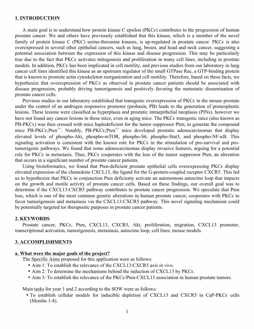
1. INTRODUCTION
A main goal is to understand how protein kinase C epsilon (PKCε) contributes to the progression of human prostate cancer. We and others have previously established that this kinase, which is a member of the novel family of protein kinase C (PKC) serine-threonine kinases, is up-regulated in prostate cancer. PKCε is also overexpressed in several other epithelial cancers, such as lung, breast, and head and neck cancer, suggesting a potential association between the expression of this kinase and disease progression. This may be particularly true due to the fact that PKCε activates mitogenesis and proliferation in many cell lines, including in prostate models. In addition, PKCε has been implicated in cell motility, and previous studies from our laboratory in lung cancer cell lines identified this kinase as an upstream regulator of the small GTPase Rac, a GTP-binding protein that is known to promote actin cytoskeleton reorganization and cell motility. Therefore, based on these facts, we hypothesize that overexpression of PKCε as observed in prostate cancer patients should be associated with disease progression, probably driving tumorigenesis and positively favoring the metastatic dissemination of prostate cancer cells.
Previous studies in our laboratory established that transgenic overexpression of PKCε in the mouse prostate under the control of an androgen responsive promoter (probasin, PB) leads to the generation of preneoplastic lesions. These lesions were classified as hyperplasia and prostatic intraepithelial neoplasia (PINs), however we have not found any cancer lesions in these mice, even in aging mice. The PKCε transgenic mice (also known as PB-PKCε) was then crossed with mice haplodeficient for the tumor suppressor Pten, to generate the compound mice PB-PKCε;Pten+/-. Notably, PB-PKCε;Pten+/- mice developed prostatic adenocarcinomas that display elevated levels of phospho-Akt, phospho-mTOR, phospho-S6, phospho-Stat3, and phospho-NF-κB. This signaling activation is consistent with the known role for PKCε in the stimulation of pro-survival and pro-tumorigenic pathways. We found that some adenocarcinomas display invasive features, arguing for a potential role for PKCε in metastasis. Thus, PKCε cooperates with the loss of the tumor suppressor Pten, an alteration that occurs in a significant number of prostate cancer patients.
Using bioinformatics, we found that Pten-deficient prostate epithelial cells overexpressing PKCε display elevated expression of the chemokine CXCL13, the ligand for the G-protein-coupled receptor CXCR5. This led us to hypothesize that PKCε in conjunction Pten deficienty activate an autonomous autocrine loop that impacts on the growth and motile activity of prostate cancer cells. Based on these findings, our overall goal was to determine if the CXCL13:CXCR5 pathway contributes to prostate cancer progression. We speculate that Pten loss, which is one of the most common genetic alterations in human prostate cancer, cooperates with PKCε to favor tumorigenesis and metastasis via the CXCL13:CXCR5 pathway. This novel signaling mechanism could be potentially targeted tor therapeutic purposes in prostate cancer patients.
2. KEYWORDSProstate cancer, PKCε, Pten, CXCL13, CXCR5, Akt, proliferation, migration, CXCL13 promoter,
transcriptional activation, tumorigenesis, metastasis, autocrine loop, cell lines, mouse models.
3. ACCOMPLISHMENTS
a. What were the major goals of the project?The Specific Aims proposed for this application were as follows: • Aim 1: To establish the relevance of the CXCL13:CXCR5 axis in vivo.• Aim 2: To determine the mechanisms behind the induction of CXCL13 by PKCε.• Aim 3: To establish the relevance of the PKCε/Pten-CXCL13 association in human prostate tumors.
Main tasks for year 1 and 2 according to the SOW were as follows: • To establish cellular models for inducible depletion of CXCL13 and CXCR5 in CaP-PKCε cells
(Months 1-8).

2
• To assess the effect of CXCL13 and CXCR5 inducible silencing on the tumorigenic activity of CaP-PKCε cells (Months 3-8).
• To assess the effect of CXCL13 and CXCR5 inducible silencing on metastasis (Months 8-14). • To determine if CXCL13 and CXCR5 are required for the formation of preneoplastic lesions in PB-
PKCε mice (Months 1-36). • To establish the signaling pathways involved in PKCε-induced release of CXCL13 from prostate cells
(Months 14-20). • To determine if PKCε-mediated CXCL13 induction involves transcriptional activation of the CXCL13
promoter (Months 21-27). • To determine the expression of PKCε and Pten in human prostate tumors (Months 16-25)
b. What was accomplished under these goals?
During the second year of funding, we continued our work on the role of the CXCL13:CXCR5 axis in prostate cancer progression. We had previously generated prostate epithelial cell lines that overexpress PKCε. However, as described in our previous annual report, these cell lines unexpectedly lost the expression of PKCε, even if they were stable cell lines. As indicated last year, we had to construct again the PKCε lentivirus and subsequently generate the corresponding cell lines, and this problem caused a significant delay in our research during the first year. Nevertheless, we solved this issue and successfully generated cell lines again.
In total we had 8 isogenic cell lines derived from parental P2 and P8 cells. Cell lines overexpressing PKCε were named P2-PKCε and P8-PKCε. Pten-deficient cell lines (CaP2 and CaP8) overexpressing PKCε were named CaP2-PKCε and CaP8-PKCε. For validation purposes, we confirmed again that Pten-deficient cell lines overexpressing PKCε display the previously observed phenotype (enhanced motile activity and signaling, specifically Akt and Erk activation in response to growth factors). Establishing a role for the CXCL13:CXCR5 axis in growth and tumorigenesis:
Since PKCε overexpression and Pten loss individually and synergically promote the secretion of CXCL13 from prostate epithelial cells, and considering that CXCL13 or CXCR5 RNAi impairs the growth and motile capacity of CaP8-PKCε cells in culture, we speculated that the phenotype driven by PKCε in vivo is dependent on CXCL13 and CXCR5. Although the original plan involved the use of an inducible silencing approach, we decided to initiate the experiments using what we had proposed as an alternative approach, which was the use of non-inducible silencing by means of shRNA lentiviruses.
First, we infected P8, P8-PKCε, CaP8, and CaP8-PKCε cells with different shRNA lentiviruses for CXCR5, and selected with puromycin. As a control, we used a “non-target control” (NTC) shRNA lentivirus. We achieved significant silencing of CXCR5 in all for cell lines (Fig. 1).
CaP8-PKCε cells, which display the highest growth rate in culture, significantly reduced their proliferative capacity upon CXCR5 RNAi depletion. A comparable inhibitory effect of CXCR5 RNAi was observed in cells overexpressing PKCε (P8-PKCε) or lacking Pten (CaP8) (Fig. 2).
F i g . 1 . C X C R 5 R N A i deplet ion from murine prostate epithelial cell lines. P8, P8-PKCε, CaP8, and C a P 8 - P K C ε c e l l s w e r e infected with either CXCR5 or non-target control (NTC) shRNA len t i v i ruses and selected with puromycin. CXCR5 express ion was determined by Western blot.

3
We then assessed the effect of knocking down CXCR5 on the ability of cells to grow in soft agar. Consistent with experiments in liquid media, we found a significant reduction in the formation of colonies in soft agar in CaP8-PKCε cells subject to CXCR5 RNAi (Fig. 3).
We carried out similar experiments in cells subject to CXCL13 depletion. We used shRNA lentivirus for
CXCL13. We observed that stable silencing of CXCL13 expression impaired the ability of CaP8-PKCε cells to grow in soft agar (Fig. 4).
Fig. 2. Proliferation of P8, P8-PKCε, CaP8, and CaP8-PKCε cells infected with either CXCR5 or NTC shRNA lentiviruses. Cell growth was determined using the MTT assay 1, 2, and 3 days after seeding.
Fig. 3. Effect of CXCR5 RNAi on anchorage-independent growth of CaP8-PKCε cells in soft agar. Top: representative experiment. Bottom left: quantification of colonies/field. *, p< 0.05. Bottom right: Western blot for CXCR5 expression.
Fig. 4. CXCL13 depletion impairs anchorage-independent growth. CaP8-PKCε cells were infected with CXCL13 or non-target control (NTC) shRNA lentiviruses and selected with puromycin. Left: CXCL13 protein levels in the culture medium, as determined by ELISA. Results normalized to cells stably transduced with NTC shRNA are expressed as mean ± S.D. of triplicate measurements. *, p< 0.05. Middle: Representative images of growth in soft agar. Right: quantification of colonies/field. *, p< 0.05.

4
In order to assess the effect of CXCR5 and CXCL13 depletion on the tumorigenic activity of CaP8-PKCε cells, we used athymic nude mice. CaP8-PKCε cells (parental, NTC, CXCL13 shRNA, and CXCR5 shRNA, using two different shRNAs in each case) were inoculated s.c. into ~8-week-old athymic male nude mice. Tumor growth was monitored for 35 days. As shown in Fig. 5, CaP8-PKCε cells readily formed tumors in nude mice. However, when cells were infected with shRNA lentiviruses for either CXCL13 or CXCR5, tumor growth was markedly reduced. Therefore, the tumorigenic activity of CaP8-PKCε cells is dependent upon the expression of CXCL13 and CXCR5, thus underscoring the requirement of an autocrine CXCR5-CXCL13 axis in growth and tumorigenic activity of these cells.
Establishing a role for the CXCL13:CXCR5 axis in signaling and metastasis:
Metastatic cells display highly motile and invasive phenotypes, and there is a known activation of the Rac pathway that correlates with motility and metastasis. Based on our preliminary data, PKCε overexpression and Pten loss individually and synergically confer highly motile and invasive properties to prostate epithelial cells. Indeed, CaP8-PKCε cells display a very high migratory capacity in Boyden chambers, both in the absence and presence of Matrigel. We hypothesize that the high motile/invasive/metastatic capacity of CaP8-PKCε cells is mediated by the CXCL13:CXCR5 axis. We had previously showed that conditioned medium (CM) collected from prostate epithelial cells overexpressing PKCε and/or Pten depleted confers a migratory response when added to naïve P8 cells. Indeed, we had indicated that the activity of the different CM varied as CM-CaP8-PKCε > CM-P8-PKCε ~ CM-CaP8 > CM-P8 cells, which was proportional to the CXCL13 production that we found in each cell line.
To determine if this pro-migratory effect is causally related to the released CXCL13, CaP8-PKCε cells were subject to CXCL13 RNAi depletion, which as expected reduced CXCL13 mRNA levels (as determined by Q-PCR) and protein release (as determined by ELISA). Notably, CM from CXCL13-depleted CaP8-PKCε cells lost its ability to induce a migratory response when added to naïve P8 cells. This effect was rescued by exogenous addition of CXCL13 (Fig. 6).
Fig. 5. Effect of CXCR5 and CXCL13 RNAi on tumorigenesis. CaP8-PKCε cells were infected with CXCR5, CXCL13 or non-target control (NTC) shRNA lentiviruses and selected with puromycin. Left: representative pictures of nude mice 35 days after s.c. inoculation. Right: Tumor volume, expressed as mean ± S.D. (n= 5 mice/group).
Fig. 6. Effect of CXCL13 RNAi on migratory activity of conditioned medium (CM) from CaP8-PKCε cells. CM was collected from CaP8-PKCε cells subject to either CXCL13 or non-target control (NTC) RNAi. Left: CXCL13 mRNA levels in CaP8-PKCε cells (Q-PCR) and CXCL13 protein levels in CM-CaP8-PKCε cells (ELISA). Results normalized to NTC were expressed as mean ± S.D. (n=3). Middle: P8 cell migration was determined after treatment with CM from CaP8-PKCε cells subject to either CXCL13 or NTC RNAi. Reconstitution with exogenous recombinant CXCL13 (100 ng/ml) added to the CM was done where indicated. Right: Quantification of migrating cells. *, p< 0.05 and **, p< 0.01.

5
Similar results were observed when CM from CXCL13-depleted CaP8-PKCε cells was added to either P8-PKCε or CaP8 cells (Fig. 7).
Notably, when CXCR5 expression in the recipient naïve P8 cells was silenced, the pro-migratory activity of
CM-CaP8-PKCε was essentially lost (Fig. 8), further hinting at the important role of CXCL13. To further establish if an autocrine CXCL13:CXCR5 loop mediates effects driven by PKCε overexpression
and Pten loss, we examined the effect of silencing either CXCL13 or CXCR5 on the migratory capacity of CaP8-PKCε cells. Notably, in cells subject to CXCL13 RNAi the migratory activity of CaP8-PKCε cells was essentially abolished (Fig. 9).
These results were validated using a neutralizing anti-murine CXCL13 antibody, which dose-dependently inhibited migration of CaP8-PKCε cells compared to a non-specific isotype control antibody (Fig. 10).
In addition, when we silenced CXCR5 expression from PKCε overexpressing and/or Pten-depleted cells,
migratory activity was inhibited (Fig. 11). Thus, enhanced production of CXCL13 as a consequence of PKCε overexpression and/or Pten loss plays a role in driving the migratory phenotype of CaP8-PKCε cells. This also suggests that CXCL13 and CXCR5 may have an important role in metastatic dissemination.
Fig. 7. Conditioned medium (CM) from cells with PKCε overexpression or Pten loss induces migration. P8-PKCε and CaP8 cell migration was determined after treatment with CM from CaP8-PKCε cells subject to either CXCL13 or NTC RNAi. Reconstitution with exogenous recombinant CXCL13 (100 ng/ml) added to the CM was done where indicated. Left: representative images. Middle: Quantification of migrating CaP8 cells. Right: Quantification of migrating P8-PKCε cells. *, p< 0.05.
Fig. 8. Effect of CXCR5 RNAi depletion in P8 cells on the migratory activity of conditioned medium (CM) from CaP8-PKCε cells. P8 cells subjected to either CXCR5 or NTC RNAi were treated with CM collected from CaP8-PKCε cells. Left: CXCR5 mRNA levels in P8 (recipient) cells were determined by Q-PCR. Results normalized to NTC were expressed as mean ± S.D. (n=3). Middle: representative images. Right: Quantification of migrating cells. *, p< 0.05.
Fig. 9. Effect of CXCL13 RNAi on migratory activity of CaP8-PKCε cells. Migration of CaP8-PKCε cells subject to either CXCL13 or non-target control (NTC) RNAi. Left: representative images. Right: Quantification of migrating cells. *, p< 0.05.
Fig. 10. Effect of an anti-CXCL13 neutralizing antibody on the migratory activity of CaP8-PKCε cells. Cells were incubated with either a neutralizing anti-murine CXCL13 antibody or a non-specific isotype control antibody, and migration assessed using a Boyden chamber. Left: representative images. Right: Quantification of migrating cells. *, p< 0.05 and **, p< 0.01.

6
Generation of PB-PKCε;CXCL13-/- and PB-PKCε;CXCR5-/- mice:
A main objective was to determine if CXCL13 and CXCR5 were required for the formation of preneoplastic lesions in PB-PKCε mice. We had previously reported that expression of PKCε in the mouse prostate confers preneoplastic lesions. Therefore, our goal was to generate mouse models of prostate-specific PKCε overexpression in CXCL13- or CXCR5-null backgrounds.
We ordered two mouse strains for CXCL13 and CXCR5 deficiency from the Jackson Laboratory: B6.129X1-Cxcl13tm1Cys/J and B6.129S2(Cg)-Cxcr5tm1Lipp/J, respectively. The CXCR5-deficient mice has been delivered and we are currently expanding the colony. Regarding the CXCL13-deficient mice, cryorecovery was needed, which delayed the shipping of the mice. This last strain recently arrived to the laboratory, and we are currently in the process of expanding and genotyping the mice.
Assessment of the role of PKCε in metastatic dissemination: In our laboratory we were characterizing the role of PKCε in metastasis, particularly to the bone, which is a preferred site for metastasis of prostate cancer cells. We optimized a model for studying bone metastasis using PC3-ML cells, which we hope now to adapt to CaP8-PKCε cells. Importantly, we established that PKCε is required for the formation of bone metastatic foci, which was determined by silencing PKCε from PC3-ML cells. This model could also be adapted to assess the role for CXCL13 and CXCR5 in metastasis, which would help us to dissect the role of this pathway in the context of PKCε overexpression and Pten loss. c. What opportunities for training and professional development has the project provided?
Nothing to report. d. How were the results disseminated to communities of interest?
The P.I. presented results related to this project at several venues, as follows: 1. FASEB Summer Research Conference “Cell Signaling in Cancer: from mechanisms to therapy”, Steamboat Springs , CO. June 2016. 2. Universidad de La Plata, Argentina. June 2016. 3. University of Cambridge , Cambridge, UK. August 2016. 4. Tel Aviv University, Tel Aviv, Israel. August 2016 5. Cancer Spring Symposium from the Chilean Advanced Center for Chronic Diseases, Olmue, Chile. September 2016. 4. IMPACT a. What was the impact on the development of the principal discipline(s) of the project?
Although CXCL13 has been primarily studied in the context of B cell migration, lymphoma, HIV and autoimmune diseases, emerging evidence indicates that this chemokine can be produced by prostate cancer cells. As we established that PKCε overexpression and Pten deficiency induce the expression of CXCL13 in prostate epithelial cells and prostate cancer cells produce significant levels of this chemokine, we believe that our studies can have significant impact not only in the understanding of the molecular basis of the disease but
Fig. 11. Effect of CXCR5 RNAi on the migratory activity of prostate epithelial cells with PKCε overexpression and/or Pten loss. P8, P8-PKCε, CaP8, and CaP8-PKCε cells infected with either CXCR5 or NTC shRNA lentiviruses were assessed for migration in Boyden chambers. Left: representative images. Right: Quantification of migrating cells. *, p< 0.05 and **, p< 0.01.

7
also in therapeutics, as CXCR5 antagonists could be potentially inhibitors of prostate cancer cell growth and metatatic dissemination. It is also conceivable that CXCL13 could be used as a prognostic marker for prostate cancer progression.
b. What was the impact on other disciplines?
In the last years there were several studies suggesting potential roles for CXCL13 and its receptor CXCR5 in the progression of cancer, not only prostate cancer. For example, CXCL13 seems to have a role in breast and head and neck cancer. Therefore, our studies may reveal an important pathway that is regulated by PKCε overexpression and Pten loss, which are alterations that also occur in other cancers, and this certainly can be translated into the development of novel therapeutic approaches for a broad number of malignancies. It is also important to notice that recent studies suggested potential roles for CXCL13 produced by the tumor microenvironment, thus our studies may be expanded to learn important novel signaling interactions that contribute to cancer progression through non-autonomous mechanisms. c. What was the impact on technology transfer?
Nothing to report. d. What was the impact on society beyond science and technology?
Nothing to report. 5. CHANGES/PROBLEMS a. Changes in approach and reasons for change
As described in our first annual report, we had found unexpected issues in the maintenance of the stable expression of PKCε in CaP8-PKCε cells. It took a while to solve this problem, and due to the delay that this caused we estimate that it would be very hard to complete all the proposed work in the estimated time-frame for this proposal (3 years). Therefore, our goal now is to focus our experimental efforts and allocate resources to address key questions in the remaining time for this grant. Hopefully this approach will provide us significant information on fundamental questions that should address the potential role of the CXCL13:CXCR5 pathway in prostate cancer progression, particularly tumorigenesis and aspects of the metastatic cascade, such as migration. One approach to save time was the use of traditional RNAi approaches rather than inducible, as this last methodology is more laborious, expensive, and time-consuming. Indeed, we achieved great success with non-inducible RNAi and were able to address very imporant experimental questions. Due to the long-term nature of some of our proposed experiments, such as studies involving animal crossings, metastasis in mice and human specimens, we hope to focus primarily on short-term questions, such as those involving the analysis of signaling mechanisms. b. Actual or anticipated problems or delays and actions or plans to resolve them
This has been explained above. c. Changes that had a significant impact on expenditures
No significant changes. d. Significant chages in use or care of human subjects, vertebrates, biohazards, and/or selecte agents.
Nothing to report. 6. PRODUCTS
Gutierrez-Uzquiza, A., Lopez-Haber, C., Jernigan, D.L., Fatatis, A., Kazanietz, M.G. PKCε is an essential
mediator of prostate cancer bone metastasis. Mol Cancer Res. 13:1336-1346 (2015).

8
7. PARTICIPANTS AND OTHER COLLABORATING ORGANIZATIONS a. What individuals have worked on the project?
There are no changes with regards to personnel. Personnel includes Marcelo G. Kazanietz (P.I.), Michael Feldman (Co-investigator), Rachana Garg (Research Associate), and Cynthia Lopez-Haber (Technician). b. Has there been a change in the active other support of the PD/PI(s) or senior key personnel since the last reporting period?
The P.I. has been awarded a new R01 project during the last year: R01 CA196232 (P.I. Kazanietz), NCI, NIH (01/16-12/20) “Effectors of protein kinase C-mediated tumor progression”. The main goal in this proposal is to characterize COX-2 and mPGES-1 as PKC effectors in prostate cancer, using cellular and animal models. Pharmacological and inducible shRNA approaches will be used to determine if COX-2 is required for the tumorigenicity of PKCε overexpressing/Pten depleted prostate epithelial cells. The role of individual p110 PI3Ks in COX-2/mPGES-1/PGF2 induction will be studied. There is no overlap with current proposal. c. What other organizations were involved as partners?
Nothing to report. 8. SPECIAL REPORTING REQUIREMENTS
Not applicable. 9. APPENDICES Not applicable.