Embed Size (px)
Citation preview

1
Antibodies: Role in MS and Relationship to Triggers
Antibodies: Role in MS and Relationship to Triggers
Jeffrey L. Bennett, MD, PhD
ProfessorDepartments of Neurology and Ophthalmology
University of Colorado Denver
Jeffrey L. Bennett, MD, PhD
ProfessorDepartments of Neurology and Ophthalmology
University of Colorado Denver
B Cells and Antibody in MS LesionsB Cells and Antibody in MS Lesions
Type II: Mixed T and B cell infiltrate
• ~ 50% Lesions of RRMS lesions
Ig-Complement deposition• Hallmark of active pathology
Type II: Mixed T and B cell infiltrate
• ~ 50% Lesions of RRMS lesions
Ig-Complement deposition• Hallmark of active pathology
Acute ON Nerve: IgG DepositionOptic Nerve Biopsy
Acute ON Nerve: IgG DepositionOptic Nerve BiopsyAcute MS Plaque: IgG Deposition
Brain BiopsyAcute MS Plaque: IgG Deposition
Brain Biopsy

2
Oligoclonal IgG in MS CSFOligoclonal IgG in MS CSF
Kabat et al. (1942)• Elevated amount of antibodies in MS CSF
Yahr et al. (1954)• Intrathecal synthesis
Link (1967)• Oligoclonal bands (OCB)
Kabat et al. (1942)• Elevated amount of antibodies in MS CSF
Yahr et al. (1954)• Intrathecal synthesis
Link (1967)• Oligoclonal bands (OCB)
pH
9.3
8.6
8.0
6.0
4.5
Cry
ptoc
occa
lm
enin
gitis
MS
Ser
um
MS
CS
F
Nor
mal
Oligoclonal IgGSensitive marker of disease
95% of MS patientsOCBs do not disappear
Present in all stages of disease
Oligoclonal IgGDisease Directed Against Reference
Subacute sclerosing panencephalitis Measles virus Vandvik et al., 1976
Cryptococcal meningitis Cryptococcus Porter et al., 1977
Mumps meningitis Mumps virus Vandvik et al., 1978
Chronic rubella panencephalitis Rubella virus Coyle et al., 1981
(HSV) encephalitis HSV glycoprotein B Grimaldi et al., 1988
Progressive multifocal JC virus Sindic et al., 1997leukoencephalopathy
Neurosyphilis Treponema pallidum Vartdal et al., 1981
VZV vasculopathy VZV Burgoon et al., 2003
Theiler’s virus-induced demyelination Theiler’s virus Roos et al., 1987
Theiler’s virus Pachner et al., 2007
Coronavirus-induced demyelination JHM coronavirus Dorries et al., 1987
Oligoclonal Bands in Neurologic DiseaseOligoclonal Bands in Neurologic Disease
Multiple Sclerosis Negative for major myelin proteins and common virusesNegative for major myelin proteins and common viruses

3
What is the Nosology of CNS Demyelinating Disorders?
What is the Nosology of CNS Demyelinating Disorders?
CNS
Demyelinating Disease
CNS
Demyelinating Disease
Multiple SclerosisMultiple
Sclerosis
Neuromyelitis Optica
Neuromyelitis Optica
Schilder
Disease
Schilder
DiseaseMarburg DiseaseMarburg Disease
BaloConcentric Sclerosis
BaloConcentric Sclerosis
Multiple Sclerosis
Schilder’sDisease
Marburg Disease
BaloConcentric Sclerosis
Neuromyelitis Optica
A Candidate Approach?Myelin Proteins
MOGMBPPLPαB-crystallin
Cell Adhesion Moleculesneurofascincontactin
Glycolipidsgalactocerebrosidessulfatides
Foreign Antigens“God, Collings, I hate to start a Monday with a case like this.”
EAE

4
Reindl et al. J Neuroimmunol 180:50 (2006).
Reindl et al. J Neuroimmunol 180:50 (2006).
Frequency of Anti-Myelin Antibodies Are Assay Dependent

5
Myelin Antigen Arrays: High Throughput Antigen Screening
Robinson et al. Nat Biotechnol 21:1033 (2003).
Enhanced Dynamic Range
Epitope Spreading in EAEMyelin Proteome Microarray
Myelin Antigen Arrays: Application in Multiple Sclerosis
Quintana et al. Neurology 78:532 (2012).
• 334 myelin and inflammation related CNS Ags• CSF: MS vs. OND (non-inflammatory controls)• 115 Ags > 3SD over OND mean; 29 with ↑ freq
• Antibody Index: Ag (CSF/Ser) / Albumin (CSF/Ser)• Suggestive of intrathecal synthesis

6
Immunoprecipitation• Serum Ab; 2D PAGE• Proteomics
Kir 4.1• inwardly rectifying K(+) channel• Astrocytes and oligodendrocytes-Kir4.1 Ab
– 47% of MS patients– 0.9% of OND
Immunoprecipitation• Serum Ab; 2D PAGE• Proteomics
Kir 4.1• inwardly rectifying K(+) channel• Astrocytes and oligodendrocytes-Kir4.1 Ab
– 47% of MS patients– 0.9% of OND
Antibody Index (AI)• AI > 2 considered intrathecal synthesis• 3/30 positive
Antibody Index (AI)• AI > 2 considered intrathecal synthesis• 3/30 positive
FACS Sort of Normal CSF
CD19+
CD3 +
T Cells (CD3)
B C
ells
(C
D1
9)

7
FACS: CSF B and Plasma Cells in ON, MS & NMOFACS: CSF B and Plasma Cells in ON, MS & NMO
Light Scattering
CD19+ B Cell Population104
CD19
3.7% 0.4%
88.7%7.2%
CD3
100
101
102
103
100 101 102 103 104
FITC Comp
PE
Co
mp
CD138+ Plasma Cell Population
CD138
CD3
3.5%
6.7%
0.1%
89.7%
100
101
102
103
104
100 101 102 103 104
FITC Comp
PE
Co
mp
0 64 128 192 2560
64
128
192
256
FSC
SS
C
• Recover Ab-secreting B cells (plasma cells) from CSF • Clone and produce rAbs that duplicate their Ag specificity
CSF as a Surrogate to Study Humoral Immunity in CNS Demyelinating Disease
B cells cultured from MS CSF produce oligoclonal bands
(CSF Cells) Culture in vitro Oligoclonal banding
CS
F
(
)IEF
Strategy
Nat Med, vol. 14, pp. 688–693, (2008).

8
Isolation of Single Plasma or B Cells by FACS
Patient CSF
Pellet CSF cellsand label with conjugate antibodies
CD19-APCCD138-PECD3-FITCCD14-APC cy7
FACS
Deposit single CD138+ CD3- plasma cell
Reverse Transcriptase PCR and PCR Amplification
RT-PCR
LEADER V-REGION D J CH1
1 2 3FR 1 FR 3
Primary PCR
Nested PCR
CH3
VH Leader 1 CH1
cDNA Synthesis
Fr1 CH2
VH PCR Products
Vk PCR Products
Electrophoresis of PCR product
DNA Sequence Positive VH and Vk products
9
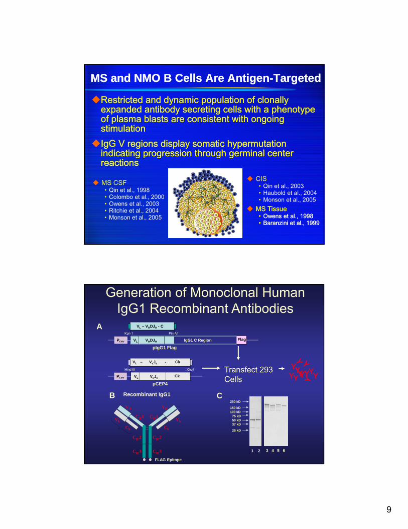
MS and NMO B Cells Are Antigen-TargetedMS and NMO B Cells Are Antigen-Targeted
Restricted and dynamic population of clonally expanded antibody secreting cells with a phenotype of plasma blasts are consistent with ongoing stimulation
IgG V regions display somatic hypermutation indicating progression through germinal center reactions
Restricted and dynamic population of clonally expanded antibody secreting cells with a phenotype of plasma blasts are consistent with ongoing stimulation
IgG V regions display somatic hypermutation indicating progression through germinal center reactions
MS Tissue• Owens et al., 1998• Baranzini et al., 1999
MS Tissue• Owens et al., 1998• Baranzini et al., 1999
MS CSF• Qin et al., 1998• Colombo et al., 2000• Owens et al., 2003• Ritchie et al., 2004• Monson et al., 2005
CIS• Qin et al., 2003• Haubold et al., 2004• Monson et al., 2005
C
Transfect 293 Cells
YYY
YYYY
YY Y
Y
Recombinant IgG1B
CH3
CH 2CH 2
CH3
CH1
Ck
Vk
VH
Ck
Vk
VH
CH1
FLAG Epitope
250 kD
150 kD100 kD
75 kD50 kD37 kD
25 kD
1 2 3 4 5 6
A
VHDJHPCMV IgG1 C RegionVL
VL – VHDJH - C
Flag
pIgG1 Flag
VJPCMV CkVL
VL – VJ - Ck
pCEP4
Xho1Hind III
Pin A1Kpn 1
Generation of Monoclonal Human IgG1 Recombinant Antibodies

10
Staining of HEK 293 Cells Expressing MOG with MS CSF rAbs
MS03-1 # 5 & 15 MS04-2 #30 & 33 MS03-7 #10 & 13 ONO3-3 #69 & 77 ON04-7 #18 & #57
8-18C5 mAb 8-18C5 (Mock) 2B7 mAb Hu 2B7 rAb MS02-19 # 28 & 53
Owens et al. Ann Neurol (2009) vol. 65 (6) pp. 639-49
MS CSF rAbs Bind to Glial Cell Antigens
Courtesy of G. Owens

11
MS04-2#30 binds white matter antigen in CNSMS04-2#30 binds white matter antigen in CNS
Human optic nerve
Cerebellum
Cerebrum
Hind brain
A
a b c d
B
a b c d
C
a b c d
E
a b c d
F
a b c d
D
a b c d
a b c d
G.
Spinal cord outgrowth
Courtesy of K. Blauth and G. Owens
MS05-3#38 binds neuronal targets in the cerebellum
A
B
C
a b c d
a b c d
a b c d
P
G
M
Courtesy of K. Blauth and G. Owens
12
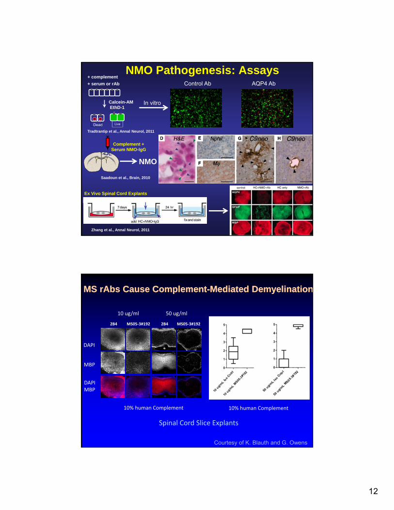
NMO Pathogenesis: Assays
Complement +Serum NMO-IgG
NMO
Saadoun et al., Brain, 2010
Ex Vivo Spinal Cord Explants
Zhang et al., Annal Neurol, 2011
+ complement
+ serum or rAb
Calcein-AMEthD-1
LiveDead
In vitro
Control Ab AQP4 Ab
Tradtrantip et al., Annal Neurol, 2011
MS rAbs Cause Complement-Mediated DemyelinationMS rAbs Cause Complement-Mediated Demyelination
Courtesy of K. Blauth and G. Owens
MS05‐3#1922B42B4 MS05‐3#192
DAPI
MBP
DAPIMBP
10 ug/ml 50 ug/ml
10% human Complement 10% human Complement
Spinal Cord Slice Explants

13
rAb: Glycolipid bindingrAb: Glycolipid binding
Source % Glycolipid Reactive Specificity*
Multiple Sclerosis(10 patients 73 rAbs)
27%(5 patients, 20 rAbs)
• Sulfatide• Sulfatide complexes• Cholesterol
Inflammatory Controls(6 patients, 27 rAbs)
33%(3 patients, 9 rAbs)
• Sulfatide• Lipid complexes
Brennan et al., J. Neuroimmunol, 238: 87 (2011)
Sulfatide Lipids Inhibit Axon OutgrowthSulfatide Lipids Inhibit Axon Outgrowth
Winzeler et al., J Neurosci, 31:6481 (2011)
Sulfatide Specifically Inhibits Axonal Outgrowth
O4 Ab (Anti-Sulfatide) Antibody Restores Neurite Outgrowth

14
B Cells Are Multifunctional at Neuro-immune InterfaceB Cells Are Multifunctional at Neuro-immune Interface
Produce Antibodies
Instruct T Cells• Antigen Presentation
Activate Immune Cells• Antigen Presenting Cells
Immunoregulation
Produce Antibodies
Instruct T Cells• Antigen Presentation
Activate Immune Cells• Antigen Presenting Cells
Immunoregulation
McFarland. N Engl J Med, 358: 664-5
Dependent on B cell Receptor
B Cells Are Antigen-Presenting CellsB Cells Are Antigen-Presenting Cells
Protein Antigens
Antigen Internalization BCR (Ag) Dependent Costimulatory Molecule
Expression
Protein Antigens
Antigen Internalization BCR (Ag) Dependent Costimulatory Molecule
Expression
Constant et al. J Immunol 155: 3734 (1995).

15
Secretion of Toxins and CytokinesSecretion of Toxins and Cytokines
Secretory products of multiple sclerosis B cells are cytotoxic to oligodendroglia in vitro
Lisak et al., J Neuroimmunol, vol. 246: 85 (2012).
B cell depletion may ameliorate disease through ablation of IL-6–
producing B cells Barr et al., J Exp Med, vol. 209: 1001 (2012).
Acknowledgments
FundingNIHGuthy Jackson
Universitätsmedizin Göttingen
Christine Stadelmann
Stefan Nessler
Claudia Wrzos
Alan Verkman
Hua Zhang
Lukmanee Tradtrantip
Puay Phan
Julien Ratelade
Greg Owens
Corey Reiter
Alanna Ritchie
Scott Wemlinger
Kevin Blauth
John Soltys
Hannah Schumann
Markus Kowarik
Greg Owens
Corey Reiter
Alanna Ritchie
Scott Wemlinger
Kevin Blauth
John Soltys
Hannah Schumann
Markus Kowarik