Embed Size (px)
Citation preview
Int.J.Curr.Microbiol.App.Sci (2014) 3(12): 276-283
276
Original Research Article
Bacteria undergo programmed cell death upon low dose gamma radiation exposure
Surbhi Wadhawan, Satyendra Gautam* and Arun Sharma
Food Technology Division, Bhabha Atomic Research Centre, Mumbai - 400085, India
*Corresponding author
A B S T R A C T
Introduction
Microbial contamination is a menace causing food spoilage, food borne illnesses and other human and animal diseases. Controlling microbial contamination is quite challenging. Food contamination can be overcome by various physical and chemical antimicrobial treatments. Radiation processing is one of the physical treatments used to sanitize food and has been approved by various international bodies such as WHO, FAO and IAEA (Farkas, 2006). In general, radiation treatment kills the microrganisms by damaging its DNA and
inhibiting their reproductive capability. Cells either succumb to DNA damage or respond in other ways which suppress their growth and induces the process of damage repair.
Recently, bacterial cultures have been found to display population behavior such as altruism, quorum sensing and biofilm formation. In the current scenario, it is known that bacteria die due to radiation exposure, but how does the cell respond to lethal or sub-lethal doses of genotoxic treatments like gamma radiation still
ISSN: 2319-7706 Volume 3 Number 12 (2014) pp. 276-283 http://www.ijcmas.com
K e y w o r d s
Radiation, Bacteria, Programmed cell death, FACS
A subpopulation of Salmonella enterica sv. Typhimurium, Bordetella bronchiseptica, Xanthomonas campestris and Bacillus subtilis was found to undergo caspase dependent programmed cell death (PCD) upon exposure to gamma radiation. Irradiating these bacteria at their respective D10 dose in the presence of inhibitor of caspase-3 or poly (ADP ribose) polymerase (PARP) increased the cell survival significantly. FACS analysis indicated phosphatidylserine externalization (PS) in these bacteria upon radiation exposure. A PCD negative mutant of Xanthomonas lacking caspase-3-like activity and displaying reduced PS externalization was found to be comparatively less susceptible to radiation than its wild type counterpart. Together these findings established the involvement of PCD in a subpopulation of radiation exposed bacteria.
Int.J.Curr.Microbiol.App.Sci (2014) 3(12): 276-283
277
remains to be resolved. To address this issue, in the current study different bacterial cultures namely, Salmonella enterica sv. Typhimurium, Bordetella bronchiseptica, Xanthomonas campestris pv. glycines and Bacillus subtilis were exposed to various doses of gamma radiation and their radiation sensitivity was determined. Also, the nature of death (programmed or necrotic) was ascertained by exposing these cells to radiation in the presence of cell permeable inhibitor of caspase-3 or poly (ADP ribose) polymerase (PARP), the key enzymes reported to be involved in the process of PCD in other systems.
Additionally, the level of caspase-3-like activity, status of phosphatidylserine (PS) externalization (an important marker of PCD) and intracellular level of ROS were also evaluated in radiation treated bacterial cultures. Also, a PCD and caspase negative mutant of Xanthomonas campestris pv. glycines, XcgM42, was used during the course of this study .
Results and Discussion
Radiation treatment caused cell death in bacteria
With an increase in radiation dose enhanced cell killing was observed in different bacteria, however, the extent of death varied among different genera. Salmonella enterica sv. Typhimurium was found to be comparatively moderately radiation sensitive and its D10 was found to be 148 Gy (Fig. 1C). D10 indicates decimal reduction dose, i.e. the dose required to kill 90% of the cell population (Vincent et al., 1990). Many isolates of S. Typhimurium have been found to be pathogenic to humans. Bordetella bronchiseptica and Bacillus subtilis were found to be comparatively more resistant with a D10 of 318 and 330 Gy respectively
(Fig. 1A and B). Some strains of Bordetella have been found to be opportunistic pathogens in humans and animals. Bacillus subtilis is a soil saprophytic bacterium but also causes food spoilage. A pathogen of soybean plant, Xanthomonas campestris pv. glycines, was found to be radiation sensitive with D10 of 66 Gy whereas the D10 for XcgM42, a caspase and PCD negative mutant of X. campestris (Gautam and Sharma, 2002a) was found to be 77 Gy, 17% higher than its wild type counterpart (Fig. 1D). Increase in D10 or in other words radioresistance of this mutant indicated that possibly caspase mediated PCD plays a significant role in radiation induced cell death (RICD) in this organism. Although the possible existence of caspase/metacaspase-like domain containing proteins has been reported from different bacterial species by many authors (Koonin and Aravind, 2002; Ning et al., 2002; Sahoo et al., 2006; Jimenez et al., 2009), still the exact sequence of its gene and protein has not been resolved.
Radiation treatment resulted in activation of caspase-3-like protein in bacteria
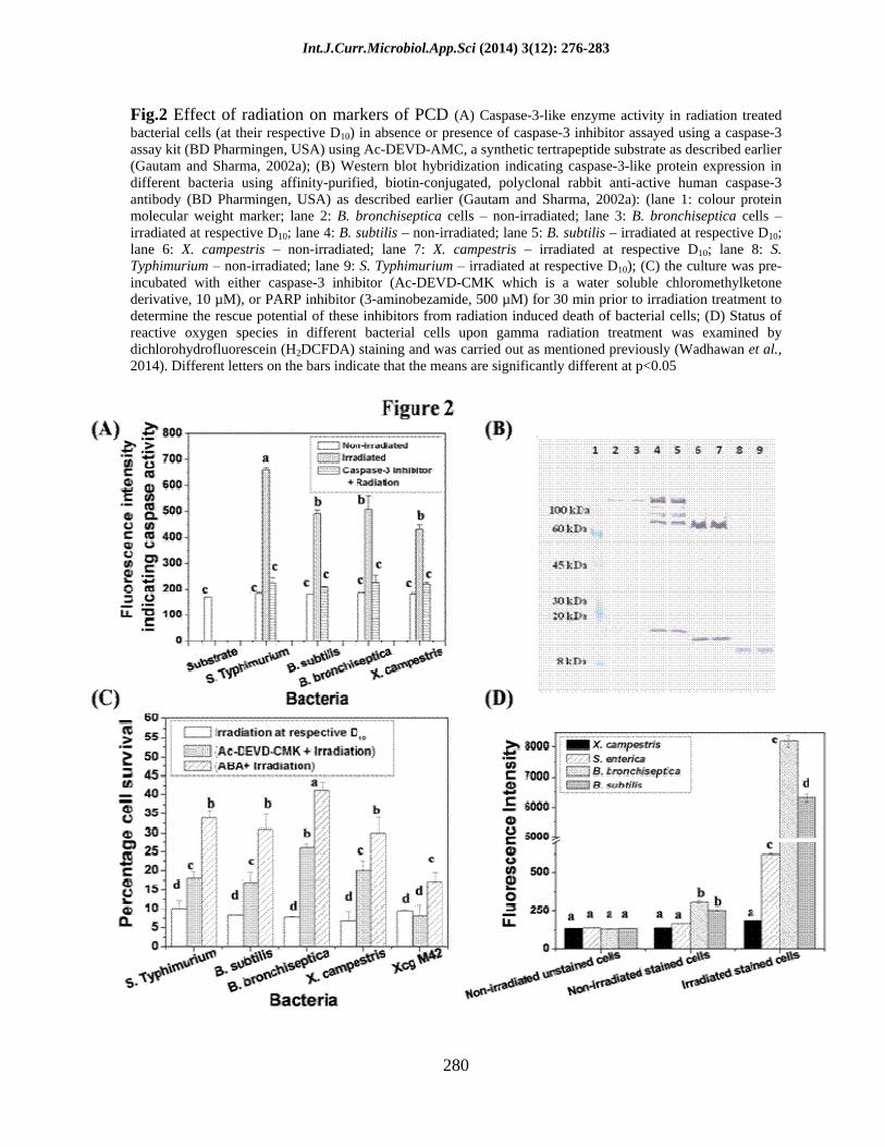
Activation of caspase-3 enzyme is an important event in case of eukaryotic cells undergoing PCD (Michelin et al., 2004; Yuan and Horvitz, 2004; Elmore, 2007). Here too, radiation exposure was found to significantly induce caspase-3-like activity in these bacteria (Fig. 2A). The activity of this enzyme was found to be negligible in all the non-irradiated bacterial cultures. Upon radiation treatment it was found to increase by 3.5 fold in S. enterica sv. Typhimurium cells, 2.7 fold in B. subtilis and B. bronchiseptica and 2.3 fold in X. campestris. However, the caspase-3-like activity dropped significantly in the cells pre-incubated with Ac-DEVD-CMK, a cell permeable caspase-3 inhibitor. This strongly
Int.J.Curr.Microbiol.App.Sci (2014) 3(12): 276-283
278
correlated with the increase in cell survival found in these bacteria when exposed to radiation in the presence of caspase-3 inhibitor (as discussed later in Fig. 2C). This clearly implies that radiation induced cell death (RICD) in a subpopulation of bacteria was caspase dependent. However, after gamma radiation exposure no significant increase in caspase-3-like protein (CLP) biosynthesis was observed in these bacteria as analyzed by immunoblotting using polyclonal anti-caspase-3 antibody (Fig. 2B). The molecular weight of CLP detected by the caspase-3 antibody varied in different bacterial strains. CLP was found to be 15 kDa in S. enterica sv. Typhimurium (Fig. 2B, lanes 8 and 9). The detected molecular weight is quite smaller than the reported molecular weight of caspase in different organisms. In case of B. subtilis and B. bronchiseptica a protein band of ~150 kDa (Fig. 2B, lanes 2 5) and in X. campestris ~90 kDa protein band was detected by anti-caspase-3 antibody (Fig. 2B, lanes 6 and 7). The presence of caspase-3-like protein has been reported earlier from this laboratory in X. campestris (Gautam and Sharma 2002b; Gautam and Sharma, 2005; Gautam et al., 2005; Raju et al., 2006; Wadhawan et al., 2010; Wadhawan et al., 2014; Bayles, 2014) and in B. subtilis by other authors (Sahoo et al., 2006).
Inhibition of radiation induced cell death (RICD) by cell permeable caspase-3 inhibitor
Several small peptide inhibitors mimicking the recognition sequence of caspases alkylate the cysteine residue in the active site of caspase and irreversibly inactivate it (Schotte et al., 1999). Based on this, the effect of an irreversible caspase-3 inhibitor on the survival of radiation treated bacterial population was investigated. Interestingly, the cell survival in all these bacteria was
found to improve remarkably when cells were incubated with the caspase-3 inhibitor 30 min prior to radiation treatment at their respective D10 (Fig. 2C). This resulted in two fold increase in survival in the case of S. enterica sv. Typhimurium and B. subtilis cells and a threefold increase in survival of B. bronchiseptica and X. campestris cells. Unlike its wild type counterpart, no increase in cell survival of radiation treated XcgM42 cultures (PCD and caspase negative mutant) was observed even after pre-incubation with caspase-3 inhibitor (Fig. 2C). There are similar reports of reversal of PCD induced by radiation or other stress in eukaryotic cells and thymocytes in the presence of peptide based caspase inhibitors (Toyooka et al., 1998). Caspase has also been reported to be activated upon radiation exposure in other systems like neural cell precursors and HeLa cells (Michelin et al., 2004; Kim et al., 2003). Probably, a fraction of the dying population undergoes caspase dependent cell death (i.e. those which were rescued in the presence of caspase-3 inhibitor) and the rest of the cells die by some other mechanism like necrosis due to acute cellular damage.
PARP inhibitor also protects bacteria from RICD
Poly (ADP-ribose) polymerase (PARP) has also been reported to be involved in DNA repair and PCD in different systems (Giansanti et al., 2010). It catalyzes the cleavage of NAD+ into ADP and ADP-ribose and attaches several molecules of the latter to the target protein in a process called poly ADP-ribosylation. PARP is activated by DNA breaks, and can deplete NAD+ and ATP of a cell in an attempt to repair the damaged DNA. ATP depletion in a cell could lead to necrosis. Presence of PARP inhibitor, 3-aminobenzamide (3-ABA), was found to offer varying degrees of protection against RICD in these bacteria (Fig. 2C).

Int.J.Curr.Microbiol.App.Sci (2014) 3(12): 276-283
279
Fig.1 Effect of radiation treatment at different doses (upto 1 kGy) on the viability of bacteria (A) Bacillus subtilis, (B) Bordetella bronchiseptica, (C) Salmonella enterica sv. Typhimurium, (D) Xanthomonas campestris pv. glycines (wild type) and a PCD and caspase negative mutant (Xcg M42) (earlier obtained by a random mutagenesis using 1-methyl-2-nitro-1-nitrosoguanidine (MNNG) mutagenesis, Gautam and Sharma, 2002a). An aliquot (1 ml) of log phase grown culture of above mentioned bacteria was withdrawn and serially diluted using saline (0.85%) to achieve the cell density of ~106 cfu/ml. The cell suspension was irradiated at different doses in a Gamma Chamber (Co60 source, dose rate - 5 Gy/min). The viable plate count was determined ~1h after irradiation.
Int.J.Curr.Microbiol.App.Sci (2014) 3(12): 276-283
280
Fig.2 Effect of radiation on markers of PCD (A) Caspase-3-like enzyme activity in radiation treated bacterial cells (at their respective D10) in absence or presence of caspase-3 inhibitor assayed using a caspase-3 assay kit (BD Pharmingen, USA) using Ac-DEVD-AMC, a synthetic tertrapeptide substrate as described earlier (Gautam and Sharma, 2002a); (B) Western blot hybridization indicating caspase-3-like protein expression in different bacteria using affinity-purified, biotin-conjugated, polyclonal rabbit anti-active human caspase-3 antibody (BD Pharmingen, USA) as described earlier (Gautam and Sharma, 2002a): (lane 1: colour protein molecular weight marker; lane 2: B. bronchiseptica cells
non-irradiated; lane 3: B. bronchiseptica cells
irradiated at respective D10; lane 4: B. subtilis
non-irradiated; lane 5: B. subtilis
irradiated at respective D10;
lane 6: X. campestris
non-irradiated; lane 7: X. campestris
irradiated at respective D10; lane 8: S. Typhimurium
non-irradiated; lane 9: S. Typhimurium
irradiated at respective D10); (C) the culture was pre-incubated with either caspase-3 inhibitor (Ac-DEVD-CMK which is a water soluble chloromethylketone derivative, 10 µM), or PARP inhibitor (3-aminobezamide, 500 µM) for 30 min prior to irradiation treatment to determine the rescue potential of these inhibitors from radiation induced death of bacterial cells; (D) Status of reactive oxygen species in different bacterial cells upon gamma radiation treatment was examined by dichlorohydrofluorescein (H2DCFDA) staining and was carried out as mentioned previously (Wadhawan et al., 2014). Different letters on the bars indicate that the means are significantly different at p<0.05
Int.J.Curr.Microbiol.App.Sci (2014) 3(12): 276-283
281
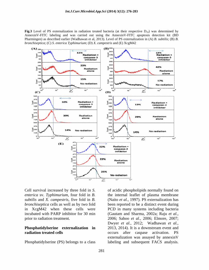
Fig.3 Level of PS externalization in radiation treated bacteria (at their respective D10) was determined by AnnexinV-FITC labeling and was carried out using the AnnexinV-FITC apoptosis detection kit (BD Pharmingen) as described earlier (Wadhawan et al, 2013). Level of PS externalization in (A) B. subtilis; (B) B. bronchiseptica; (C) S. enterica Typhimurium; (D) X. campestris and (E) XcgM42
Cell survival increased by three fold in S. enterica sv. Typhimurium, four fold in B. subtilis and X. campestris, five fold in B. bronchiseptica cells as well as by two fold in XcgM42 when these cells were incubated with PARP inhibitor for 30 min prior to radiation treatment.
Phosphatidylserine externalization in radiation treated cells
Phosphatidylserine (PS) belongs to a class
of acidic phospholipids normally found on the internal leaflet of plasma membrane (Naito et al., 1997). PS externalization has been reported to be a distinct event during PCD in many systems including bacteria (Gautam and Sharma, 2002a; Raju et al., 2006; Sahoo et al., 2006; Elmore, 2007; Dwyer et al., 2012; Wadhawan et al., 2013, 2014). It is a downstream event and occurs after caspase activation. PS externalization was assayed by annexinV labeling and subsequent FACS analysis.
Int.J.Curr.Microbiol.App.Sci (2014) 3(12): 276-283
282
The extent of PS externalization increased to 30, 42, 23 and 29% in radiation treated cultures of S. enterica sv. Typhimurium, B. bronchiseptica, B. subtilis and X. campestris, respectively (Fig. 3A D).
PS externalization reduced significantly in cells incubated for 30 min with caspase-3 inhibitor prior to radiation exposure and was similar to that found in control non-irradiated cells (Fig. 3A D). Also, no change in PS externalization level was detected in radiation treated culture of XcgM42 (Fig. 3E), a PCD negative mutant of Xanthomonas indicating the requirement of functional caspase-3 protein for this event to occur.
Gamma radiation exposure resulted in enhanced reactive oxygen species (ROS) generation
An increase in ROS level was observed when these bacterial cells (Xanthomonas campestris, Bacillus subtilis, Bordetella bronchiseptica and Salmonella enterica sv. Typhimurium) were exposed to radiation (at their half D10 dose). The ROS level was determined by H2DCFDA (2, 7 dichlorohydrofluorescein) staining. This increase in intracellular level of ROS in radiation exposed bacterial cells was found to be least in the case of Xanthomonas (~1.5 fold), four fold in Salmonella, and highest in Bordetella (27 fold) followed by Bacillus (25 fold) (Fig. 2D). The observation indicates that Xanthomonas is comparatively more sensitive to oxidative stress. This observation also explains the probable reason of difference in D10 values of these bacteria. Unlike Bacillus and Bordetella which have higher D10, Xanthomonas succumbs to death even at lower ROS level making it comparatively more radiosensitive. This could be due to the differences in the antioxidant defense mechanisms of these bacteria.
The findings of the current study indicated the activation of inherent caspase-3-like activity in different bacteria upon radiation treatment resulting in induction of programmed cell death. This also indicated the evolutionary conserved nature of PCD among different organisms which serves as one of the mechanisms of cell death upon radiation treatment.
Acknowledgements
Authors thank Mr. A. P. Janardhan for his help in performing flow cytometry.
References
Bayles, K.W. 2014. Bacterial programmed cell death: making sense of a paradox. Nat. Rev. Microbiol., 12: 63 69.
Dwyer, D.J., Camacho, D.M., Kohanski, M.A., Callura, J.M., Collins, J.J. 2012. Antibiotic-induced bacterial cell death exhibits physiological and biochemical hallmarks of apoptosis. Mol. Cell., 46: 561 572.
Elmore, S. 2007. Apoptosis: a review of programmed cell death. Toxicol. Pathol., 35: 495 516.
Farkas, J. 2006. Irradiation for better foods. Trends Food Sci. Tech., 17: 148 152.
Gautam, S., Sharma, A. 2002a. Involvement of caspase-3-like protein in rapid cell death of Xanthomonas. Mol. Microbiol., 44: 393 401.
Gautam, S., Sharma, A. 2002b. Rapid cell death in Xanthomonas campestris pv. glycines. J. Gen. Appl. Micribiol., 48: 67 76.
Gautam, S., Sharma, A. 2005. Programmed cell death: an overview. In: Chakraborty, C., (Ed.), Advances in biochemistry and biotechnology. Daya Publishing House, India. Pp. 122 157.
Gautam, S., Sharma, A., Kobayashi, I.,
Int.J.Curr.Microbiol.App.Sci (2014) 3(12): 276-283
283
2005. Survival and death in bacteria. In: Yamada, M., (Ed.), Programmed cell death in microorganisms. Research Sign Post, India, Pp. 1 39.
Giansanti, V., Dona, F., Tillhon, M. And Scovassi, A.I. 2010. PARP inhibitors: New tools to protect from in ammation. Biochem. Pharmacol., 80: 1869 1877.
Jimenez, C., Capasso, J.M., Edelstein, C.L., Rivard, C.J., et al. 2009. Different ways to die: cell death modes of the unicellular chlorophyte Dunaliella viridis exposed to various environmental stresses are mediated by the caspase-like activity DEVDase. J. Exp. Bot., 60: 815 828.
Kim, K.Y., Seol, J.Y., Jeon, G., Nam, M.J. 2003. The combined treatment of aspirin and radiation induces apoptosis by the regulation of bcl-2 and caspase-3 in human cervical cancer cell. Cancer Lett., 189: 157166.
Koonin, E.V., Aravind, L. 2002. Origin and evolution of eukaryotic apoptosis: the bacterial connection. Cell Death Diff., 9: 394 404.
Michelin, S., Perez, M.D.R., Dubner, D., Gisone, P. 2004. Increased activity and involvement of caspase-3 in radiation-induced apoptosis in neural cells precursors from developing rat brain. NeuroToxicol., 25: 387 398.
Naito, M., Nagashima, K., Mashima, T., Tsuruo, T. 1997. Phosphatidylserine externalization is a downstream event of interleukin-1 converting enzyme family protease activation during apoptosis. Blood, 89: 2060 2066.
Ning, S.B., Guo, H.L., Wang, L., Song, Y.C. 2002. Salt stress induces programmed cell death in prokaryotic organism Anabaena. J. Appl. Microbiol., 93: 15 28.
Raju, K.K, Gautam, S., Sharma, A. 2006.
Molecules involved in the modulation of rapid cell death in Xanthomonas. J. Bact., 188: 5408 5416.
Sahoo, S., Rao, K.K., Suraishkumar, G.K. 2006. Reactive oxygen species induced by shear stress mediate cell death in Bacillus subtilis. Biotechnol. Bioeng., 94: 118 127.
Schotte, P., Declercq, W., Huffel, S.V., Vandenabeele, P., Beyaert, R. 1999. Non-specific effects of methyl ketone peptide inhibitors of caspases. FEBS Lett., 442: 117 121.
Toyooka, K., Tai, X.G., Park, C., Yashiro, Y., et al. 1998. A caspase inhibitor protects thymocytes from diverse signal-mediated apoptosis but not from clonal deletion in fetal thymus organ culture. Immunol. Lett., 63: 8389.
Vincent, F., Tibi, A., Goury, V., Darbord, J.C. 1990. Combined treatment using irradiation and heat: Susceptibility of Bacillus, Salmonella, Staphylococcus and Clostridium. Radiat. Phys. Chem., 35: 279 283.
Wadhawan, S., Gautam, S., Sharma, A. 2013. A component of gamma radiation induced cell death in E. coli is programmed and interlinked with activation of caspase-3 and SOS response. Arch. Microbiol., 195: 54557.
Wadhawan, S., Gautam, S., Sharma, A. 2010. Metabolic stress-induced programmed cell death in Xanthomonas. FEMS Microbiol. Lett., 312: 176 183.
Wadhawan, S., Gautam, S., Sharma, A. 2014. Involvement of proline oxidase (PutA) in programmed cell death of Xanthomonas. PLoS ONE, 9(5): e96423.
Yuan, J., Horvitz, H.R. 2004. A first insight into the molecular mechanisms of apoptosis. Cell, 116: 53 56.