Embed Size (px)
Citation preview
Bacterial-Fungal Interactions Highlighted using Microbiomics:
Potential Application for Plant Growth Enhancement
Veronica Artursson Faculty of Natural Resources and Agricultural Sciences
Department of Microbiology Uppsala
Doctoral thesis Swedish University of Agricultural Sciences
Uppsala 2005
Acta Universitatis Agriculturae Sueciae 2005:127 ISSN 1652-6880 ISBN 91-576-6926-0 © 2005 Veronica Artursson, Uppsala Tryck: SLU Service/Repro, Uppsala 2005
Abstract
Artursson, V. 2005. Bacterial-Fungal Interactions Highlighted using Microbiomics: Potential Application for Plant Growth Enhancement. Doctoral dissertation. ISSN 1652-6880, ISBN 91-576-6926-0. Arbuscular mycorrhizal (AM) fungi and bacteria can interact synergistically to stimulate plant growth through a range of mechanisms that include improved nutrient acquisition and inhibition of fungal plant pathogens. These interactions may be of crucial importance within sustainable, low-input agricultural cropping systems that rely on biological processes rather than agrochemicals to maintain plant health. The first goal of the present work was to further develop and optimise a method, bromodeoxyuridine immunocapture, suitable for the identification of actively growing bacteria in soil containing abundant AM fungi. DNA was extracted from soil that had been incubated with BrdU for 2 days, and the DNA was isolated by immunocapture of the BrdU-containing DNA. The actively growing bacteria in the community were identified by 16S rRNA gene PCR amplification and DNA sequence analysis. One of the actively growing bacteria, Bacillus cereus strain VA1, was isolated from the soil, tagged with green fluorescent protein (GFP) and shown to clearly attach to AM fungal hyphae. It was subsequently shown, however, that this bacterial strain had preferences for non-vital AM fungal hyphae, whereas one of the control strains, Paenibacillus brasilensis PB177, actually showed greater attachment to vital hyphae. A second objective was to study the impact of specific AM fungi and plant type on the actively growing soil bacterial communities, by using the BrdU method in combination with a fingerprinting technique (e.g. terminal restriction fragment length polymorphism, T-RFLP). This microbiomics (e.g. molecular tools for analysis of complex microbial communities) approach revealed distinct differences in bacterial community composition within the treatments, and the putative identities of the dominant bacterial species, activated as a result of Glomus mosseae inoculation, were found to be mostly uncultured bacteria and Paenibacillus sp., indicating the great significance of using an approach not relying on the culturability of the bacteria. Finally, Paenibacillus brasilensis PB177, previously shown to be associated with AM fungi, was shown to inhibit growth of several phytopathogenic fungi. In summary, the results of these studies provide novel valuable insights to consider when developing combined microbial inocula (e.g. bacteria and AM fungi) for enhancing plant growth within sustainable agriculture in the future. Keywords: arbuscular mycorrhizal fungi, phytopathogenic fungi, Paenibacillus, bromodeoxyuridine immunocapture, GFP, T-RFLP, microbiomics. Author´s address: Veronica Artursson, Department of Microbiology, Swedish University of Agricultural Sciences, Box 7025, SE-750 07 Uppsala, Sweden.
Till Mamma och Pappa
~ …The dreams you dare to dream really do come true from “Over the Rainbow”, E.Y. Harburg
Contents
Introduction 7
Arbuscular mycorrhiza 11
-The symbiosis 11 -The fungi 14 -Host plants 15
AM fungal associated bacteria 17
-Plant growth promoting rhizobacteria (PGPR) 17 -Bacteria enhancing nitrogen bioavailability 20 -Bacteria enhancing phosphorus bioavailability 21 -AM fungal endosymbiotic bacteria 22
Microbiomics tools 25
-Green fluorescent protein tagging 25 -Bromodeoxyuridine immunocapture 27 -Terminal Restriction Fragment Length Polymorphism (T-RFLP) 28
Specific AM fungal-bacterial interactions 30
-Specificity between AM fungi and bacteria 31 -Physical interactions 32
Interactions with plant pathogenic fungi 34
-Inhibition of pathogenic fungi by bacteria and/or AM fungi 34 -Paenibacillus brasilensis PB177 36
Conclusions 39
Future perspectives 39
References 41
Acknowledgements 49
Appendix
Papers I-IV This thesis is based on the following publications, which are referred to by their Roman numerals: I. Artursson, V. & Jansson, J.K. 2003. Use of Bromodeoxyuridine immunocapture
to identify active bacteria associated with arbuscular mycorrhizal hyphae. Applied and environmental microbiology 69, 6208-6215.
II. Artursson, V., Finlay, R.D. & Jansson, J.K. 2005. Combined bromodeoxyuridine immunocapture and terminal restriction fragment length polymorphism analysis highlights differences in the active soil bacterial metagenome due to Glomus mosseae inoculation or plant species. Environmental microbiology 7, 1952-1966.
III. Toljander, J.F., Artursson, V., Paul, L.R., Jansson, J.K. & Finlay, R.D. Attachment of different soil bacteria to arbuscular mycorrhizal fungal extraradical hyphae is determined by hyphal vitality and fungal species. FEMS Letters, in press, doi: 10.1111/j.1574-6968.2005.00003.x.
IV. von der Weid, I., Artursson, V., Seldin, L. & Jansson, J.K. Antifungal and root surface colonization properties of GFP-tagged Paenibacillus brasilensis PB177. World journal of microbiology and biotechnology, in press, doi: 10.1007/s11274-005-8123-3.
Papers are reprinted with permission from the respective publisher. My contributions to the papers included in this thesis have been as follows: I. Planned the experiments together with my supervisor and contributed many of
the ideas. Did all the laboratory work. Did most of the writing of the manuscript, including the first draft.
II. Planned the experiments together with my supervisors and contributed many of the ideas. Did all the laboratory work. Did most of the writing of the manuscript, including the first draft.
III. Planned the experiments together with the other authors. Performed most of the laboratory work together with Jonas Toljander. Minor part in writing of the manuscript.
IV. Did some complementary experiments to strengthen the findings of this study that was initiated by the first author and her supervisors, Lucy Seldin and Janet Jansson. Helped with revision of the manuscript prior to submission.
Additional publication: Artursson, V., Finlay, R.D. & Jansson, J.K. 2005. Interactions between arbuscular mycorrhizal fungi and bacteria and their potential for stimulating plant growth. Environmental microbiology, in press, doi:10.1111/j.1462-2920.2005.00942x.

7
“The study of plants without their mycorrhizas is the study of artifacts. The majority of plants, strictly speaking, do not have roots; they have mycorrhizas.” BEG Committee, 1993
Introduction
Microorganisms are the most abundant and the most diverse group of living organisms on Earth, yet most of them are unknown to us, representing our largest unexplored biological resource. Microorganisms live in a wide variety of environments, among them the soil biota of which they are the most frequently detected members. In a recently published study, Gans, Wolinsky & Dunbar (2005) estimate that 10 grams of unpolluted soil will typically contain 8.3 x 10⁶ different species of bacteria, corresponding to almost 10 million species in that very small amount of soil. As these new calculations revealed far more bacterial species in soil than anyone realised, the next obvious challenge would be to identify those species and their potentially unknown key functions within the ecosystem. Additionally, it would be very interesting to correlate species diversity with how well plants grow, potentially providing information about the ecological consequences of losing large amounts of bacterial biodiversity in nature. The soil microbiota include species responsible for nutrient mineralization and cycling, antagonists (biological control agents against plant pests and diseases), species that produce substances capable of modifying plant growth, and species that form mutually beneficial (symbiotic) relationships with plant roots. This latter group includes mycorrhizal fungi, various actinomycetes, and some bacteria, of which the majority are known to be able to stimulate plant growth through direct or indirect interactions. However, the beneficial traits of root-colonizing bacteria and mycorrhizal fungi have been mainly evaluated separately. Only recently have the synergistic effects of these microorganisms been explored with respect to their combined beneficial impacts on plants (see Artursson, Finlay & Jansson, 2005, for a review). Optimally, this fairly new branch of research will ultimately lead to an extended use of biological inoculants within low input, sustainable agriculture. One pre-requisite for reaching this goal, however, is the improved knowledge about biological interactions (e.g. mycorrhizal fungi – bacteria - plant roots) occurring in the soil. Interactions, such as competition, antagonism, synergism and symbiosis, may be critical in determining the stability and spread of introduced microbial inoculants (genetically modified or not), as well as their effects on the indigenous microbial communities within natural ecosystems. For example, it is important to identify and isolate mycorrhizal associated bacteria that have potential as beneficial inoculants within low-input cropping systems, and also to determine the influence of these bacteria, together with mycorrhizal fungi, on interactions with fungal pathogens. Potential associations between bacterial isolates and mycorrhizal fungi should ultimately be studied both in sterile and non-sterile soil samples, followed by analyses of bacterial effects on mycorrhizal establishment and fungal root colonization, and subsequently on plant growth and development. Hopefully, this will lead to an increased understanding and
8
manipulation of naturally occurring microbial populations necessary for low-input cropping systems that are dependent upon biofertilisation and biocontrol, rather than inorganic fertilizers and fungicides, for maintaining crop health and productivity. In turn, this should lead to a more widespread ecological crop production with greater agricultural output and better profit margins, encouraging a higher environmental awareness. Both ectomycorrhizal (Garbaye, 1994) and endomycorrhizal (Meyer & Linderman, 1986) fungi can interact with different bacterial species, however this thesis work focuses on the interactions involving arbuscular mycorrhizal (AM) fungi, included in the endomycorrhizal group. The interactions between AM fungi and bacteria usually occur in the zone of soil surrounding the roots and fungal hyphae; commonly referred to as the “mycorrhizosphere”, or in the more delimited “hyphosphere”, referring only to the zone of soil surrounding individual fungal hyphae (Fig. 1; Rambelli, 1973).

9
Fig. 1. Schematic view of different soil zones; e.g. the rhizosphere, the mycorrhizosphere, and the hyphosphere. The drawing is not to scale and underestimates the relative surface area of the extraradical mycorrhizal mycelium. The insert shows a microscopic image typical of the area represented by the black square. The surface area of AM fungal hyphae may exceed that of the roots by 1-3 orders of magnitude, probably providing an important niche for soil bacteria through the plant-derived carbon compounds released by the hyphae. It is very likely that this specific nutrient-rich environment provides a potential resource for generating great natural genetic bacterial diversity. On the other hand, it could create a site where very specific microbial interactions occur, e.g. clear preferences between certain bacterial groups and AM fungal hyphal species, due to competition for carbon compounds or species-specific fungal hyphal exudates. However, irrespective of how great or complex the bacterial diversity is in the mycorrhizal hyphosphere, it is nevertheless a very influential environment for biological life, and it surely deserves more scientific attention than it has received so far. To date, there is little information on the mechanisms controlling interactions of bacteria with AM fungi and plant roots in the mycorrhizosphere. However, a

10
number of possible alternatives have been proposed. Some bacteria have been shown to directly affect AM fungal germination and growth rate (Carpenter-Boggs, Loynachan & Stahl, 1995; Daniels & Trappe, 1980; Mayo, Davis & Motta, 1986; Mosse, 1959) and thus the beneficial impact to the plant could be through the AM association. Other bacteria can directly influence the physiology of the plants, for example by increasing root cell permeability. In addition to interacting directly to beneficially influence the mycorrhizal relationship and/or plant growth (Garbaye, 1994; Linderman, 1988; Linderman, 1992; Vivas et al., 2003), specific bacteria together with AM fungi may create a more indirect synergism that supports plant growth (Barea, 1997), including nutrient acquisition (Barea, Azcon & Azcon-Aguilar, 2002), inhibition of plant pathogenic fungi (Budi et al., 1999), and enhancement of root branching (Gamalero et al., 2002). There is much scope for further experimental analyses of the underlying mechanisms of bacterial-AM fungal interactions with plant roots, and the investigations of their roles in more detail will probably be one of the biggest challenges for future mycorrhizal research. However, it is important to keep in mind that if these associations are as specific as suggested, the underlying mechanisms are probably extremely complex and complicated to evaluate, due to the close intimacy of the microorganisms involved in the interactions. In addition to the, above mentioned, proven effects of bacteria on AM fungi, the AM fungi themselves have also been shown to have an impact on the composition of bacterial communities (paper II). This impact may be relayed through the plant root since mycorrhizal establishment has been shown to change the chemical composition of root exudates, which in turn often is a source of nutrients to associated bacteria in the mycorrhizosphere (Azcón-Aguilar & Bago, 1994; Barea, 1997; Barea, 2000; Gryndler, 2000; Harley & Smith, 1983; Linderman, 1992; Linderman, 2000; Smith et al., 1994). Changes in these nutrient-rich root exudates (and hence hyphal exudates) might naturally represent a driving force for bacterial growth. This in turn would result in stimulation of microbial activity, which might be an important parameter to consider in the future when screening for mycorrhizal associated bacteria. Actively growing organisms within a community are clearly those that have the potential to exert the greatest effect on their immediate proximity, and such bacterial groups do usually include those species that are involved in particular physiological responses within the specific environment evaluated, such as plant growth promotion via AM fungi. Two main groups of bacteria interact with AM fungi in the mycorrhizosphere: saprophytes and symbionts, both groups potentially consisting of detrimental, neutral and beneficial bacteria (Barea, Azcon & Azcon-Aguilar, 2002; Johansson, Paul & Finlay, 2004). However, the main focus of this thesis has been on interactions between bacteria and AM fungi with proven or potentially synergistic properties that may lead to stimulation of plant growth. In paper I, an actively growing bacterium was isolated from soil containing abundant AM fungi, and this bacterium as well as a number of bacterial control strains, were then evaluated due to their ability to associate with AM fungal hyphae in the mycorrhizosphere (papers I and III). The impact of specific AM fungi and plant species on the actively growing bacterial communities in soil was studied (paper II), and a
11
bacterial strain previously shown to be associated with AM fungi (paper III), was studied for its ability to inhibit growth of several phytopathogenic fungi (paper IV). In general, the results of these studies provide valuable insights about the suitability of using combined microbial inocula (eg. bacteria and AM fungi) for enhancing plant growth within sustainable agriculture in the future.
Arbuscular mycorrhiza
The Symbiosis Arbuscular mycorrhizas are the most common underground symbiotic associations, which are formed between roots of most terrestrial plants and fungi of the phylum Glomeromycota. The AM symbiosis is biotrophic (the fungus needs the plant to survive) and normally mutualistic, the interactions being characterized by bi-directional movement of nutrients, where carbon (C) flows to the fungus and different nutrients move to the plant. When a nutrient is deficient in soil solution, the critical root parameter controlling its uptake is surface area. AM fungal extraradical hyphae have the potential to greatly increase this absorbing surface area of the root (hyphae usually extend for several centimeters away from the surface of the root), thereby contributing to a more efficient nutrient uptake. However, to be most efficient in their uptake, the AM fungal hyphae must be distributed beyond the nutrient depletion zone that develops around the roots when nutrients are removed from the soil more rapidly than they are replaced by diffusion (Sylvia, 2002). One of the most important plant nutrients, phosphate, is a poorly-mobile ion and its depletion zone is usually seen as a sharp and narrow area close to the root. Because of its narrowness, the fungal hyphae can easily bridge the depletion zone and grow into surrounding soil with an adequate supply of phosphorus, thereby substantially improving the P status of the plant. These processes, when AM fungal hyphae absorb P from the soil and translocate it to the root, are much faster than diffusion of P through the soil. Consequently, hyphal transfer overcomes the reductions in rate of uptake which result from the development of the depletion zones around the roots (Smith & Read, 1997). Micronutrients, such as zinc and copper, can also easily be made more available to the symbiotic plants through the AM fungal hyphae because of the difficulty of these nutrients to diffuse back into the depletion zones (Sylvia, 2002). However, for more mobile nutrients, for example nitrate (NO3
-) , the mass flow of the soil solution to roots allows the uptake to be maintained at rates dependent on the root absorbing power, resulting in that being the factor limiting the nitrate uptake. Consequently, no mycorrhizal effect should be expected, except in very dry soil where the mobility is reduced. On the other hand, NH4
+, commonly found in soils with slow nitrification rates, is a relatively non-mobile ion, resulting in easily developed depletion zones even in moist soil and as with P, diffusion rather than root absorbing power, limits the rate of uptake, giving AM fungi an important role in NH4
+ uptake rate (Smith & Read, 1997).
12
The AM fungi, which are obligate symbionts, are believed to obtain almost all of their C from their autotrophic plant partners. About 20 % of the total C assimilated by plants may be transferred to the fungal partner (Pearson & Jakobsen, 1993), probably in the form of glucose which subsequently is transformed into fungal sugars thereby preventing its return to the host. This transfer of organic C to the fungus has sometimes been considered a drain on the host. However, the host plant may increase photosynthetic activity as a result of mycorrhizal colonization (Miller et al., 2002), to compensate for the C “lost” to the fungus. In an ecosystem, the flow of C to the fungal partner serves several important functions. For example, it is utilized in growth and maintenance of the intra- and extra-radical mycelium, hence in turn promoting nutrient uptake. Consequently, a greatly increased growth of extraradical mycelium and production of spores are often detected once the AM fungal root colonization is established. The AM fungi may also directly influence the C dynamics in soil through the growth and turnover of extraradical hyphae. These hyphae have been considered having a relatively short residence time, and Staddon et al. (2003) recently confirmed this idea by showing that the turnover rate of extraradical hyphae attached to plants, averaged five to six days. The authors suggested that C flow from the host plants to the AM fungi in soil may consequently be respired quite rapidly back to the atmosphere, indicating a quick pathway for atmospheric C to enter the soil C cycle (Zhu & Miller, 2003). Therefore, the large C deposit into the surrounding soil, both that released from dying hyphae but also that actively released from living extraradical hyphae, will contribute to the development of a specific rhizosphere microbial community, the mycorrhizosphere or more specifically, the hyphosphere, which provides an important niche for other beneficial microorganisms. Initiation of the ′so-called′ AM fungal root colonization, begins with hyphae growing toward the plant root and extensively around it, where they subsequently attach to the root surface. The hyphal tips start swelling, forming specific structures called appressoria between the epidermal root cells, followed by the development of infective penetration pegs emerging as hyphal tips from the appressoria (Mandelbaum & Piche, 2000). No appressoria are formed on dead roots or on various artificial fibres (Giovannetti et al., 1993), however, indicating that the formation of this structure occurs only as a result of fungal recognition of a potential host plant. AM fungal hyphae, bearing the above mentioned penetration pegs in their ends, penetrate the adjacent epidermal root cell walls followed by the cortical cell walls, to enter the root. The hyphae cross the hypodermis and start branching in the outer cortex. At the penetration stage, the plant seems to recognize the fungal attachment, and it has been shown that the epidermal cells adjacent to the penetrating hyphae, get slightly thicker walls (Garriock, Peterson & Ackerley, 1989) at this stage, probably as a defence response to the fungal penetration. However, the thickenings do not contain callose or lignin (substances commonly produced in plant cells as a response to wounding or pathogen infection, providing the plant with a defensive structure) and do consequently not prevent the penetration of fungal hyphae through the walls (Harrison & Dixon, 1994).
13
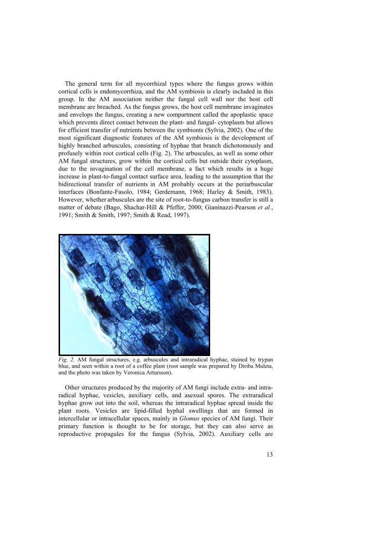
The general term for all mycorrhizal types where the fungus grows within cortical cells is endomycorrhiza, and the AM symbiosis is clearly included in this group. In the AM association neither the fungal cell wall nor the host cell membrane are breached. As the fungus grows, the host cell membrane invaginates and envelops the fungus, creating a new compartment called the apoplastic space which prevents direct contact between the plant- and fungal- cytoplasm but allows for efficient transfer of nutrients between the symbionts (Sylvia, 2002). One of the most significant diagnostic features of the AM symbiosis is the development of highly branched arbuscules, consisting of hyphae that branch dichotomously and profusely within root cortical cells (Fig. 2). The arbuscules, as well as some other AM fungal structures, grow within the cortical cells but outside their cytoplasm, due to the invagination of the cell membrane, a fact which results in a huge increase in plant-to-fungal contact surface area, leading to the assumption that the bidirectional transfer of nutrients in AM probably occurs at the periarbuscular interfaces (Bonfante-Fasolo, 1984; Gerdemann, 1968; Harley & Smith, 1983). However, whether arbuscules are the site of root-to-fungus carbon transfer is still a
matter of debate (Bago, Shachar-Hill & Pfeffer, 2000; Gianinazzi-Pearson et al., 1991; Smith & Smith, 1997; Smith & Read, 1997).
Fig. 2. AM fungal structures, e.g. arbuscules and intraradical hyphae, stained by trypan blue, and seen within a root of a coffee plant (root sample was prepared by Diriba Muleta, and the photo was taken by Veronica Artursson). Other structures produced by the majority of AM fungi include extra- and intra-radical hyphae, vesicles, auxiliary cells, and asexual spores. The extraradical hyphae grow out into the soil, whereas the intraradical hyphae spread inside the plant roots. Vesicles are lipid-filled hyphal swellings that are formed in intercellular or intracellular spaces, mainly in Glomus species of AM fungi. Their primary function is thought to be for storage, but they can also serve as reproductive propagules for the fungus (Sylvia, 2002). Auxiliary cells are
14
clustered swellings on extraradical hyphae that are produced only by certain AM fungi, such as Scutellospora and Gigaspora species. The function of these structures is unknown, however. Reproductive spores can be formed as hyphal swellings either in the root or, more commonly, in the soil and contain lipids, cytoplasm and many nuclei. Spores are mainly formed when nutrients are remobilised from roots where the AM association are senescing, and function as storage structures, resting stages and propagules (Brundrett et al., 1996). Overall, the AM symbiosis can, especially in infertile soils, contribute to improved plant growth and reproduction, through the increased nutrient uptake by their AM fungal partner. As a result, mycorrhizal plants are usually more competitive and better able to tolerate environmental stresses than are non-mycorrhizal plants. The Fungi Arbuscular mycorrhizal fungi belong to the fungal phylum Glomeromycota (Schüssler, Schwarzott & Walker, 2001) which currently comprises approximately 150 described species distributed among ten genera, most of which have been defined mainly by spore morphology. The genera which include most of the described species are Acaulospora, Gigaspora, Glomus and Scutellospora (http://www.tu-darmstadt.de/fb/bio/bot/schuessler/amphylo/amphylogeny.html; 19-Oct-2005). A large proportion of the AM fungi were previously placed in the phylum Zygomycota, but several facts indicate that they form a monophyletic group distinct from other Zygomycotan lineages, among them their symbiotic habit, their apparent lack of zygospores and their rRNA gene phylogeny (http://tolweb.org/tree?group=Glomeromycota; 19-Oct-2005). Based on these indications, Schüssler, Schwarzott & Walker (2001) erected the phylum Glomeromycota. AM fungi are thought to be the oldest group of asexual multicellular organisms. Fossil records have shown that they are at least 460 million years old with indications that their origins may be twice as old (Redecker, Kodner & Graham, 2000; Schüssler, 2002). The AM fungi are considered asexual organisms, since no evidence exist that they are able to reproduce sexually. Therefore mutation and possibly heterokaryosis probably provide the main basis for the variation necessary to permit adaptation to environmental changes and continuing evolution (Smith & Read, 1997). The AM fungi are coenocytic (lacking septa) and multinuclear. There are conflicting reports however, whether the nuclei in the mycelium and spores of a single AM fungus are genetically identical or not (Hijri & Sanders, 2005; Kuhn, Hijri & Sanders, 2001; Pawlowska & Taylor, 2004), and hence there is a possibility that AM fungi possess more than one genome, presupposed that heterokaryosis exists among the nuclei (Hijri & Sanders, 2005). As mutualistic symbionts, AM fungi are able to grow within plant roots without causing disease symptoms. They are obligate symbionts because no one has been
15
successful in growing glomeromycotan fungi separately from their symbiotic plant host yet. Thus, if no host root is found by the germinating hypha of a spore, growth ceases and the cytoplasm may be retracted within the spore. In order to propagate AM fungi, the most common approach is therefore to use a suitable host plant in pot cultures grown in a greenhouse (http://tolweb.org/tree?group=Glomeromycota; 19-Oct-2005). However, to obtain an uncontaminated inoculum of AM fungi, monoxenic root organ cultures can be used, meaning that the fungus is grown on excised, transformed plant roots growing on a sterile medium in a Petri dish (Fortin et al., 2002). Almost all AM fungal species tested have been shown to grow in monoxenic culture. However, several species, for example Glomus mosseae, have only been shown to be able to produce mycelia but no mature spores, thereby failing in a complete life cycle. The only study where this fungal species has been reported to produce spores is the one performed by Raman, Sahadevan & Srinivaan (2001). However, the amount of produced spores was sparse, making this material unusable for large experiments and distribution purposes. A first prerequisite for successfully culturing AM fungi in vitro, is therefore the capacity of the fungi to complete their life cycle, with the production of sufficient amounts of spores and intraradical structures characteristic of the genera considered (Declerck, Séguin & Dalpé, 2005). The second prerequisite for obtaining properly produced monoxenic cultures of AM fungi is explained by Declerck, Séguin & Dalpé (2005) as the capacity of the fungal material to be subcultured, i.e. cultured continuously, under the same monoxenic conditions favouring multiplication of the material, necessary for the distribution and durability of the strain (Declerck, Séguin & Dalpé, 2005). When considering these two prerequisites, only ten AM fungal species have been successfully grown axenically and maintained over several generations (strictly referring to published papers), corresponding to 5.5 % of the approximate 180 described species (Declerck, Séguin & Dalpé, 2005). However, conversely of the AM fungal biomass produced in pot cultures, the fungal material produced in monoxenic cultures should not contain any microorganisms or other contaminants, making this method the most appropriate for propagating fungi for molecular biological experiments, and the one that preferentially should be used when at all possible considering the AM fungal species needed. Host plants The range of potential host plants for AM fungi is extremely wide, and about 80 % of plant families from all phyla of land plants are included in this group. Some members of most families of angiosperms and gymnosperms, together with ferns, lycopods and bryophytes, develop AM symbiosis. The plant families Brassicaceae and Chenopodiaceae, however, include species that do not usually form mycorrhizal symbiosis, among them sugar beet and rape (Tester, Smith & Smith, 1987). Taxonomic specificity or host range indicates whether or not a given species of fungus can form mycorrhizal relationships with more than one species of host
16
plant or whether or not a given species of host associates mycorrhizally with more than one species of fungus (Smith & Read, 1997). There is no clear evidence that any absolute specificity exists between taxa of AM fungi and taxa of potential host plants, and it is very likely that an AM fungus isolated from one species of host plant will colonize any other species that has been shown to be capable of forming AM symbioses, thus combining wide host range with permanence of association (Smith & Read, 1997). The lack of specificity might result in the fact that a single plant species can be colonized by many different fungi and that individual plants can be linked below ground by common mycorrhizal mycelia (Newman et al., 1994). The function of these common mycelia are not fully understood, although it is known that one plant might, for example, depend on the mycelium for uptake of mineral nutrients whereas another plant is critical for the C nutrition of that mycelium. This consequently indicates that interactions between different host plants could occur through their fungal symbionts even in the potential absence of actual interplant transfer. Although AM fungi most likely have a broad potential host range, a certain degree of plant host specificity (e.g. preference) should not be totally excluded. Many of the experiments on which the above described specificity issues were based, were performed with individual isolates of fungal species that were grown separately, apart from competitive interactions (Bever et al., 2001). However, when whole communities are considered a certain degree of specificity between AM fungi and their host plants has been indicated. For example, Bever et al. (1996) found that isolates of different AM fungal species sporulated differentially, with the relative dominance of fungal species being reversed, depending on the plant species with which they were associated (Bever, et al., 1996). Hetrick and Bloom (1986) studied the influence of five host plants on colonization and spore production of AM fungi. They showed that the composition of the AM fungal community was strongly influenced by the host species through differential effects on hyphal growth and sporulation. Additional studies have indicated similar results, such as the one by McGonigle & Fitter (1990) who reported that AM fungal communities varied with the host species present. In turn, such a specificity of fungal response to different species of host plants might potentially contribute to the maintenance of diversity within an AM fungal community (Bever, et al., 2001). Additionally, single species of AM fungi used in pot culture experiments should be tested for their optimal plant symbiont in order to subsequently ensure maximum abundance, and consequently influence, of the fungus in the soil.
17
AM fungal associated bacteria
Different types of bacteria have been found to associate with different host plants colonized by arbuscular mycorrhizal (AM) fungi (Johansson, Paul & Finlay, 2004). Several of these associations have potentially beneficial functions, including those where the bacterial groups described below are involved. Plant growth promoting rhizobacteria (PGPR) PGPR are usually in contact with the root surface, or rhizoplane, and increase plant yield by one or more mechanisms such as improved mineral nutrition, disease suppression, or phytohormone production (Broek & Vanderleyden, 1995; Défago & Keel, 1995; Kloepper et al., 1991; Lugtenberg, De Weger & Bennett, 1991; Weller, 1988). An additional possibility is that the beneficial effects of some PGPR bacteria are due to their interactions with AM fungi. Some reports have shown that PGPR have a strong stimulatory impact on the growth of AM fungi (Linderman, 1997). For example, increased mycelial growth from G. mosseae spores caused by an unidentified PGPR has been reported by Azcón (1987). These results suggest that selected PGPR and AM fungi could be co-inoculated to optimize the formation and functioning of the AM symbiosis. Apart from having effects on AM fungal growth, PGPR have been suggested to possess a variety of other direct mechanisms to support the mycorrhizal symbiosis. Garbaye (1994) proposed the term “mycorrhization helper bacteria” for rhizobacteria that increased the ability of the root to establish symbiotic interactions with ectomycorrhizal fungi. He suggested a number of possible mechanisms for the helper effect, including stimulation of root development, enhanced susceptibility of the root to ectomycorrhizal fungal colonization, or enhancement of the recognition process between root and fungus. Several reports have also demonstrated enhanced AM fungal colonization levels in roots in the presence of PGPR. For example, association of Pseudomonas putida with indigenous AM fungi resulted in a clear growth enhancement of clover plants (Meyer & Linderman, 1986), suggesting that some PGPR may have properties that support both mycorrhizal establishment and function. In addition, Sanchez et al. (2004) showed that a fluorescent pseudomonad and an AM fungus (G. mosseae) had similar impacts on plant gene induction, supporting the hypothesis that some plant cell programmes may be shared during root colonization by these beneficial microorganisms. We previously found that the rhizosphere-associated bacterium, Paenibacillus brasilensis strain PB177, is able to stimulate AM fungal clover root colonization while co-inoculated with G. mosseae in sterilized soil (Artursson & Jansson, unpublished). The differences in plant growth performance between the treatments in this experiment were only evaluated visually, however, and in order to study this issue in more detail, an additional pot culture experiment was initiated (Artursson, Johansson & Jansson, unpublished). In this assay we found that the growth (shoot- and root dry weight) of wheat plants decreased in soil where G.
18
mosseae or Glomus intraradices and P. brasilensis were co-inoculated, compared to controls without the bacterium but with either of the fungi still present. However, the AM fungal root colonization measured 12 weeks after sowing, was significantly higher in the pot cultures containing the bacterial inoculum compared to those without bacterial amendments, which is in agreement with the results of the first experiment. The fact that P. brasilensis repeatedly increased AM fungal root colonization whilst decreasing plant growth suggests that this bacterium may have stimulated the colonization to such a high extent that the large amount of AM fungi within the roots became deleterious to the plants, rather than beneficial. The amount of inoculated Paenibacillus bacteria in the second experiment was 109 cells per gram soil, corresponding to relatively high numbers of bacteria, giving further support to our hypothesis regarding high abundance of AM fungal biomass leading to plant pathogenicity. Indeed, negative, as well as neutral, plant growth responses as a result of mycorrhizal root colonization have previously been reported to occur (Fitter, 1991; Koide, 1985; Modjo & Hendrix, 1986). A common factor for these situations could be that the net costs of the mycorrhizal association exceed their net benefits (Johnson, Graham & Smith, 1997), potentially resulting in growth depression of AM plants due to the high carbon cost and low nutrient gain. For example, the use of fertilizers or insufficient light supply are both parameters which might lead to higher costs than benefits of the mycorrhizal symbiosis to the plant (Johnson, Graham & Smith, 1997). Consequently, in the experiments performed in our lab and described above, inoculation with P. brasilensis might have led to increased AM fungal colonization and biomass, resulting in a higher carbon cost for the plant compared to the net amount of nutrients received from its fungal partner. This, in turn, could potentially be detected as reduced plant growth, as was the case in our experiment (Artursson, Johansson & Jansson, unpublished). Specific interactions between AM fungi and PGPR most likely occur, and certain groups of bacteria have been shown to be established to a much higher extent in the mycorrhizosphere compared to other groups. This was shown for example by Andrade et al. (1997) who found that bacteria of the genera Arthrobacter and Bacillus were most frequent in the hyphosphere, whereas Pseudomonas spp. were most abundant in the rhizosphere of Sorghum bicolor. Similarly, Mansfeld-Giese, Larsen & Bödker (2002) identified isolates of Paenibacillus spp. to be more frequently established in the mycorrhizosphere and hyphosphere of cucumber plants (Cucumis sativus) colonized with G. intraradices, than in the rhizosphere of non-mycorrhizal plants. Consequently, the authors suggested that bacteria of the genera Paenibacillus might live in intimate association with the mycelium of G. intraradices (Mansfeld-Giese, Larsen & Bödker, 2002). The same trend considering preferences between AM fungi and gram positive bacteria was seen in paper II, where several bacteria of the genera Paenibacillus were found to be stimulated by G. mosseae. On the other hand, the majority of the bacteria associated with AM fungi in this study could not be identified, since their 16S rRNA gene sequences could not be matched with any previously known species, indicating that conclusions regarding AM fungal preferences for gram positive bacteria based solely on the results obtained in this
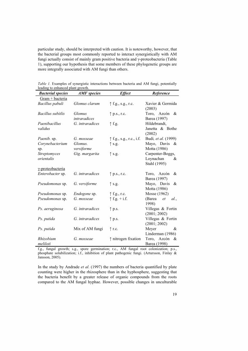
19
particular study, should be interpreted with caution. It is noteworthy, however, that the bacterial groups most commonly reported to interact synergistically with AM fungi actually consist of mainly gram positive bacteria and γ-proteobacteria (Table 1), supporting our hypothesis that some members of these phylogenetic groups are more integrally associated with AM fungi than others. Table 1. Examples of synergistic interactions between bacteria and AM fungi, potentially leading to enhanced plant growth. Bacterial species AMF species Effect Reference Gram + bacteria
Bacillus pabuli Glomus clarum ↑ f.g., s.g., r.c. Xavier & Germida (2003)
Bacillus subitlis Glomus intraradices
↑ p.s., r.c. Toro, Azcón & Barea (1997)
Paenibacillus validus
G. intraradices ↑ f.g. Hildebrandt, Janetta & Bothe (2002)
Paenib. sp. G. mosseae ↑ f.g., s.g., r.c., i.f. Budi, et al. (1999) Corynebacterium sp.
Glomus. versiforme
↑ s.g. Mayo, Davis & Motta (1986)
Streptomyces orientalis
Gig. margarita ↑ s.g. Carpenter-Boggs, Loynachan & Stahl (1995)
γ-proteobacteria Enterobacter sp. G. intraradices ↑ p.s., r.c. Toro, Azcón &
Barea (1997) Pseudomonas sp. G. versiforme ↑ s.g. Mayo, Davis &
Motta (1986) Pseudomonas sp. Endogone sp. ↑ f.g., r.c. Mosse (1962) Pseudomonas sp. G. mosseae ↑ f.g. + i.f. (Barea et al.,
1998) Ps. aeruginosa G. intraradices ↑ p.s. Villegas & Fortin
(2001; 2002) Ps. putida G. intraradices ↑ p.s. Villegas & Fortin
(2001; 2002) Ps. putida Mix of AM fungi ↑ r.c. Meyer &
Linderman (1986) Rhizobium meliloti
G. mosseae ↑ nitrogen fixation Toro, Azcón & Barea (1998)
f.g., fungal growth; s.g., spore germination; r.c., AM fungal root colonization; p.s., phosphate solubilization; i.f., inhibition of plant pathogenic fungi. (Artursson, Finlay & Jansson, 2005). In the study by Andrade et al. (1997) the numbers of bacteria quantified by plate counting were higher in the rhizosphere than in the hyphosphere, suggesting that the bacteria benefit by a greater release of organic compounds from the roots compared to the AM fungal hyphae. However, possible changes in unculturable
20
taxa were not evaluated in that study. In paper II, we used molecular tools to bypass the problems commonly encountered with culture-based approaches to visualize changes in actively growing bacterial community compositions as a result of G. mosseae inoculation or plant species. We found that mostly “uncultured bacteria” and Paenibacillus sp. were active in the G. mosseae inoculated soil, suggesting that many species of interest may be missed if relying on culturing alone. In addition to direct stimulation or inhibition of particular bacterial taxa by organic compounds released from roots or hyphae, it is also possible that there are indirect effects. For example, it is known that AM fungi influence soil aggregates through exudation of glycoproteins such as glomalin (Zhu & Miller, 2003) and Andrade et al. (1998) demonstrated that there were differences in the bacteria associated with water-stable soil aggregates compared with the non-stable soil fraction (Artursson, Finlay & Jansson, 2005). Bacteria enhancing nitrogen bioavailability Nitrogen-fixing bacteria are known to improve the bioavailability of nitrogen to plants, and this capability may be enhanced when plants are also colonized by AM fungi (Barea, Azcon & Azcon-Aguilar, 2002). For N2 fixing rhizobia, the mycorrhizal and root nodule symbioses are typically synergistic both with regard to infection rate and their impact on mineral nutrition and growth of the plant. Although, AM fungi may contribute to an increased nutrient status in the mycorrhizosphere, by decomposing organic N compounds, plants may have a greater benefit through additional nitrogen provided through N2 fixation. Toro, Azcón & Barea (1998) used the 15N/14N ratio in plant shoots to show that N2 fixation rates in Rhizobium meliloti inoculated mycorrhizal alfalfa plants were higher than the corresponding rates in non-mycorrhizal plants. One explanation for increased N2 fixation in mycorrhizal plants is that when both nitrogen and phosphorus are limiting, AM fungi can improve phosphorus uptake by the plant which in turn would result in more energy available for nitrogen fixation by rhizobia (Fitter & Garbaye, 1994; Kucey & Paul, 1982). In support of this hypothesis, it has been found that the enhanced N2-fixing ability in mycorrhizal plants compared to non-mycorrhizal plants, usually disappears if the non-mycorrhizal plants are supplied with a readily available P source (Karandashov & Bucher, 2005; Smith & Read, 1997). The uptake of other essential micronutrients from the soil by the AM fungal hyphae might also play a role in general plant growth improvement as well as in more indirect effects upon the N2-fixing system. However, although the main mycorrhizal effect in enhancing N2 fixation is apparently mediated by a generalized stimulation of host nutrition, more specific effects may take place at the root or nodule level (Barea, Azcón & Azcón-Aguilar, 1992). Interactions between AM fungi and rhizobia may, for example, occur at either the pre-colonization stages, when both microorganisms are localized in the mycorrhizosphere, or during the development of the tripartite symbiosis (Azcón-Aguilar & Barea, 1992). In addition, AM fungi may interact with both symbiotic and free-living N2-fixing bacteria (Barea, 1997).
21
Organic forms of nitrogen may also be made more available by bacteria associated with mycorrhizal fungal hyphae. Recent experiments by Hodge, Campbell & Fitter (2001) demonstrated that the AM fungus Glomus hoi was able to enhance decomposition and increase plant N capture from grass leaves. However, further research is still needed to distinguish between the direct capacity of AM fungi to mobilise organic substrates and their possible, indirect effects on decomposition and plant nutrient uptake, caused by stimulation of decomposers and subsequent uptake of their decomposition products by mycorrhizal hyphae (Artursson, Finlay & Jansson, 2005).
Bacteria enhancing phosphorus bioavailability Bacteria may also support the AM symbiosis by increasing bioavailable phosphate. In soil with low P bioavailability, free-living phosphate-solubilizing bacteria may release phosphate ions from sparingly soluble inorganic and organic P compounds in soil (Kucey, Janzen & Leggett, 1989), and thereby contribute with an increased soil phosphate pool available for the extraradical AM fungal hyphae to pass on to the plant (Smith & Read, 1997). The inorganic form of P may be held firmly in crystal lattices of largely insoluble forms, and may also be chemically bonded to the surface of clay minerals and unavailable to plants. Organic P is also largely unavailable to plants until it is converted to an inorganic form for example, by phosphate-solubilizing bacteria. Soluble P entering the soil after mineralization by such bacteria results in localized and short-term increases in the concentration of phosphate ions in the soil solution, which AM fungal hyphae and subsequently plants may benefit from. Organic P may be mineralized by bacteria that secrete phosphatases whereas inorganic P may be released by bacteria that excrete organic acids (Smith & Read, 1997). Several studies have demonstrated synergistic interactions between phosphate-solubilizing bacteria and AM fungi (Barea, Azcón-Aguilar & Azcón, 1997; Kim, Jordan & McDonald, 1998). For example, Toro, Azcón & Barea (1997) studied phosphate limited systems containing plants, AM fungi and phosphate-solubilizing bacteria. Their study revealed that the bacteria promoted mycorrhizal establishment whereas the mycorrhizal symbiosis increased the size of the phosphate-solubilizing bacterial population. The treatments inoculated with both AM fungi and bacteria significantly increased plant biomass and N and P accumulation in plant tissues, compared to their controls which were not dually inoculated. Using 32P isotopic dilution approaches they found that dually inoculated plants displayed lower specific activities (32P/31P) than control plants, indicating that AM fungi and phosphate-solubilizing bacteria interacted to make use of P sources otherwise unavailable to plants (Artursson, Finlay & Jansson, 2005).
22
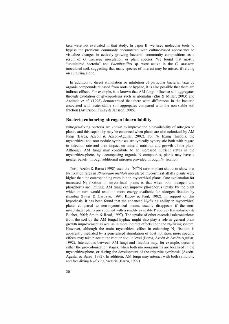
AM fungal endosymbiotic bacteria Endocellular bacteria are reported in only a few fungi including some Glomeromycota species (AM fungi and Geosiphon pyriforme) (Bianciotto et al., 1996a; Bianciotto et al., 2000; de Boer et al., 2005; Perotto & Bonfante, 1997; Scannerini & Bonfante, 1991; Schüssler & Kluge, 2001) and in the ectomycorrhizal basidiomycete Laccaria bicolor (Bertaux et al., 2003). Regarding the AM fungi, their cytoplasm harbours bacteria-like organisms (Fig. 3), which have been observed by microscopy in several of these fungal species (Glomus versiforme, Acaulospora laevis, Gigaspora margarita) (Bonfante, Balestrini & Mendgen, 1994; MacDonald & Chandler, 1981; Mosse, 1970; Scannerini & Bonfante, 1991).
Fig. 3. Schematic view of endocellular bacteria Ca. Glomeribacter gigasporarum living in AM fungi. (A) Bacteria (green rods) inside the cytoplasm of an AM fungal spore (circles within the spore represent its many nuclei). (B) The expected total genome size of Ca. Glomeribacter gigasporarum is 1.4 Mb, consisting of a ca. 750-kb chromosome and an additional 650-kb replicon. The drawing is not to scale and underestimates the relative surface area of the extraradical mycorrhizal mycelium (Artursson, Finlay & Jansson, 2005). Further investigation of these structures, including the demonstration of their prokaryotic nature, was long regarded as a task too complicated because they could not be cultured. However, by using morphological observations in combination with molecular analyses, Bianciotto et al. (1996a) succeeded in showing that they actually were of true bacterial origin. They also demonstrated the AM fungal endosymbiotic properties of these bacteria, that they were able to
23
complete their life cycles within fungal cells, and that the bacterial cells were gram-negative and rod-shaped. Several additional characteristics of the endosymbiotic bacterial genome have since been reported (Minerdi, Bianciotto & Bonfante, 2002; Minerdi, Fani & Bonfante, 2002; Minerdi et al., 2001; Ruiz-Lozano & Bonfante, 1999; Ruiz-Lozano & Bonfante, 2000). Endosymbiotic bacteria have been detected in several members of the Gigasporaceae; actually the only fungal species in this family among the evaluated ones, reported not to contain such bacteria was Gigaspora rosea (Bianciotto, et al., 2000). In the five other species belonging to the Gigasporaceae, intracellular bacteria were detected through all the steps of the fungal life cycle: spores, germtubes, and extra- and intraradical hyphae, except arbuscules (Bianciotto, et al., 1996a). The AM fungus most extensively studied for its endosymbiotic bacteria is Gig. margarita isolate BEG 34, which was also the first fungus in which these prokaryotic cells were further investigated (Bianciotto, et al., 1996a). Recent studies have indicated an average of about 20,000 bacteria per Gig. margarita spore (Bianciotto et al., 2004; Jargeat et al., 2004). These bacteria were initially assigned to the genus Burkholderia on the basis of their 16S ribosomal RNA gene sequence, but were recently reassigned to a new taxon termed Candidatus Glomeribacter gigasporarum (Bianciotto et al., 2003). In spite of several attempts, these bacteria have never been grown on cell-free media (Bianciotto, et al., 2004; Jargeat, et al., 2004; MacDonald & Chandler, 1981; Scannerini & Bonfante, 1991), which is the reason why they are assigned to the provisional Candidatus designation for uncultured bacteria (Murray & Schleifer, 1994; Murray & Stackebrandt, 1995). The physiological role of the endosymbiotic bacteria in AM fungi is unknown, as is their potential role in the mycorrhizal symbiosis (Jargeat, et al., 2004). However, some hints about such roles were derived from a genomic library developed from Gig. margarita spores, shown to also represent the genome of the bacterial endosymbiont (van Buuren et al., 1999). Among the bacterial genes isolated from this library and from genomic spore DNA were several interesting finds, including a putative phosphate transporter gene, pst (Ruiz-Lozano & Bonfante, 1999), a vacB-like gene involved in host cell colonization by enteroinvasive, pathogenic bacteria (Shigella flexneri and Escherichia coli) (Ruiz-Lozano & Bonfante, 2000), three nif-genes (nifH, nifD, and nifK) (Minerdi, et al., 2001), the mcpA (Minerdi, Fani & Bonfante, 2002) and cheY (Minerdi, Bianciotto & Bonfante, 2002) genes which are involved in chemotaxis, a kinase gene (prkA) and a spoVR gene (Minerdi, Bianciotto & Bonfante, 2002) which is involved in coat formation of bacterial endospores. Jargeat et al. (2004) tried to verify the presence of these genes in Ca. Glomeribacter gigasporarum, using DNA obtained from pure genomic preparations, but were not able to PCR amplify several of the genes. They concluded that the original genomic library derived from Gig. margarita spores may have been contaminated with foreign bacterial DNA, which also seemed to be confirmed when further screening of the library was performed (Jargeat, et al., 2004). However, the pst and the vacB genes were still detected and these might be of particular interest for future determination of the potential role of the bacterium in the mycorrhizal symbiosis.
24
Until recently the mode of transmission of the endosymbionts to succeeding generations of the AM fungi was not established. Two alternatives include permanent and cyclical endosymbioses. A permanent symbiosis remains stable over time whereas a cyclical one involves regular reassociation events. For example, each AM fungal colonization event requires a reassociation of the fungal propagule with its host plant (Bianciotto, et al., 2004), and is therefore considered to represent a cyclical symbiosis. Similar modes of transmission are also found for G. pyriforme, in which cyanobacteria penetrate the fungi through an endocytotic process (Schüssler & Kluge, 2001). Conversely, Bianciotto et al. (2004) demonstrated that cells of Ca. Glomeribacter gigasporarum were vertically transmitted through five fungal vegetative generations of Gig. margarita spores. The asexual reproduction typical of AM fungi and the coenocytic nature of their mycelium may facilitate the migration of the endosymbiotic bacteria from spores to hyphae, and thereby allow for the vertical transmission to take place. Active bacterial proliferation was demonstrated to occur in the fungal mycelium, and the authors suggested that these bacteria are obligate endocellular components of their AM fungal host, and thus represent a permanent endosymbiosis unlike the majority of endosymbioses present in the plant kingdom (Bianciotto, et al., 2004). Ca. Glomeribacter gigasporarum has a surprisingly small genome size for a bacterium, only around 1.4 Mb in total consisting of a ca. 750-kb chromosome and an additional replicon of ca. 650-kb (Jargeat, et al., 2004). However, small genomes are often a feature of obligate endocellular bacterial species, a fact which might lend additional support to the hypotheses discussed above regarding the vertical transmission of Ca. Glomeribacter gigasporarum bacteria within AM fungi, and their obligate endocellular nature. Considering processes like reductive evolution where only those genes absolutely essential for survival in an intracellular environment should be retained (Dale et al., 2002), Ca. Glomeribacter gigasporarum represents a typical candidate for a permanent endosymbiont with its small genome size. One of the major future challenges within this research area, is to reveal the functional significance of AM fungal endobacteria. One important step in this direction is to be able to remove the bacteria from the fungal cytoplasm, enabling comparisons of fungal effects on plants, in the presence and absence of the bacterial symbiont. Recently, Bonfante and co-workers obtained spores that were devoid of bacteria after successive vegetative generations, resulting in cured spores (P. Bonfante, personal communication). These present an excellent tool for further elucidation of the impact of the endosymbiont on the fungus and subsequently on plants (Artursson, Finlay & Jansson, 2005).

25
Microbiomics tools
Studies of interactions between AM fungi and bacteria will greatly benefit from application of new molecular approaches, which might enable valuable insights into the mechanisms of these associations, as well as important information regarding fungal and bacterial community structures and metabolic activities. Such molecular tools facilitating the characterization of complex microbial communities are defined in this thesis as “microbiomics”, and the following approaches are all included in this term. Green fluorescent protein tagging One popular technique for monitoring specific microorganisms is by tagging them with so-called “marker genes” or “biomarkers” (Jansson, 2003; Jansson & de Bruijn, 1999). The organisms can then be tracked based on their unique phenotype conferred by the marker gene or, alternatively, the marker gene DNA can be detected directly (Unge, 2000). One of the most widely used marker genes today is the green fluorescent protein (GFP) which was originally isolated from the jellyfish Aequorea victoria. GFP is a 27-k Dalton monomeric protein consisting of 238 amino acids, with a cylindrical structure encapsulating the chromophore element in the centre. Once expressed by the cell, autocatalytic oxidation and cyclization of the GFP amino acids at positions 65 to 67 leads to the formation of the chromophore. However, to fluoresce the chromophore also needs further interactions with other parts of the protein (Yang, Moss & Phillips, 1996). The oxidation reaction only requires the presence of molecular oxygen, and the subsequent fluorescence does not require any additional gene products, substrates or other factors to work properly (http://dias.umist.ac.uk/NJG/Abstracts/GFP1/Analcom.htm; 27-Oct-2005), making this naturally fluorescent protein convenient to use for expression in various prokaryotic and eukaryotic organisms (Chalfie et al., 1994). The wild type GFP has two excitation peaks, a major one at 395 nm (in the long UV range) and a smaller one at 475 nm (blue) with its emission peak at 509 nm (green). One problem with this wildtype protein, however, is that the excitation at 395 nm, causes fairly rapid quenching of the fluorescence (photobleaching), due to photoisomerisation (Cubitt et al., 1995). Additional drawbacks with the wildtype GFP include the protein being trapped in inclusion bodies which are non-fluorescent (Heim, Prasher & Tsien, 1994), and slow folding of the protein (approximately 4 hours). To alleviate these problems, several GFP mutants have been constructed, with shifted excitation and emission wavelengths, higher fluorescence intensities, better solubility and faster folding (Unge, 2000). Among the most widely used mutants are the red-shifted GFPs, having their excitation maximum around 490 nm with their emission remaining at 509 nm. Such variants with increased excitation at longer wavelengths due to ionisation of the chromophore, have been especially important due to their higher fluorescence intensities (Tsien, 1998), and it has been suggested that this increase is due to more efficient protein folding or chromophore formation.
26
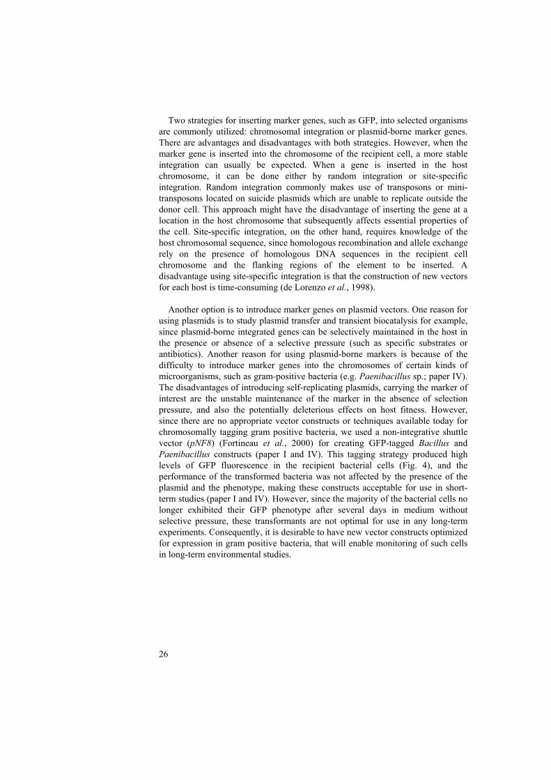
Two strategies for inserting marker genes, such as GFP, into selected organisms are commonly utilized: chromosomal integration or plasmid-borne marker genes. There are advantages and disadvantages with both strategies. However, when the marker gene is inserted into the chromosome of the recipient cell, a more stable integration can usually be expected. When a gene is inserted in the host chromosome, it can be done either by random integration or site-specific integration. Random integration commonly makes use of transposons or mini-transposons located on suicide plasmids which are unable to replicate outside the donor cell. This approach might have the disadvantage of inserting the gene at a location in the host chromosome that subsequently affects essential properties of the cell. Site-specific integration, on the other hand, requires knowledge of the host chromosomal sequence, since homologous recombination and allele exchange rely on the presence of homologous DNA sequences in the recipient cell chromosome and the flanking regions of the element to be inserted. A disadvantage using site-specific integration is that the construction of new vectors for each host is time-consuming (de Lorenzo et al., 1998). Another option is to introduce marker genes on plasmid vectors. One reason for using plasmids is to study plasmid transfer and transient biocatalysis for example, since plasmid-borne integrated genes can be selectively maintained in the host in the presence or absence of a selective pressure (such as specific substrates or antibiotics). Another reason for using plasmid-borne markers is because of the difficulty to introduce marker genes into the chromosomes of certain kinds of microorganisms, such as gram-positive bacteria (e.g. Paenibacillus sp.; paper IV). The disadvantages of introducing self-replicating plasmids, carrying the marker of interest are the unstable maintenance of the marker in the absence of selection pressure, and also the potentially deleterious effects on host fitness. However, since there are no appropriate vector constructs or techniques available today for chromosomally tagging gram positive bacteria, we used a non-integrative shuttle vector (pNF8) (Fortineau et al., 2000) for creating GFP-tagged Bacillus and Paenibacillus constructs (paper I and IV). This tagging strategy produced high levels of GFP fluorescence in the recipient bacterial cells (Fig. 4), and the performance of the transformed bacteria was not affected by the presence of the plasmid and the phenotype, making these constructs acceptable for use in short-term studies (paper I and IV). However, since the majority of the bacterial cells no longer exhibited their GFP phenotype after several days in medium without selective pressure, these transformants are not optimal for use in any long-term experiments. Consequently, it is desirable to have new vector constructs optimized for expression in gram positive bacteria, that will enable monitoring of such cells in long-term environmental studies.
27
Fig. 4. Bacillus cereus strain VA1; the wildtype (left) and the GFP-tagged strain (right).
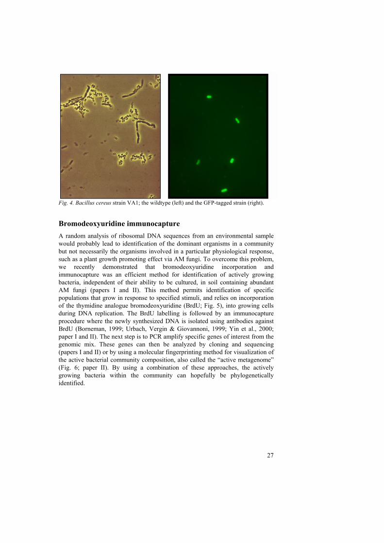
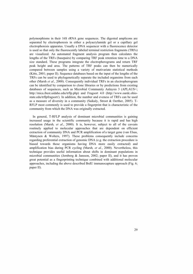
Bromodeoxyuridine immunocapture A random analysis of ribosomal DNA sequences from an environmental sample would probably lead to identification of the dominant organisms in a community but not necessarily the organisms involved in a particular physiological response, such as a plant growth promoting effect via AM fungi. To overcome this problem, we recently demonstrated that bromodeoxyuridine incorporation and immunocapture was an efficient method for identification of actively growing bacteria, independent of their ability to be cultured, in soil containing abundant AM fungi (papers I and II). This method permits identification of specific populations that grow in response to specified stimuli, and relies on incorporation of the thymidine analogue bromodeoxyuridine (BrdU; Fig. 5), into growing cells during DNA replication. The BrdU labelling is followed by an immunocapture procedure where the newly synthesized DNA is isolated using antibodies against BrdU (Borneman, 1999; Urbach, Vergin & Giovannoni, 1999; Yin et al., 2000; paper I and II). The next step is to PCR amplify specific genes of interest from the genomic mix. These genes can then be analyzed by cloning and sequencing (papers I and II) or by using a molecular fingerprinting method for visualization of the active bacterial community composition, also called the “active metagenome” (Fig. 6; paper II). By using a combination of these approaches, the actively growing bacteria within the community can hopefully be phylogenetically identified.
28
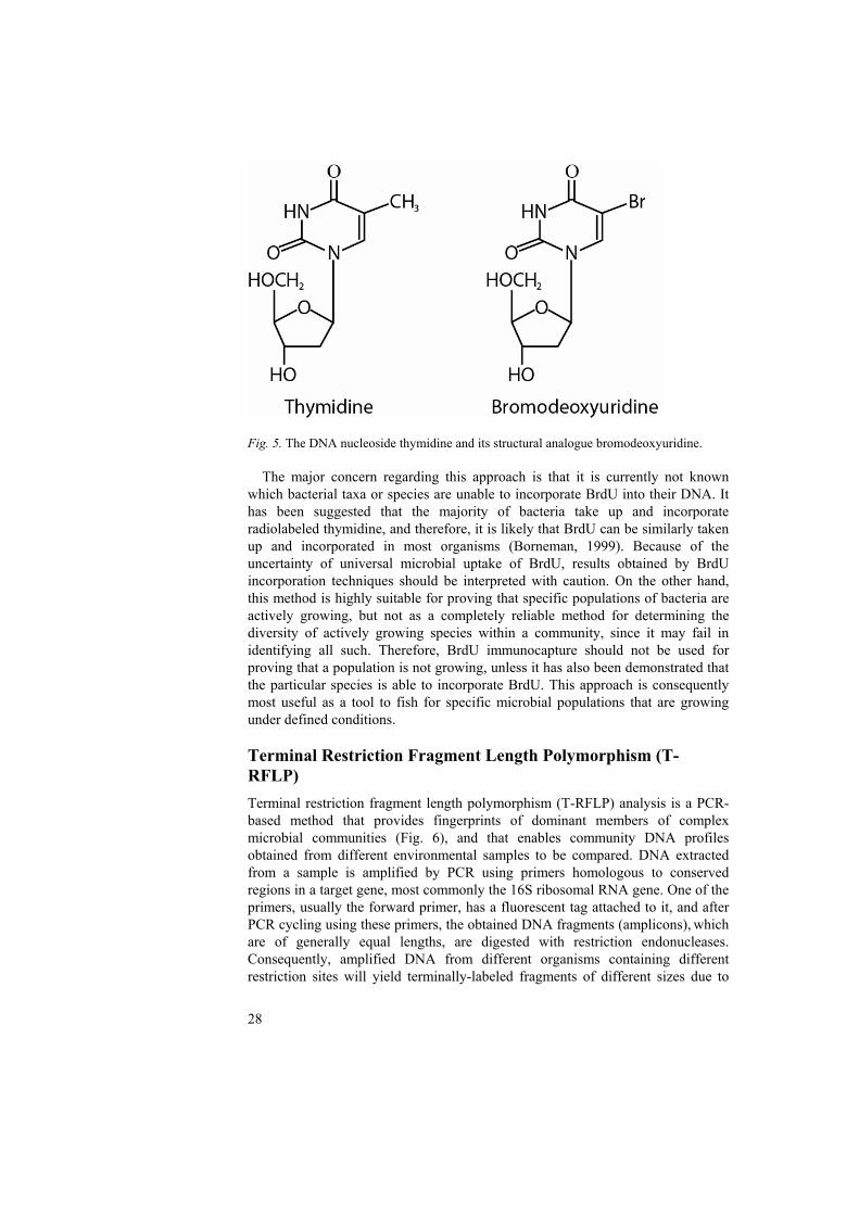
Fig. 5. The DNA nucleoside thymidine and its structural analogue bromodeoxyuridine. The major concern regarding this approach is that it is currently not known which bacterial taxa or species are unable to incorporate BrdU into their DNA. It has been suggested that the majority of bacteria take up and incorporate radiolabeled thymidine, and therefore, it is likely that BrdU can be similarly taken up and incorporated in most organisms (Borneman, 1999). Because of the uncertainty of universal microbial uptake of BrdU, results obtained by BrdU incorporation techniques should be interpreted with caution. On the other hand, this method is highly suitable for proving that specific populations of bacteria are actively growing, but not as a completely reliable method for determining the diversity of actively growing species within a community, since it may fail in identifying all such. Therefore, BrdU immunocapture should not be used for proving that a population is not growing, unless it has also been demonstrated that the particular species is able to incorporate BrdU. This approach is consequently most useful as a tool to fish for specific microbial populations that are growing under defined conditions. Terminal Restriction Fragment Length Polymorphism (T-RFLP) Terminal restriction fragment length polymorphism (T-RFLP) analysis is a PCR-based method that provides fingerprints of dominant members of complex microbial communities (Fig. 6), and that enables community DNA profiles obtained from different environmental samples to be compared. DNA extracted from a sample is amplified by PCR using primers homologous to conserved regions in a target gene, most commonly the 16S ribosomal RNA gene. One of the primers, usually the forward primer, has a fluorescent tag attached to it, and after PCR cycling using these primers, the obtained DNA fragments (amplicons), which are of generally equal lengths, are digested with restriction endonucleases. Consequently, amplified DNA from different organisms containing different restriction sites will yield terminally-labeled fragments of different sizes due to
29
polymorphisms in their 16S rRNA gene sequences. The digested amplicons are separated by electrophoresis in either a polyacrylamide gel or a capillary gel electrophoresis apparatus. Usually a DNA sequencer with a fluorescence detector is used so that only the fluorescently labeled terminal restriction fragments (TRFs) are visualized. An automated fragment analysis program then calculates the lengths of the TRFs (basepairs) by comparing TRF peak retention time to a DNA size standard. These programs integrate the electropherograms and return TRF peak height and area. The patterns of TRF peaks can then be numerically compared between samples using a variety of mutivariate statistical methods (Kitts, 2001; paper II). Sequence databases based on the input of the lengths of the TRFs can be used to phylogenetically separate the included organsims from each other (Marsh et al., 2000). Consequently individual TRFs in an electropherogram can be identified by comparison to clone libraries or by predictions from existing databases of sequences, such as Microbial Community Anlaysis 3 (APLAUS+; http://mica.ibest.uidaho.edu/trflp.php) and Fragsort 4.0 (http://www.oardc.ohio-state.edu/trflpfragsort/). In addition, the number and eveness of TRFs can be used as a measure of diversity in a community (Saikaly, Stroot & Oerther, 2005). T-RFLP most commonly is used to provide a fingerprint that is characteristic of the community from which the DNA was originally extracted.
In general, T-RFLP analysis of dominant microbial communities is gaining increased usage in the scientific community because it is rapid and has high resolution (Marsh, et al., 2000). It is, however, subject to all of the caveats
routinely applied to molecular approaches that are dependent on efficient extraction of community DNA and PCR amplification of a target gene (van Elsas, Mäntynen & Wolters, 1997). These problems consequently include concerns regarding preferential extraction of genomic DNA (e.g. the extraction procedure is biased towards those organisms having DNA more easily extracted) and
amplification bias during PCR cycling (Marsh, et al., 2000). Nevertheless, this technique provides useful information about shifts in dominant populations in microbial communities (Jernberg & Jansson, 2002; paper II), and it has proven great potential as a fingerprinting technique combined with additional molecular approaches, including the above described BrdU immunocapture approach (Fig. 6; paper II).
30
Fig. 6. Schematic drawing of a suitable approach, involving BrdU immunocapture, T-RFLP and clone libraries, for identifying actively growing bacterial populations within soil bacterial communities (paper II). Specific AM fungal-bacterial interactions
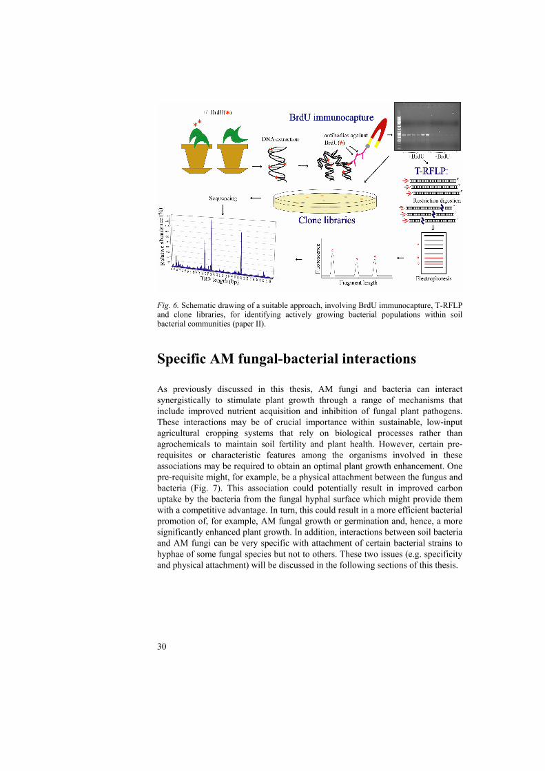
As previously discussed in this thesis, AM fungi and bacteria can interact synergistically to stimulate plant growth through a range of mechanisms that include improved nutrient acquisition and inhibition of fungal plant pathogens. These interactions may be of crucial importance within sustainable, low-input agricultural cropping systems that rely on biological processes rather than agrochemicals to maintain soil fertility and plant health. However, certain pre-requisites or characteristic features among the organisms involved in these associations may be required to obtain an optimal plant growth enhancement. One pre-requisite might, for example, be a physical attachment between the fungus and bacteria (Fig. 7). This association could potentially result in improved carbon uptake by the bacteria from the fungal hyphal surface which might provide them with a competitive advantage. In turn, this could result in a more efficient bacterial promotion of, for example, AM fungal growth or germination and, hence, a more significantly enhanced plant growth. In addition, interactions between soil bacteria and AM fungi can be very specific with attachment of certain bacterial strains to hyphae of some fungal species but not to others. These two issues (e.g. specificity and physical attachment) will be discussed in the following sections of this thesis.
31
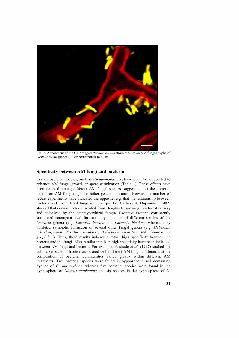
Fig. 7. Attachment of the GFP-tagged Bacillus cereus strain VA1 to an AM fungal hypha of Glomus dussii (paper I). Bar corresponds to 4 μm. Specificity between AM fungi and bacteria Certain bacterial species, such as Pseudomonas sp., have often been reported to enhance AM fungal growth or spore germination (Table 1). These effects have been detected among different AM fungal species, suggesting that the bacterial impact on AM fungi might be rather general in nature. However, a number of recent experiments have indicated the opposite, e.g. that the relationship between bacteria and mycorrhizal fungi is more specific. Garbaye & Duponnois (1992) showed that certain bacteria isolated from Douglas fir growing in a forest nursery and colonized by the ectomycorrhizal fungus Laccaria laccata, consistently stimulated ectomycorrhizal formation by a couple of different species of the Laccaria genera (e.g. Laccaria laccata and Laccaria bicolor), whereas they inhibited symbiotic formation of several other fungal genera (e.g. Hebeloma cylindrosporum, Paxillus involutus, Telephora terrestris and Cenococcum geophilum). Thus, these results indicate a rather high specificity between the bacteria and the fungi. Also, similar trends in high specificity have been indicated between AM fungi and bacteria. For example, Andrade et al. (1997) studied the culturable bacterial fraction associated with different AM fungi and found that the composition of bacterial communities varied greatly within different AM treatments. Two bacterial species were found in hyphospheric soil containing hyphae of G. intraradices, whereas five bacterial species were found in the hyphosphere of Glomus etunicatum and six species in the hyphosphere of G.
32
mosseae, under identical conditions (Andrade, et al., 1997). Thus, these results indicate preferential combinations of AM fungi and bacteria. Since less than 10 % of the total bacterial community members in soil are estimated to be cultured to date, molecular approaches are necessary to get a better representative picture of the bacterial communities in natural soil that respond to the presence of certain AM fungi. Therefore, we chosed to combine two molecular approaches, bromodeoxyuridine (BrdU) immunocapture and terminal restriction fragment length polymorphism (T-RFLP), which in combination with sequence information from clone libraries, enabled the identification of actively growing populations, within the total bacterial community (paper II). Distinct differences in active bacterial community compositions were found in the soil according to G. mosseae inoculation, supporting the results obtained by Andrade et al. (1997) regarding a high AM fungal – bacterial specificity. The putative identities of the dominant bacterial species that were activated as a result of G. mosseae inoculation were found to be mostly uncultured bacteria and Paenibacillus species (paper II), suggesting that there remains a great amount of work to further our knowledge on the bacterial species associated with different AM fungal species. The Paenibacillus genus, in particular, warrants further attention with respect to its impact on AM fungi and plant growth. Paenibacillus strains have previously been shown to be associated with both G. mosseae (Budi, et al., 1999) and G. intraradices (Hildebrandt, Janetta & Bothe, 2002; Mansfeld-Giese, Larsen & Bödker, 2002), and to stimulate AM formation. In addition, we previously demonstrated that strains of Paenibacillus tightly adhered to hyphae of the AM fungi G. dussii, Glomus sp. and G. intraradices, whereas a number of bacterial control strains did not colonize to the same extent (papers I and III). Further investigation of the specificity between AM fungi and Paenibacillus strains is therefore of interest for the future application of these microorganisms, for example in the context of mixed microbial incoula. One possible explanation for the noted stimulation of certain bacterial species by specific AM fungi (Andrade, et al., 1997; paper II) may be that those bacteria are activated by species-specific fungal exudates. Therefore, it would be of interest to further elucidate the potential fungal mechanisms for attraction of specific bacteria. Physical interactions Several PGPR have been shown to be excellent root colonizers (Barea, Azcon & Azcon-Aguilar, 2002; Lugtenberg & Dekkers, 1999) and a number of surface components have been demonstrated to play a role in the physical interactions between such bacteria and plant roots (Bianciotto & Bonfante, 2002). However, little information is available concerning the extent to which PGPR colonize AM fungal hyphae. Bianciotto et al. (1996b) reported that some Rhizobium and Pseudomonas species attached to germinated AM fungal spores and hyphae under sterile conditions, and that the degree of attachment varied with the bacterial strain. However, no specificity for either fungal or inorganic surfaces could be detected among the bacteria tested. Based on their results, these authors suggested that interactions between rhizobacteria and AM fungi were mediated by soluble factors or physical contact.
33
Several bacteria reported to be good root colonizers, for example some Pseudomonas spp., are also capable of adhering to AM fungal hyphal surfaces, suggesting that the mechanisms involved could be fairly similar. Close cell-to-cell contact between for example, rhizobia and their host plant roots is an important prerequisite for the formation of the nodules during endosymbiosis, and one may speculate whether similar correlations exist between attachment of bacteria to AM fungal hyphal surfaces and changes in fungal growth or performance. To further evaluate this issue, a number of mycorrhizal associated PGPR were tested for their ability to attach to hyphae of ectomycorrhizal fungi. Although some of the bacteria adhered to the fungal mycelium (Sen et al., 1996), in another study positive effects of other rhizobacteria on ectomycorrhizal fungal development and establishment were observed even when attachment did not occur (Garbaye, 1994). One possible way that attachment could benefit both partners, would be through facilitation of certain metabolic interactions, such as nutrient and carbon exchange and this would rely on close cell contact between the bacterial and fungal components. We recently demonstrated that a Bacillus cereus strain, isolated from a Swedish soil containing abundant AM fungi, attached to hyphae of the AM fungus G. dussii at significantly higher levels than a number of bacterial control strains (paper I; Fig. 7), indicating that the colonization ability varies considerably between different bacteria. To further examine the biological features of bacterial attachment to AM fungal hyphae, we compared the attachment of five different gfp-tagged bacterial strains [Paenibacillus brasilensis PB177 (pnf8), Bacillus cereus VA1 (pnf8), Pseudomonas fluorescens SBW25::gfp/lux, Arthrobacter chlorophenolicus A6G, and Paenibacillus peoriae BD62 (pnf8)] to vital and non-vital hyphae of the AM fungi Glomus sp. MUCL 43205 and G. intraradices MUCL 43194 (paper III). This study indicated major differences between the bacterial strains in their ability to attach to different physiological states of hyphae and also that this can be influenced by the AM fungal species involved. A. chlorophenolicus strain A6G did not attach to any hyphae, whereas the four other bacterial strains did to a varying degree. Only P. brasilensis PB177 (pnf8) showed a higher degree of attachment to vital hyphae than to non-vital hyphae of both Glomus species tested. P. fluorescens SBW25::gfp/lux showed a higher level of attachment to vital compared with non-vital hyphae of Glomus sp. MUCL 43205, whereas this relationship was the opposite for attachment to hyphae of G. intraradices MUCL 43194. Both B. cereus and P. peoriae, on the other hand, showed a higher degree of attachment to non-vital hyphae compared to vital hyphae, independent of the AM fungus considered (paper III). Consequently, bacterial attachment to AM fungal hyphae does not necessarily mean that the bacterial-fungal association is beneficial to the AM fungus or the plant, but might also indicate a saprophytic lifestyle of the bacterium involved, benefiting from released carbon sources of dying hyphae (paper III). Since the effect of electrostatic attraction was diminished by washing the hyphae with strong salt solution before examination by microscopy, our results support those suggested by Bianciotto et al. (1996b) regarding a two-step mechanism. During the first stage of this proposed mechanism a weak binding will occur, often governed by general physicochemical parameters such as electrostatic attraction, whereas the second,
34
more stable binding can be explained by mechanisms involving the production of cellulose fibrils or other bacterial extracellular polymers. In support of this hypothesis, Bianciotto et al. (2001) studied bacterial mutants inhibited in extracellular polysaccharide production and found that they were less able to attach to AM fungal hyphal surfaces compared to the wild type strain. Additionally, to determine whether proteins were involved in the bacterial attachment to AM fungal hyphae, we treated the B. cereus and P. brasilensis strains with proteinase K (Artursson and Jansson, unpublished). However, this treatment did not affect the extent of attachment, futher supporting the hypothesis discussed above, concerning carbohydrates such as polysaccharides as one of the responsible factors for bacterial attachment to AM fungal hyphae, rather than, for example, polar flagella or amino acids. Since the significance of bacterial attachment for mycorrhizal functioning, especially within the AM symbiosis, is still not clear, the next step would be to evaluate whether there generally is a clear correlation between bacterial attachment to living mycorrhizal fungal hyphae and enhanced mycorrhizal fungal growth or performance. If that was shown to be the case, attachment properties should be an important feature to consider when screening for AM fungal compatible bacterial inoculants. Interactions with plant pathogenic fungi
Microbial inoculants can be used as alternative means for controlling pests and diseases in sustainable agriculture, permitting the reduced use of pesticides that could otherwise pose threats to human health and non-targeted organisms (Johansson, Paul & Finlay, 2004). In addition to testing the ability of microbial inoculants (e.g. AM fungi and bacteria) to enhance plant growth, it is therefore also critical to evaluate their single and synergistic potential to inhibit growth of plant pathogenic fungi, in turn indirectly leading to improved plant performance and better environmental conditions. Inhibition of pathogenic fungi by bacteria and/or AM fungi There are indications that AM fungi and bacteria, synergistically, are able to antagonize soil-borne fungal plant pathogens. For example, Citernesi et al. (1996) studied bacteria isolated from the mycorrhizosphere of G. mosseae kept in pot cultures for 17 years, and found that several of those were actively antagonistic against Fusarium and Phytophtora growing in vitro. The authors thus concluded that it would be possible to use AM fungi as vehicles for selected microorganisms in biocontrol of soil-borne fungal pathogens. In another study, Filion, St-Arnaud & Fortin (1999) tested the crude extract obtained from the growth medium of the AM fungus G. intraradices, on the growth of two bacteria and on the sporulation of two pathogenic fungi. Their results indicated that growth of Pseudomonas chlororaphis (a biocontrol agent) and conidial germination of Trichoderma harzianum (a mycoparasite) were stimulated in the presence of the AM fungal

35
extract, whereas growth of Clavibacter michiganensis (a plant pathogen) was not affected and germination of Fusarium oxysporum (a plant pathogen) was reduced. The measured effects were directly correlated with the extract concentration of G. intraradices and no significant influence of pH on growth or germination was noted. These results suggest that unspecified substances released by G. intraradices into the growth medium were the main factors explaining the differential growth response of the microorganisms tested (Filion, St-Arnaud & Fortin, 1999). The volume of biocontrol literature concerning bacterial antagonism to pathogenic fungi continues to increase at a rapid rate, stimulated by the growing ease with which molecular techniques can now be applied to answer queries concerning distribution, occurrence and relative importance of the specific modes of action involved (Whipps, 2001). For example, there are numerous reports of the production of antifungal metabolites produced by bacteria, including ammonia, butyrolactones and 2,4-diacetylphloroglucinol (DAPG). When releasing great amounts of such antifungal metabolite-producing bacterial biocontrol agents into nature, one concern might be the potentially negative effects they will have on the non-targeted indigenous AM fungal community. Barea et al. (1998) tested the compatibility of DAPG-producing Pseudomonas strains with the formation and functioning of AM associations. Surprisingly, they found that Pseudomonas sp., isolated from the rhizosphere of mature sugar beets (Fenton et al., 1992), stimulated both G. mosseae mycelial development from spores germinating in soil, and AM fungal root colonization of tomato plants (Barea, et al., 1998). These results suggest that DAPG is very specific, exhibiting antifungal activity against pathogenic fungi but not against the tested AM fungus. If such strains of Pseudomonas can be used as biocontrol inoculants without having concerns about their ecological impact on beneficial indigenous soil microbial populations (e.g. AM fungi), it might significantly facilitate the introduction and utilization of new biocontrol products on the market. It is well known that some AM fungi, also without bacterial contribution, can protect plants against fungal root pathogens (Newsham, Fitter & Watkinson, 1995; Niemira, Hammerschmidt & Safir, 1996). The underlying mechanisms however, are not very well understood, although a few alternatives have been suggested, such as improvement of plant nutrition and competition for photosynthates, or changes in root exudation leading to less carbohydrates available to pathogens (Azcon-Aguilar & Barea, 1996). It is important to keep in mind that the efficiency of biocontrol strains under field conditions is likely to be affected by several factors, including pH, temperature, water content, and interactions with other microorganisms. Therefore, more studies are required to better understand complex biological interactions before introducing potential inoculum candidates into the field.
36
Paenibacillus brasilensis PB177 Paenibacillus brasilensis was recently described as a new nitrogen-fixing species isolated from the maize rhizosphere in Brazil. In addition, P. brasilensis strain PB177 was shown to produce antimicrobial substances, suggesting its potentially important role in nature; not only by fixing nitrogen but also by preventing plant diseases (von der Weid et al., 2002). Thus, we aimed to thoroughly evaluate the interactions between P. brasilensis PB177, pathogenic fungi, and plants (e.g. maize), by studying the potential antagonism of the bacterium towards phytopathogenic fungi causing diseases in maize plants (paper IV). GFP was used to tag P. brasilensis PB177, enabling the monitoring of the bacterial-fungal interactions in situ on maize seeds and roots. Complimentary in vitro assays were also performed to further evaluate the interactions, confirming the production and secretion of an antifungal compound by P. brasilensis strain PB177 able to inhibit growth of all five phytopathogenic fungal strains tested in the present plate assay (e.g. Diplodia macrospora, Fusarium moniliforme, Fusarium oxysporum, Rhizoctonia solani, and Verticillium dahliae) (Fig. 8). Two of these fungi, one more and one less susceptible (D. macrospora and F. moniliforme, respectively) to the compound produced by the bacterium, were chosen for further studies in which paired cultures of bacteria and fungi in liquid medium were employed. This study demonstrated that the presence of bacteria led to a quick death of hyphae of both fungal species, starting with the attachment of bacteria to fungal hyphae, followed by hyphal deformation, the enlargement of cytoplasmic vacuoles and ultimately, loss of cellular impermeability to trypan blue (used as the indicator dye for damaged cells), suggesting that the presence of bacteria caused a strong inhibition of fungal growth.
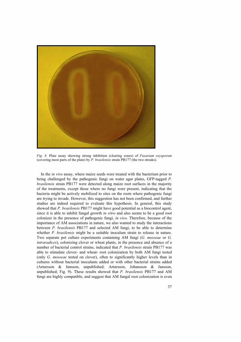
37
Fig. 8. Plate assay showing strong inhibition (clearing zones) of Fusarium oxysporum (covering most parts of the plate) by P. brasilensis strain PB177 (the two streaks). In the in vivo assay, where maize seeds were treated with the bacterium prior to being challenged by the pathogenic fungi on water agar plates, GFP-tagged P. brasilensis strain PB177 were detected along maize root surfaces in the majority of the treatments, except those where no fungi were present, indicating that the bacteria might be actively mobilized to sites on the roots where pathogenic fungi are trying to invade. However, this suggestion has not been confirmed, and further studies are indeed required to evaluate this hypothesis. In general, this study showed that P. brasilensis PB177 might have good potential as a biocontrol agent, since it is able to inhibit fungal growth in vitro and also seems to be a good root colonizer in the presence of pathogenic fungi, in vivo. Therefore, because of the importance of AM associations in nature, we also wanted to study the interactions between P. brasilensis PB177 and selected AM fungi, to be able to determine whether P. brasilensis might be a suitable inoculum strain to release in nature. Two separate pot culture experiments containing AM fungi (G. mosseae or G. intraradices), colonizing clover or wheat plants, in the presence and absence of a number of bacterial control strains, indicated that P. brasilensis strain PB177 was able to stimulate clover- and wheat- root colonization by both AM fungi tested (only G. mosseae tested on clover), often to significantly higher levels than in cultures without bacterial inoculants added or with other bacterial strains added (Artursson & Jansson, unpublished; Artursson, Johansson & Jansson, unpublished; Fig. 9). These results showed that P. brasilensis PB177 and AM fungi are highly compatible, and suggest that AM fungal root colonization is even
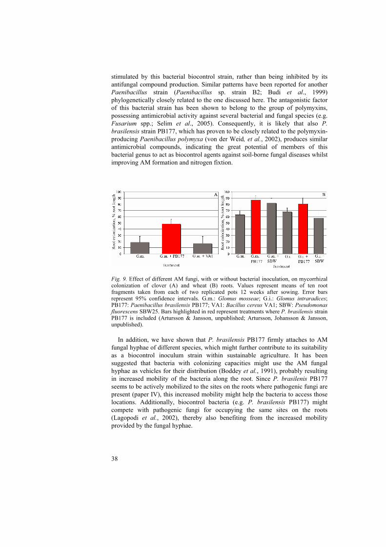
38
stimulated by this bacterial biocontrol strain, rather than being inhibited by its antifungal compound production. Similar patterns have been reported for another Paenibacillus strain (Paenibacillus sp. strain B2; Budi et al., 1999) phylogenetically closely related to the one discussed here. The antagonistic factor of this bacterial strain has been shown to belong to the group of polymyxins, possessing antimicrobial activity against several bacterial and fungal species (e.g. Fusarium spp.; Selim et al., 2005). Consequently, it is likely that also P. brasilensis strain PB177, which has proven to be closely related to the polymyxin-producing Paenibacillus polymyxa (von der Weid, et al., 2002), produces similar antimicrobial compounds, indicating the great potential of members of this bacterial genus to act as biocontrol agents against soil-borne fungal diseases whilst improving AM formation and nitrogen fixtion.
Fig. 9. Effect of different AM fungi, with or without bacterial inoculation, on mycorrhizal colonization of clover (A) and wheat (B) roots. Values represent means of ten root fragments taken from each of two replicated pots 12 weeks after sowing. Error bars represent 95% confidence intervals. G.m.: Glomus mosseae; G.i.: Glomus intraradices; PB177: Paenibacillus brasilensis PB177; VA1: Bacillus cereus VA1; SBW: Pseudomonas fluorescens SBW25. Bars highlighted in red represent treatments where P. brasilensis strain PB177 is included (Artursson & Jansson, unpublished; Artursson, Johansson & Jansson, unpublished). In addition, we have shown that P. brasilensis PB177 firmly attaches to AM fungal hyphae of different species, which might further contribute to its suitability as a biocontrol inoculum strain within sustainable agriculture. It has been suggested that bacteria with colonizing capacities might use the AM fungal hyphae as vehicles for their distribution (Boddey et al., 1991), probably resulting in increased mobility of the bacteria along the root. Since P. brasilenis PB177 seems to be actively mobilized to the sites on the roots where pathogenic fungi are present (paper IV), this increased mobility might help the bacteria to access those locations. Additionally, biocontrol bacteria (e.g. P. brasilensis PB177) might compete with pathogenic fungi for occupying the same sites on the roots (Lagopodi et al., 2002), thereby also benefiting from the increased mobility provided by the fungal hyphae.
39
Conclusions
The major findings of this thesis are:
• BrdU immunocapture is a suitable method for identifying actively growing soil bacteria associated with AM fungi (papers I and II), especially in combination with other microbiomics tools (papers I and II).
• The actively growing soil bacterial community composition is significantly changed according to G. mosseae inoculation (paper II), indicating influence of AM fungi on specific members of the active bacterial community.
• Most of the bacteria actively responding to G. mosseae inoculation, were identified as Paenibacillus sp. and previously uncultured species (paper II). In addition, Paenibacillus bacteria were shown to beneficially interact with different AM fungi (paper I, II and III), which indicates that this is one of the most interesting AM fungal associated bacterial genera that should receive attention in the future.
• Of five different GFP-tagged soil bacterial strains tested, only P. brasilensis PB177 showed greater attachment to vital hyphae than non-vital hyphae of both Glomus species tested (e.g. Glomus sp. MUCL 43205 and G. intraradices MUCL 43194) (paper III), further supporting the intimate relationship suggested (paper II), between Paenibacillus bacteria and AM fungi.
• P. brasilensis PB177 was shown to inhibit growth of a number of phytopathogenic fungi (paper IV), whilst stimulating AM fungal clover- and wheat- root colonization (Artursson & Jansson, unpublished; Artursson, Johansson & Jansson, unpublished), indicating its potential as a future biocontrol inoculum strain, perhaps together with compatible strains of AM fungi.
Future perspectives
Although there have been a substantial number of studies on interactions between AM fungi and bacteria, the underlying mechanisms of these associations are not very well understood, and the proposed mechanisms still need further experimental confirmation. More insight into these mechanisms will enable optimization of the effective use of AM fungi in combination with their bacterial partners as a tool for increasing crop yields. In order to better study bacterial – fungal interactions in soil it is beneficial to have means to specifically identify the active bacterial populations in the complex soil community, since these have the potential to exert the greatest effect on their immediate environment. Therefore, one important aspect for the future is to continue to optimize and apply the methods used in this thesis (e.g. BrdU immunocapture in combination with T-RFLP) to facilitate the understanding of the

40
soil microbial complexity. The application of such microbiomics techniques should help to provide more information about the role of each fungal/bacterial group on growth, survival and fitness of plants, and this information should in turn be used to design better inoculants for maintenance of plant health and production within low-input, sustainable cropping systems. In addition, as more genome sequence information is produced and made available, the associations between AM fungi and bacteria can be evaluated in increasing detail. For example, the genome of the AM fungus G. intraradices is currently being sequenced, and this information will hopefully provide more clues about AM fungal genome organisation, fungal evolution and AM fungal-bacterial specificity issues, including those dealt with in the present thesis.
41
References
Andrade, G., Mihara, K.L., Linderman, R.G. & Bethlenfalvay, G.J. 1997. Bacteria from rhizosphere and hyphosphere soils of different arbuscular-mycorrhizal fungi. Plant and soil 192, 71-79.
Andrade, G., Mihara, K.L., Linderman, R.G. & Bethlenfalvay, G.J. 1998. Soil aggregation status and rhizobacteria in the mycorrhizosphere. Plant and soil 202, 89-96.
Artursson, V., Finlay, R.D. & Jansson, J.K. 2005. Interactions between arbuscular mycorrhizal fungi and bacteria and their potential for stimulating plant growth. Environmental microbiology doi:10.1111/j.1462-2920.2005.00942x
Azcón-Aguilar, C. & Bago, B. 1994. Physiological characteristics of the host plant promoting an undisturbed functioning of the mycorrhizal symbiosis. In Impact of arbuscular mycorrhizas on sustainable agriculture and natural ecosystems. Edited by S. Gianinazzi & H. Schüepp. ALS, Birkhäuser Verlag. Basel. 47-60. pp.
Azcon-Aguilar, C. & Barea, J.M. 1996. Arbuscular mycorrhizas and biological control of soil-borne plant pathogens - An overview of the mechanisms involved. Mycorrhiza 6, 457-464.
Azcón-Aguilar, C. & Barea, J.M. 1992. Interactions between mycorrhizal fungi and other rhizosphere microorganisms. In Mycorrhizal functioning: an integrative plant-fungal process. Edited by M.J. Allen. Routledge, Chapman & Hall Inc. New York. 163-198. pp.
Azcón, R. 1987. Germination and hyphal growth of Glomus mosseae in vitro: effects of rhizosphere bacteria and cell-free culture media. Soil biology and biochemistry 19, 417-419.
Bago, B., Shachar-Hill, Y. & Pfeffer, P.E. 2000. Dissecting carbon pathways in arbuscular mycorrhizas with NMR spectroscopy. In Current advances in mycorrhizae research. Edited by G.K. Podila & D.D.J. Douds. APS press. St. Paul. 111-126. pp.
Barea, J.M. 1997. Mycorrhiza-bacteria interactions on plant growth promotion. In Plant growth promoting rhizobacteria. Edited by A. Ogoshi, K. Kobayashi, Y. Homma, F. Kodama, N. Kondo & S. Akino. OECD Press. Paris. 150-158. pp.
Barea, J.M. 2000. Rhizosphere and mycorrhiza of field crops. In Biological resource management: Connecting science and policy (OECD). Edited by J.P. Toutant, E. Balazs, E. Galante, J.M. Lynch, J.S. Schepers, D. Werner & P.A. Werry. INRA, Editions and Springer. 110-125. pp.
Barea, J.M., Andrade, G., Bianciotto, V.V., Dowling, D., Lohrke, S., Bonfante, P., O'Gara, F. & Azcon-Aguilar, C. 1998. Impact on arbuscular mycorrhiza formation of pseudomonas strains used as inoculants for biocontrol of soil-borne fungal plant pathogens. Applied and environmental microbiology 64, 2304-7.
Barea, J.M., Azcón-Aguilar, C. & Azcón, R. 1997. Interactions between mycorrhizal fungi and rhizosphere microorganisms within the context of sustainable soil-plant systems. In Multitrophic interactions in terrestrial systems. Edited by A.C. Gange & V.K. Brown. Blackwell Science. Oxford. 65-77. pp.
Barea, J.M., Azcon, R. & Azcon-Aguilar, C. 2002. Mycorrhizosphere interactions to improve plant fitness and soil quality. Antonie Van Leeuwenhoek 81, 343-51.
Barea, J.M., Azcón, R. & Azcón-Aguilar, C. 1992. Vesicular-arbuscular mycorrhizal fungi in nitrogen-fixing systems. In Methods in microbiology. Edited by J.R. Norris, D.J. Read & A.K. Varma. Academic Press. London. 391-416. pp.
Bertaux, J., Schmid, M., Prevost-Boure, N.C., Churin, J.L., Hartmann, A., Garbaye, J. & Frey-Klett, P. 2003. In situ identification of intracellular bacteria related to Paenibacillus spp. in the mycelium of the ectomycorrhizal fungus Laccaria bicolor S238N. Applied and environmental microbiology 69, 4243-8.
Bever, J.D., Morton, J., Antonovics, J. & Schultz, P.A. 1996. Host-dependent sporulation and species diversity of mycorrhizal fungi in a mown grassland. Journal of ecology 75, 1965-1977.
Bever, J.D., Schultz, P.A., Pringle, A. & Morton, J.B. 2001. Arbuscular mycorrhizal fungi: more diverse than meets the eye, and the ecological tale of why. BioScience 51, 923-932.
42
Bianciotto, V., Andreotti, S., Balestrini, R., Bonfante, P. & Perotto, S. 2001. Extracellular polysaccharides are involved in the attachment of Azospirillum brasilense and Rhizobium leguminosarum to arbuscular mycorrhizal structures. European journal of histochemistry 45, 39-49.
Bianciotto, V., Bandi, C., Minerdi, D., Sironi, M., Tichy, H.V. & Bonfante, P. 1996a. An obligately endosymbiotic mycorrhizal fungus itself harbors obligately intracellular bacteria. Applied and environmental microbiology 62, 3005-10.
Bianciotto, V. & Bonfante, P. 2002. Arbuscular mycorrhizal fungi: a specialised niche for rhizospheric and endocellular bacteria. Antonie Van Leeuwenhoek 81, 365-71.
Bianciotto, V., Genre, A., Jargeat, P., Lumini, E., Becard, G. & Bonfante, P. 2004. Vertical transmission of endobacteria in the arbuscular mycorrhizal fungus Gigaspora margarita through generation of vegetative spores. Applied and environmental microbiology 70, 3600-8.
Bianciotto, V., Lumini, E., Bonfante, P. & Vandamme, P. 2003. 'Candidatus glomeribacter gigasporarum' gen. nov., sp. nov., an endosymbiont of arbuscular mycorrhizal fungi. International journal of systematic and evolutionary microbiology 53, 121-4.
Bianciotto, V., Lumini, E., Lanfranco, L., Minerdi, D., Bonfante, P. & Perotto, S. 2000. Detection and identification of bacterial endosymbionts in arbuscular mycorrhizal fungi belonging to the family Gigasporaceae. Applied and environmental microbiology 66, 4503-9.
Bianciotto, V., Minerdi, D., Perotto, S. & Bonfante, P. 1996b. Cellular interactions between arbuscular mycorrhizal fungi and rhizosphere bacteria. Protoplasma 193, 123-131.
Boddey, R.M., Urquiaga, S., Reis, V. & Doberreiner, J. 1991. Biological nitrogen fixation associated with sugar cane. Plant and soil 137, 111-117.
Bonfante-Fasolo, P. 1984. Anatomy and morphology of VA mycorrhizae. In VA mycorrhiza. Edited by C.L. Powell & D.J. Bagyaraj. CRC press. Boca Raton. 5-33. pp.
Bonfante, P., Balestrini, R. & Mendgen, K. 1994. Storage and secretion processes in the spore of Gigaspora margarita Baecker & Hall as revealed by high-pressure freezing and free substitution. New phytologist 128, 93-101.
Borneman, J. 1999. Culture-independent identification of microorganisms that respond to specified stimuli. Applied and environmental microbiology 65, 3398-400.
Broek, A.V. & Vanderleyden, J. 1995. Genetics of the Azospirillum-plant root association. Critical reviews in plant sciences 14, 445-466.
Brundrett, M., Bougher, N., Dell, B., Grove, T. & Malajczuk, N. 1996. Working with mycorrhizas in forestry and agriculture. ACIAR Monographs. Canberra pp.
Budi, S.W., van Tuinen, D., Martinotti, G. & Gianinazzi, S. 1999. Isolation from the Sorghum bicolor mycorrhizosphere of a bacterium compatible with arbuscular mycorrhiza development and antagonistic towards soilborne fungal pathogens. Applied and environmental microbiology 65, 5148-50.
Carpenter-Boggs, L., Loynachan, T.E. & Stahl, P.D. 1995. Spore germination of Gigaspora margarita stimulated by volatiles of soil-isolated actinomycetes. Soil biology and biochemistry 27, 1445-1451.
Chalfie, M., Tu, Y., Euskirchen, G., Ward, W.W. & Prasher, D.C. 1994. Green fluorescent protein as a marker for gene expression. Science 263, 802-5.
Citernesi, A.S., Fortuna, P., Filippi, C., Bagnoli, G. & Giovannetti, M. 1996. The occurence of antagonistic bacteria in Glomus mosseae pot cultures. Agronomie 16, 671-677.
Cubitt, A.B., Heim, R., Adams, S.R., Boyd, A.E., Gross, L.A. & Tsien, R.Y. 1995. Understanding, improving and using green fluorescent proteins. Trends in biochemical sciences 20, 448-55.
Dale, C., Plague, G.R., Wang, B., Ochman, H. & Moran, N.A. 2002. Type III secretion systems and the evolution of mutualistic endosymbiosis. Proceedings of the national academy of science of the U.S.A. 99, 12397-12402.
Daniels, B.A. & Trappe, J.M. 1980. Factors affecting spore germination of the vesicular-arbuscular mycorrhizal fungus, Glomus epigaeus. Mycologia 72, 457-471.
de Boer, W., Folman, L.B., Summerbell, R.C. & Boddy, L. 2005. Living in a fungal world: impact of fungi on soil bacterial niche development. FEMS microbiology reviews 29, 795-811.
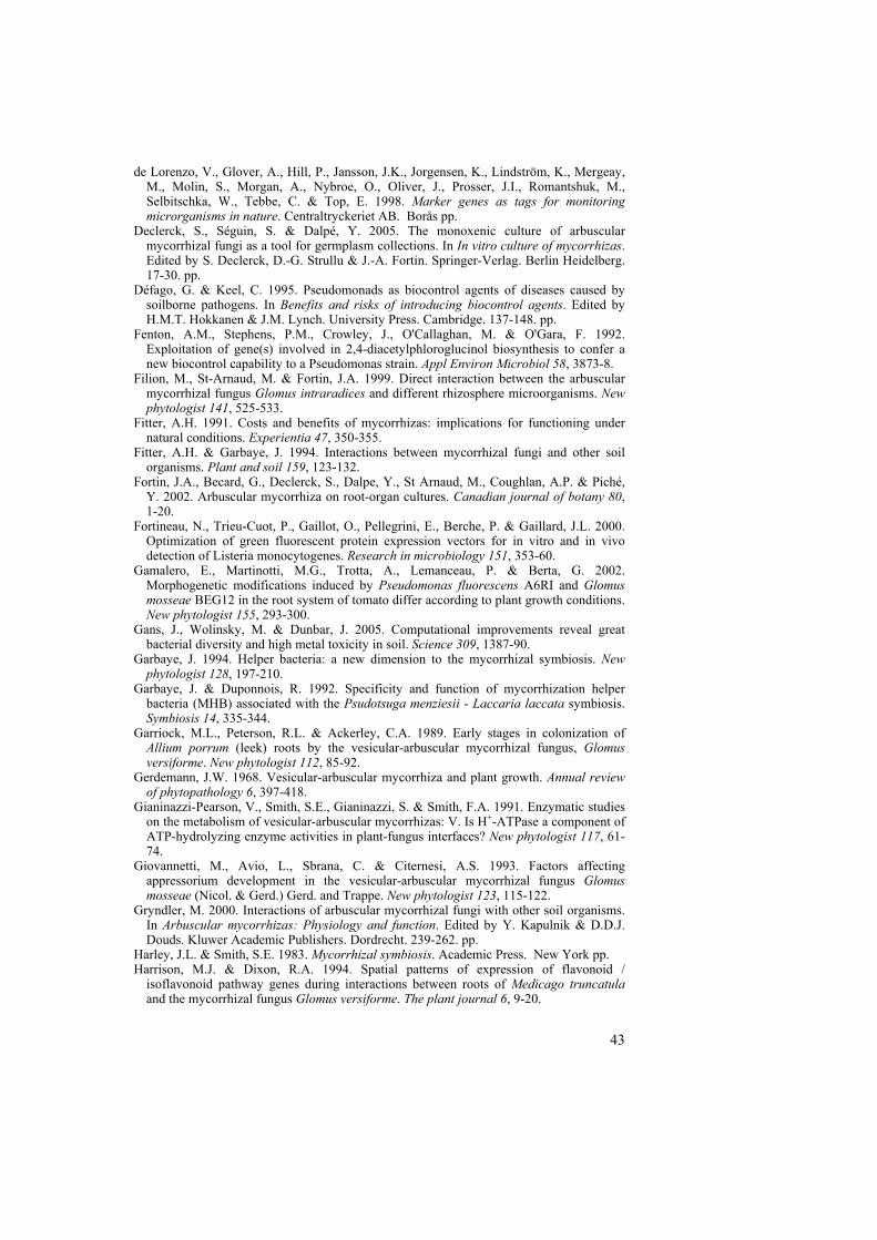
43
de Lorenzo, V., Glover, A., Hill, P., Jansson, J.K., Jorgensen, K., Lindström, K., Mergeay, M., Molin, S., Morgan, A., Nybroe, O., Oliver, J., Prosser, J.I., Romantshuk, M., Selbitschka, W., Tebbe, C. & Top, E. 1998. Marker genes as tags for monitoring microrganisms in nature. Centraltryckeriet AB. Borås pp.
Declerck, S., Séguin, S. & Dalpé, Y. 2005. The monoxenic culture of arbuscular mycorrhizal fungi as a tool for germplasm collections. In In vitro culture of mycorrhizas. Edited by S. Declerck, D.-G. Strullu & J.-A. Fortin. Springer-Verlag. Berlin Heidelberg. 17-30. pp.
Défago, G. & Keel, C. 1995. Pseudomonads as biocontrol agents of diseases caused by soilborne pathogens. In Benefits and risks of introducing biocontrol agents. Edited by H.M.T. Hokkanen & J.M. Lynch. University Press. Cambridge. 137-148. pp.
Fenton, A.M., Stephens, P.M., Crowley, J., O'Callaghan, M. & O'Gara, F. 1992. Exploitation of gene(s) involved in 2,4-diacetylphloroglucinol biosynthesis to confer a new biocontrol capability to a Pseudomonas strain. Appl Environ Microbiol 58, 3873-8.
Filion, M., St-Arnaud, M. & Fortin, J.A. 1999. Direct interaction between the arbuscular mycorrhizal fungus Glomus intraradices and different rhizosphere microorganisms. New phytologist 141, 525-533.
Fitter, A.H. 1991. Costs and benefits of mycorrhizas: implications for functioning under natural conditions. Experientia 47, 350-355.
Fitter, A.H. & Garbaye, J. 1994. Interactions between mycorrhizal fungi and other soil organisms. Plant and soil 159, 123-132.
Fortin, J.A., Becard, G., Declerck, S., Dalpe, Y., St Arnaud, M., Coughlan, A.P. & Piché, Y. 2002. Arbuscular mycorrhiza on root-organ cultures. Canadian journal of botany 80, 1-20.
Fortineau, N., Trieu-Cuot, P., Gaillot, O., Pellegrini, E., Berche, P. & Gaillard, J.L. 2000. Optimization of green fluorescent protein expression vectors for in vitro and in vivo detection of Listeria monocytogenes. Research in microbiology 151, 353-60.
Gamalero, E., Martinotti, M.G., Trotta, A., Lemanceau, P. & Berta, G. 2002. Morphogenetic modifications induced by Pseudomonas fluorescens A6RI and Glomus mosseae BEG12 in the root system of tomato differ according to plant growth conditions. New phytologist 155, 293-300.
Gans, J., Wolinsky, M. & Dunbar, J. 2005. Computational improvements reveal great bacterial diversity and high metal toxicity in soil. Science 309, 1387-90.
Garbaye, J. 1994. Helper bacteria: a new dimension to the mycorrhizal symbiosis. New phytologist 128, 197-210.
Garbaye, J. & Duponnois, R. 1992. Specificity and function of mycorrhization helper bacteria (MHB) associated with the Psudotsuga menziesii - Laccaria laccata symbiosis. Symbiosis 14, 335-344.
Garriock, M.L., Peterson, R.L. & Ackerley, C.A. 1989. Early stages in colonization of Allium porrum (leek) roots by the vesicular-arbuscular mycorrhizal fungus, Glomus versiforme. New phytologist 112, 85-92.
Gerdemann, J.W. 1968. Vesicular-arbuscular mycorrhiza and plant growth. Annual review of phytopathology 6, 397-418.
Gianinazzi-Pearson, V., Smith, S.E., Gianinazzi, S. & Smith, F.A. 1991. Enzymatic studies on the metabolism of vesicular-arbuscular mycorrhizas: V. Is H+-ATPase a component of ATP-hydrolyzing enzyme activities in plant-fungus interfaces? New phytologist 117, 61-74.
Giovannetti, M., Avio, L., Sbrana, C. & Citernesi, A.S. 1993. Factors affecting appressorium development in the vesicular-arbuscular mycorrhizal fungus Glomus mosseae (Nicol. & Gerd.) Gerd. and Trappe. New phytologist 123, 115-122.
Gryndler, M. 2000. Interactions of arbuscular mycorrhizal fungi with other soil organisms. In Arbuscular mycorrhizas: Physiology and function. Edited by Y. Kapulnik & D.D.J. Douds. Kluwer Academic Publishers. Dordrecht. 239-262. pp.
Harley, J.L. & Smith, S.E. 1983. Mycorrhizal symbiosis. Academic Press. New York pp. Harrison, M.J. & Dixon, R.A. 1994. Spatial patterns of expression of flavonoid /
isoflavonoid pathway genes during interactions between roots of Medicago truncatula and the mycorrhizal fungus Glomus versiforme. The plant journal 6, 9-20.
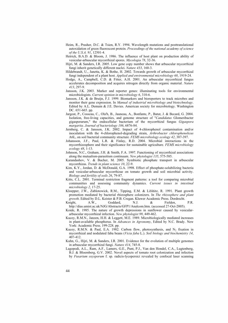
44
Heim, R., Prasher, D.C. & Tsien, R.Y. 1994. Wavelength mutations and posttranslational autoxidation of green fluorescent protein. Proceedings of the national academy of science of the U.S.A. 91, 12501-4.
Hetrick, B.A.D. & Bloom, J. 1986. The influence of host plant on production ability of vesicular-arbuscular mycorrhizal spores. Mycologia 78, 32-36.
Hijri, M. & Sanders, I.R. 2005. Low gene copy number shows that arbuscular mycorrhizal fungi inherit genetically different nuclei. Nature 433, 160-3.
Hildebrandt, U., Janetta, K. & Bothe, H. 2002. Towards growth of arbuscular mycorrhizal fungi independent of a plant host. Applied and environmental microbiology 68, 1919-24.
Hodge, A., Campbell, C.D. & Fitter, A.H. 2001. An arbuscular mycorrhizal fungus accelerates decomposition and acquires nitrogen directly from organic material. Nature 413, 297-9.
Jansson, J.K. 2003. Marker and reporter genes: illuminating tools for environmental microbiologists. Current opinion in microbiology 6, 310-6.
Jansson, J.K. & de Bruijn, F.J. 1999. Biomarkers and bioreporters to track microbes and monitor their gene expression. In Manual of industrial microbiology and biotechnology. Edited by A.L. Demain & J.E. Davies. American society for microbiology. Washington DC. 651-665. pp.
Jargeat, P., Cosseau, C., Ola'h, B., Jauneau, A., Bonfante, P., Batut, J. & Becard, G. 2004. Isolation, free-living capacities, and genome structure of "Candidatus Glomeribacter gigasporarum," the endocellular bacterium of the mycorrhizal fungus Gigaspora margarita. Journal of bacteriology 186, 6876-84.
Jernberg, C. & Jansson, J.K. 2002. Impact of 4-chlorophenol contamination and/or inoculation with the 4-chlorophenol-degrading strain, Arthrobacter chlorophenolicus A6L, on soil bacterial community structure. FEMS microbiology ecology 42, 387-397.
Johansson, J.F., Paul, L.R. & Finlay, R.D. 2004. Microbial interactions in the mycorrhizosphere and their significance for sustainable agriculture. FEMS microbiology ecology 48, 1-13.
Johnson, N.C., Graham, J.H. & Smith, F.A. 1997. Functioning of mycorrhizal associations along the mutualism-parasitism continuum. New phytologist 135, 575-585.
Karandashov, V. & Bucher, M. 2005. Symbiotic phosphate transport in arbuscular mycorrhizas. Trends in plant science 10, 22-9.
Kim, K.Y., Jordan, D. & McDonald, G.A. 1998. Effect of phosphate-solubilizing bacteria and vesicular-arbuscular mycorrhizae on tomato growth and soil microbial activity. Biology and fertility of soils 26, 79-87.
Kitts, C.L. 2001. Terminal restriction fragment patterns: a tool for comparing microbial communities and assessing community dynamics. Current issues in intestinal microbiology 2, 17-25.
Kloepper, J.W., Zablotowick, R.M., Tipping, E.M. & Lifshitz, R. 1991. Plant growth promotion mediated by bacterial rhizosphere colonizers. In The rhizosphere and plant growth. Edited by D.L. Keister & P.B. Cregan. Kluwer Academic Press. Dordrecht.
Knight, A.W., Goddard, N.J. & Fielden, P.R. http://dias.umist.ac.uk/NJG/Abstracts/GFP1/Analcom.htm. (accessed 27-Oct-2005).
Koide, R. 1985. The nature of growth depressions in sunflower caused by vesicular-arbuscular mycorrhizal infection. New phytologist 99, 449-462.
Kucey, R.M.N., Janzen, H.H. & Leggett, M.E. 1989. Microbiologically mediated increases in plant-available phosphorus. In Advances in Agronomy, Edited by N.C. Brady. New York: Academic Press. 199-228. pp.
Kucey, R.M.N. & Paul, E.A. 1982. Carbon flow, photosynthesis, and N2 fixation in mycorrhizal and nodulated faba beans (Vicia faba L.). Soil biology and biochemistry 14, 407-412.
Kuhn, G., Hijri, M. & Sanders, I.R. 2001. Evidence for the evolution of multiple genomes in arbuscular mycorrhizal fungi. Nature 414, 745-8.
Lagopodi, A.L., Ram, A.F., Lamers, G.E., Punt, P.J., Van den Hondel, C.A., Lugtenberg, B.J. & Bloemberg, G.V. 2002. Novel aspects of tomato root colonization and infection by Fusarium oxysporum f. sp. radicis-lycopersici revealed by confocal laser scanning
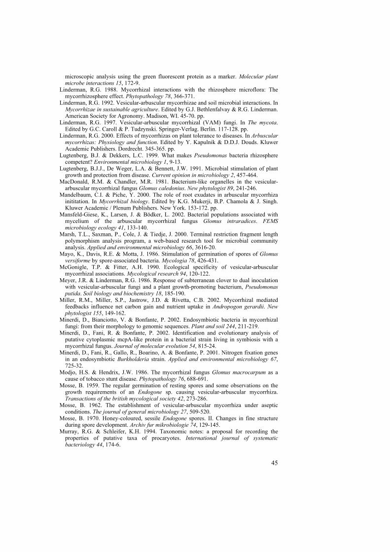
45
microscopic analysis using the green fluorescent protein as a marker. Molecular plant microbe interactions 15, 172-9.
Linderman, R.G. 1988. Mycorrhizal interactions with the rhizosphere microflora: The mycorrhizosphere effect. Phytopathology 78, 366-371.
Linderman, R.G. 1992. Vesicular-arbuscular mycorrhizae and soil microbial interactions. In Mycorrhizae in sustainable agriculture. Edited by G.J. Bethlenfalvay & R.G. Linderman. American Society for Agronomy. Madison, WI. 45-70. pp.
Linderman, R.G. 1997. Vesicular-arbuscular mycorrhizal (VAM) fungi. In The mycota. Edited by G.C. Caroll & P. Tudzynski. Springer-Verlag. Berlin. 117-128. pp.
Linderman, R.G. 2000. Effects of mycorrhizas on plant tolerance to diseases. In Arbuscular mycorrhizas: Physiology and function. Edited by Y. Kapulnik & D.D.J. Douds. Kluwer Academic Publishers. Dordrecht. 345-365. pp.
Lugtenberg, B.J. & Dekkers, L.C. 1999. What makes Pseudomonas bacteria rhizosphere competent? Environmental microbiology 1, 9-13.
Lugtenberg, B.J.J., De Weger, L.A. & Bennett, J.W. 1991. Microbial stimulation of plant growth and protection from disease. Current opinion in microbiology 2, 457-464.
MacDonald, R.M. & Chandler, M.R. 1981. Bacterium-like organelles in the vesicular-arbuscular mycorrhizal fungus Glomus caledonius. New phytologist 89, 241-246.
Mandelbaum, C.I. & Piche, Y. 2000. The role of root exudates in arbuscular mycorrhiza inititation. In Mycorrhizal biology. Edited by K.G. Mukerji, B.P. Chamola & J. Singh. Kluwer Academic / Plenum Publishers. New York. 153-172. pp.
Mansfeld-Giese, K., Larsen, J. & Bödker, L. 2002. Bacterial populations associated with mycelium of the arbuscular mycorrhizal fungus Glomus intraradices. FEMS microbiology ecology 41, 133-140.
Marsh, T.L., Saxman, P., Cole, J. & Tiedje, J. 2000. Terminal restriction fragment length polymorphism analysis program, a web-based research tool for microbial community analysis. Applied and environmental microbiology 66, 3616-20.
Mayo, K., Davis, R.E. & Motta, J. 1986. Stimulation of germination of spores of Glomus versiforme by spore-associated bacteria. Mycologia 78, 426-431.
McGonigle, T.P. & Fitter, A.H. 1990. Ecological specificity of vesicular-arbuscular mycorrhizal associations. Mycological research 94, 120-122.
Meyer, J.R. & Linderman, R.G. 1986. Response of subterranean clover to dual inoculation with vesicular-arbuscular fungi and a plant growth-promoting bacterium, Pseudomonas putida. Soil biology and biochemistry 18, 185-190.
Miller, R.M., Miller, S.P., Jastrow, J.D. & Rivetta, C.B. 2002. Mycorrhizal mediated feedbacks influence net carbon gain and nutrient uptake in Andropogon gerardii. New phytologist 155, 149-162.
Minerdi, D., Bianciotto, V. & Bonfante, P. 2002. Endosymbiotic bacteria in mycorrhizal fungi: from their morphology to genomic sequences. Plant and soil 244, 211-219.
Minerdi, D., Fani, R. & Bonfante, P. 2002. Identification and evolutionary analysis of putative cytoplasmic mcpA-like protein in a bacterial strain living in symbiosis with a mycorrhizal fungus. Journal of molecular evolution 54, 815-24.
Minerdi, D., Fani, R., Gallo, R., Boarino, A. & Bonfante, P. 2001. Nitrogen fixation genes in an endosymbiotic Burkholderia strain. Applied and environmental microbiology 67, 725-32.
Modjo, H.S. & Hendrix, J.W. 1986. The mycorrhizal fungus Glomus macrocarpum as a cause of tobacco stunt disease. Phytopathology 76, 688-691.
Mosse, B. 1959. The regular germination of resting spores and some observations on the growth requirements of an Endogone sp. causing vesicular-arbuscular mycorrhiza. Transactions of the british mycological society 42, 273-286.
Mosse, B. 1962. The establishment of vesicular-arbuscular mycorrhiza under aseptic conditions. The journal of general microbiology 27, 509-520.
Mosse, B. 1970. Honey-coloured, sessile Endogone spores. II. Changes in fine structure during spore development. Archiv fur mikrobiologie 74, 129-145.
Murray, R.G. & Schleifer, K.H. 1994. Taxonomic notes: a proposal for recording the properties of putative taxa of procaryotes. International journal of systematic bacteriology 44, 174-6.
46
Murray, R.G. & Stackebrandt, E. 1995. Taxonomic note: implementation of the provisional status Candidatus for incompletely described procaryotes. International journal of systematic bacteriology 45, 186-7.
Newman, E.I., Devoy, C.L.N., Easen, N.J. & Fowles, K.J. 1994. Plant species that can be linked by VA mycorrhizal fungi. New phytologist 126, 691-693.
Newsham, K.K., Fitter, A.H. & Watkinson, A.R. 1995. Arbuscular mycorrhiza protect an annual grass from root pathogenic fungi in the field. Journal of ecology 83, 991-1000.
Niemira, B.A., Hammerschmidt, R. & Safir, G.R. 1996. Postharvest suppression of potato dry rot (Fusarium sambucinum) in prenuclear minitubers by arbuscular mycorrhizal fungal inoculum. American potato journal 73, 509-515.
Pawlowska, T.E. & Taylor, J.W. 2004. Organization of genetic variation in individuals of arbuscular mycorrhizal fungi. Nature 427, 733-7.
Pearson, J.N. & Jakobsen, I. 1993. Symbiotic exchange of carbon and phosphorus between cucumber and three arbuscular mycorrhizal fungi. New phytologist 124, 481-488.
Perotto, S. & Bonfante, P. 1997. Bacterial associations with mycorrhizal fungi: close and distant friends in the rhizosphere. Trends in microbiology 5, 496-501.
Raman, N., Sahadevan, C. & Srinivasan, V. 2001. Growth of AM fungi on in vitro root organ culture of Sorghum vulgare and Saccharum officinarum. Indian journal of experimental biology 39, 1293-8.
Rambelli, A. 1973. The rhizosphere of mycorrhizae. In Ectomycorrhizae. Edited by G.L. Marks & K. T.T. Academic Press. New York. 299-343. pp.
Redecker, D. http://tolweb.org/tree?group=Glomeromycota. (accessed 19-Oct-2005). Redecker, D., Kodner, R. & Graham, L.E. 2000. Glomalean fungi from the Ordovician.
Science 289, 1920-1. Ruiz-Lozano, J.M. & Bonfante, P. 1999. Identification of a putative P-transporter operon in
the genome of a Burkholderia strain living inside the arbuscular mycorrhizal fungus Gigaspora margarita. Journal of bacteriology 181, 4106-9.
Ruiz-Lozano, J.M. & Bonfante, P. 2000. A Burkholderia Strain Living Inside the Arbuscular Mycorrhizal Fungus Gigaspora margarita Possesses the vacB Gene, Which Is Involved in Host Cell Colonization by Bacteria. Microbial ecology 39, 137-144.
Saikaly, P.E., Stroot, P.G. & Oerther, D.B. 2005. Use of 16S rRNA gene terminal restriction fragment analysis to assess the impact of solids retention time on the bacterial diversity of activated sludge. Applied and environmental microbiology 71, 5814-22.
Sanchez, L., Weidmann, S., Brechenmacher, L., Batoux, M., van Tuinen, D., Lemanceau, P., Gianinazzi, S. & Gianinazzi-Pearson, V. 2004. Common gene expression in Medicago truncatula roots in response to Pseudomonas fluorescens colonization, mycorrhiza development and nodulation. New phytologist 161, 855-863.
Scannerini, S. & Bonfante, P. 1991. Bacteria and bacteria like objects in endomycorrhizal fungi (Glomaceae). In Symbiosis as a source of evolutionary innovation: speciation and morphogenesis. Edited by L. Margulis & R. Fester. MIT Press. Cambridge, Massachusetts. 273-287. pp.
Schüssler, A. http://www.tu-darmstadt.de/fb/bio/bot/schuessler/amphylo/amphylogeny.html. (accessed 19-Oct-2005).
Schüssler, A. 2002. Molecular phylogeny, taxonomy, and evolution of Geosiphon pyriformis and arbuscular mycorrhizal fungi. Plant and soil 244, 75-83.
Schüssler, A. & Kluge, M. 2001. Geosiphon pyriforme, and endocytosymbiosis between fungus and cyanobacteria, and its meaning as a model system for arbuscular mycorrhizal research. In The mycota IX. Edited by B. Hock. Springer Verlag. Berlin. 151-161. pp.
Schüssler, A., Schwarzott, D. & Walker, C. 2001. A new fungal phylum, the Glomeromycota: phylogeny and evolution. Mycological research 105, 1413-1421.
Selim, S., Negrel, J., Govaerts, C., Gianinazzi, S. & van Tuinen, D. 2005. Isolation and partial characterization of antagonistic peptides produced by Paenibacillus sp. strain B2 isolated from the Sorghum mycorrhizosphere. Applied and environmental microbiology 71, 6501-6507.
Sen, R., Nurmiaho-Lassila, K., Haahtela, K. & Korhonen, K. 1996. Specificity and mode of primary attachment of Pseudomonas fluorescens strains to the cell walls of ectomycorrhizal fungi. In Mycorrhizas in integrated systems: from genes to plant
47
development. Edited by C. Azcón-Aguilar & J.M. Barea. ECSC-EC-EAEC Press. Brussels.
Smith, F.A. & Smith, S.E. 1997. Structural diversity in (vesicular)-arbuscular mycorrhizal symbioses. New phytologist 137, 373-388.
Smith, S.E., Gianinazzi-Pearson, V., Koide, R. & Cairney, J.W.G. 1994. Nutrient transport in mycorrhizas: structure, physiology and consequences for efficiency of the symbiosis. In Management of mycorrhizas in agriculture, horticulture and forestry. Edited by A.D. Robson, L.K. Abbott & N. Malajczuk. Kluwer Academic Publishers. Dordrecht. 103-113. pp.
Smith, S.E. & Read, D.J. 1997. Mycorrhizal symbiosis. Academic Press. San Diego pp. Staddon, P.L., Ramsey, C.B., Ostle, N., Ineson, P. & Fitter, A.H. 2003. Rapid turnover of
hyphae of mycorrhizal fungi determined by AMS microanalysis of 14C. Science 300, 1138-40.
Sylvia, D.M. 2002. Mycorrhizal symbioses. In Principles and applications of soil microbiology. Edited by D.M. Sylvia, J.J. Fuhrmann, P.G. Hartel & D.A. Zuberer. Prentice Hall. New Jersey. 408-426. pp.
Tester, M., Smith, S.E. & Smith, F.A. 1987. The phenomenon of "nonmycorrhizal" plants. Canadian journal of botany 65, 419-431.
Toro, M., Azcón, R. & Barea, J.M. 1997. Improvement of arbuscular mycorrhiza development by inoculation of soil with phosphate-solubilizing rhizobacteria to improve rock phosphate bioavailability (32P) and nutrient cycling. Applied and environmental microbiology 63, 4408-4412.
Toro, M., Azcón, R. & Barea, J.M. 1998. The use of isotopic dilution techniques to evaluate the interactive effects of Rhizobium genotype, mycorrhizal fungi, phosphate-solubilizing rhizobacteria and rock phosphate on nitrogen and phosphorus acquisition by Medicago sativa. New phytologist 138, 265-273.
Tsien, R.Y. 1998. The green fluorescent protein. Annual review of biochemistry 67, 509-44. Unge, A. (2000). Marker genes: molecular tools for in situ monitoring of bacterial
distribution and activity in environmental samples, Stockholm University. Urbach, E., Vergin, K.L. & Giovannoni, S.J. 1999. Immunochemical detection and isolation
of DNA from metabolically active bacteria. Applied and environmental microbiology 65, 1207-13.
van Buuren, M.L., Lanfranco, L., Longato, S., Minerdi, D., Harrison, M.J. & Bonfante, P. 1999. Construction and characterization of genomic libraries of two endomycorrhizal fungi: Glomus versiforme and Gigaspora margarita. Mycological research 103, 955-960.
van Elsas, J.D., Mäntynen, V. & Wolters, A.C. 1997. Soil DNA extraction and assessment of the fate of Mycobacterium chlorophenolicum strain PCP-1 in different soils by 16S ribosomal RNA gene sequence based most-probable-number PCR and immunofluorescence. Biology and fertility of soils 24, 188-195.
Weller, D.M. 1988. Biological control of soilborne plant pathogens in the rhizosphere with bacteria. Annual review of phytopathology 26, 379-407.
Whipps, J.M. 2001. Microbial interactions and biocontrol in the rhizosphere. Journal of experimental botany 52, 487-511.
Villegas, J. & Fortin, J.A. 2001. Phosphorus solubilization and pH changes as a result of the interactions between soil bacteria and arbuscular mycorrhizal fungi on a medium containing NH4
+ as nitrogen source. Canadian journal of botany 79, 865-870. Villegas, J. & Fortin, J.A. 2002. Phosphorus solubilization and pH changes as a result of the
interactions between soil bacteria and arbuscular mycorrhizal fungi on a medium containing NO3
- as nitrogen source. Canadian journal of botany 80, 571-576. Vivas, A., Azcon, R., Biro, B., Barea, J.M. & Ruiz-Lozano, J.M. 2003. Influence of
bacterial strains isolated from lead-polluted soil and their interactions with arbuscular mycorrhizae on the growth of Trifolium pratense L. under lead toxicity. Canadian journal of microbiology 49, 577-88.
von der Weid, I., Duarte, G.F., van Elsas, J.D. & Seldin, L. 2002. Paenibacillus brasilensis sp. nov., a novel nitrogen-fixing species isolated from the maize rhizosphere in Brazil. International journal of systematic and evolutionary microbiology 52, 2147-2153.

48
Xavier, L.J.C. & Germida, J.J. 2003. Bacteria associated with Glomus clarum spores influence mycorrhizal activity. Soil biology and biochemistry 35, 471-478.
Yang, F., Moss, L.G. & Phillips, J.G.N. 1996. The molecular structure of green fluorescent protein. Nature biotechnology 14, 1246-1251.
Yin, B., Crowley, D., Sparovek, G., De Melo, W.J. & Borneman, J. 2000. Bacterial functional redundancy along a soil reclamation gradient. Applied and environmental microbiology 66, 4361-5.
Zhu, Y.G. & Miller, R.M. 2003. Carbon cycling by arbuscular mycorrhizal fungi in soil-plant systems. Trends in plant science 8, 407-9.
49
Acknowledgements
I would like to acknowledge the people that have contributed to the present thesis, including those that have supported me in either a professional or personal way (or both)! In particular, I wish to thank:
my main supervisor Janet Jansson for being such an encouraging scientific role model to me. Thank you for treating me as an equal, whilst also being honest, trusting and open-minded regarding my thoughts and ideas. It has been a true privilege working with you, taking part in strategic planning of pioneering projects (!) and inspiring discussions. Additionally, I have really appreciated our more informal moments, where everything from “academic careers” to “pregnancy planning” were dealt with.
my assistant supervisor Roger Finlay. Your great competence in mycorrhizal research has been invaluable to me, especially during the later part of my project, and your contribution to our common papers has significantly improved their quality. I have also truly enjoyed your great ability to create a cheerful atmosphere, with your good mood and contagious laughter!
my friend and fellow PhD-student Jonas Toljander for fruitful collaboration. We had very ambitious collaboration plans in the beginning of our project; it has not always been an easy task to coordinate our experiments and ideas, however. Now, in the end, I am glade that our common efforts resulted in at least, one good paper. I wish you all the best in the future!
Leslie Paul at the Dept. of Forest Mycology and Pathology, SLU, for the collaboration in paper III, and Irene von der Weid and Lucy Seldin at the Instituto de Microbiologia, Universidade Federal do Rio de Janeiro, Brazil, for the collaboration in paper IV. Also thanks to Björn Lindahl at the Dept. of Forest Mycology and Pathology, SLU, for help with the statistical analyses in paper III.
Silvio Gianinazzi at INRA, Université de Bourgogne, France, for your encouraging support and generous collaboration suggestions. Thank you for believing in me, and for inviting me to participate in your COST action. Also thanks for letting me come to your lab in Dijón. I really appreciated that!
50
Guillaume Bécard at CNRS/Université Paul Sabatier, France, for letting me stay in your lab. Your great expertise in the mycorrhizal field is impressive, and I am so grateful that you allocated time to share some of your experiences with me. I had such a pleasant time in your group, feeling very well taken care of. Many thanks to all the group members and also to your lovely family!
the former and present members of Janet´s research group: Agneta, Anna, Berndt, Cia, Elisabeth, Fredrik, Jocke, Johan, Karin, Karolina, Lisa, Lotta, Lu, Magnus, Maria, Matilda, Mei, Mia, Ninwe, Riccardo, Sara and Åsa, for creating a stimulating and cheerful working environment. I would like to express my special gratitude to the following people in the group; Åsa for being such a great roommate. It has been a true pleasure sharing everything from “common problems” to “funny pranks” with you! Also thanks for all the beautiful illustrations that you have made throughout the years, and that I have had the pleasure to use for various purposes (including this thesis)! Karin for being so helpful and sympathetic, no matter what! It has been truly uplifting having you nextdoor! Mia for being such a caring and encouraging person with common interests. I am so grateful to you Mia for your great support during my thesis writing! Lotta for creating a cheerful atmosphere, by being yourself and arranging plenty of pleasant parties! Cia for answering all my questions when I started as a PhD student; your help was invaluable! Special thanks also to Maria who keeps our lab in such a good shape, always willing to help! You are truly important to us!
Anna-Ida Johnsson, my newest roommate; it has been really nice getting to know you, and I wish you all the best with your continuing PhD studies! I also wish to thank Martin Nilsson, my first roommate here at SLU, for introducing me to the department in such a talented way. I forgive you for not giving me the window (!).
all other members at the Dept. of Microbiology, for creating a pleasant atmosphere, and funny lunch/coffee-breaks! Special thanks to Barbro and Anki for your help with administrative matters and Ingemar for your help with various practical issues. I would also like to thank Urban Pettersson at the Dept. of Plant Biology and Forest Genetics, for being so helpful with my greenhouse experiments. Additionally, I have enjoyed the company of all the fellow “big bellies” at the department; you make my steps feel a little less slow-moving..!
Michael Anderson at the Dept. of Plant and Soil Sciences, Oklahoma State University, US, and Richard Bélanger at the Dept. de Phytologie, Université Laval, Canada, for regularly cheering me up with long, amusing e-mails. I really appreciate you offering me so much help, and giving me so many valuable tips for a successful future career!
51
all my friends outside work. Special thanks to Carro & Emanuel N., Ingrid & Jan-Ola, Micke and Jenny for being good friends. We do not see each other very often nowadays, but still we are having so much fun whenever we do! Thanks to Johan and Emanuel R. for the many pleasant moments in Nynäshamn. You are very important to me even though I am not very good to keep in touch! I also wish to thank our fairly new friends here in Uppsala; Johannes & Klara, Jenny and Per-Olof. Our get-togethers already mean a lot to me!
Haldo & Rakel, Peter & Catarina, Mathias, Emelie, Peter & Kathie, Sean, Evelina, Annelie and Kailey. Thanks for letting me be part of your wonderful families!
Mormor & Morfar for your never-ending support and interest in me. Thank you for all the wonderful times during my childhood! Also thanks to Kerstin, Gunnar, Kristofer and Kajsa for many pleasant moments.
my dear family; Mamma, Pappa, Camillah and Ulrica for always being there for me. Thank you for your encouragement and enormous support; without your belief in me I would not be where I am today. My feelings for you cannot be described in words!
Per-Olov for being who you are; including my best friend and greatest supporter. You mean the world to me, and I feel so well favoured having you and all our pets in my life. I am really looking forward to our common future! This work was financed by grants from the Swedish Research Council for Environment, Agricultural Sciences and Spatial Planning (FORMAS). How much better to get wisdom than gold, To choose understanding rather than silver! Proverbs 16:16

52


![[BIOTA CONVENTION]. PROGRAMMEbiotaph.org/wp-content/uploads/2012/04/BIOTA-CONVENTION-2012... · BIOTA Hymn Ateneo de Naga University Choir ... Bicol University 9:30 AM SNACK ... [BIOTA](https://img.pdfslide.net/doc/110x75/5b786cae7f8b9a7f378b8034/biota-convention-biota-hymn-ateneo-de-naga-university-choir-bicol-university.jpg)