Embed Size (px)
Citation preview
UvA-DARE is a service provided by the library of the University of Amsterdam (http://dare.uva.nl)
UvA-DARE (Digital Academic Repository)
Bacterial meningitis in adults: Host and pathogen factors, treatment and outcomeHeckenberg, S.G.B.
Link to publication
Citation for published version (APA):Heckenberg, S. G. B. (2013). Bacterial meningitis in adults: Host and pathogen factors, treatment and outcome.
General rightsIt is not permitted to download or to forward/distribute the text or part of it without the consent of the author(s) and/or copyright holder(s),other than for strictly personal, individual use, unless the work is under an open content license (like Creative Commons).
Disclaimer/Complaints regulationsIf you believe that digital publication of certain material infringes any of your rights or (privacy) interests, please let the Library know, statingyour reasons. In case of a legitimate complaint, the Library will make the material inaccessible and/or remove it from the website. Please Askthe Library: http://uba.uva.nl/en/contact, or a letter to: Library of the University of Amsterdam, Secretariat, Singel 425, 1012 WP Amsterdam,The Netherlands. You will be contacted as soon as possible.
Download date: 13 Apr 2019

Chapter 3
Naturally occurring lipid A mutants in Neisseria meningitidis
from patients with invasive meningococcal disease are
associated with reduced coagulopathy
Floris Fransen
Sebastiaan G.B. Heckenberg
Hendrik Jan Hamstra
Moniek Feller
Claire J.P. Boog
Jos P.M. van Putten
Diederik van de Beek
Arie van der Ende*
Peter van der Ley*
*Both authors contributed equally
PLoS Pathogens, 2009;5(4):e1000396
24407 Heckenberg.indd 31 26-02-13 09:17
Chapter 3
32
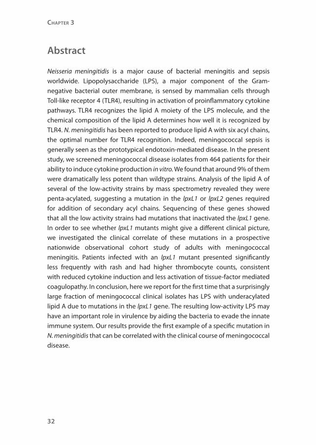
Abstract
Neisseria meningitidis is a major cause of bacterial meningitis and sepsis
worldwide. Lipopolysaccharide (LPS), a major component of the Gram-
negative bacterial outer membrane, is sensed by mammalian cells through
Toll-like receptor 4 (TLR4), resulting in activation of proinflammatory cytokine
pathways. TLR4 recognizes the lipid A moiety of the LPS molecule, and the
chemical composition of the lipid A determines how well it is recognized by
TLR4. N. meningitidis has been reported to produce lipid A with six acyl chains,
the optimal number for TLR4 recognition. Indeed, meningococcal sepsis is
generally seen as the prototypical endotoxin-mediated disease. In the present
study, we screened meningococcal disease isolates from 464 patients for their
ability to induce cytokine production in vitro. We found that around 9% of them
were dramatically less potent than wildtype strains. Analysis of the lipid A of
several of the low-activity strains by mass spectrometry revealed they were
penta-acylated, suggesting a mutation in the lpxL1 or lpxL2 genes required
for addition of secondary acyl chains. Sequencing of these genes showed
that all the low activity strains had mutations that inactivated the lpxL1 gene.
In order to see whether lpxL1 mutants might give a different clinical picture,
we investigated the clinical correlate of these mutations in a prospective
nationwide observational cohort study of adults with meningococcal
meningitis. Patients infected with an lpxL1 mutant presented significantly
less frequently with rash and had higher thrombocyte counts, consistent
with reduced cytokine induction and less activation of tissue-factor mediated
coagulopathy. In conclusion, here we report for the first time that a surprisingly
large fraction of meningococcal clinical isolates has LPS with underacylated
lipid A due to mutations in the lpxL1 gene. The resulting low-activity LPS may
have an important role in virulence by aiding the bacteria to evade the innate
immune system. Our results provide the first example of a specific mutation in
N. meningitidis that can be correlated with the clinical course of meningococcal
disease.
24407 Heckenberg.indd 32 25-02-13 11:11
Lipid A mutAnts in meningococcAL diseAse
33
Ch
apt
er 3
Introduction
Neisseria meningitidis is a major cause of bacterial meningitis and sepsis
worldwide.1 While it is a frequent commensal of the human upper respiratory
tract, in some individuals the bacterium spreads to the bloodstream
causing meningitis and/or sepsis, serious conditions with high morbidity
and mortality. As in all Gram-negative bacteria, lipopolysaccharide (LPS) is
a major component of the outer membrane of N. meningitidis. It is now well
established that LPS is sensed by mammalian cells through Toll-like receptor
4 (TLR4), in combination with coreceptors MD-2 and CD14.2 Activation of this
complex leads to recruitment of the adapters MyD88, Mal, TRIF, and TRAM to
the cytoplasmic domain of TLR4.3 These adapters initiate signal transduction
pathways that lead to induction of innate immunity. These pathways are
classified in a so called “MyD88-dependent” pathway involving MyD88 and
Mal, and a “MyD88-independent” pathway involving TRIF and TRAM. Hallmarks
of MyD88-dependent and MyD88-independent signaling are induction of pro-
inflammatory cytokines and type I IFN respectively. While the response to LPS
can be beneficial to the host by containing a beginning infection, it can also
be detrimental when excessive stimulation occurs through growth of large
numbers of bacteria in the bloodstream as happens during sepsis.2, 4, 5
TLR4 recognizes the lipid A moiety of the LPS molecule.2 The chemical
composition of the lipid A determines how well it is recognized by TLR4 and
consequently it determines the biological activity of the LPS. N. meningitidis
has been reported to produce lipid A with six acyl chains, the optimal number
for TLR4 recognition.6 Indeed purified LPS of this bacterium is highly active and
plasma concentrations of LPS in patients with meningococcal disease correlate
strongly with mortality risk.7 LPS is also important in the activation of the
coagulation system through upregulation of tissue factor. Excessive activation
of the coagulation system can lead to disseminated intravascular coagulation
(DIC), the most feared complication of invasive meningococcal disease.1 DIC
is clinically characterized by hypotension, petechial rash, and depletion of
thrombocytes and coagulation factors.
Uniquely among Gram-negative bacteria, N. meningitidis can grow without
LPS, as was shown by us when we constructed a mutant with an inactivated
lpxA gene, required for the first step in LPS biosynthesis.8 In addition, we have
previously shown that insertional inactivation of the lpxL1 or lpxL2 genes
24407 Heckenberg.indd 33 25-02-13 11:11
Chapter 3
34
required for addition of secondary acyl chains leads to reduced biological
activity of meningococcal LPS.9, 10 The possibility that such mutations might
also occur naturally was suggested to us by a report showing that the group
Y strain HF13 was defective in signaling through the MyD88-independent
pathway and TLR4.11
Here we report that strain HF13 has penta-acylated lipid A due to a mutation
in its lpxL1 gene. Screening of a selection of clinical isolates revealed lpxL1
mutations in approximately 13% of meningococcal disease isolates of all
major serogroups and clonal complexes. Several different kinds of mutations
were found. We also found evidence for on-and-off switching of lpxL1 in vivo
in humans. Importantly, patients with meningococcal meningitis that were
infected with an lpxL1 mutant strain had less severe systemic inflammation and
reduced coagulopathy.
Materials and Methods
Ethics statement
This observational study with anonymous patient data was carried out in
accordance with the Dutch privacy legislation. Written informed consent to use
data made anonymous was obtained from the patient (if possible) or from the
patient’s legal representative.
N. meningitidis strains
Strain HF13 was a kind gift from M. Kilian. The constructed lpxA and lpxL1
mutants were generated in the H44/76 strain as previously described.8, 9 All
other strains were selected from the collection of the Netherlands Reference
Laboratory for Bacterial Meningitis. Details about year of isolation, serogroup,
genotype and anatomical site of isolation are presented in supplementary table
3.1. Meningococci were cultured in GC broth or on GC plates (Difco laboratories)
supplemented with 1% (vol/vol) Vitox (Oxoid) at 37ºC in humified atmosphere
of 5% CO2.12 Bacteria were suspended in PBS and the A
620 was determined.
The bacteria were heat inactivated at 56ºC for 30 min. Serogrouping were
performed as described elsewhere.13 MLST was performed as described by
Maiden et al.14
24407 Heckenberg.indd 34 25-02-13 11:11
Lipid A mutAnts in meningococcAL diseAse
35
Ch
apt
er 3
Lipid A structure
Bacteria were grown as described above and suspended in isobutyric acid-
ammonium hydroxide 1M (5:3, v/v). Lipid A was extracted as described
previously with slight modifications.15 The lipid A structure was analyzed by
nanoelectrospray tandem mass spectrometry (MS/MS) on a Finnigan LCQ in
the negative (MS) or positive (MS/MS) ion mode.16
Sequencing
DNA was extracted from boiled cultures of N. meningitidis. Sequencing of
lpxL1 was carried out using primers 344-2 and 670-1 (supplementary table
3.2) and BigDyeTerminator chemistry (Applied Biosystems) according to
the instructions of the manufacturer. The primers used to obtain sequences
upstream and downstream of lpxL1 are presented in supplementary table 3.2.
Sequence traces were obtained with ABI Big-dyes and an ABI 3730 sequencer.
Cell lines and PBMCs
PBMC from HLA-oligotyped donors after leukapheresis were isolated by
centrifugation of buffy coat cells on Ficoll-Hypaque (Pfizer) and were used
after cryopreservation. For experiments and/or maintenance, the human
monocyte cell line Mono-mac-6 (MM6), the mouse macrophage cell line
J774A.1, and PBMCs were suspended in IMDM (Gibco BRL) supplemented
with 100 units/ml penicillin, 100 μg/ml streptomycin, 300 μg/ml l-glutamine
(Gibco BRL), and 10% heat-inactivated fetal calf serum (FCS) (Gibco BRL).
For experiments and maintenance of HEK-293 cells stably transfected with
human TLR4A, MD-2, and CD14 (Invivogen), DMEM (Gibco BRL) was used,
supplemented with 10% FCS, 10 µg/ml blasticidin (Invivogen), and 50 µg/ml
Hygromycin B (Invivogen).
ELISA
Depending on the experiment either J774A.1, MM6, PBMCs, or HEK-293 hTLR4/
MD-2/CD14 cells were used. Different plates and quantities of cells were used:
1.106 cells in 1 ml medium per well in 12-well plates, 9.104-5.105 cells in 250-
1000 µl medium per well in 24-well plates, and 1.105-3.105 cells in 200-300 µl
medium per well in 96-well plates. Cells were stimulated with bacteria and
incubated o/n at 37 °C in a humidified atmosphere containing 5% CO2. Cytokine
concentrations in the culture supernatants were quantified with ELISA. Mouse
24407 Heckenberg.indd 35 25-02-13 11:11
Chapter 3
36
IP-10 was determined with mouse IP-10 ELISA kit (R&D systems) and human IL-
6, TNF-α, IL-1β, and IL-8 with PeliPairTM reagent sets (Sanquin).
Meningitis cohort study
The Dutch Meningitis Cohort Study included 258 patients with meningococcal
meningitis; from 254 patients the bacterial strain was stored in the Netherlands
Reference Laboratory for Bacterial Meningitis.17 Inclusion and exclusion criteria
have been described extensively elsewhere.13 In summary, eligible patients
were older than 16 years, had bacterial meningitis confirmed by culture of
cerebrospinal fluid (CSF), and were listed in the database of the Netherlands
Reference Laboratory for Bacterial Meningitis from October 1998 to April
2002. This laboratory receives CSF isolates from about 85% of all patients with
bacterial meningitis in the Netherlands. The treating physician was contacted,
and informed consent was obtained from all participating patients or their
legally authorized representatives. This observational study with anonymous
patient data was carried out in accordance with the Dutch privacy legislation.
Patients underwent a neurologic examination at discharge, and outcome
was graded with the Glasgow Outcome Scale. This measurement scale is well
validated with scores varying from 1 (indicating death) to 5 (good recovery). A
favorable outcome was defined as a score of 5, and an unfavorable outcome as
a score of 1-4. Focal neurologic deficits were defined as focal cerebral deficits
(aphasia, monoparesis, or hemiparesis) or cranial nerve palsies. Serogrouping,
MLST, and susceptibility testing of meningococcal isolates were performed by
the Netherlands Reference Laboratory for Bacterial Meningitis.
Statistics
The Mann-Whitney U test was used to identify differences between groups in
continuous variables, and dichotomous variables were compared by the chi-
square or Fisher exact test. All statistical tests were 2-tailed, and a p value less
than 0.05 was regarded as significant.
List of accession numbers/ID numbers for genes mentioned in the text
Please see supplementary table 3.3 for accession numbers.
24407 Heckenberg.indd 36 25-02-13 11:11

Lipid A mutAnts in meningococcAL diseAse
37
Ch
apt
er 3
Results
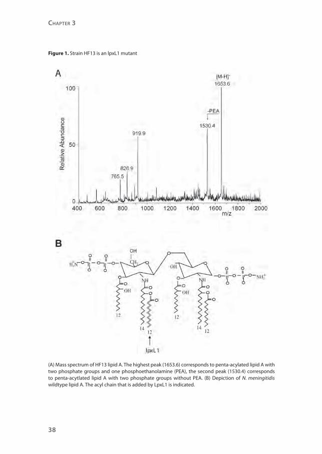
Strain HF13 is a natural lpxL1 mutant
Mogensen et al. demonstrated that the serogroup Y strain HF13 is defective
in TLR4 activation and initiation of MyD88-independent signaling.11 Reduced
biological activity of meningococcal LPS is associated with altered lipid A
structure.9, 10 Therefore, the lipid A structure of strain HF13 was assessed by mass
spectrometry (Figure 1A). The spectrum shows major peaks that correspond
with lipid A with only five acyl chains. One of the two secondary C12
acyl chains
is absent, but the spectrum is not conclusive on which one, since the C12
acyl
chains have the same mass. This result implies that in strain HF13 either lpxL1
or lpxL2 is inactive, as we previously found that the addition of the secondary
C12
acyl chains to lipid A requires active lpxL1 and lpxL2.9 Sequence analyses of
both genes showed a normal lpxL2 sequence, but the lpxL1 sequence contained
one adenosine deletion in a poly adenosine tract, leading to a frameshift and a
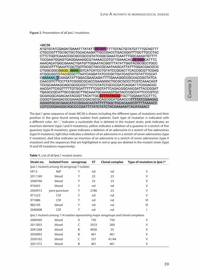
premature stop of the translated protein (Figure 2, Table 1).
The inactivated lpxL1 gene in strain HF13 results in a penta-acylated lipid
A lacking the secondary acyl chain at the 2’-position in lipid A, while N.
meningitidis typically has a hexa-acylated lipid A (Figure 1B). These results
provide an explanation for the inability of strain HF13 to activate TLR4 and to
initiate MyD88-independent signaling.
Mutations in lpxL1 are present in several serogroups and clonal complexes
To evaluate the distribution of lpxL1 mutations among meningococcal isolates
from patients, we initially screened a panel of 56 serogroup Y meningococcal
isolates for their capacity to induce the MyD88-independent cytokine IP-10
in the mouse macrophage cell line J774A.1 (supplementary figure 3.1). As
controls, strain H44/76 and HF13 were included. Of 56 serogroup Y isolates,
eight strains induced like HF13 little or no IP-10. Sequence analyses of lpxL1
of these isolates revealed that they all had mutations in lpxL1, resulting in an
inactive gene. Five strains had one adenosine deletion in a poly A tract just
like strain HF13 (type V mutation, Figure 2, Table 1), two strains had a deletion
of ten nucleotides (type VI mutation), and one strain had an insertion of the
insertion element IS1301 (Type I mutation).
24407 Heckenberg.indd 37 25-02-13 11:11
Chapter 3
38
Figure 1. Strain HF13 is an lpxL1 mutant
(A) Mass spectrum of HF13 lipid A. The highest peak (1653.6) corresponds to penta-acylated lipid A with two phosphate groups and one phosphoethanolamine (PEA), the second peak (1530.4) corresponds to penta-acytlated lipid A with two phosphate groups without PEA. (B) Depiction of N. meningitidis wildtype lipid A. The acyl chain that is added by LpxL1 is indicated.
24407 Heckenberg.indd 38 25-02-13 11:11
Lipid A mutAnts in meningococcAL diseAse
39
Ch
apt
er 3
Figure 2. Presentation of all lpxL1 mutations
The lpxL1 gene sequence of strain MC58 is shown including the different types of mutations and their position in the gene found among isolates from patients. Each type of mutation is indicated with a different color. An ‘_’ indicates a nucleotide that is deleted in the mutant strain, pink indicates an insertion element (type I and II mutations), yellow indicates a deletion of a guanine in a stretch of five guanines (type III mutation), green indicates a deletion of an adenosine in a stretch of five adenosines (type IV mutation), light blue indicates a deletion of an adenosine in a stretch of seven adenosines (type V mutation), dark blue indicates an insertion of an adenosine in a stretch of seven adenosines (type V mutation) and the sequences that are highlighted in red or gray are deleted in the mutant strain (type VI and VII mutations respectively).
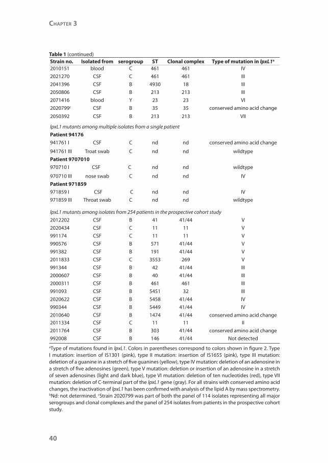
Table 1. List of all lpxL1 mutant strains
Strain no. Isolated from serogroup ST Clonal complex Type of mutation in lpxL1a
lpxL1 mutants among 56 serogroup Y isolates
HF13 Ndb Y nd nd V
2011169 blood Y 23 23 V
2040760 blood Y 23 23 V
970455 blood Y nd nd V
2050913 joint puncture Y 2786 23 V
971523 CSF Y nd nd V
971886 CSF Y nd nd VI
982195 blood Y nd nd VI
2040608 CSF Y nd nd I
lpxL1 mutants among 114 isolates representing major serogroups and clonal complexes
2000569 blood X 750 750 V
2011833 blood C 3553 269 V
2041268 blood B 4926 35 V
2050093 blood B 461 461 V
2030162 blood C 337 41/44 V
2051372 blood B 461 461 V
24407 Heckenberg.indd 39 25-02-13 11:11
Chapter 3
40
Strain no. Isolated from serogroup ST Clonal complex Type of mutation in lpxL1a
2010151 blood C 461 461 IV
2021270 CSF C 461 461 III
2041396 CSF B 4930 18 III
2050806 CSF B 213 213 III
2071416 blood Y 23 23 VI
2020799c CSF B 35 35 conserved amino acid change
2050392 CSF B 213 213 VII
lpxL1 mutants among multiple isolates from a single patient
Patient 94176
941761 I CSF C nd nd conserved amino acid change
941761 III Troat swab C nd nd wildtype
Patient 9707010
970710 I CSF C nd nd wildtype
970710 III nose swab C nd nd IV
Patient 971859
971859 I CSF C nd nd IV
971859 III Throat swab C nd nd wildtype
lpxL1 mutants among isolates from 254 patients in the prospective cohort study
2012202 CSF B 41 41/44 V
2020434 CSF C 11 11 V
991174 CSF C 11 11 V
990576 CSF B 571 41/44 V
991382 CSF B 191 41/44 V
2011833 CSF C 3553 269 V
991344 CSF B 42 41/44 III
2000607 CSF B 40 41/44 III
2000311 CSF B 461 461 III
991093 CSF B 5451 32 III
2020622 CSF B 5458 41/44 IV
990344 CSF B 5449 41/44 IV
2010640 CSF B 1474 41/44 conserved amino acid change2011334 CSF C 11 11 II
2011764 CSF B 303 41/44 conserved amino acid change
992008 CSF B 146 41/44 Not detected
aType of mutations found in lpxL1. Colors in parentheses correspond to colors shown in figure 2. Type I mutation: insertion of IS1301 (pink), type II mutation: insertion of IS1655 (pink), type III mutation: deletion of a guanine in a stretch of five guanines (yellow), type IV mutation: deletion of an adenosine in a stretch of five adenosines (green), type V mutation: deletion or insertion of an adenosine in a stretch of seven adenosines (light and dark blue), type VI mutation: deletion of ten nucleotides (red), type VII mutation: deletion of C-terminal part of the lpxL1 gene (gray). For all strains with conserved amino acid changes, the inactivation of lpxL1 has been confirmed with analysis of the lipid A by mass spectrometry. bNd: not determined. cStrain 2020799 was part of both the panel of 114 isolates representing all major serogroups and clonal complexes and the panel of 254 isolates from patients in the prospective cohort study.
Table 1 (continued)
24407 Heckenberg.indd 40 25-02-13 11:11
Lipid A mutAnts in meningococcAL diseAse
41
Ch
apt
er 3
These results prompted us to investigate the distribution of lpxL1 mutations
among meningococci of the major serogroups and clonal complexes.
Previously, we have shown that at higher dilutions an lpxL1 mutant induces
less pro-inflammatory cytokines than wildtype N. meningitidis.9, 10 To identify
meningococcal isolates with mutations in lpxL1, isolates were tested on their
capacity to induce IL-6 in the human monocytic cell line Mono Mac 6 (MM6). Of
114 isolates, representing all major serogroups and clonal complexes, 13 were
found to induce low amounts of IL-6 (supplementary figure 3.2). Sequence
analyses of lpxL1 showed that 12 isolates had a mutation in lpxL1, rendering the
gene inactive (Figure 2, Table 1). Of these strains, 10 had an insertion or deletion
in a polyadenosine or polyguanosine tract (type III, IV and V mutations); six of
these had the same mutation as found in the majority of mutant serogroup
Y strains. One strain had a type VI mutation, like in the two aforementioned
serogroup Y strains. One strain had a deletion of the C-terminal part of the gene
(type VII mutation). The remaining strain (2020799) had apparently no mutation
in lpxL1 that would lead to its inactivation. However, closer examination of its
putative amino acid sequence showed that one amino acid was altered at a
position conserved in all known lpxL1 homologues. Therefore, the LpxL1
protein of this strain is probably nonfunctional. Indeed, we confirmed that
strain 2020799 had penta-acylated lipid A by mass spectrometry (data not
shown). As a control, also lpxL1 of 34 strains that induced a normal level of
IL-6 was sequenced. As expected, these strains had no mutations in lpxL1 (data
not shown). Together, seven unique lpxL1 mutations were found among this
panel of different serogroups and different clonal complexes, indicating that
inactivation of lpxL1 must have occurred multiple times independently. The
results show that lpxL1 mutations are not associated with serogroup or clonal
complexes and occur also among the serogroup B and C strains, which are
prevalent among isolates from patients with meningococcal disease in Europe.
Screening of lpxL1 mutations in a panel of multiple isolates per patient
Most of the identified lpxL1 mutations were in nucleotide repeats of adenosines
and guanosines, the type III, IV and V mutations (Figure 2, Table 1). These
sequences are prone to cause slippage of the DNA polymerase during DNA
replication, leading to reversible frameshift mutations. This slipped-strand
mispairing is the most common mechanism of translational phase variation,
the process of random and reversible on-and-off switching of a gene. Phase
24407 Heckenberg.indd 41 25-02-13 11:11
Chapter 3
42
variation creates a phenotypically diverse population, allowing the bacterium
to adapt to different microenvironments within the human host. To investigate
whether N. meningitidis can switch lpxL1 on-and-off we screened a panel of
strains obtained from different anatomical locations within individual patients:
isolates from the blood and/or cerebrospinal fluid (CSF) as well as from the
throat and/or nose of 40 patients were used. The MM6 cell line was stimulated
with these strains and IL-6 production was measured with ELISA. Three strains
induced low levels of IL-6 compared to wildtype N. meningitidis (supplementary
figure 3.3). These isolates were from three different patients. Two strains were
isolated from the cerebrospinal fluid and one strain was isolated from the
throat. The other isolates of these patients induced normal levels of IL-6. The
lpxL1 genes of all isolates of these three patients were sequenced and found
to be mutated in the isolates that induced low IL-6, but not in the isolates that
induced normal IL-6 (Figure 2, Table 1). Two strains had a type IV mutation,
which potentially is reversible. The third strain had a point mutation leading to
substitution of a conserved amino acid. These results suggest that in the host
the expression status of lpxL1 of meningococci is subject to phase variation.
lpxL1 mutants induce less pro-inflammatory cytokines in a TLR4-dependent manner
The identified lpxL1 mutations occurred in strains of widely varying genetic
background, and it is therefore conceivable that other factors besides altered
LPS contribute to their reduced cytokine induction. To investigate this, titrations
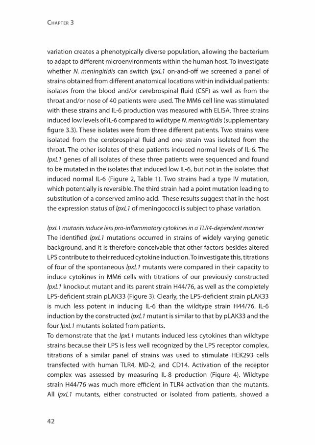
of four of the spontaneous lpxL1 mutants were compared in their capacity to
induce cytokines in MM6 cells with titrations of our previously constructed
lpxL1 knockout mutant and its parent strain H44/76, as well as the completely
LPS-deficient strain pLAK33 (Figure 3). Clearly, the LPS-deficient strain pLAK33
is much less potent in inducing IL-6 than the wildtype strain H44/76. IL-6
induction by the constructed lpxL1 mutant is similar to that by pLAK33 and the
four lpxL1 mutants isolated from patients.
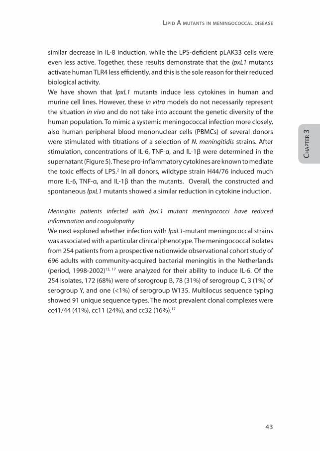
To demonstrate that the lpxL1 mutants induced less cytokines than wildtype
strains because their LPS is less well recognized by the LPS receptor complex,
titrations of a similar panel of strains was used to stimulate HEK293 cells
transfected with human TLR4, MD-2, and CD14. Activation of the receptor
complex was assessed by measuring IL-8 production (Figure 4). Wildtype
strain H44/76 was much more efficient in TLR4 activation than the mutants.
All lpxL1 mutants, either constructed or isolated from patients, showed a
24407 Heckenberg.indd 42 25-02-13 11:11
Lipid A mutAnts in meningococcAL diseAse
43
Ch
apt
er 3
similar decrease in IL-8 induction, while the LPS-deficient pLAK33 cells were
even less active. Together, these results demonstrate that the lpxL1 mutants
activate human TLR4 less efficiently, and this is the sole reason for their reduced
biological activity.
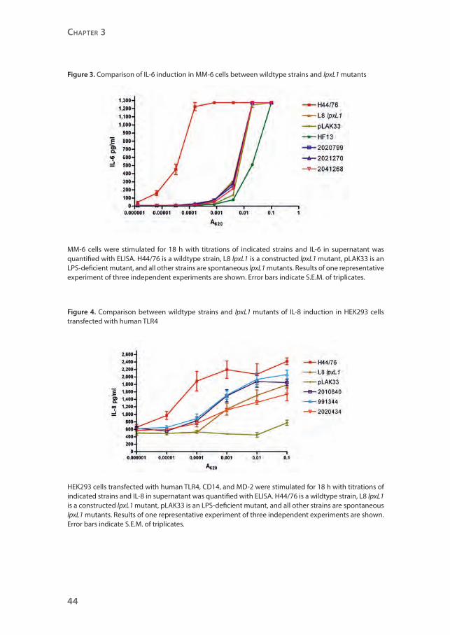
We have shown that lpxL1 mutants induce less cytokines in human and
murine cell lines. However, these in vitro models do not necessarily represent
the situation in vivo and do not take into account the genetic diversity of the
human population. To mimic a systemic meningococcal infection more closely,
also human peripheral blood mononuclear cells (PBMCs) of several donors
were stimulated with titrations of a selection of N. meningitidis strains. After
stimulation, concentrations of IL-6, TNF-α, and IL-1β were determined in the
supernatant (Figure 5). These pro-inflammatory cytokines are known to mediate
the toxic effects of LPS.2 In all donors, wildtype strain H44/76 induced much
more IL-6, TNF-α, and IL-1β than the mutants. Overall, the constructed and
spontaneous lpxL1 mutants showed a similar reduction in cytokine induction.
Meningitis patients infected with lpxL1 mutant meningococci have reduced
inflammation and coagulopathy
We next explored whether infection with lpxL1-mutant meningococcal strains
was associated with a particular clinical phenotype. The meningococcal isolates
from 254 patients from a prospective nationwide observational cohort study of
696 adults with community-acquired bacterial meningitis in the Netherlands
(period, 1998-2002)13, 17 were analyzed for their ability to induce IL-6. Of the
254 isolates, 172 (68%) were of serogroup B, 78 (31%) of serogroup C, 3 (1%) of
serogroup Y, and one (<1%) of serogroup W135. Multilocus sequence typing
showed 91 unique sequence types. The most prevalent clonal complexes were
cc41/44 (41%), cc11 (24%), and cc32 (16%).17
24407 Heckenberg.indd 43 25-02-13 11:11
Chapter 3
44
Figure 3. Comparison of IL-6 induction in MM-6 cells between wildtype strains and lpxL1 mutants
MM-6 cells were stimulated for 18 h with titrations of indicated strains and IL-6 in supernatant was quantified with ELISA. H44/76 is a wildtype strain, L8 lpxL1 is a constructed lpxL1 mutant, pLAK33 is an LPS-deficient mutant, and all other strains are spontaneous lpxL1 mutants. Results of one representative experiment of three independent experiments are shown. Error bars indicate S.E.M. of triplicates.
Figure 4. Comparison between wildtype strains and lpxL1 mutants of IL-8 induction in HEK293 cells transfected with human TLR4
HEK293 cells transfected with human TLR4, CD14, and MD-2 were stimulated for 18 h with titrations of indicated strains and IL-8 in supernatant was quantified with ELISA. H44/76 is a wildtype strain, L8 lpxL1 is a constructed lpxL1 mutant, pLAK33 is an LPS-deficient mutant, and all other strains are spontaneous lpxL1 mutants. Results of one representative experiment of three independent experiments are shown. Error bars indicate S.E.M. of triplicates.
24407 Heckenberg.indd 44 25-02-13 11:11
Lipid A mutAnts in meningococcAL diseAse
45
Ch
apt
er 3
Figure 5. Comparison between wildtype strains and lpxL1 mutants in pro-inflammatory cytokine induction in PBMCs
PBMCs from three different donors were stimulated with titrations of the indicated strains and IL-6, TNF-α, and IL-1β were quantified in the supernatant 18 h after stimulation. H44/76 is a wildtype strain, L8 lpxL1 is a constructed lpxL1 mutant, pLAK33 is an LPS-deficient mutant, and all other strains are spontaneous lpxL1 mutants. Results of one representative experiment of two independent experiments are shown. Error bars indicate S.E.M. of triplicates.
24407 Heckenberg.indd 45 25-02-13 11:11
Chapter 3
46
MM6 cells were stimulated with these strains and IL-6 induction was assessed
(supplementary figure 3.4). The isolates of 17 patients (7%) showed a decreased
IL-6 induction and sequencing revealed mutations in lpxL1 in all but one
(Figure 2, Table 1). Twelve isolates had a type III, IV or V mutation. Three strains
had a point mutation leading to substitution of an essential amino acid, and
one strain had an IS1655 insertion. In one strain (992008) we were unable to
identify a mutation in lpxL1 that could lead to gene inactivation or inactive
gene product. Further analyses with mass spectrometry to determine the mass
of its lipid A and silver staining of a Tricine-SDS-PAGE gel to analyze the size
and quantity of its LPS, demonstrated that LPS was not detectable in this strain
(results not shown). The responsible mutation remains to be identified. There
were no overall differences in lpxL1 mutation frequency between serogroups
(P=0.85) and clonal complexes (P=0.56).
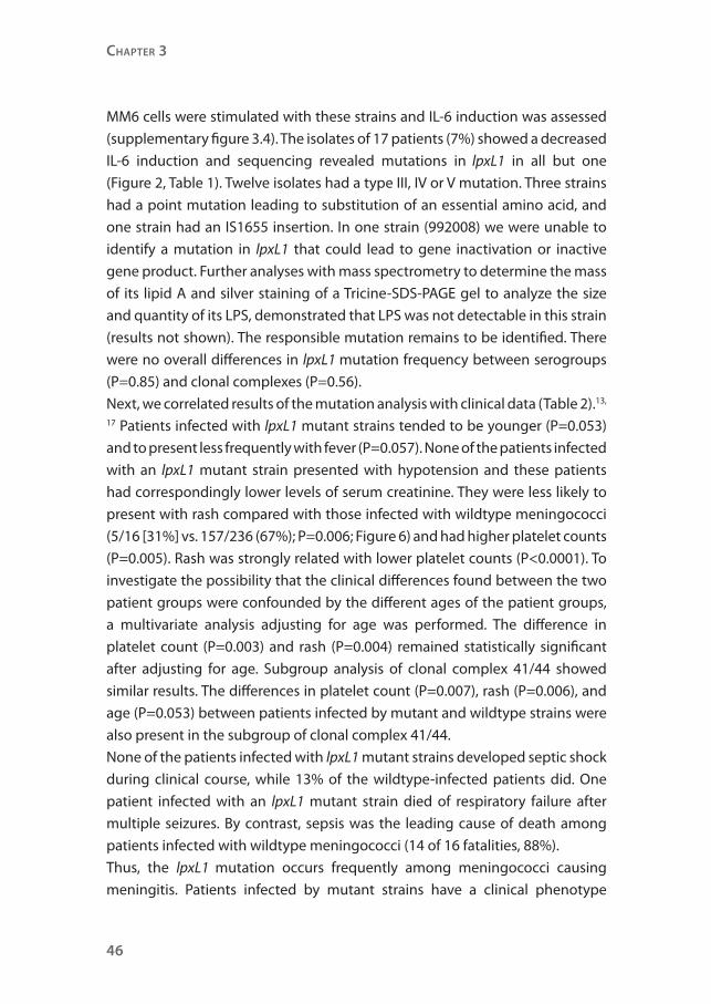
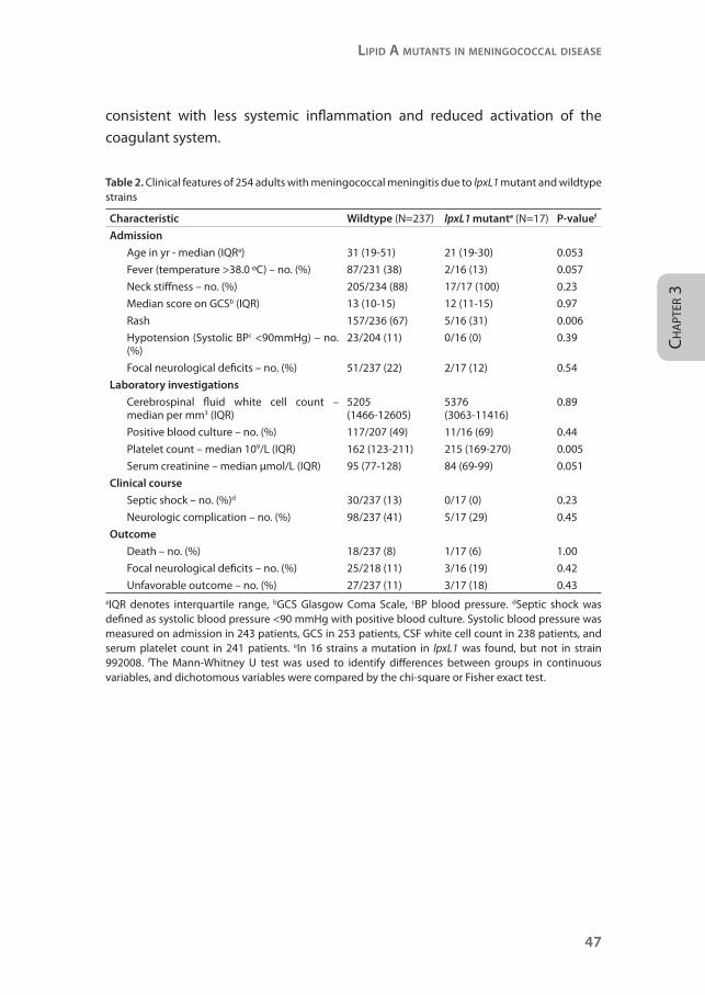
Next, we correlated results of the mutation analysis with clinical data (Table 2).13,
17 Patients infected with lpxL1 mutant strains tended to be younger (P=0.053)
and to present less frequently with fever (P=0.057). None of the patients infected
with an lpxL1 mutant strain presented with hypotension and these patients
had correspondingly lower levels of serum creatinine. They were less likely to
present with rash compared with those infected with wildtype meningococci
(5/16 [31%] vs. 157/236 (67%); P=0.006; Figure 6) and had higher platelet counts
(P=0.005). Rash was strongly related with lower platelet counts (P<0.0001). To
investigate the possibility that the clinical differences found between the two
patient groups were confounded by the different ages of the patient groups,
a multivariate analysis adjusting for age was performed. The difference in
platelet count (P=0.003) and rash (P=0.004) remained statistically significant
after adjusting for age. Subgroup analysis of clonal complex 41/44 showed
similar results. The differences in platelet count (P=0.007), rash (P=0.006), and
age (P=0.053) between patients infected by mutant and wildtype strains were
also present in the subgroup of clonal complex 41/44.
None of the patients infected with lpxL1 mutant strains developed septic shock
during clinical course, while 13% of the wildtype-infected patients did. One
patient infected with an lpxL1 mutant strain died of respiratory failure after
multiple seizures. By contrast, sepsis was the leading cause of death among
patients infected with wildtype meningococci (14 of 16 fatalities, 88%).
Thus, the lpxL1 mutation occurs frequently among meningococci causing
meningitis. Patients infected by mutant strains have a clinical phenotype
24407 Heckenberg.indd 46 25-02-13 11:11
Lipid A mutAnts in meningococcAL diseAse
47
Ch
apt
er 3
consistent with less systemic inflammation and reduced activation of the
coagulant system.
Table 2. Clinical features of 254 adults with meningococcal meningitis due to lpxL1 mutant and wildtype strains
Characteristic Wildtype (N=237) lpxL1 mutante (N=17) P-valuef
Admission
Age in yr - median (IQRa) 31 (19-51) 21 (19-30) 0.053
Fever (temperature >38.0 ºC) – no. (%) 87/231 (38) 2/16 (13) 0.057
Neck stiffness – no. (%) 205/234 (88) 17/17 (100) 0.23
Median score on GCSb (IQR) 13 (10-15) 12 (11-15) 0.97
Rash 157/236 (67) 5/16 (31) 0.006
Hypotension (Systolic BPc <90mmHg) – no. (%)
23/204 (11) 0/16 (0) 0.39
Focal neurological deficits – no. (%) 51/237 (22) 2/17 (12) 0.54
Laboratory investigations
Cerebrospinal fluid white cell count – median per mm3 (IQR)
5205 (1466-12605)
5376 (3063-11416)
0.89
Positive blood culture – no. (%) 117/207 (49) 11/16 (69) 0.44
Platelet count – median 109/L (IQR) 162 (123-211) 215 (169-270) 0.005
Serum creatinine – median µmol/L (IQR) 95 (77-128) 84 (69-99) 0.051
Clinical course
Septic shock – no. (%)d 30/237 (13) 0/17 (0) 0.23
Neurologic complication – no. (%) 98/237 (41) 5/17 (29) 0.45
Outcome
Death – no. (%) 18/237 (8) 1/17 (6) 1.00
Focal neurological deficits – no. (%) 25/218 (11) 3/16 (19) 0.42
Unfavorable outcome – no. (%) 27/237 (11) 3/17 (18) 0.43aIQR denotes interquartile range, bGCS Glasgow Coma Scale, cBP blood pressure. dSeptic shock was defined as systolic blood pressure <90 mmHg with positive blood culture. Systolic blood pressure was measured on admission in 243 patients, GCS in 253 patients, CSF white cell count in 238 patients, and serum platelet count in 241 patients. eIn 16 strains a mutation in lpxL1 was found, but not in strain 992008. fThe Mann-Whitney U test was used to identify differences between groups in continuous variables, and dichotomous variables were compared by the chi-square or Fisher exact test.
24407 Heckenberg.indd 47 25-02-13 11:11
Chapter 3
48
Figure 6. Clinical correlate of lpxL1 mutations in meningococcal meningitis
(A) Frequency of rash in patients presenting with meningitis infected by lpxL1 wildtype and mutant strains. (B,C) Platelet counts on admission for lpxL1 wildtype and mutant strains (B) and patients presenting with and without rash (C). Horizontal bars reflect medians. The Mann-Whitney U test was used to identify differences between groups in continuous variables, and dichotomous variables were compared by the chi-square or Fisher exact test.
24407 Heckenberg.indd 48 25-02-13 11:11
Lipid A mutAnts in meningococcAL diseAse
49
Ch
apt
er 3
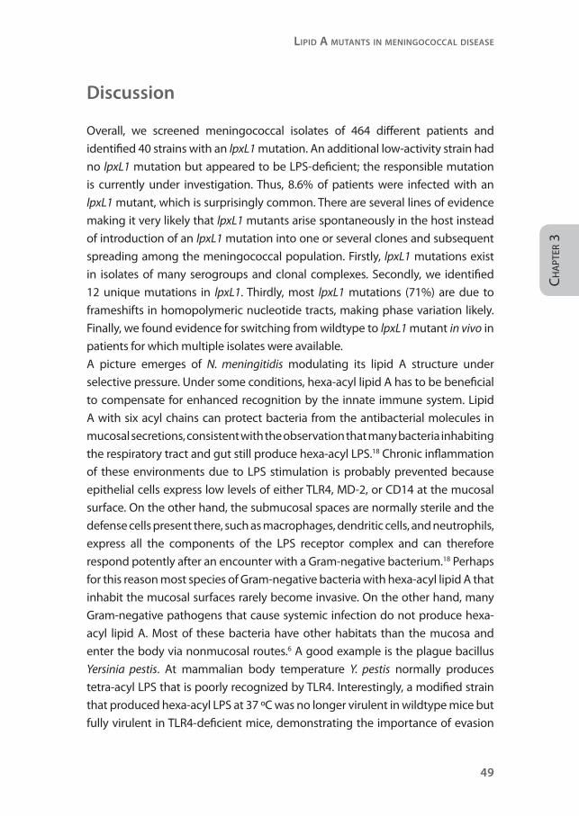
Discussion
Overall, we screened meningococcal isolates of 464 different patients and
identified 40 strains with an lpxL1 mutation. An additional low-activity strain had
no lpxL1 mutation but appeared to be LPS-deficient; the responsible mutation
is currently under investigation. Thus, 8.6% of patients were infected with an
lpxL1 mutant, which is surprisingly common. There are several lines of evidence
making it very likely that lpxL1 mutants arise spontaneously in the host instead
of introduction of an lpxL1 mutation into one or several clones and subsequent
spreading among the meningococcal population. Firstly, lpxL1 mutations exist
in isolates of many serogroups and clonal complexes. Secondly, we identified
12 unique mutations in lpxL1. Thirdly, most lpxL1 mutations (71%) are due to
frameshifts in homopolymeric nucleotide tracts, making phase variation likely.
Finally, we found evidence for switching from wildtype to lpxL1 mutant in vivo in
patients for which multiple isolates were available.
A picture emerges of N. meningitidis modulating its lipid A structure under
selective pressure. Under some conditions, hexa-acyl lipid A has to be beneficial
to compensate for enhanced recognition by the innate immune system. Lipid
A with six acyl chains can protect bacteria from the antibacterial molecules in
mucosal secretions, consistent with the observation that many bacteria inhabiting
the respiratory tract and gut still produce hexa-acyl LPS.18 Chronic inflammation
of these environments due to LPS stimulation is probably prevented because
epithelial cells express low levels of either TLR4, MD-2, or CD14 at the mucosal
surface. On the other hand, the submucosal spaces are normally sterile and the
defense cells present there, such as macrophages, dendritic cells, and neutrophils,
express all the components of the LPS receptor complex and can therefore
respond potently after an encounter with a Gram-negative bacterium.18 Perhaps
for this reason most species of Gram-negative bacteria with hexa-acyl lipid A that
inhabit the mucosal surfaces rarely become invasive. On the other hand, many
Gram-negative pathogens that cause systemic infection do not produce hexa-
acyl lipid A. Most of these bacteria have other habitats than the mucosa and
enter the body via nonmucosal routes.6 A good example is the plague bacillus
Yersinia pestis. At mammalian body temperature Y. pestis normally produces
tetra-acyl LPS that is poorly recognized by TLR4. Interestingly, a modified strain
that produced hexa-acyl LPS at 37 ºC was no longer virulent in wildtype mice but
fully virulent in TLR4-deficient mice, demonstrating the importance of evasion
24407 Heckenberg.indd 49 25-02-13 11:11
Chapter 3
50
of TLR4 activation for this bacterium.19 N. meningitidis seems to be one of the
exceptions to the general rule that Gram-negative bacteria with hexa-acyl lipid
A do not cause systemic disease. However, our observation that a proportion
of clinical isolates have penta-acylated LPS suggests that evasion of TLR4
activation might aid the bacterium to circumvent host defences after crossing
the nasopharyngeal epithelium. The hypothesis that TLR4 plays an important
role in the prevention of meningococcal disease corroborates with the finding
that subjects with rare TLR4 mutations have an increased risk for developing the
disease.20 If the assumption is correct that hexa-acyl LPS gives the bacterium
an advantage on mucosal surfaces and that non hexa-acyl LPS is better for
bacteria in submucosal spaces, one would expect that the frequency of lpxL1
mutants is lower in meningococcal isolates from the respiratory tract compared
to meningococcal isolates from the cerebrospinal fluid or blood.
Mogensen et al. showed that strain HF13 is specifically defective in activation of
the MyD88-independent pathway, but not in inducing the MyD88-dependent
pathway.11 However, we demonstrate that strain HF13 and other lpxL1 mutants
are also defective in inducing the MyD88-dependent cytokines IL-6, TNF-α,
and IL-1β. Our experiments indicate that lpxL1 mutants or purified lpxL1 LPS
compared to wildtype controls are not specifically deficient in inducing the
MyD88-dependent vs. independent pathway. This apparent discrepancy might
be explained by the dose of bacteria used. If cells are stimulated with a high dose
of bacteria the difference between lpxL1 mutant and wildtype is only detectable
for the MyD88-independent pathway. This is because LPS is the only bacterial
component capable of inducing the MyD88-independent pathway, while
many other bacterial components can induce the MyD88-dependent pathway
(e.g. TLR2 ligands). When cells are stimulated with lower doses of bacteria the
difference in induction of the MyD88-dependent pathway becomes apparent,
because LPS is by far the most active component of the bacterium and the other
non-TLR4 ligands that can activate the MyD88-dependent pathway are diluted
too far to be still active.
The relatively high frequency of phase variation raises the question whether
the lpxL1 mutations might have arisen in vitro after isolation from the patient.
Previously, we have performed extensive research on the phase variation of
porA in N. meningitidis. In this gene, homopolymeric nucleotide tracts are found
in the promoter (polyguanidine) and in the coding region (polyadenine). The
frequencies by which these sequences vary in length are 10-3.12, 21 Others showed
24407 Heckenberg.indd 50 25-02-13 11:11
Lipid A mutAnts in meningococcAL diseAse
51
Ch
apt
er 3
phase variation of capsule expression caused by insertion of IS1301 in the siaA
gene with a frequency of phase variation of 9×10−4.22, 23 In vitro selection of porA
phase variants and siaA phase variants have not been reported. Meningococcal
isolates received by the Netherlands Reference Laboratory for Bacterial Meningitis
(NRLBM) are low passages (up to 2 passages). We sequenced the lpxL1 gene of 20
individual colonies of a culture of a mutant isolate (971859 I) and of 25 individual
colonies of a culture of isolate 971859 III and found in each instance the same
sequence, i.e. 20 mutant sequences and 25 wildtype sequences, respectively.
Therefore, we estimate the frequency of phase switching to be less than 2.2 x
10-2. In addition, we sequenced lpxL1 of DNA extracted from a swap taken from
4 different quadrants of another culture plate of isolate 971859 III. All 4 lpxL1
sequences were homogeneous and identical. Thus we are confident that the
discovered lpxL1 mutations are not caused by in vitro phase variation.
Infection with lpxL1-mutant meningococcal strains is associated with a particular
clinical phenotype, which consisted of less systemic inflammation and reduced
activation of the coagulant system, reflected in less fever, higher serum platelet
counts, and lower numbers with rash. Moreover, our in vitro data have shown
that lpxL1 mutants induce much less pro-inflammatory cytokines than wildtype
strains. The coagulation system is activated through upregulation of tissue factor.1
It has been demonstrated that LPS upregulates tissue factor on monocytes and
endothelial cells.24-26 Furthermore, in particular the pro-inflammatory cytokine
IL-6 appears to mediate in vivo expression of tissue factor. 27, 28 Finally, IL-1β and
TNF-α inhibit anticoagulant pathways by downregulating thrombomodulin at
the endothelial surface and by increasing plasminogen activator inhibitor type-1
(PAI-1).29, 30 Thus, our finding that patients infected with an lpxL1 mutant show
less activation of the coagulation system is consistent with our results that show
that lpxL1 LPS is less potent and that lpxL1 mutants induce less pro-inflammatory
cytokines. Remarkably, the lpxL1 mutants induced the same degree of CSF
leukocytosis as wildtype strains. There are several explanations for “normal” CSF
white cell counts in patients infected by mutant strains. Patients in the cohort
all had positive CSF cultures; almost all had clinical signs of meningitis and CSF
leukocytosis. Likely, leukocytosis is not only mediated by lipid A, but also by other
microbial constituents.
It should be noted that not all groups of patients were included in our analysis
of clinical patient data. The study only included adults with meningitis. Patients
younger than 16 years or patients with sepsis only were not included. Therefore,
24407 Heckenberg.indd 51 25-02-13 11:11
Chapter 3
52
our results are potentially biased by excluding these patient groups. Patients
with meningitis often have a less severe form of the disease, as reflected by the
overall low mortality of 8% in our study. However, patients with sepsis have
very serious symptoms resulting from high concentrations of bacteria in the
circulation. Mortality rates in these patients can be as high as 50%. Also, patients
younger than 16 years are an import group, because rates for meningococcal
disease are highest for young children.1 It would be interesting to see whether
lpxL1 mutants also exist in these patients groups, and if so, if these patients
have a different clinical course compared to patients infected with a wildtype
strain. These additional data are needed to fully understand the impact of lpxL1
mutations on meningococcal disease.
Meningococcal sepsis is generally seen as the prototypical endotoxin-mediated
disease. Here we report for the first time that meningococcal lipid A mutants
which are defective in TLR4 activation occur naturally. Their frequency is
unexpectedly high, suggesting an important role in virulence for the resulting
low-activity LPS. Our results suggest that in most cases this mutation has occurred
through phase variation, and may give the bacteria an advantage because
they are less well sensed by the innate immune system. Patients infected with
these mutant strains endure milder symptoms with less systemic inflammation
and reduced activation of the coagulant system, showing that our findings are
clinically relevant. Importantly, these results with lpxL1 also provide the first
example of a specific bacterial mutation which can be associated with the clinical
course of meningococcal disease. More generally, it shows how there can be an
underestimated heterogeneity in the TLR4-activating capacity of pathogenic
bacteria.
Acknowledgements
M. Kilian kindly provided N. meningitidis group Y strain HF13. This publication
made use of the Neisseria Multi Locus Sequence Typing website (http://pubmlst.
org/neisseria/) developed by Keith Jolley and Man-Suen Chan and sited at the
University of Oxford 31. The development of this site has been funded by the
Wellcome Trust and European Union. We thank M. Bertayli, H.D. Meiring, and J.
ten Hove for experimental assistance.
24407 Heckenberg.indd 52 25-02-13 11:11
Lipid A mutAnts in meningococcAL diseAse
53
Ch
apt
er 3
Supplementary material
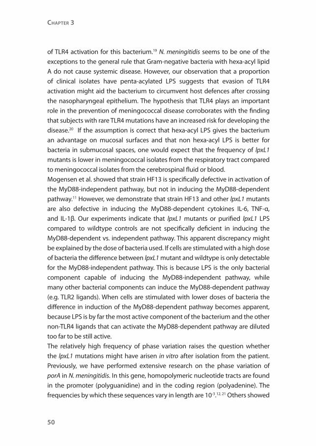
Supplementary figure 3.1. Screening of N. meningitidis group Y clinical isolates on cytokine induction
J774A.1 cells were stimulated for 3 h with a panel of group Y strains (0.1 OD) and IP-10 in the supernatant was determined with ELISA.
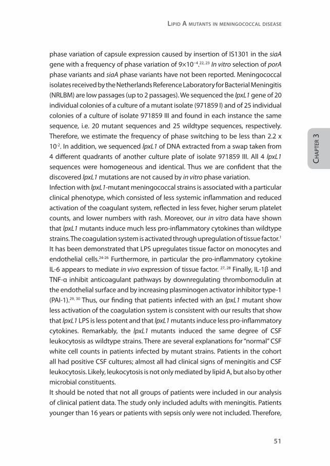
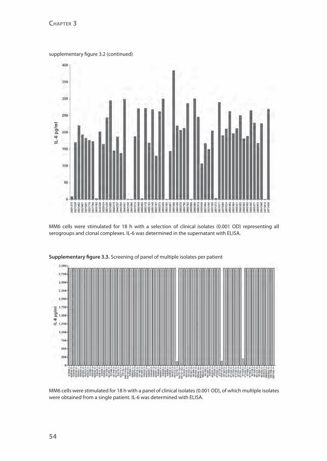
Supplementary figure 3.2. Screening of panel of N. meningitidis clinical isolates representing all major serogroups and clonal complexes
24407 Heckenberg.indd 53 26-02-13 09:17
Chapter 3
54
MM6 cells were stimulated for 18 h with a selection of clinical isolates (0.001 OD) representing all serogroups and clonal complexes. IL-6 was determined in the supernatant with ELISA.
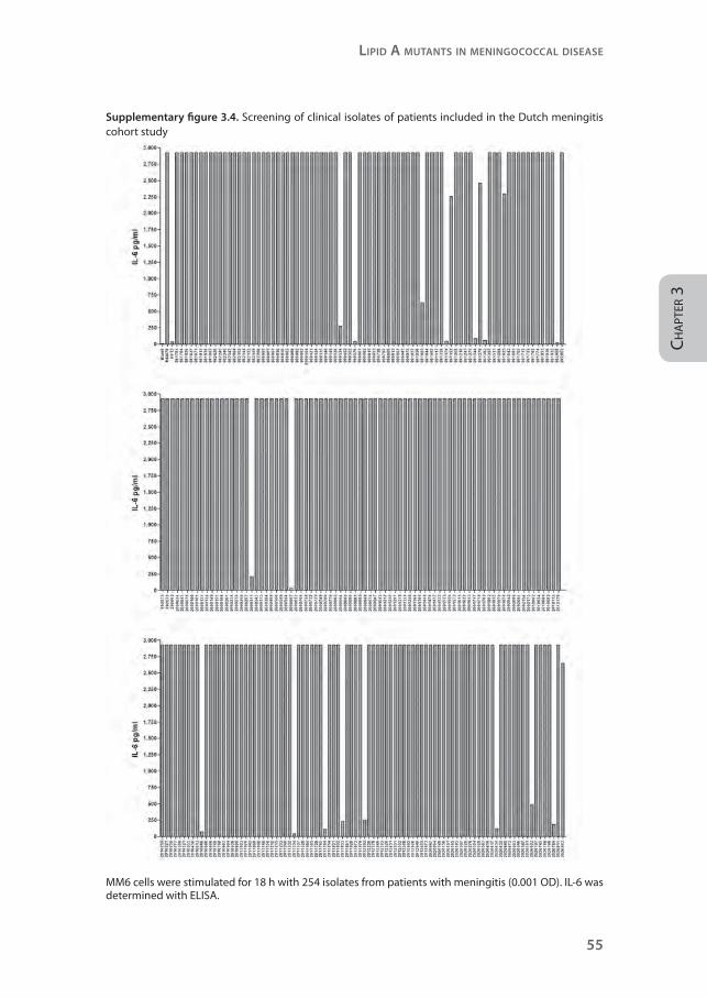
Supplementary figure 3.3. Screening of panel of multiple isolates per patient
MM6 cells were stimulated for 18 h with a panel of clinical isolates (0.001 OD), of which multiple isolates were obtained from a single patient. IL-6 was determined with ELISA.
supplementary figure 3.2 (continued)
24407 Heckenberg.indd 54 25-02-13 11:11
Lipid A mutAnts in meningococcAL diseAse
55
Ch
apt
er 3
Supplementary figure 3.4. Screening of clinical isolates of patients included in the Dutch meningitis cohort study
MM6 cells were stimulated for 18 h with 254 isolates from patients with meningitis (0.001 OD). IL-6 was determined with ELISA.
24407 Heckenberg.indd 55 25-02-13 11:11
Chapter 3
56
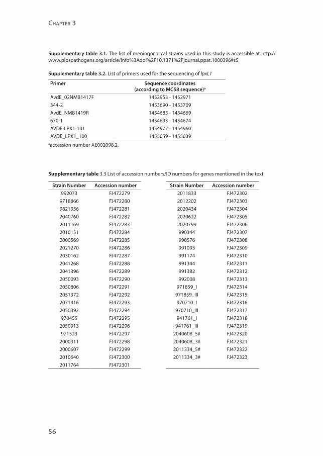
Supplementary table 3.1. The list of meningococcal strains used in this study is accessible at http://www.plospathogens.org/article/info%3Adoi%2F10.1371%2Fjournal.ppat.1000396#s5
Supplementary table 3.2. List of primers used for the sequencing of lpxL1
Primer Sequence coordinates(according to MC58 sequence)a
AvdE_02NMB1417F 1452953 - 1452971
344-2 1453690 - 1453709
AvdE_NMB1419R 1454685 - 1454669
670-1 1454693 - 1454674
AVDE-LPX1-101 1454977 - 1454960
AVDE_LPX1_100 1455059 - 1455039aaccession number AE002098.2.
Supplementary table 3.3 List of accession numbers/ID numbers for genes mentioned in the text
Strain Number Accession number
992073 FJ472279
9718866 FJ472280
9821956 FJ472281
2040760 FJ472282
2011169 FJ472283
2010151 FJ472284
2000569 FJ472285
2021270 FJ472286
2030162 FJ472287
2041268 FJ472288
2041396 FJ472289
2050093 FJ472290
2050806 FJ472291
2051372 FJ472292
2071416 FJ472293
2050392 FJ472294
970455 FJ472295
2050913 FJ472296
971523 FJ472297
2000311 FJ472298
2000607 FJ472299
2010640 FJ472300
2011764 FJ472301
Strain Number Accession number
2011833 FJ472302
2012202 FJ472303
2020434 FJ472304
2020622 FJ472305
2020799 FJ472306
990344 FJ472307
990576 FJ472308
991093 FJ472309
991174 FJ472310
991344 FJ472311
991382 FJ472312
992008 FJ472313
971859_I FJ472314
971859_III FJ472315
970710_I FJ472316
970710_III FJ472317
941761_I FJ472318
941761_III FJ472319
2040608_5# FJ472320
2040608_3# FJ472321
2011334_5# FJ472322
2011334_3# FJ472323
24407 Heckenberg.indd 56 25-02-13 11:11
Lipid A mutAnts in meningococcAL diseAse
57
Ch
apt
er 3
References
1. Stephens DS, Greenwood B, Brandtzaeg P. Epidemic meningitis, meningococcaemia, and Neisseria meningitidis. Lancet 2007;369(9580):2196-2210.
2. Beutler B, Rietschel ET. Innate immune sensing and its roots: the story of endotoxin. Nat Rev Immunol 2003;3(2):169-176.
3. Palsson-McDermott EM, O’Neill LA. Signal transduction by the lipopolysaccharide receptor, Toll-like receptor-4. Immunology 2004;113(2):153-162.
4. Parrillo JE. Pathogenetic mechanisms of septic shock. N Engl J Med 1993;328(20):1471-1477.
5. Russell JA. Management of sepsis. N Engl J Med 2006;355(16):1699-1713.
6. Munford RS, Varley AW. Shield as signal: lipopolysaccharides and the evolution of immunity to gram-negative bacteria. PLoS Pathog 2006;2(6):e67.
7. Brandtzaeg P, Bjerre A, Ovstebo R, Brusletto B, Joo GB, Kierulf P. Neisseria meningitidis lipopolysaccharides in human pathology. J Endotoxin Res 2001;7(6):401-420.
8. Steeghs L, den Hartog R, den Boer A, Zomer B, Roholl P, van der Ley P. Meningitis bacterium is viable without endotoxin. Nature 1998;392(6675):449-450.
9. van der Ley P, Steeghs L, Hamstra HJ, ten Hove J, Zomer B, van Alphen L. Modification of lipid A biosynthesis in Neisseria meningitidis lpxL mutants: influence on lipopolysaccharide structure, toxicity, and adjuvant activity. Infect Immun 2001;69(10):5981-5990.
10. Steeghs L, Tommassen J, Leusen JH, van de Winkel JG, van der Ley P. Teasing apart structural determinants of ‘toxicity’ and ‘adjuvanticity’: implications for meningococcal vaccine development. J Endotoxin Res 2004;10(2):113-119.
11. Mogensen TH, Paludan SR, Kilian M, Ostergaard L. Two Neisseria meningitidis strains with different ability to stimulate Toll-like receptor 4 through the MyD88-independent pathway. Scand J Immunol 2006;64(6):646-654.
12. van der Ende A, Hopman CT, Dankert J. Deletion of porA by recombination between clusters of repetitive extragenic palindromic sequences in Neisseria meningitidis. Infect Immun 1999;67(6):2928-2934.
13. van de Beek D, de Gans J, Spanjaard L, Weisfelt M, Reitsma JB, Vermeulen M. Clinical features and prognostic factors in adults with bacterial meningitis. N Engl J Med 2004;351(18):1849-1859.
14. Maiden MC, Bygraves JA, Feil E et al. Multilocus sequence typing: a portable approach to the identification of clones within populations of pathogenic microorganisms. Proc Natl Acad Sci U S A 1998;95(6):3140-3145.
15. El Hamidi A, Tirsoaga A, Novikov A, Hussein A, Caroff M. Microextraction of bacterial lipid A: easy and rapid method for mass spectrometric characterization. J Lipid Res 2005;46(8):1773-1778.
16. Wilm M, Mann M. Analytical properties of the nanoelectrospray ion source. Anal Chem 1996;68(1):1-8.
17. Heckenberg SG, de Gans J, Brouwer MC et al. Clinical Features, Outcome, and Meningococcal Genotype in 258 Adults With Meningococcal Meningitis: A Prospective Cohort Study. Medicine (Baltimore) 2008;87(4):185-192.
18. Munford RS. Sensing gram-negative bacterial lipopolysaccharides: a human disease determinant? Infect Immun 2008;76(2):454-465.
19. Montminy SW, Khan N, McGrath S et al. Virulence factors of Yersinia pestis are overcome by a strong lipopolysaccharide response. Nat Immunol 2006;7(10):1066-1073.
20. Smirnova I, Mann N, Dols A et al. Assay of locus-specific genetic load implicates rare Toll-like receptor 4 mutations in meningococcal susceptibility. Proc Natl Acad Sci U S A 2003;100(10):6075-6080.
21. van der Ende A, Hopman CT, Zaat S, Essink BB, Berkhout B, Dankert J. Variable expression of class 1 outer membrane protein in Neisseria meningitidis is caused by variation in the spacing between the -10 and -35 regions of the promoter. J Bacteriol 1995;177(9):2475-2480.
24407 Heckenberg.indd 57 25-02-13 11:11
Chapter 3
58
22. Hammerschmidt S, Hilse R, van Putten JP, Gerardy-Schahn R, Unkmeir A, Frosch M. Modulation of cell surface sialic acid expression in Neisseria meningitidis via a transposable genetic element. EMBO J 1996;15(1):192-198.
23. Weber MV, Claus H, Maiden MC, Frosch M, Vogel U. Genetic mechanisms for loss of encapsulation in polysialyltransferase-gene-positive meningococci isolated from healthy carriers. Int J Med Microbiol 2006;296(7):475-484.
24. Meszaros K, Aberle S, Dedrick R et al. Monocyte tissue factor induction by lipopolysaccharide (LPS): dependence on LPS-binding protein and CD14, and inhibition by a recombinant fragment of bactericidal/permeability-increasing protein. Blood 1994;83(9):2516-2525.
25. Li A, Chang AC, Peer GT, Hinshaw LB, Taylor FB, Jr. Comparison of the capacity of rhTNF-alpha and Escherichia coli to induce procoagulant activity by baboon mononuclear cells in vivo and in vitro. Shock 1996;5(4):274-279.
26. Drake TA, Cheng J, Chang A, Taylor FB, Jr. Expression of tissue factor, thrombomodulin, and E-selectin in baboons with lethal Escherichia coli sepsis. Am J Pathol 1993;142(5):1458-1470.
27. Bjerre A, Ovstebo R, Kierulf P, Halvorsen S, Brandtzaeg P. Fulminant meningococcal septicemia: dissociation between plasma thrombopoietin levels and platelet counts. Clin Infect Dis 2000;30(4):643-647.
28. Levi M, van der Poll T, Buller HR. Bidirectional relation between inflammation and coagulation. Circulation 2004;109(22):2698-2704.
29. Nawroth PP, Stern DM. Modulation of endothelial cell hemostatic properties by tumor necrosis factor. J Exp Med 1986;163(3):740-745.
30. van der Poll T, de Jonge E, Levi M. Regulatory role of cytokines in disseminated intravascular coagulation. Semin Thromb Hemost 2001;27(6):639-651.
31. Jolley KA, Chan MS, Maiden MC. mlstdbNet - distributed multi-locus sequence typing (MLST) databases. BMC Bioinformatics 2004;5:86.
24407 Heckenberg.indd 58 25-02-13 11:11